Orthopantomography
30
PANORAMIC RADIOGRAPHY
-
Upload
kmct-dental-college -
Category
Healthcare
-
view
277 -
download
5
Transcript of Orthopantomography
PANORAMIC RADIOGRAPHY
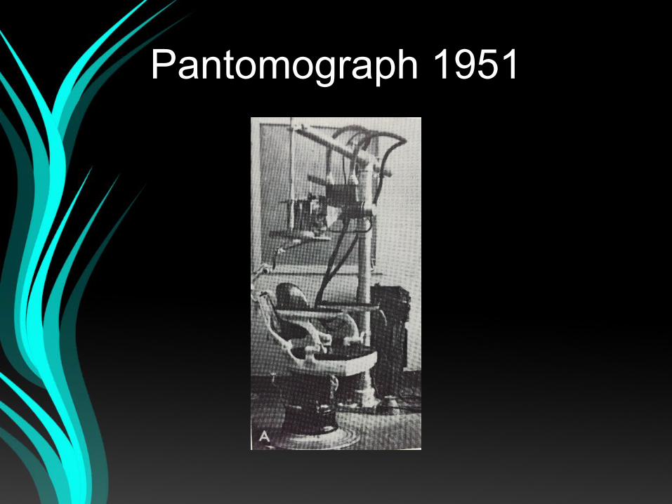
Pantomograph 1951
Pantomography
Rotational radiography
History
– Dr. H. Numata (1933)– Dr. Y.V. Paatero (1946)
OPG
• Ortho - straight
• Panoramic - An obstructed or a complete view of the object in every direction
• Tomography – An exray technique for making radiographs of layers of tissue in depth, without the interference of tissue above and below that level
Single image of facial structures that
includes maxillary
and mandibular arches
and their supporting
structures.
Advantages…
• Broad anatomic coverage
• Low patient radiation dose
• Convenience of examination
• Used in patients unable to open mouth
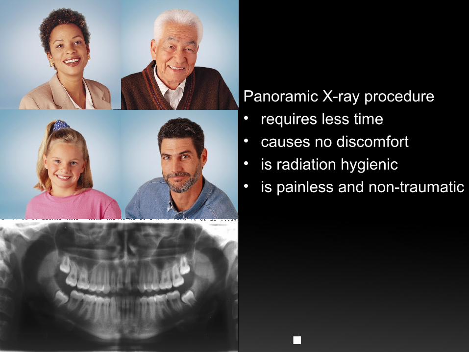
Panoramic X-ray procedure
• requires less time
• causes no discomfort• is radiation hygienic• is painless and non-traumatic
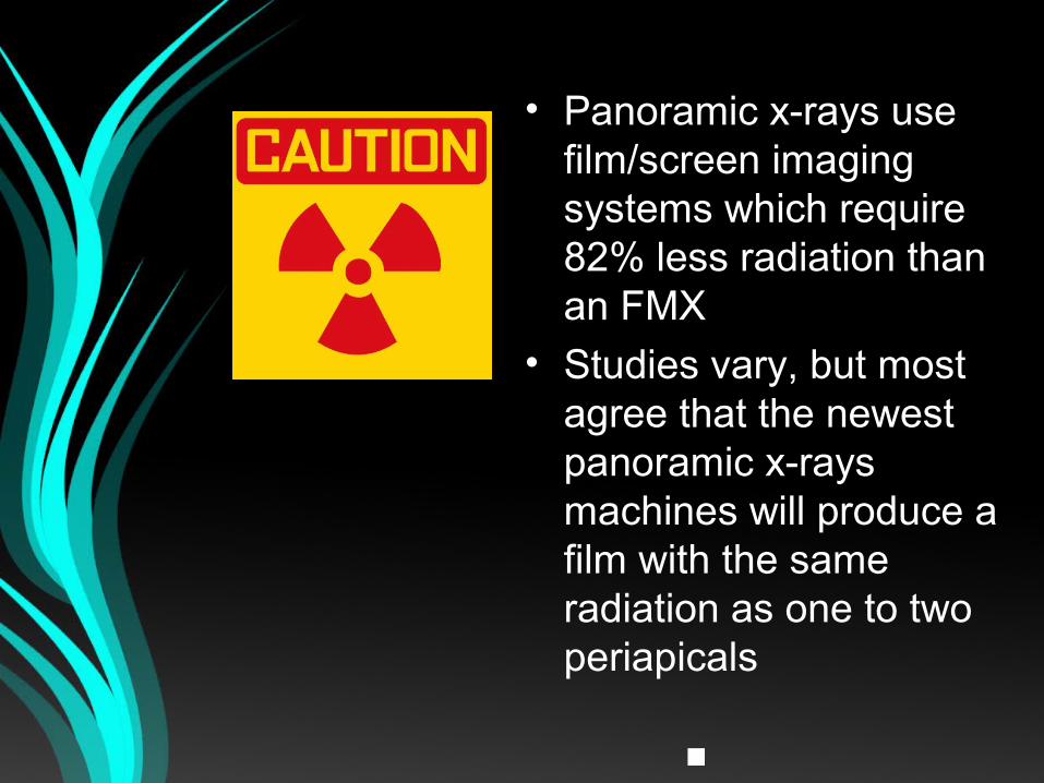
• Panoramic x-rays use film/screen imaging systems which require 82% less radiation than an FMX
• Studies vary, but most agree that the newest panoramic x-rays machines will produce a film with the same radiation as one to two periapicals
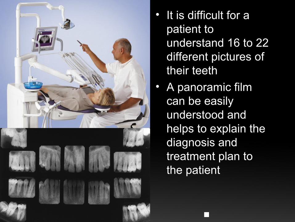
• It is difficult for a patient to understand 16 to 22 different pictures of their teeth
• A panoramic film can be easily understood and helps to explain the diagnosis and treatment plan to the patient
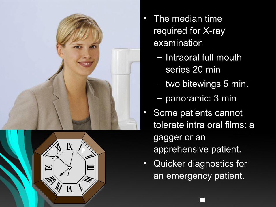
• The median time required for X-ray examination
– Intraoral full mouth series 20 min
– two bitewings 5 min.
– panoramic: 3 min
• Some patients cannot tolerate intra oral films: a gagger or an apprehensive patient.
• Quicker diagnostics for an emergency patient.
Disadvantages
• Does not show fine anatomic details
• Magnification
• Distortion
• Overlapped image of teeth
• Expensive
Indications
• Evaluation of trauma
• Third molars
• Large lesions
• Tooth development
• Developmental anomalies
• Intolerant to intraoral procedures
OPG IS A
SCREENING RADIOGRAPH
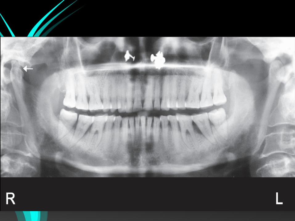
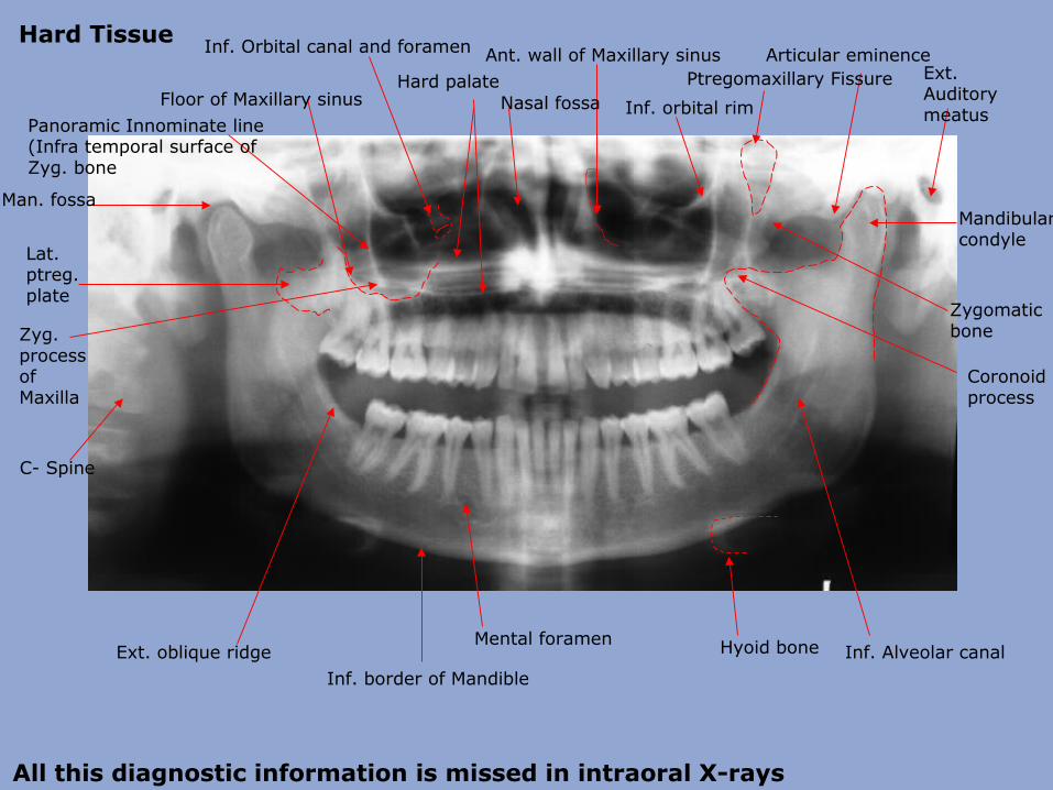
Ext. Auditory meatus
Mandibular condyle
Articular eminence
Coronoid process
Zygomatic bone
Ptregomaxillary Fissure
Inf. orbital rimFloor of Maxillary sinus
Ant. wall of Maxillary sinusHard palate
Nasal fossa
Inf. Orbital canal and foramen
Zyg. process of Maxilla
Panoramic Innominate line (Infra temporal surface of Zyg. bone
Lat. ptreg. plate
Man. fossa
Inf. border of Mandible
C- Spine
Mental foramen Hyoid bone Inf. Alveolar canalExt. oblique ridge
All this diagnostic information is missed in intraoral X-rays
Hard Tissue
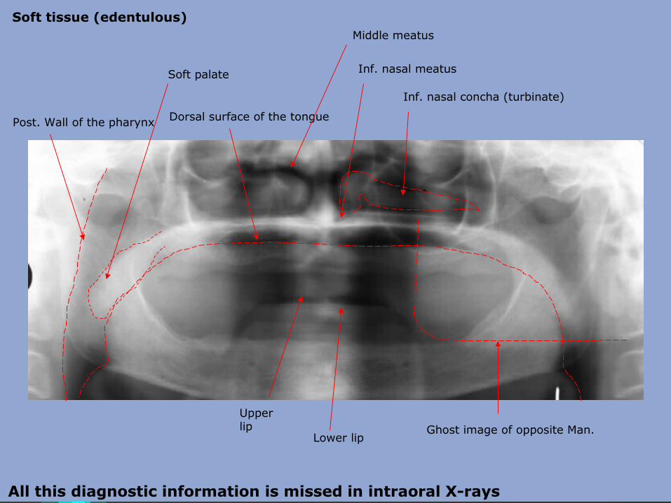
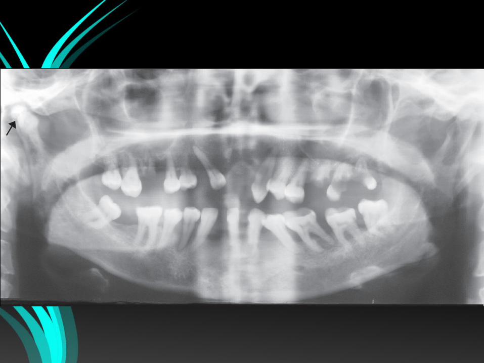
Inf. nasal concha (turbinate)
Inf. nasal meatus
Dorsal surface of the tonguePost. Wall of the pharynx
Soft palate
Lower lip
Upper lip
Middle meatus
Ghost image of opposite Man.
Soft tissue (edentulous)
All this diagnostic information is missed in intraoral X-rays
Factors for interpretation
• Principles of image formation
• Techniques of patient positioning
• Radiographic appearance of normal anatomic structures.
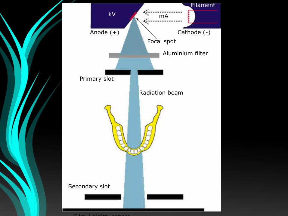
Primary slot
Aluminium filter
Secondary slot
Film / digital sensor
Filament
Focal spot
mAkV
Anode (+) Cathode (-)
Radiation beam
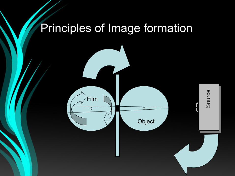
Principles of Image formation
Film
Object
Sou
rce
Principle
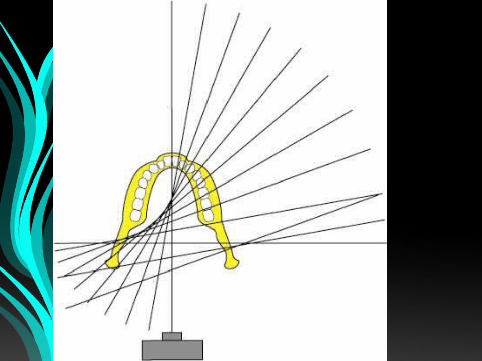
• X ray source is rotated, so the central ray constantly passes through the centre of rotation of disc 1.
• Both disc 2 and the film collimator rotate about the centre of disc 1.
• While disc 2 moves, film on this disc rotates past the slit.
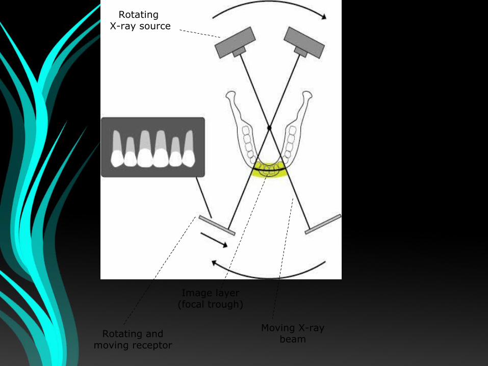
Rotating X-ray source
Rotating andmoving receptor
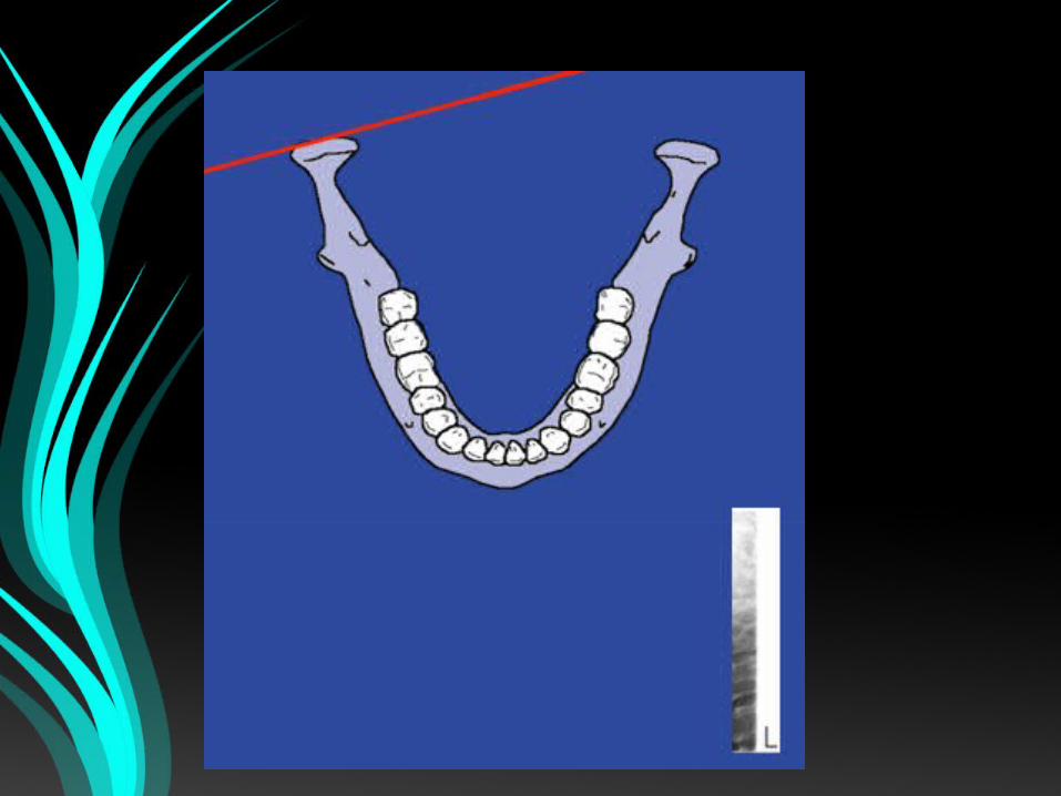
Image layer(focal trough)
Moving X-raybeam
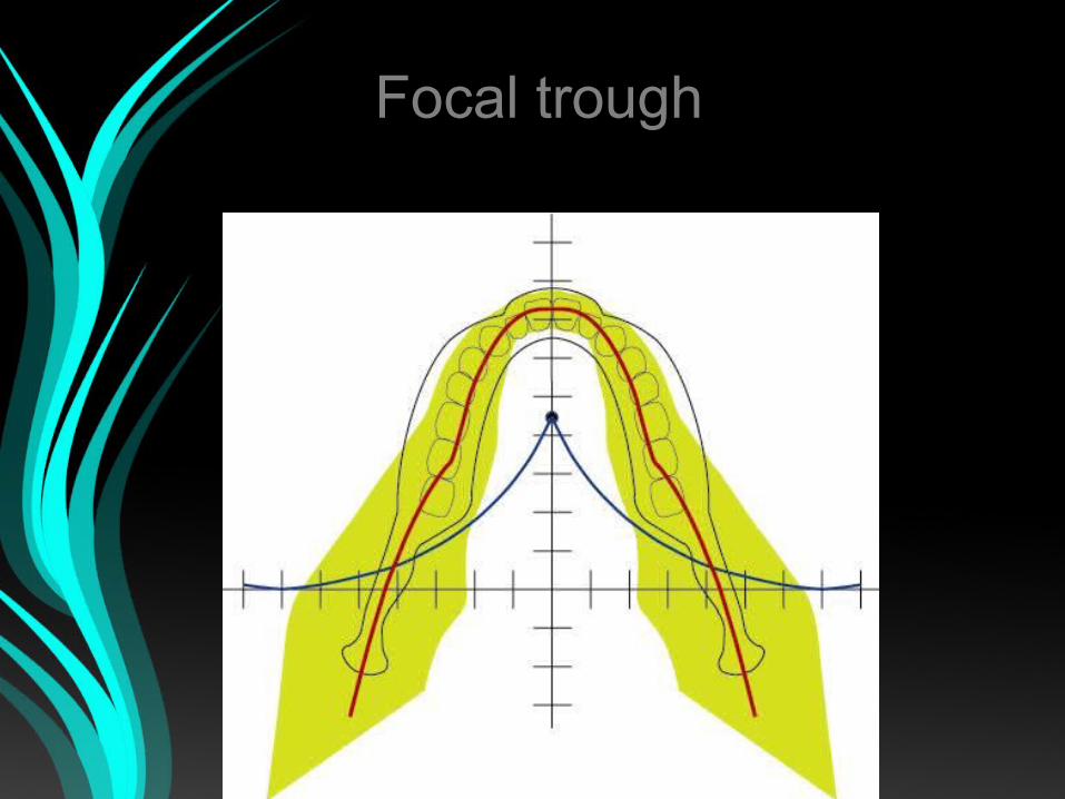
Focal trough
• Focal trough is a three dimensional
image layer in which structures are
reasonably well defined on panoramic
radiograph.
Focal troughFocal trough
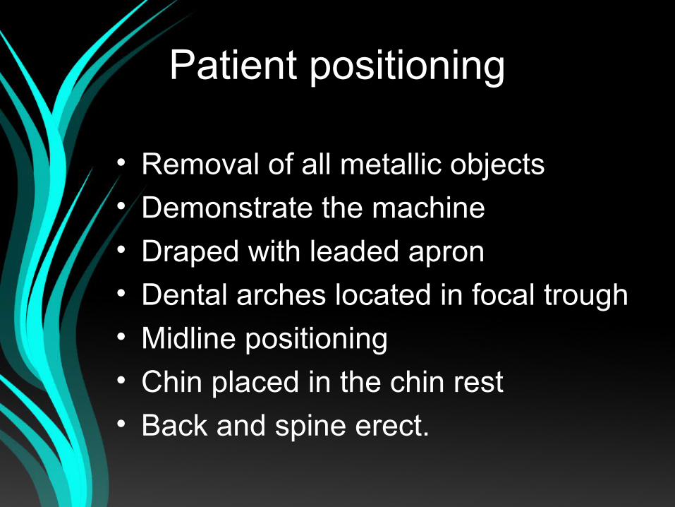
Patient positioning
• Removal of all metallic objects
• Demonstrate the machine
• Draped with leaded apron
• Dental arches located in focal trough
• Midline positioning
• Chin placed in the chin rest
• Back and spine erect.