ORT Aluminum Stress Affects Growth and ...
7
HORTSCIENCE 52(11):1601–1607. 2017. doi: 10.21273/HORTSCI12268-17 Aluminum Stress Affects Growth and Physiological Characteristics in Oil Tea Liyuan Huang, Jun Yuan, Hui Wang, and Xiaofeng Tan 1 Key Laboratory of Non-Wood Forest Product of State Forestry Administration, The Key Laboratory of Cultivation and Protection for Non-wood Forest Trees, Ministry of Education, Central South University of Forestry and Technology, Changsha 410004, China Genhua Niu 1 Texas A&M AgriLife Research Center at El Paso, Texas A&M University System, 1380 A&M Circle, El Paso, TX 79927 Additional index words. Camellia oleifera, root morphology, photosynthetic characteristics, antioxidative enzymes, cell ultrastructure Abstract. High concentration of aluminum ion (Al 3+ ) in acidic soil often negatively affects plant growth. To deepen understanding of the mechanisms of physiological response to Aluminum (Al) toxicity, changes in physiology and cell ultrastructure of oil tea (Camellia oleifera) were investigated under different Al levels. Oil tea plants were grown in pots filled with sand and treated with Al at 0, 0.5, 1.25, 2.0, or 4.0 mM. Results showed that Al at 0.5–2.0 mM improved plant growth, whereas Al at 4.0 mM inhibited root growth and damaged cell ultrastructure. Net photosynthetic rate (P n ), stomatal conductance (g s ), transpiration rate (T r ), and photochemical efficiency increased as Al concentration increased from 0 to 2.0 mM; however, all parameters mentioned previously decreased at 4.0 mM. The activities of superoxide dismutase (SOD), peroxidase (POD), and catalase (CAT) in leaves treated with 2.0 mM Al reached the maximum, which were 29%, 63%, and 28% higher than that of control. When Al was £2.0 mM, the content of soluble sugar and soluble protein increased with increasing Al concentration. These results may indicate that oil tea adapted to Al stress through osmotic adjustment and through increasing antioxidant enzyme system. In summary, Al at low concentration (0.5–2.0 mM) improved growth and physiological performance, whereas 4.0 mM negatively impacted performance of oil tea. Al is one of the most abundant metal elements in the earth’s crust (Pilon-Smits et al., 2009). Its main forms presenting in soil are Al oxides and aluminosilicates. These forms of Al are usually unavailable for any toxic reactions in plants (Mukhopadyay et al., 2012). However, when soil pH decreases (pH < 5.0), Al solubilizes into mobile ionic forms (such as Al 3+ ) (Kochian et al., 2004). Al 3+ is considered to be a major limiting factor to plant growth in acid soils (Horst et al., 2010). In China, acid soil covering an area of 0.203 million km 2 , consisting of 21% of the total arable land area (Qian et al., 2014). In recent years, the acid soil area was increased by increasing commercial ammonia/ammonium fertilizers application (Zhang and Raun, 2006). Therefore, Al tox- icity concern is widespread and tremendous attention has been paid to exploring and understanding its resistance mechanism. The initial and most dramatic symptom of Al toxicity is the inhibition of root growth and alteration of root morphology (Sivaguru and Horst, 1998). Within 40 h of hydropon- ics, the inhibition of root growth in Zea mays induced by Al (50 mM) could be measured (Ryan et al., 1993). Previous studies found that Al (400 mM) could inhibit the root growth by decreasing the root volume, number of lateral roots, and activity in Triticum aesti- vum and Camellia sinensis (Ghanati et al., 2005; Zakir Hossain et al., 2006). Because of the inhibition of root growth in T. aestivum, water and nutrient uptake were inhibited (Vitorello et al., 2005). Another primary effect of Al toxicity is the excessive accu- mulation of reactive oxygen species (ROS), such as the hydroxyl radical (OH – ), hydrogen peroxide (H 2 O 2 ), and the superoxide anion radical (O 2 – ) (Bartels and Sunkar, 2005), which may break the balance between gen- eration and scavenging of the ROS. To de- toxify Al-induced ROS, plants employ enzymatic antioxidants, such as POD, SOD, and CAT (Boscolo et al., 2003; Inostroza- Blancheteau et al., 2012). Al stress (0.4 mM) can destroy the ultrastructure of root cell in Dimocarpus longan and T. aestivum plants (Li et al., 2006). Physical changes, including increased vacuole size, destroyed nuclei and translocation of the amyloplasts in close proximity were observed in soybean that were grown under microgravity conditions (Klymchuk et al., 2003). By contrast, however, some plants can grow well on acid soils and take up large amounts of Al from the soil in their shoots without showing any symptom of Al toxicity. For example, buckwheat leaves can accumu- late as much as 10 g · kg –1 Al in the leaves in 12 weeks (Shen et al., 2006). Al (0.5 mM)- induced improvements of the root activity contributed to a growth enhancement in Melastoma (Watanabe et al., 2005). P n ,T r , g s , and growth of C. sinensis were enhanced by Al at 15 mM (Mukhopadyay et al., 2012). A similar beneficial effect of Al on plant growth was reported in Miconia albicans (Haridasan, 1988) and Pinus radiate (Huang and Bachelard, 1993). Oil tea is a unique woody edible oil tree in China and is one of the four major woody edible oil crops in the world (Gao et al., 2015). The extracted camellia oil has several favorable characteristics, including high oleic acid content typically exceeding 80% with low saturated fats content, which are very healthy (Yang et al., 2016). Oil tea is mainly distributed in acid soil in southern China. It was found that mature leaves of oil tea accumulate more than 13.5 g · kg –1 Al, which is considered a typical Al hyper- accumulator (Zeng et al., 2011). Unlike tea and other plants, limited information is avail- able for oil tea on its responses to abiotic stress, such as Al toxicity. Therefore, the objective of this study was to understand the physiological mechanisms relating to oil tea in response to Al stress. Specifically, the effect of Al stress on the growth, root activity, leaf photosynthesis, chlorophyll fluorescence pa- rameters, osmoregulation substance, antioxi- dative enzymes, and cell ultrastructure of oil tea were investigated in a pot experiment. Materials and Methods Plant materials and treatments. This ex- periment was conducted in Central South University of Forestry and Technology (CSUFT) in Hunan (Changsha, China; lat. 28°8#14$N, long. 112°59#32$E). The 1- year-old oil tea seedlings were cultured by grafting a shoot of C. oleifera ‘Huajin’ onto germinated hypocotyl of seeds collected from a widely cultivar (C. oleifera ‘XLC15’) as a rootstock. The healthy and uniform seed- lings were selected and transplanted to plastic pots (10 cm diameter · 20 cm height) con- taining 2 kg sand (average particle size: 0.5– 1.0 mm), two plants per pot. The grafted plants were acclimatized for 6 months (from 5 Jan. to 30 June 2015) outdoor before the Al treatment. Plants were watered every day, while the nutrient solution was given every 3–4 d. The nutrient solution was modified Hoagland solution, which composed of macronutrients: Ca(NO 3 ) 2 , KNO 3 , NH 4 NO 3 , KH 2 PO 4 , and MgSO 4 at 6, 5, 1, 1, and 4.0 mM, respectively, and micronutrients: KI, HBO 3 , MnSO 4 , ZnSO 4 , Na 2 MoO 4· 2H 2 O, CuSO 4 , CoCl 2 , and Fe-EDTA at 5, 100, 147, 53, 103, 0.15, 0.19, and 50 mM, respectively. After an acclimatization period of 6 months, Al was added to the nutrient solution Received for publication 6 July 2017. Accepted for publication 26 Sept. 2017. This study was supported by National Science Foundation of China (No.31400582) and Forestry Industry Research Special Funds for Public Wel- fare Projects (No.201404702-1). 1 Corresponding authors. E-mail: tanxiaofengcm@ 126.com or [email protected]. HORTSCIENCE VOL. 52(11) NOVEMBER 2017 1601
Transcript of ORT Aluminum Stress Affects Growth and ...
HORTSCIENCE 52(11):1601–1607. 2017. doi: 10.21273/HORTSCI12268-17
Aluminum Stress Affects Growth andPhysiological Characteristics in Oil TeaLiyuan Huang, Jun Yuan, Hui Wang, and Xiaofeng Tan1
Key Laboratory of Non-Wood Forest Product of State ForestryAdministration, The Key Laboratory of Cultivation and Protection forNon-wood Forest Trees, Ministry of Education, Central South Universityof Forestry and Technology, Changsha 410004, China
Genhua Niu1
Texas A&M AgriLife Research Center at El Paso, Texas A&M UniversitySystem, 1380 A&M Circle, El Paso, TX 79927
Additional index words. Camellia oleifera, root morphology, photosynthetic characteristics,antioxidative enzymes, cell ultrastructure
Abstract. High concentration of aluminum ion (Al3+) in acidic soil often negatively affectsplant growth. To deepen understanding of the mechanisms of physiological response toAluminum (Al) toxicity, changes in physiology and cell ultrastructure of oil tea (Camelliaoleifera) were investigated under different Al levels. Oil tea plants were grown in potsfilled with sand and treated with Al at 0, 0.5, 1.25, 2.0, or 4.0mM. Results showed that Al at0.5–2.0 mM improved plant growth, whereas Al at 4.0 mM inhibited root growth anddamaged cell ultrastructure. Net photosynthetic rate (Pn), stomatal conductance (gs),transpiration rate (Tr), and photochemical efficiency increased as Al concentrationincreased from 0 to 2.0 mM; however, all parameters mentioned previously decreased at4.0 mM. The activities of superoxide dismutase (SOD), peroxidase (POD), and catalase(CAT) in leaves treated with 2.0 mM Al reached the maximum, which were 29%, 63%,and 28% higher than that of control. When Al was £2.0 mM, the content of soluble sugarand soluble protein increased with increasing Al concentration. These results mayindicate that oil tea adapted to Al stress through osmotic adjustment and throughincreasing antioxidant enzyme system. In summary, Al at low concentration (0.5–2.0mM)improved growth and physiological performance, whereas 4.0 mM negatively impactedperformance of oil tea.
Al is one of the most abundant metalelements in the earth’s crust (Pilon-Smitset al., 2009). Its main forms presenting insoil are Al oxides and aluminosilicates. Theseforms of Al are usually unavailable for anytoxic reactions in plants (Mukhopadyay et al.,2012). However, when soil pH decreases(pH < 5.0), Al solubilizes into mobile ionicforms (such as Al3+) (Kochian et al., 2004).Al3+ is considered to be a major limitingfactor to plant growth in acid soils (Horstet al., 2010). In China, acid soil covering anarea of 0.203 million km2, consisting of�21% of the total arable land area (Qianet al., 2014). In recent years, the acid soil areawas increased by increasing commercialammonia/ammonium fertilizers application(Zhang and Raun, 2006). Therefore, Al tox-icity concern is widespread and tremendousattention has been paid to exploring andunderstanding its resistance mechanism.
The initial and most dramatic symptom ofAl toxicity is the inhibition of root growth
and alteration of root morphology (Sivaguruand Horst, 1998). Within 40 h of hydropon-ics, the inhibition of root growth in Zea maysinduced by Al (50 mM) could be measured(Ryan et al., 1993). Previous studies foundthat Al (400 mM) could inhibit the root growthby decreasing the root volume, number oflateral roots, and activity in Triticum aesti-vum and Camellia sinensis (Ghanati et al.,2005; Zakir Hossain et al., 2006). Because ofthe inhibition of root growth in T. aestivum,water and nutrient uptake were inhibited(Vitorello et al., 2005). Another primaryeffect of Al toxicity is the excessive accu-mulation of reactive oxygen species (ROS),such as the hydroxyl radical (OH–), hydrogenperoxide (H2O2), and the superoxide anionradical (O2
–) (Bartels and Sunkar, 2005),which may break the balance between gen-eration and scavenging of the ROS. To de-toxify Al-induced ROS, plants employenzymatic antioxidants, such as POD, SOD,and CAT (Boscolo et al., 2003; Inostroza-Blancheteau et al., 2012). Al stress (0.4 mM)can destroy the ultrastructure of root cell inDimocarpus longan and T. aestivum plants(Li et al., 2006). Physical changes, includingincreased vacuole size, destroyed nuclei andtranslocation of the amyloplasts in closeproximity were observed in soybean thatwere grown under microgravity conditions(Klymchuk et al., 2003).
By contrast, however, some plants cangrow well on acid soils and take up largeamounts of Al from the soil in their shootswithout showing any symptom of Al toxicity.For example, buckwheat leaves can accumu-late as much as 10 g·kg–1 Al in the leaves in12 weeks (Shen et al., 2006). Al (0.5 mM)-induced improvements of the root activitycontributed to a growth enhancement inMelastoma (Watanabe et al., 2005). Pn, Tr,gs, and growth of C. sinensis were enhancedby Al at 15 mM (Mukhopadyay et al., 2012).A similar beneficial effect of Al on plantgrowth was reported in Miconia albicans(Haridasan, 1988) and Pinus radiate (Huangand Bachelard, 1993).
Oil tea is a unique woody edible oil tree inChina and is one of the four major woodyedible oil crops in the world (Gao et al.,2015). The extracted camellia oil has severalfavorable characteristics, including higholeic acid content typically exceeding 80%with low saturated fats content, which arevery healthy (Yang et al., 2016). Oil tea ismainly distributed in acid soil in southernChina. It was found that mature leaves of oiltea accumulate more than 13.5 g·kg–1 Al,which is considered a typical Al hyper-accumulator (Zeng et al., 2011). Unlike teaand other plants, limited information is avail-able for oil tea on its responses to abioticstress, such as Al toxicity. Therefore, theobjective of this study was to understand thephysiologicalmechanisms relating to oil tea inresponse toAl stress. Specifically, the effect ofAl stress on the growth, root activity, leafphotosynthesis, chlorophyll fluorescence pa-rameters, osmoregulation substance, antioxi-dative enzymes, and cell ultrastructure of oiltea were investigated in a pot experiment.
Materials and Methods
Plant materials and treatments. This ex-periment was conducted in Central SouthUniversity of Forestry and Technology(CSUFT) in Hunan (Changsha, China; lat.28�8#14$N, long. 112�59#32$E). The 1-year-old oil tea seedlings were cultured bygrafting a shoot of C. oleifera ‘Huajin’ ontogerminated hypocotyl of seeds collected froma widely cultivar (C. oleifera ‘XLC15’) asa rootstock. The healthy and uniform seed-lings were selected and transplanted to plasticpots (10 cm diameter · 20 cm height) con-taining 2 kg sand (average particle size: 0.5–1.0 mm), two plants per pot. The graftedplants were acclimatized for 6 months (from5 Jan. to 30 June 2015) outdoor before the Altreatment. Plants were watered every day,while the nutrient solution was given every3–4 d. The nutrient solution was modifiedHoagland solution, which composed ofmacronutrients: Ca(NO3)2, KNO3, NH4NO3,KH2PO4, andMgSO4 at 6, 5, 1, 1, and 4.0 mM,respectively, and micronutrients: KI, HBO3,MnSO4, ZnSO4, Na2MoO4·2H2O, CuSO4,CoCl2, and Fe-EDTA at 5, 100, 147, 53,103, 0.15, 0.19, and 50 mM, respectively.
After an acclimatization period of 6months, Al was added to the nutrient solution
Received for publication 6 July 2017. Accepted forpublication 26 Sept. 2017.This study was supported by National ScienceFoundation of China (No.31400582) and ForestryIndustry Research Special Funds for Public Wel-fare Projects (No.201404702-1).1Corresponding authors. E-mail: [email protected] or [email protected].
HORTSCIENCE VOL. 52(11) NOVEMBER 2017 1601
and was supplied in the form of AlCl3 inconcentrations of 0 (control), 0.5, 1.25, 2, and4.0 mM for 90 d (from 2 July to 30 Sept.2015). The nutrient solution was adjusted topH 4.2 · 1 MHCl or NaOH. The seedlings wereirrigated with 300 of mL nutrient solution
every other day. The 100-mL DI water wasprovided on the day without nutrient solution,if the temperature and solar radiation werehigh to ensure sufficient watering. All potswere placed outdoor on CSUFT campus ina completely randomized design. There were
20 pots (two plants per pot) per treatment.During the experimental period, the averagetemperature was 20 to 23 �C at night, 24 to28 �C during the day, and relative humiditywas 60% to 80%.
Measurement of gas exchange andchlorophyll fluorescence parameters. ThePn, gs, intercellular CO2 concentration (Ci),and Tr were measured using the third fullyexpanded mature leaves (one leaf per plant;five plants per replicate). The measurementswere taken at 9:00–10:00 on a clear day in thelast week of the experiment, using a Li-6400portable photosynthesis analyzer (Lincoln,NE). The environmental conditions of theleaf chamber were controlled at PPF densityat 1600 mmol·m–2·s–1, temperature at 27 �C,and CO2 concentration at 400 mmol·mol–1.
Chlorophyll Fluorescence parameterswere measured using LI-6400–40LCF (LI-COR, Lincoln, NE). Leaves were dark-adapted for 30 min before the fluorescencemeasurements. The minimal Chl fluores-cence (F0) level was measured after applyinga far-red pulse for 6 s, and the maximalfluorescence (Fm) was registered after ap-plying a 0.8 s saturating flash. The photo-chemical efficiency (Fv/Fm) was expressedas: Fv/Fm = (Fm – F0)/Fm (Genty et al.,1989).
Measurement of plant growth. At the endof the experiment, five pots per treatmentwere harvested to determine plant growth.The remaining plant materials were usedfor physiological and biochemical analysis,with three replicates (three pots). The plantwas washed with distilled water, then the
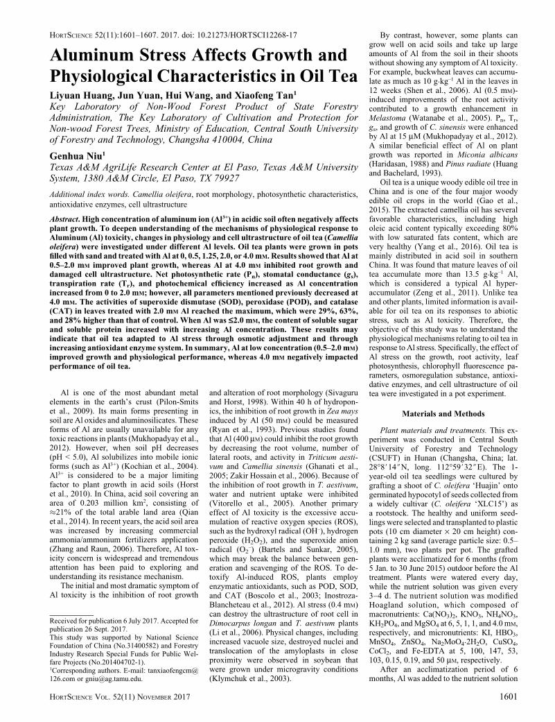
Fig. 1. Seedling height (A), stem diameter (B), dry weight (C), and root to shoot ratio (D) of oil tea(Camellia oleifera) as affected by aluminum. Vertical bars indicate standard errors. Different letters onthe top of bars indicate significant difference at P < 0.05 by least significant difference test.
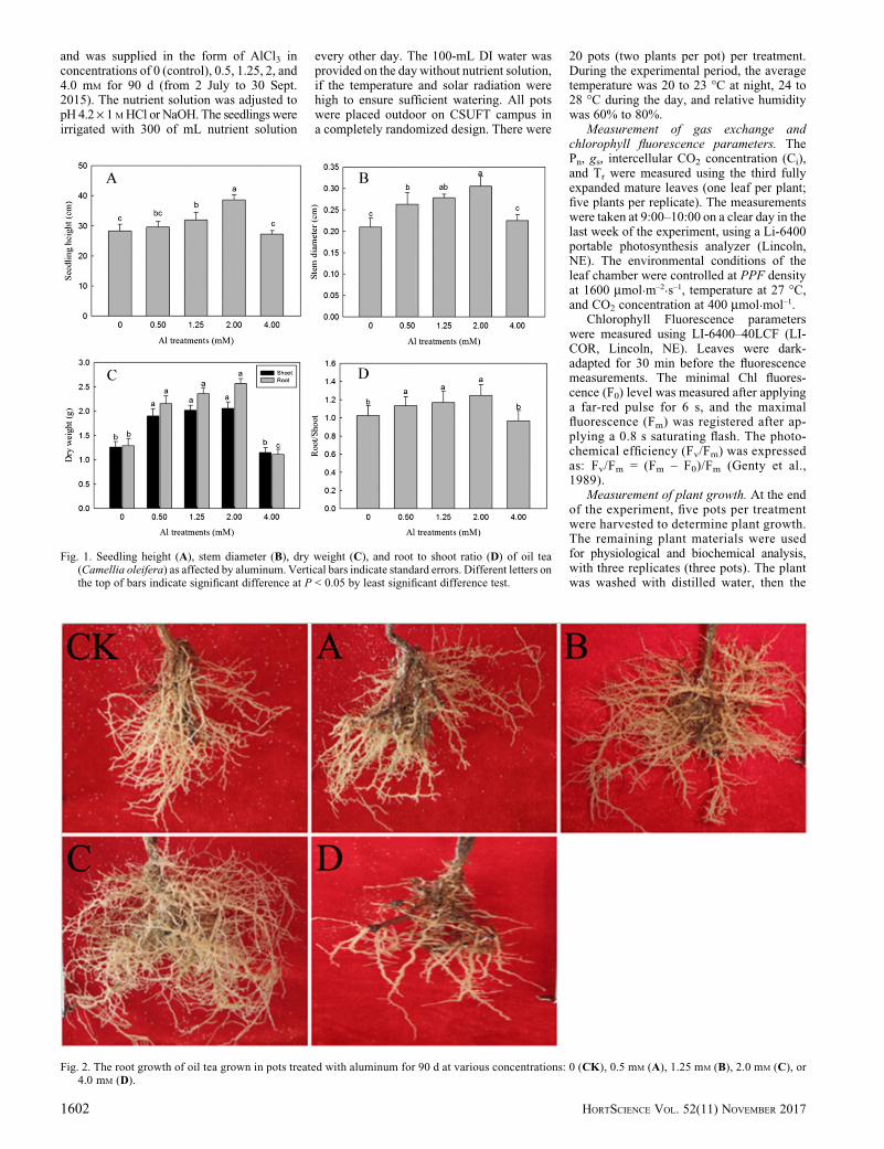
Fig. 2. The root growth of oil tea grown in pots treated with aluminum for 90 d at various concentrations: 0 (CK), 0.5 mM (A), 1.25 mM (B), 2.0 mM (C), or4.0 mM (D).
1602 HORTSCIENCE VOL. 52(11) NOVEMBER 2017

stem diameter and plant height were mea-sured. Plant roots and shoots were separatedand dried in an oven at 65 �C for 48 h, and dryweights were recorded.
Assay of root activity. The root activitywas assayed according to Yu et al. (2011).The root tips (4–6 mm) were homogenized
with 5 mL of 50 mg·L–1 a-naphtylamine and5 mL of phosphate buffer solution (PBS)(pH = 7.0). The homogenized mixture wasshaken gently and placed in the thermo-tank for24 h. Four solutions, 2 mL of a-naphtylaminesolution, 10 mL of distilled water, 1 mL of1% p-aminobenzene sulfonic acid, and1 mL of sodium nitrite, were added to themixture with the volume of the mixed solu-tion at 25 mL and then kept at room temper-ature. The reaction solution absorbance wasassayed at 510 nm using spectrophotometer(ultraviolet-1100; Mapada, Shanghai, China,same spectrophotometer was used for thefollowing assay).
Assay of soluble sugar content. The solu-ble sugar content in the leaves was assayedaccording to Irigoyen et al. (1992) with somemodifications, using glucose as a standard.Dry leaves were ground to powder and thenhomogenized with 5 mL of 95% ethanol.Alcoholic leaf extract preserved in refrigera-tor was mixed with 5 mL anthrone (200 mganthrone, 100 mL sulphuric acid). Then, the
samples were placed in a boiling water bath
for 10 min. The reaction solution absorbance
was assayed at 595 nm.
Assay of soluble protein content. Thesoluble protein content in the leaf wasassayed according to Bradford (1976) usingbovine serum albumin as a standard. Thefresh leaves from each Al treatment werewashed with distilled water, dried with papertowel, and were ground in a mortar with 5 mL0.5 M PBS. The homogenate was centrifugedat 5000 gn for 10 min. Then, the supernatantabsorbance was assayed at 625 nm.
Assay of malondialdehyde (MDA)content. The MDA content in the leaf wasassayed according to Jia et al. (2015). Thefresh leaves were washed in distilled water,dried with paper towel, and were ground in anice mortar with 5 mL of 0.5 M PBS by grinding.The homogenate with 5 mL of 0.5% (w/v) 2-thiobarbituric acid was prepared in 5% trichloro-acetic acid. The homogenate was placed ina boiling water bath for 10 min, then quicklycooled in an ice bath and centrifuged at 3000 gnfor 15 min. The supernatant liquor absorbancewas assayed at 532, 600, and 450 nm. TheMDAcontent was calculated using the formula of[6.452 · (OD532 – OD600) – (0.56 · OD450)] ·plant tissue extraction compound (L)/the sampleweight (g).
Assay of antioxidant enzyme activities.Leaves of oil tea were homogenized usinga mortar and pestle in a medium containing50 mM PBS (pH = 7.0), 0.1 mmol·L–1 EDTA-Na2, and 1% (w/v) Polyvinyl Pyrolidone. Thehomogenate was centrifuged at 15,000 gn for15 min at 4 �C.
Superoxide dismutase (SOD, EC1.15.1.1)activity was assayed according to Paridaet al. (2004). The inhibition of the photo-chemical reduction of nitro blue tetrazolium(NBT) was spectrophotometrically measuredat 560 nm. One unit of SOD activity wasdefined as the amount of enzyme required tocause a 50% inhibition of the reduction ofNBT. SOD activity was assayed at 560 nm.
Peroxidase (POD, EC1.11.1.7) activitywas assayed according to Farooq et al.(2013) with somemodifications. The reactionmixture comprised 1 mL enzyme extract,1 mL 1% (v/v) guaiacol, 6.9 mL PBS, 1 mL0.18% (v/v) H2O2, then reaction at 25 �C for15 min, inhibited the reaction by 5% (v/v)metaphosphoric acid. POD activity wasassayed at 470 nm.
Catalase (CAT, EC1.11.1.6) activitywas assayed according to Parida et al.(2004). The reaction mixture comprised0.1 mL enzyme extract, 1 mL PBS, 1.7 mLdistilled water, and 0.2 mL of 0.2 M H2O2. TheCAT activity assay was based on the spectro-photometric measurement of H2O2 absor-bance decrease at 240 nm.
Transmission electron microscope. After90 d of Al treatment, young leaf pieces withoutveins (from 1 to 2 cm2) and root tips (from 0.2 to0.5 cm) were collected from randomly selectedplants and then fixed overnight in 2.5% glutar-aldehyde (v/v) in 0.1 M PBS (pH = 7.4) andcleaned three times with the same PBS (35 mineach time). The samples were postfixed in 1%OsO4 for 12 h, and then dehydrated in a gradedseries of acetone (30%, 50%, 70%, 80%,90%, 95%, and 100%). After dehydration, the
Fig. 3. Effects of aluminum stress on root activityof oil tea seedlings. Vertical bars indicatestandard errors. Different letters on the top ofbars indicate significant difference at P < 0.05by least significant difference test.
Fig. 4. Effects of aluminum (Al) stress on net photosynthetic rate (Pn, A), stomatal conductance (gs, B),intracellular CO2 concentration (Ci, C), transpiration rate (Tr, D) of oil tea seedlings grown in potstreated with Al at various concentrations. Vertical bars indicate standard error. Different letters on thetop of bars indicate significant difference at P < 0.05 by least significant difference test.
Table 1. Effect of aluminum (Al) stress on photochemical efficiency (Fv/Fm), relative quantum efficiencyof PSII photochemistry (FPSII), and nonphotochemical quenching (NPQ) of oil tea grown in potstreated with Al at different concentrations.
Chlorophyll fluorescence parameters
Al concn (mM)
0 0.50 1.25 2.00 4.00
Fv/Fm 0.806 bz 0.810 a 0.811 a 0.813 a 0.801 cFPSII 0.155 ab 0.160 a 0.161 a 0.145 b 0.126 cNPQ 2.948 b 2.845 bc 2.757 c 3.229 a 2.369 dzMeans within the same row followed by different letters are significantly different at P < 0.05 by leastsignificant difference test.
HORTSCIENCE VOL. 52(11) NOVEMBER 2017 1603

samples were embedded in Epon812 resinovernight and then heated at 70 �C for 9 h.The ultrathin sections (80 nm) of samples wereprepared and mounted on copper grids to beviewed under electron microscope (TEX-1230EX; JEOL, Tokyo, Japan) at 60.0 kV voltages.
Experimental design and statisticalanalysis.Theexperiment followeda completelyrandomized design with three (physiologicaland biochemical analyses) or five replicates(growth andmorphological parameters). Datawere analyzed by one-way analysis of varianceusing the statistical program SPSS 17.0 (SPSS,USA). Means were compared among the treat-ments using the least significant difference testat 5% probability level.
Results
Plant growth and development. Thegrowth of oil tea was not adversely affected
in the 0.5 to 2.0 mM Al treatments (Fig. 1).Plants were taller in the 2.0 mM, followed bythose in the 1.25 mM Al treatment. Stemdiameter was greater in the 2.0 mM than thosein 0, 0.5, and 4.0 mM Al treatments. No
differences were found in shoot and rootdry weight and root/shoot ratio among Alconcentrations of 0.5, 1.25, and 2.0 mM. Inthe 4.0 mM Al treatment, the root dry weightwas significantly lower than that of other
Table 2. Effect of aluminum (Al) stress on soluble sugar, soluble protein, malondialdehyde (MDA),superoxide dismutase (SOD), peroxidase (POD), and catalase (CAT) of oil tea grown in pots treatedwith Al at different concentrations.
Index
Al concn (mM)
0 0.50 1.25 2.00 4.00
Soluble sugar (mg·g–1) 76.0b cz 80.0 b 88.1 b 97.6 a 70.0 cSoluble protein (mg·g–1) 4.25 c 6.13 b 6.70 ab 7.19 a 4.50 cMDA (mmol·g–1) 13.20 b 12.00 c 13.11 b 13.38 b 15.35 aSOD activity (U·g–1 FW·min–1) 121.34 c 130.09 b 138.56 b 149.21 a 111.86 dCAT activity (mg H2O2·g
–1 FW·min–1) 78.10 b 80.23 b 93.24 a 99.75 a 70.17 cPOD activity (U·g–1 FW·min–1) 0.89 c 0.93 c 1.32 b 1.45 a 0.80 czMeans within the same row followed by different letters are significantly different at P < 0.05 by leastsignificant difference test.
Fig. 5. Electron micrographs of oil tea root tip cell under aluminum (Al) stress. (A–C) The oil tea seedling without Al treatment showing intact structuralconfiguration of root tip cells with a big nucleus. (D–F) The oil tea seedling with 2.0 mM Al treatment showing slight changes in the root tips in comparisonwith control. The cell wall became protrusive, the intercellular spaces increased, and the mitochondria bulged outward. (G–I) The oil tea seedling with 4.0 mM
Al treatment showing the vesicles around the root tip cell wall, the broken plasma membrane and the decreased cell inclusion. CW = cell wall; N = nucleus;AP = amoeboid folds; IS = intercellular space; M = mitochondria; V = vesicles; PM = plasmalemma.
1604 HORTSCIENCE VOL. 52(11) NOVEMBER 2017
treatments. The lateral roots were well de-veloped at 2.0 mM Al treatments (Fig. 2C).By contrast, growth of lateral roots wereinhibited at 4.0 mMAl treatments with shorterand fewer lateral roots (Fig. 2D). There wereno significant differences among CK, A, andB (Fig. 2).
Root activity. The root activity was thegreatest in the 2.0 mM Al treatment (82.3%increase compared with control), followed bythose in the 0.5 and 1.25 mM Al treatments(Fig. 3). No difference was found betweencontrol and Al at 0.5 mM concentration. Incontrast to the control, 20% decline wasnoticed in the root activity in response to4.0 mM Al.
Gas exchanges and chlorophyll fluorescenceparameters. The Pn, gs, and Tr were 23.4% to27.3%, 29.6% to 32.6%, and 16.3% to 18.5%,respectively, higher at Al concentrations of1.25 and 2.0 mM compared with the control(Fig. 4). No differences were found in Pn andTr between the control and Al at 0.5 mM
concentration and between Al at 1.25 and2.0 mM. Al at 4.0 mM significantly reduced Pnby 14.8%, gs by 17.1% and Tr by 13.9%compared with their respective controls. Nodifferences were found in Ci among Altreatments.
Chlorophyll fluorescence parameters ofoil tea were influenced by Al treatments(Table 1). Fv/Fm at Al concentrations of 0.5,1.25, and 2.0 mM was similar, which wasslightly higher than that of the control, and Al
at 4.0 mM resulted in the lowest Fv/Fm.However, all Fv/Fm values were above 0.8.No differences were found in relative quan-tum efficiency of PSII photochemistry(FPSII) among Al concentration of 0.5,1.25 mM, and the control. The nonphoto-chemical quenching (NPQ) was greatest inthe 2.0 mM, followed by those in the controland 0.5 mM Al treatments. Al at 4.0 mM
resulted in the lowest FPSII and NPQ.Osmoregulation substance and antioxidative
enzymes. The content of soluble sugar wasgreatest in the 2.0 mM Al treatment, 28.4%increase compared with control (Table 2). Nodifferences were found between the controland Al at 0.5, 1.25 mM, and between controland Al at 4.0 mM. The content of solubleprotein was greater in 2.0 mM than those incontrol, 0.5 and 4.0 mM Al treatments. TheMDA content increased by 16.3% over thecontrol in response to 4.0 mM Al treatment.There were no differences among 0, 1.25, and2.0 mM Al treatments, whereas MDA waslowest at Al of 0.5 mM.
Al significantly altered the activities ofantioxidant enzymes-SOD, POD, and CAT inoil tea leaves (Table 2). The activity of SODwas higher at Al of 2.0 mM and followed bythose at Al concentration of 0.5 and 1.25 mM,whereas Al at 4.0 mM resulted in the lowestSOD. Likewise, the activities of CAT werehigher at Al of 1.25 and 2.0 mM treatments,followed by control and Al at 0.5 mM,whereas Al at 4.0 mM had the lowest CAT.
The activities of PODwere greatest in 2.0 mM
Al treatment with a 62.9% increase comparedwith control, followed by those in the1.25 mM Al treatment. No differences werefound in POD among control, Al at 0.5 and4.0 mM concentrations.
Root tip and leaf ultrastructure. Trans-mission electron micrographs of the root tipcells are shown in Fig. 5. In the control(Fig. 5A–C), the configuration of the roottip cells was well structured and the ultra-structural characteristics were normal. Thenucleus was visible with well-developednuclear membrane and nucleolus. At Al of2.0 mM (Fig. 5D–F), there were slightchanges in the root tips in comparison withcontrol. Generally, the cell wall became pro-trusive, the intercellular spaces increased,and the mitochondria bulged outward. Themain alteration observed was the root tipappearance in 4.0 mM Al concentration(Fig. 5G–I) with disrupted cell wall. Therewere some vesicles around the cell wall andthe plasma membrane was broken. The cellinclusions were decreased. The phenomenonof the plasmolysis slightly appears in the roottip cell.
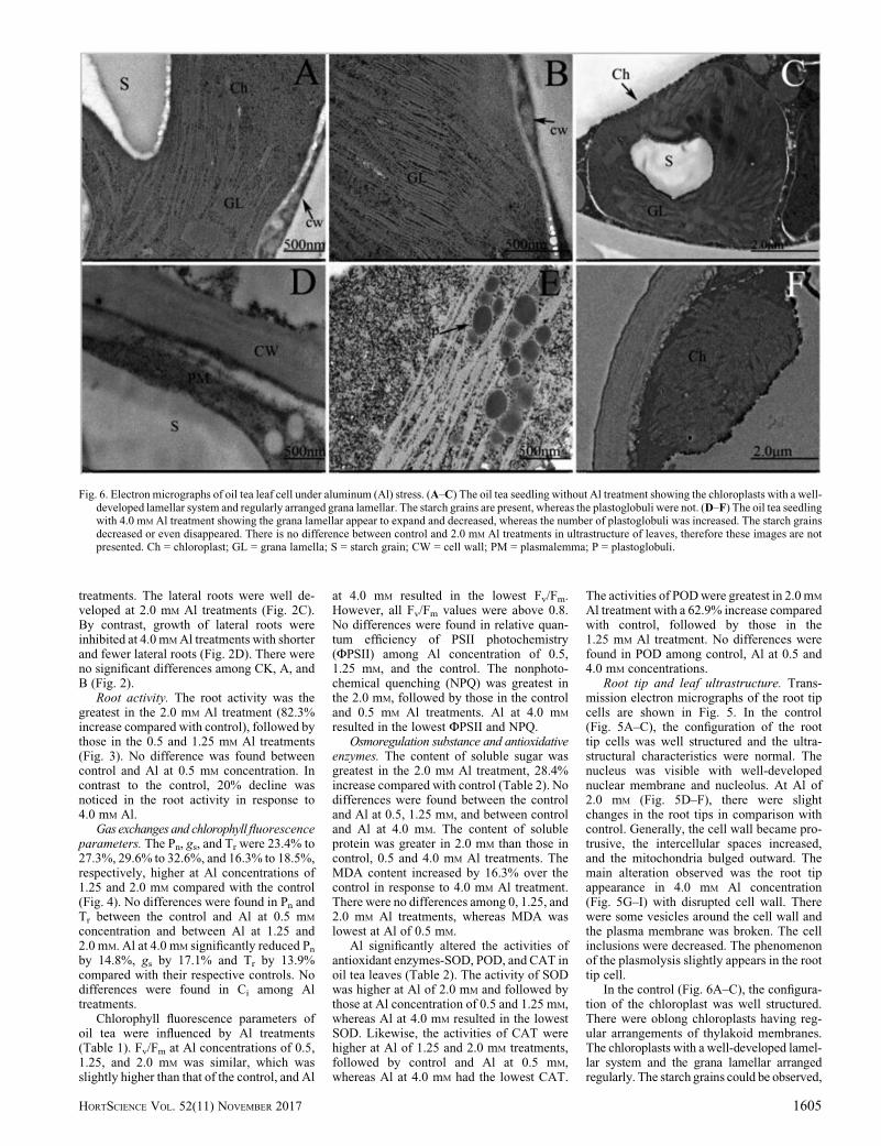
In the control (Fig. 6A–C), the configura-tion of the chloroplast was well structured.There were oblong chloroplasts having reg-ular arrangements of thylakoid membranes.The chloroplasts with a well-developed lamel-lar system and the grana lamellar arrangedregularly. The starch grains could be observed,
Fig. 6. Electron micrographs of oil tea leaf cell under aluminum (Al) stress. (A–C) The oil tea seedling without Al treatment showing the chloroplasts with a well-developed lamellar system and regularly arranged grana lamellar. The starch grains are present, whereas the plastoglobuli were not. (D–F) The oil tea seedlingwith 4.0 mM Al treatment showing the grana lamellar appear to expand and decreased, whereas the number of plastoglobuli was increased. The starch grainsdecreased or even disappeared. There is no difference between control and 2.0 mM Al treatments in ultrastructure of leaves, therefore these images are notpresented. Ch = chloroplast; GL = grana lamella; S = starch grain; CW = cell wall; PM = plasmalemma; P = plastoglobuli.
HORTSCIENCE VOL. 52(11) NOVEMBER 2017 1605

whereas the plastoglobuli were invisible. Altreatments adversely affected leaf mesophyllcellular structures as evident in the 4.0 mM Altreatments (Fig. 6D–F). The grana lamellar inchloroplast appeared to expand, and the struc-ture was loose. Compared with the control, thestructures of grana lamellar were decreased.The starch grains obviously decreased or evendisappeared, whereas the number of plasto-globuli was increased.
Discussion
Al toxicity in plants occurs in acidic soils(Matsumoto, 2000). Cocker et al. (1998)showed that Al toxicity affected the growthand physiological parameters of T. aestivum.However, in this study, addition of Al at 1.25and 2.0 mM to the nutrient solution did notinhibit growth of oil tea but instead stimu-lated growth. Kinraide (1993) reported thatalleviation of H+ production under acidicconditions by Al might be the reason behindincreases in growth.
Root activity may reflect plant toxicityresponse to heavy metal (Islam et al., 2007)because roots are directly in contact with thecontaminated environment, such as heavymetals. In this study, we found that Al at#2.0 mM could actually stimulate root activ-ity, thus maintaining high respiration meta-bolism and strengthening the uptake ofminerals and water. Similar results werereported in various plant species (Ghanatiet al., 2005, Li et al., 2004). However, at Alconcentration of 4.0mM, root activity decreased,indicating negative impact of Al on the activitiesof the root as well as the entire root system.
Photosynthesis is a highly regulated andintegrated process in plants. The photosyn-thetic process is highly sensitive to environ-ment stress (Sun et al., 2009). Previousstudies have shown that Al toxicity affectsphotosynthesis in Citrus grandis (Jiang et al.,2009). Al toxicity reduced stomatal openingand thus reduced gs, resulting in decreasedphotosynthesis of Artemisia annua (Aftabet al., 2010). In the present study, oil teareflected the similar phenomenon with regardto decreasing Pn, gs, and Tr under Al (4.0 mM)treatment. But these parameters were signif-icantly improved under Al of 1.25 and2.0 mM. These results confirmed that Al atappropriate concentrations stimulated growthand that toxicity occurred when Al washigher than threshold. Fv/Fm values can beused as a nondestructive or noninvasivetool to determine the function of photosyn-thetic apparatus. In healthy leaves, Fv/Fm iscommonly close to 0.82 (Havaux, 1993). Inthe present study, Fv/Fm values were closeto 0.82 at Al concentration from 0.5 to2.0 mM. This indicated that the oil tea hadstrong photosynthetic acclimation underAl stress.
Plants generally face oxidative damagewhen exposed to Al and other metals (Kayaet al., 2009). Al treatment can result inperoxidation by triggering a greater pro-duction of ROS. To detoxify ROS causedby Al treatment, plants employ enzymatic
antioxidants, primarily including SOD, POD,and CAT (Boscolo et al., 2003). In this study,the activity of SOD and POD were the highestat Al of 2.0 mM. Veljovic-Jovanovic et al.(2006) showed that SODs together withPODs form the first line of antioxidant de-fense against ROS. In the SOD-POD system,SODs first degrade O2
–1 into O2 and H2O2,and the latter is then degraded by POD intoH2O and O2 (Boscolo et al., 2003). Ourexperiment results revealed high CAT activityat Al concentration of 1.25 and 2.0 mM.Similarly, CAT is a key enzyme in the scav-enging of H2O2 to water andmolecular oxygenvia transfer of two electrons (Mukhopadyayet al., 2012). In accordance with earlier reportsin tea plant (Ghanati et al., 2005, Li et al.,2011), our findings suggest that inducedenzyme activities may also be involved inmodulating the resistance of oil tea plantsexposed to Al stress and exhibited stimulatoryeffect on growth. MDA concentration hasbeen used as an indicator of lipid peroxidationas well as stress level (Chaoui et al., 1997).Excess ROS reacts with lipids, proteins, andnucleic acids resulting in the rise of MDA,causing severe damage to plant cells (Halliwelland Gutteridge, 1990). Our study showed thatMDA was increased in leaves under Al of4.0 mM, indicating loss of membrane func-tions and induction of oxidative damage.
Excessive Al supply may affect the cel-lular structure. In our present study, plant roottip cells and mesophyll cell showed signifi-cant changes under Al treatments. The roottip cell wall became protrusive, and theintercellular spaces slightly increased in2.0 mM Al treatment, which might be causedby the Al accumulation. The changes in roottip cells prevented their normal physiologicalfunction from Al toxicity. The folds andprotuberance increased the surface area andserved as an adaptive strategy to providephysiological advantage for the enhancedtransport of ions and water under stress(Kurkova et al., 2002). With the increase inAl concentration (Al at 4.0 mM), the vesiclesaround the root tip cell wall and plasmamembrane were damaged. Similarly, in leafultrastructure, Al at 4.0 mM damaged thechloroplast, which resulted in a number ofplastoglobuli, disappearance of starch grains,and decreased grana lamellar. Numerousplastoglobuli distributed in the stroma isusually considered a symptom of senescencebecause these globules were regarded as lipiddroplets from thylakoid degradation (Espositoet al., 2012).
Conclusion
Based on the physiological, biochemical,and anatomical analyses, we conclude thatapplication of moderate Al (#2.0 mM) en-hanced the growth of oil tea seedlings. Theincreased osmoregulation substance and theformation of strong antioxidant enzyme sys-tem in oil tea helped maintain cell turgor andprotected cells from Al toxicity. In addition,greater root activity may provide enhancedability for oil tea in acquisition of nutrients
with limited availability in acid soil, such asphosphorus. However, Al at 4.0 mM inhibitedthe root growth, damaged the cell ultrastruc-ture, and decreased plant physiologicalactivity.
Literature Cited
Aftab, T., M.M.A. Khan,M. Idrees, andM. Naeem.2010. Effects of aluminum exposures ongrowth, photosynthetic efficiency, lipid perox-idation and artemisinin content of Artemisiaannua L. J. Phytol. 2:23–37.
Bartels, D. and R. Sunkar. 2005. Drought and salttolerance in plants. Crit. Rev. Plant Sci. 24:23–58.
Boscolo, P.R.S., M. Menossi, and R.A. Jorge.2003. Aluminum-induced oxidative stress inmaize. Phytochemistry 62:181–189.
Bradford, M.M. 1976. A rapid and sensitive methodfor the quantitation of microgram quantities ofprotein utilizing the principle of protein-dyebinding. Anal. Biochem. 72:248–254.
Chaoui, A., S. Mazhoudi, M.H. Ghorbal, and E.Ferjani. 1997. Cadmium and zinc induction oflipid peroxidation and effects on antioxidantenzyme activities in bean (Phaseolus vulgarisL.). Plant Sci. 127:139–147.
Cocker, K.M., D.E. Evans, andM.J. Hodson. 1998.The amelioration of aluminum toxicity bysilicon in wheat (Triticum aestivum L.): Malateexudation as evidence for an in planta mecha-nism. Planta 204:318–323.
Esposito, S., S. Sorbo, B. Conte, and A. Basile.2012. Effects of heavy metals on ultrastructureand HSP70s induction in the aquatic mossLeptodictyum riparium Hedw. Intl. J. Phytor-emediation 14:443–455.
Farooq, M.A., S. Ali, A. Hameed, W. Ishaque, K.Mahmood, and Z. Iqbal. 2013. Alleviation ofcadmium toxicity by silicon is related toelevated photosynthesis, antioxidant enzymes;suppressed cadmium uptake and oxidativestress in cotton. Ecotoxicol. Environ. Saf.96:242–249.
Gao, C., D.Y. Yuan, B.F. Wang, Y. Yang, D.M.Liu, and Z.Q. Han. 2015. A cytological study ofanother and pollen development in Camelliaoleifera. Genet. Mol. Res. 14:8755–8765.
Genty, B., J.M. Briantais, and N.R. Baker. 1989.The relationship between the quantum yield ofphotosynthetic electron transport and quench-ing of chlorophyll fluorescence. Biochim. Bio-phys. Acta 990:87–92.
Ghanati, F., A. Morita, and H. Yokota. 2005.Effects of aluminum on the growth of tea plantand activation of antioxidant system. Plant Soil276:133–141.
Halliwell, B. and J.M.C. Gutteridge. 1990. Role offree radicals and catalytic metal ions in humandisease: An overview. Methods Enzymol.186:1–8.
Haridasan, M. 1988. Performance of Miconiaalbicans (SW.) Triana, an aluminum accumu-lating species, in acidic and calcareous soils.Commun. Soil Sci. Plan. 19:1091–1103.
Havaux, M. 1993. Rapid photosynthetic adaptationto heat stress triggered in potato leaves bymoderately elevated temperatures. Plant CellEnviron. 16:461–467.
Horst, W.J., Y.X. Wang, and D. Eticha. 2010. Therole of the root apoplast in aluminum inducedinhibition of root elongation and in aluminumresistance of plants: A review. Ann. Bot.106:185–197.
Huang, J. and E.P. Bachelard. 1993. Effects ofaluminum on growth and cation uptake inseedlings of Eucalyptus mannifera and Pinusradiata. Plant Soil 149:121–127.
1606 HORTSCIENCE VOL. 52(11) NOVEMBER 2017
Irigoyen, J.J., D.W. Einerich, and M. S�anchez-Díaz. 1992. Water-stress induced changes inconcentrations of proline and total solublesugars in nodulated alfalfa (Medicago sativa)plants. Physiol. Plant. 84:55–60.
Inostroza-Blancheteau, C., Z. Rengel, M. Alberdi,M.D.D.L. Mora, F. Aquea, P. Arce-Johnson,and M. Reyes-Díaz. 2012. Molecular and phys-iological strategies to increase aluminum resis-tance in plants. Mol. Biol. Rpt. 39:2069–2079.
Islam, E., X.E. Yang, T.Q. Li, D. Liu, X.F. Jin, andF.H. Meng. 2007. Effect of Pb toxicity on rootmorphology, physiology and ultrastructure inthe two ecotypes of Elsholtzia argyi. J. Hazard.Mater. 147:806–816.
Jia, X., C.S. Sun, G.Y. Li, G.B. Li, and G.L. Chen.2015. Effects of progressive drought stress onthe physiology, antioxidative enzymes andsecondary metabolites of Radix Astragali. ActaPhysiol. Plant. 37:262.
Jiang, H.X., N. Tang, J.G. Zheng, Y. Li, and L.S.Chen. 2009. Phosphorus alleviates aluminum-induced inhibition of growth and photosynthe-sis in Citrus grandis seedlings. Physiol. Plant.137:298–311.
Kaya, C., A.L. Tuna, O. Sonmez, F. Ince, and D.Higgs. 2009. Mitigation effects of silicon onmaize plants grown at high zinc. J. Plant Nutr.32:1788–1798.
Kinraide, T.B. 1993. Aluminum enhancement ofplant growth in acid rooting media. A case ofreciprocal alleviation of toxicity by two toxiccations. Physiol. Plant. 88:619–625.
Klymchuk, D.O., E.L. Kordyum, T.V. Vorobyova,D.K. Chapan, and C.S. Brown. 2003. Changesin vacuolation in the root apex cells of soybeanseedling in microgravity. Adv. Space Res.31:2283–2288.
Kochian, L.V., O.A. Hoekenga, and M.A. Pineros.2004. How do crop plants tolerate acid soils?Mechanisms of aluminum tolerance and phos-phorous efficiency. Annu. Rev. Plant Biol.55:459–493.
Kurkova, E.B., N.A. Myasoedov, A.A. Kotov,L.M. Kotova, R.V. Lu�nkov, N.Z. Shamsutdinov,and Y. Balnokin. 2002. Specific structure of
root cells of the salt-accumulating halophyteSuaeda altissima L. Dokl. Biol. Sci. 387:573–576.
Li, C.L., H.M. Xu, J. Xu, X.Y. Chun, and D.J. Ni.2011. Effects of aluminum on ultrastructureand antioxidant activity in leaves of tea plant.Acta Physiol. Plant. 33:973–978.
Li, C.S., P. Liu, G.D. Xu, W.B. He, and J. Zhu.2004. Effect of acid-Al on the germination ofsoaked buckwheat seeds. J. Seed 23:9–11.
Li, C.S., P. Liu, G.D. Xu, and H.J. Lin. 2006.Ameliorating effects of exogenous organicacids on aluminum toxicity in buckwheatseedlings. J. Acta Agronomica Sinica 32:532–539.
Matsumoto, H. 2000. Cell biology of aluminumtoxicity and tolerance in higher plants. Intl.Rev. Cytol. 200:1–46.
Mukhopadyay, M., P. Bantawa, A. Das, B. Sarkar, B.Bera, P. Ghosh, and T.K. Mondal. 2012. Changesof growth, photosynthesis and alteration of leafantioxidative defence system of tea [Camelliasinensis (L.) O. Kuntze] seedlings under alumi-num stress. Biometals 25:1141–1154.
Parida, A.K., A.B. Das, and P. Mohanty. 2004.Defense potentials to NaCl in a mangrove,Bruguiera parviflora: Differential changes ofisoforms of some antioxidative enzymes. J.Plant Physiol. 161:531–542.
Pilon-Smits, E.A., C.F. Quinn, W. Tapken, M.Malagoli, and M. Schiavon. 2009. Physiologi-cal functions of beneficial elements. Curr.Opin. Plant Biol. 12:267–274.
Qian, P., R. Sun, B. Ail, R.A. Gill, L. Xu, and W.J.Zhou. 2014. Effects of hydrogen sulfide ongrowth, antioxidative capacity, and ultrastruc-tural changes in oilseed rape seedlings underaluminum toxicity. J. Plant Growth Regulat.33:526–538.
Ryan, P.R., J.M. Ditomaso, and L. Kochian. 1993.Aluminum toxicity in roots: An investigation ofspatial sensitivity and the role of the root cap. J.Expt. Bot. 44:437–446.
Shen, R.F., R.F. Chen, and J.F. Ma. 2006. Buck-wheat accumulates aluminum in leaves but notin seeds. Plant Soil 284(1):265–271.
Sivaguru, M. and W.J. Horst. 1998. The distal partof the transition zone is the most aluminum-sensitive apical root zone of maize. PlantPhysiol. 116:155–163.
Sun, C.X., H. Qi, J.J. Hao, L. Miao, J. Wang, Y.Wang, M. Liu, and L.J. Chen. 2009. Singleleaves photosynthetic characteristics of twoinsect resistant transgenic cotton (Gossypiumhirsutum L.) varieties in response to light.Photosynthetica 47:399–408.
Veljovic-Jovanovic, S., B. Kukavica, B. Stevanovic,and F. Navari-Izzo. 2006. Senescence anddrought-related changes in peroxidase andsuperoxide dismutase isoforms in leaves ofRamonda serbica. J. Expt. Bot. 57:1759–1768.
Vitorello, V.A., F.R. Capaldi, and V.A. Stefanuto.2005. Recent advances in aluminum toxicityand resistance in higher plants. Braz. J. PlantPhysiol. 17:129–143.
Watanabe, T., S. Jansen, and M. Osaki. 2005.The beneficial effect of aluminum and therole of citrate in Al accumulation in Mela-stoma malabathricum. New Phytol. 165:773–780.
Yang, C.Y., X.M. Liu, Z.Y. Chen, Y.S. Lin, andS.Y. Wang. 2016. Comparison of oil contentand fatty acid profile of ten new Camelliaoleifera cultivars. J. Lipids 2016:3982486.
Yu, H.N., P. Liu, Z.Y.Wang, W.R. Chen, and G.D.Xu. 2011. The effect of aluminum treatmentson the root growth and cell ultrastructure oftwo soybean genotypes. Crop Prot. 30:323–328.
Zakir Hossain, A.K., H. Koyama, and T. Hara.2006. Growth and cell wall properties of twowheat cultivars differing in their sensitivityto aluminum stress. J. Plant Physiol. 163:39–47.
Zeng, Q.L., R.F. Chen, X.Q. Zhao, H.Y.Wang, andR.F. Shen. 2011. Aluminum uptake and accu-mulation in the hyperaccumulator Camelliaoleifera Abel. Pedosphere 21(3):358–364.
Zhang, H. and B. Raun. 2006. Oklahoma soilfertility handbook. 6th ed. Oklahoma StateUniversity, Stillwater, OK.
HORTSCIENCE VOL. 52(11) NOVEMBER 2017 1607