Original Research Paper Volume-8 | Issue-7 | July-2018 ...
2
BUCCAL FAT PAD: WORK HORSE IN RECONSTRUCTION OF SMALL INTRA-ORAL DEFECTS Dr. Pranay Pardeshi (M.D.S) Assistant Surgeon, Department of Head & Neck Oncology, BSES MG, Hopsital, Andheri-W. Original Research Paper Medical Science Introduction Buccal fat pad in present time has become a well-accepted choice for reconstruction of intraoral defects. It is a mass of specialized adipose tissue able to enhance inter-muscular motion of cheek, thus termed ''syssarcosis'' and is important in facial contour. Its appearance is distinct from subcutaneous fat and resembles orbital fat in both form and function. The aim of the study was to evaluate and review the use of buccal fat pad (BFP) in reconstruction of intraoral defect following resection of buccal mucosa, upper & lower posterior alveolus and retromolar trigone caner. History: The first description was made by Heister in 1732 and later in 1802 by a French anatomist Xavier Bichat and referred to in medical literature as the ''boule de Bichat'' . Scammon and Goughran described the detail [2] anatomy of BFP first. Then over two centuries the application of BFP was not highlighted. Later in 1977 Egyedi was the first to report the [1] successful clinical use of the buccal fat pad. They used BFP as a pedicle graft, lined with a split thickness skin graft, for the closure of persistent oroantral and oronasal defects in four patients after resection [2] of tumors. In 1983, Neder reported the use of the BFP as a free graft for intra-oral defects. In 1995, the pedicled fat pad graft was used in . [3] four cases of palatal reconstruction of cleft patients by Hudson et al . [4] [5] In separate articles in 2000, Rapidis et al. and Hao used pedicled buccal fat pad flaps for reconstruction of medium sized post-surgical oral defects most of which were malignant lesions. Anatomy: The buccal fat pad appears at 3 months in utero and continuously [14] grows until birth . It protrudes at the anterior border of the masseter muscle and extends to the parotid duct, where it rests on the [16] buccopharyngeal fascia, which covers the buccinator muscle . There is little change in the volume of buccal fat during aging, and it is [14] approximately 10 mL . The buccal fat pad is composed of lobes and highly mobile structures (Fig.1). It has a main body and four [15] extensions: temporal, buccal, pterygoid, and pterygopalatine . The main body is surrounded by the buccinator muscle, masseter muscle, and zygomatic arch. The main body is positioned along the posterior maxilla and covered with a thin capsule. The parotid duct pierces the [16] buccinator at the anterior border of the buccal fat pad . The average volume of the fat pad is 9.6 mL (range, 8.3–11.9 mL). The average weight of the fat pad is 9.3 g (range, 8–11.5 g). When properly dissected, the buccal fat pad provides a 6 × 5 × 3 cm graft. The average [16, 17] thickness is 6 mm, and this can cover an area of 10 cm . The buccal 2 fat pad has abundant blood supplies from the maxillary artery and the superficial and deep temporal artery. There are rich capillary networks within the capsules that cover the fat pad. Arterioles enter the capsule from several directions and break up into capillary plexuses. Most of [16] the blood from the fat pad drains into the facial vein . Stensen's duct is an adjacent anatomic structure, so it is easily encountered when extracting the buccal fat pad. Thus, surgeons should take care not to damage this apparatus. Material & Methods This is a retrospective study on 22 patients who underwent intraoral st resection of malignancy at BSES MG Hospital, Mumbai from 1 st January 2014 to 1 January 2017. Patients who underwent resection of buccal mucosa, upper & lower alveolus and retromolar trigon and were reconstructed with BFP were considered for the study. Each patient was evaluated on following parameters merits, demerits, distance between host and donor site, size of defect, mouth opening and site of reconstruction. All the patients were followed up for minimum period of 1year post-operative. All patients were advised vigorous jaw stretching exercises for minimum period of 6 months post-operative. Neck dissection was done for those patients in whom it was indicated. Case Report A 42 year old male patient reported to Department of Head & Neck INDIAN JOURNAL OF APPLIED RESEARCH 11 KEYWORDS : PURPOSE- The aim of the study was to evaluate and review the use of buccal fat pad (BFP) in reconstruction of intraoral defect following resection of buccal mucosa, upper & lower posterior alveolus and retromolar trigone. MATERIALS & METHODS- Total of 22 patients who underwent resection of buccal mucosa, upper & lower alveolus and retromolar trigone and were reconstructed with BFP. Each patient was evaluated on following parameters merits, demerits, size of defect, mouth opening and site of reconstruction. RESULTS- All patients recovered within a short period of time. The yellow transposed fat turned into reddish color within a week. Patient recovered with near normal mucosa in 4 weeks. The fat graft was sufficient to cover the defect up to 4-5 cm. DISCUSSION- BFP reconstruction was considered as a quick and easy method for intra-oral reconstruction. Rapid healing without any complications added additional advantage. High blood supply and easy access make it as a first choice for reconstruction. CONCLUSION- BFP is an effective method for the reconstruction of defects up to 5 cm in diameter. ABSTRACT Dr. AshoK Mehta* (M.S, F.R.C.S, F.I.C.S) Medical Director & Senior Consultant Cancer Surgeon, BSES MG, Hopsital, Andheri-W..*Corresponding Author Dr. Rajesh Valand (M.S) Consultant ENT Surgeon, BSES MG, Hopsital, Andheri-W. Volume-8 | Issue-7 | July-2018 | PRINT ISSN No 2249-555X
Transcript of Original Research Paper Volume-8 | Issue-7 | July-2018 ...

BUCCAL FAT PAD: WORK HORSE IN RECONSTRUCTION OF SMALL INTRA-ORAL DEFECTS
Dr. Pranay Pardeshi
(M.D.S) Assistant Surgeon, Department of Head & Neck Oncology, BSES MG, Hopsital, Andheri-W.
Original Research Paper
Medical Science
IntroductionBuccal fat pad in present time has become a well-accepted choice for reconstruction of intraoral defects. It is a mass of specialized adipose tissue able to enhance inter-muscular motion of cheek, thus termed ''syssarcosis'' and is important in facial contour. Its appearance is distinct from subcutaneous fat and resembles orbital fat in both form and function. The aim of the study was to evaluate and review the use of buccal fat pad (BFP) in reconstruction of intraoral defect following resection of buccal mucosa, upper & lower posterior alveolus and retromolar trigone caner.
History:The first description was made by Heister in 1732 and later in 1802 by a French anatomist Xavier Bichat and referred to in medical literature as the ''boule de Bichat'' . Scammon and Goughran described the detail
[2]anatomy of BFP first. Then over two centuries the application of BFP was not highlighted. Later in 1977 Egyedi was the first to report the
[1]successful clinical use of the buccal fat pad. They used BFP as a pedicle graft, lined with a split thickness skin graft, for the closure of persistent oroantral and oronasal defects in four patients after resection
[2]of tumors. In 1983, Neder reported the use of the BFP as a free graft for intra-oral defects. In 1995, the pedicled fat pad graft was used in
. [3]four cases of palatal reconstruction of cleft patients by Hudson et al . [4] [5]In separate articles in 2000, Rapidis et al. and Hao used pedicled
buccal fat pad flaps for reconstruction of medium sized post-surgical oral defects most of which were malignant lesions.
Anatomy:The buccal fat pad appears at 3 months in utero and continuously
[14]grows until birth . It protrudes at the anterior border of the masseter muscle and extends to the parotid duct, where it rests on the
[16]buccopharyngeal fascia, which covers the buccinator muscle . There is little change in the volume of buccal fat during aging, and it is
[14]approximately 10 mL . The buccal fat pad is composed of lobes and highly mobile structures (Fig.1). It has a main body and four
[15]extensions: temporal, buccal, pterygoid, and pterygopalatine . The main body is surrounded by the buccinator muscle, masseter muscle, and zygomatic arch. The main body is positioned along the posterior maxilla and covered with a thin capsule. The parotid duct pierces the
[16]buccinator at the anterior border of the buccal fat pad . The average volume of the fat pad is 9.6 mL (range, 8.3–11.9 mL). The average weight of the fat pad is 9.3 g (range, 8–11.5 g). When properly dissected, the buccal fat pad provides a 6 × 5 × 3 cm graft. The average
[16, 17]thickness is 6 mm, and this can cover an area of 10 cm . The buccal 2
fat pad has abundant blood supplies from the maxillary artery and the superficial and deep temporal artery. There are rich capillary networks
within the capsules that cover the fat pad. Arterioles enter the capsule from several directions and break up into capillary plexuses. Most of
[16]the blood from the fat pad drains into the facial vein . Stensen's duct is an adjacent anatomic structure, so it is easily encountered when extracting the buccal fat pad. Thus, surgeons should take care not to damage this apparatus.
Material & MethodsThis is a retrospective study on 22 patients who underwent intraoral
st resection of malignancy at BSES MG Hospital, Mumbai from 1stJanuary 2014 to 1 January 2017. Patients who underwent resection of
buccal mucosa, upper & lower alveolus and retromolar trigon and were reconstructed with BFP were considered for the study. Each patient was evaluated on following parameters merits, demerits, distance between host and donor site, size of defect, mouth opening and site of reconstruction. All the patients were followed up for minimum period of 1year post-operative. All patients were advised vigorous jaw stretching exercises for minimum period of 6 months post-operative. Neck dissection was done for those patients in whom it was indicated.
Case Report
A 42 year old male patient reported to Department of Head & Neck
INDIAN JOURNAL OF APPLIED RESEARCH 11
KEYWORDS :
PURPOSE- The aim of the study was to evaluate and review the use of buccal fat pad (BFP) in reconstruction of intraoral defect following resection of buccal mucosa, upper & lower posterior alveolus and retromolar trigone.
MATERIALS & METHODS- Total of 22 patients who underwent resection of buccal mucosa, upper & lower alveolus and retromolar trigone and were reconstructed with BFP. Each patient was evaluated on following parameters merits, demerits, size of defect, mouth opening and site of reconstruction.RESULTS- All patients recovered within a short period of time. The yellow transposed fat turned into reddish color within a week. Patient recovered with near normal mucosa in 4 weeks. The fat graft was sufficient to cover the defect up to 4-5 cm. DISCUSSION- BFP reconstruction was considered as a quick and easy method for intra-oral reconstruction. Rapid healing without any complications added additional advantage. High blood supply and easy access make it as a first choice for reconstruction.CONCLUSION- BFP is an effective method for the reconstruction of defects up to 5 cm in diameter.
ABSTRACT
Dr. AshoK Mehta*(M.S, F.R.C.S, F.I.C.S) Medical Director & Senior Consultant Cancer Surgeon, BSES MG, Hopsital, Andheri-W..*Corresponding Author
Dr. Rajesh Valand (M.S) Consultant ENT Surgeon, BSES MG, Hopsital, Andheri-W.
Volume-8 | Issue-7 | July-2018 | PRINT ISSN No 2249-555X
Oncology, BSES MG Hospital, Mumbai with complaint of proliferative growth in left buccal mucosa.The lesion gradually increased in size over the period of 4 months. He had no known medical history or drug allergy. He is a tobacco chewer since past 15 years and consumes about 5 packets of tobacco per day. He underwent wide local excision with laser and cautery with Buccal Fat Pad reconstruction.
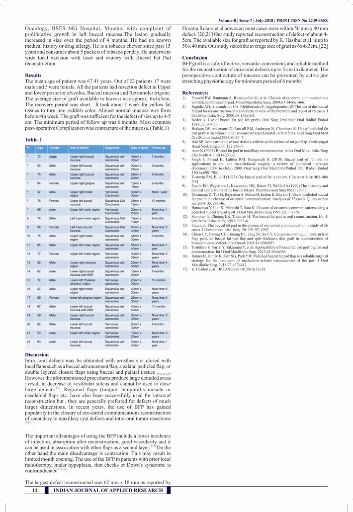
ResultsThe mean age of patient was 67.41 years. Out of 22 patients 17 were male and 5 were female. All the patients had resection defect in Upper and lower posterior alveolus, Buccal mucosa and Retromolar trigone. The average size of graft available to harvest was approx. 6x4x3cm. The recovery period was short. It took about 1 week for yellow fat tissues to turn into reddish color. Almost normal mucosa was form before 4th week. The graft was sufficient for the defect of size up to 4-5 cm. The minimum period of follow up was 6 months. Most common post-operative Complication was contracture of the mucosa. (Table.1)
Table. 1
DiscussionIntra -oral defects may be obturated with prosthesis or closed with local flaps such as a buccal advancement flap, a palatal pedicled flap, or double layered closure flaps using buccal and palatal tissues . [9,10,11,12]
However the aforementioned procedures produce large denuded areas ; result in decrease of vestibular sulcus and cannot be used to close
[13]large defects . Regional flaps (tongue, temporalis muscle or nasolabial flaps etc. have also been successfully used for intraoral reconstruction but , they are generally preferred for defects of much larger dimensions. In recent years, the use of BFP has gained popularity in the closure of oro-antral communications reconstruction of secondary to maxillary cyst defects and intra-oral tumor resections [9-11].
The important advantages of using the BFP include a lower incidence of infection, absorption after reconstruction, good vascularity and it
[16]can be used in association with other flaps as a second layer. On the other hand the main disadvantage is contraction. This may result in limited mouth opening. The use of the BFP in patients with prior local radiotherapy, malar hypoplasia, thin cheeks or Down's syndrome is
[4, 6, 17]contraindicated .
The largest defect reconstructed was 62 mm x 18 mm as reported by
Horatiu Rotaru et al however; most cases were within 50 mm x 40 mm defect. [20,21] Our study reported reconstruction of defect of about 4-5cm. The available size for graft as reported by K. Hasibul et al. is up to 50 x 40 mm. Our study stated the average size of graft as 6x4x3cm. [22]
ConclusionBFP graft is a safe, effective, versatile, convenient, and reliable method for the reconstruction of intra-oral defects up to 5 cm in diameter. The postoperative contracture of mucosa can be prevented by active jaw stretching physiotherapy for minimum period of 6 months.
References:1) Poeschl PW, Baumann A, Russmueller G, et al. Closure of oroantral communications
with Bichat's buccal fat pad. J Oral Maxillofac Surg. 2009;67:1460e1466. 2) Rapidis AD, Alexandridis CA, Eleftheriadis E, Angelopoulos AP. The use of the buccal
fat pad for reconstruction of oral defects: review of the literature and report of 15 cases. J Oral Maxillofac Surg. 2000;58:158e163.
3) Neder A. Use of buccal fat pad for grafts. Oral Surg Oral Med Oral Radiol Endod 1983;55:349–50.
4) Hudson JW, Anderson JG, Russell RM, Anderson N, Chambers K. Use of pedicled fat pad graft as an adjunct in the reconstruction of palatal cleft defects. Oral Surg Oral Med Oral Radiol Endod 1995;80:24–7.
5) Hao SP. Reconstruction of oral defects with the pedicled buccal fat pad flap. Otolaryngol Head Neck Surg 2000;122:863–7.
6) Arce K (2007) Buccal fat pad in maxillary reconstruction. Atlas Oral Maxillofac Surg Clin North Am 15(1):23–32.
7) Singh J, Prasad K, Lalitha RM, Ranganath K (2010) Buccal pad of fat and its applications in oral and maxillofacial surgery: a review of published literature (February) 2004 to (July) 2009. Oral Surg Oral Med Oral Pathol Oral Radiol Endod 110(6):698–705.
8) Tostevin PM, Ellis H (1995) The buccal pad of fat: a review. Clin Anat 8(6): 403–406 [16]
9) Stuzln JM, Wagstrom L, Kawamoto HK, Baker TJ, Wolfe SA (1990) The anatomy and clinical applications of the buccal fat pad. Plast Reconstr Surg 85(1):29–37.
10) Dolanmaz D, Tuz H, Bayraktar S, Metin M, Erdem E, Baykul T. Use of pedicled buccal fat pad in the closure of oroantral communication: Analysis of 75 cases. Quintessence Int. 2004; 35: 241-46.
11) Hanazawa Y, Itoh K, Mabashi T, Sato K. Closure of oroantral communications using a pedicled buccal fat pad graft. J Oral Maxillofac Surg 1995; 53: 771-75.
12) Samman N, Cheung LK, Tideman H. The buccal fat pad in oral reconstruction. Int. J. Oral Maxillofac. Surg. 1993; 22: 2-6.
13) Stajcic Z. The buccal fat pad in the closure of oro-antral communication: a study of 56 cases. J Craniomaxillofac. Surg. 20: 193-97, 1992.
14) Chien CY, Hwang CF, Chuang HC, Jeng SF, Su CY. Comparison of radial forearm free flap, pedicled buccal fat pad flap and split-thickness skin graft in reconstruction of buccal mucosal defect. Oral Oncol. 2005;41:694e697.
15) Toshihiro Y, Nariai Y, Takamura Y, et al. Applicability of buccal fat pad grafting for oral reconstruction. Int J Oral Maxillofac Surg. 2013;42:604e610.
16) Rotaru H, Kim MK, Kim SG, Park YW. Pedicled buccal fat pad flap as a reliable surgical strategy for the treatment of medication-related osteonecrosis of the jaw. J Oral Maxillofac Surg. 2014;73:437e442.
17) K. Hasibul et al. / JPRAS Open 10 (2016) 33e39.
12 INDIAN JOURNAL OF APPLIED RESEARCH
Volume-8 | Issue-7 | July-2018 | PRINT ISSN No 2249-555X