Original Contributions -...
13
Overexpression of the Sarcolemmal Calcium Pump in the Myocardium of Transgenic Rats Annette Hammes, Silke Oberdorf-Maass, Tobias Rother, Katja Nething, Frank Gollnick, Klaus W. Linz, Rainer Meyer, Kai Hu, Hong Han, Peter Gaudron, Georg Ertl, Sigrid Hoffmann, Ursula Ganten, Roland Vetter, Kai Schuh, Claudia Benkwitz, Hans G. Zimmer, Ludwig Neyses Abstract—The plasma membrane calmodulin– dependent calcium ATPase (PMCA) is a calcium-extruding enzyme controlling Ca 21 homeostasis in nonexcitable cells. However, its function in the myocardium is unclear because of the presence of the Na 1 /Ca 21 exchanger. We approached the question of the physiological function of the calcium pump using a transgenic “gain of function” model. Transgenic rat lines carrying the human PMCA 4 cDNA under control of the ventricle-specific myosin light chain-2 promoter were established, and expression in the myocardium was ascertained at the mRNA, protein, and functional levels. In vivo hemodynamic measurements in adult homozygous animals showed no differences in baseline and increased cardiac performance recruited by volume overload compared with controls. No differences between transgenic and control cardiomyocytes were found in patch clamp voltage dependence, activation/inactivation behavior of the L-type Ca 21 current, or fast [Ca 21 ] i transients (assessed by the Fura-2 method). To test whether the PMCA might be involved in processes other than beat-to-beat regulation of contraction/relaxation, we compared growth processes of neonatal transgenic and control cardiomyocytes. A 1.6- and 2.3-fold higher synthesis rate of total protein was seen in cells from transgenic animals compared with controls on incubation with 2% FCS for 24 hours and 36 hours, respectively. An effect of similar magnitude was observed using 20 mmol/L phenylephrine. A 1.4-fold– and 2.0-fold– higher protein synthesis peak was seen in PMCA-overexpressing cardiomyocytes after stimulation with isoproterenol for 12 hours and 24 hours, respectively. Because pivotal parts of the a- and b-adrenergic signal transduction pathways recently have been localized to caveolae, we tested the hypothesis that the PMCA might alter the amplitude of a- and b-adrenergic growth signals by virtue of its localization in caveolae. Biochemical as well as immunocytochemical studies suggested that the PMCA in large part was colocalized with caveolin 3 in caveolae of cardiomyocytes. These results indicate that the sarcolemmal Ca 21 -pump has little relevance for beat-to-beat regulation of contraction/relaxation in adult animals but likely plays a role in regulating myocardial growth, possibly through modulation of caveolar signal transduction. (Circ Res. 1998;83:877-888.) Key Words: plasma membrane Ca 21 -ATPase n myocardium n transgenic rat n contraction n cardiac growth T he plasma membrane calmodulin– dependent calcium AT- Pase (PMCA) is a ubiquitous Ca 21 -transporting enzyme extruding Ca 21 from the cell. 1– 4 So far, 4 different PMCA isoforms in human 5– 8 and rat 2,9,10 have been cloned, which are expressed in a cell type– and differentiation-specific manner. 11– 17,19 In the myocardium, the expression of the isoforms 1, 2, and 4 has been shown. 14,16,19 Whereas much insight has been gained into the expression pattern of the various PMCA isoforms and their biochemical properties, 3,4,18 studies on their physiological significance have been scant until very recently. In nonexcitable cell types, the PMCA is the only known enzyme-mediating calcium extrusion and therefore has been postulated to have a housekeeping function. 19 However, in excitable cells, which express the high capacity Na 1 /Ca 21 exchanger as an additional sarcolemmal (SL) calcium trans- porter, the functional importance of the PMCA remains unclear. In the myocardium, it has been assumed that the PMCA shares the burden of extruding calcium from the cell after each beat with the Na 1 /Ca 1 exchanger. In particular, the high calcium affinity of the PMCA has led to the speculation that it is responsible for fine tuning of calcium in the final phase of diastole. On the other hand, in isolated adult cardiomyocytes, the sodium/calcium exchanger has been demonstrated to have a much higher calcium transport activity than the PMCA. 20 Under the assumption that these in vitro studies are represen- Received December 9, 1997; accepted July 1, 1998. From the Department of Medicine, University of Wu ¨rzburg (A.H., S.O.-M., T.R., K.N., K.S., C.B., L.N.); Department of Physiology, University of Bonn (F.G., K.W.L., R.M.); Department of Medicine, Cardiology and Angiology, Medical Faculty, Mannheim, University of Heidelberg (K.H., H.H., P.G., G.E.); Max Delbru ¨ck Center for Molecular Medicine, Berlin (S.H., U.G., R.V.); and the Institute of Physiology, University of Leipzig (H.G.Z.), Germany. Present affiliation for A.H. is Developmental Genetics, Max Delbrueck Center for Molecular Medicine, Berlin, Germany. Present affiliation for R.V. is Institute of Clinical Pharmacology and Toxicology, Benjamin Franklin Medical Center, Berlin, Germany. Present affiliation for C.B. is Department of Anesthesiology, University of Wu ¨rzburg, Germany. Correspondence to Ludwig Neyses, MD, Department of Medicine, University of Wu ¨rzburg, Josef-Schneider-Str 2, D-97080 Wu ¨rzburg, Germany. © 1998 American Heart Association, Inc. 877 Original Contributions by guest on July 20, 2018 http://circres.ahajournals.org/ Downloaded from
Transcript of Original Contributions -...

Overexpression of the Sarcolemmal Calcium Pump in theMyocardium of Transgenic Rats
Annette Hammes, Silke Oberdorf-Maass, Tobias Rother, Katja Nething, Frank Gollnick,Klaus W. Linz, Rainer Meyer, Kai Hu, Hong Han, Peter Gaudron, Georg Ertl, Sigrid Hoffmann,Ursula Ganten, Roland Vetter, Kai Schuh, Claudia Benkwitz, Hans G. Zimmer, Ludwig Neyses
Abstract—The plasma membrane calmodulin–dependent calcium ATPase (PMCA) is a calcium-extruding enzymecontrolling Ca21 homeostasis in nonexcitable cells. However, its function in the myocardium is unclear because of thepresence of the Na1/Ca21 exchanger. We approached the question of the physiological function of the calcium pumpusing a transgenic “gain of function” model. Transgenic rat lines carrying the human PMCA 4 cDNA under control ofthe ventricle-specific myosin light chain-2 promoter were established, and expression in the myocardium wasascertained at the mRNA, protein, and functional levels. In vivo hemodynamic measurements in adult homozygousanimals showed no differences in baseline and increased cardiac performance recruited by volume overload comparedwith controls. No differences between transgenic and control cardiomyocytes were found in patch clamp voltagedependence, activation/inactivation behavior of the L-type Ca21 current, or fast [Ca21]i transients (assessed by the Fura-2method). To test whether the PMCA might be involved in processes other than beat-to-beat regulation ofcontraction/relaxation, we compared growth processes of neonatal transgenic and control cardiomyocytes. A 1.6- and2.3-fold higher synthesis rate of total protein was seen in cells from transgenic animals compared with controls onincubation with 2% FCS for 24 hours and 36 hours, respectively. An effect of similar magnitude was observed using20 mmol/L phenylephrine. A 1.4-fold– and 2.0-fold–higher protein synthesis peak was seen in PMCA-overexpressingcardiomyocytes after stimulation with isoproterenol for 12 hours and 24 hours, respectively. Because pivotal parts of thea- andb-adrenergic signal transduction pathways recently have been localized to caveolae, we tested the hypothesis thatthe PMCA might alter the amplitude ofa- andb-adrenergic growth signals by virtue of its localization in caveolae.Biochemical as well as immunocytochemical studies suggested that the PMCA in large part was colocalized withcaveolin 3 in caveolae of cardiomyocytes. These results indicate that the sarcolemmal Ca21-pump has little relevancefor beat-to-beat regulation of contraction/relaxation in adult animals but likely plays a role in regulating myocardialgrowth, possibly through modulation of caveolar signal transduction.(Circ Res. 1998;83:877-888.)
Key Words: plasma membrane Ca21-ATPasen myocardiumn transgenic ratn contractionn cardiac growth
The plasma membrane calmodulin–dependent calcium AT-Pase (PMCA) is a ubiquitous Ca21-transporting enzyme
extruding Ca21 from the cell.1–4 So far, 4 different PMCAisoforms in human5–8 and rat2,9,10 have been cloned, which areexpressed in a cell type– and differentiation-specific manner.11–
17,19 In the myocardium, the expression of the isoforms 1, 2, and4 has been shown.14,16,19Whereas much insight has been gainedinto the expression pattern of the various PMCA isoforms andtheir biochemical properties,3,4,18 studies on their physiologicalsignificance have been scant until very recently.
In nonexcitable cell types, the PMCA is the only knownenzyme-mediating calcium extrusion and therefore has beenpostulated to have a housekeeping function.19 However, in
excitable cells, which express the high capacity Na1/Ca21
exchanger as an additional sarcolemmal (SL) calcium trans-porter, the functional importance of the PMCA remains unclear.
In the myocardium, it has been assumed that the PMCAshares the burden of extruding calcium from the cell aftereach beat with the Na1/Ca1 exchanger. In particular, the highcalcium affinity of the PMCA has led to the speculation thatit is responsible for fine tuning of calcium in the final phaseof diastole.
On the other hand, in isolated adult cardiomyocytes, thesodium/calcium exchanger has been demonstrated to have amuch higher calcium transport activity than the PMCA.20
Under the assumption that these in vitro studies are represen-
Received December 9, 1997; accepted July 1, 1998.From the Department of Medicine, University of Wurzburg (A.H., S.O.-M., T.R., K.N., K.S., C.B., L.N.); Department of Physiology, University of
Bonn (F.G., K.W.L., R.M.); Department of Medicine, Cardiology and Angiology, Medical Faculty, Mannheim, University of Heidelberg (K.H., H.H.,P.G., G.E.); Max Delbruck Center for Molecular Medicine, Berlin (S.H., U.G., R.V.); and the Institute of Physiology, University of Leipzig (H.G.Z.),Germany. Present affiliation for A.H. is Developmental Genetics, Max Delbrueck Center for Molecular Medicine, Berlin, Germany. Present affiliationfor R.V. is Institute of Clinical Pharmacology and Toxicology, Benjamin Franklin Medical Center, Berlin, Germany. Present affiliation for C.B. isDepartment of Anesthesiology, University of Wurzburg, Germany.
Correspondence to Ludwig Neyses, MD, Department of Medicine, University of Wurzburg, Josef-Schneider-Str 2, D-97080 Wurzburg, Germany.© 1998 American Heart Association, Inc.
877
Original Contributions
by guest on July 20, 2018http://circres.ahajournals.org/
Dow
nloaded from

tative of the situation in whole heart, the PMCA may playonly a minor role in beat-to-beat regulation ofcontraction/relaxation.
Recently published results indicate that the PMCA plays arole in growth and differentiation processes. We reported thatL6 skeletal myoblasts stably overexpressing the PMCAshowed a markedly accelerated myogenic differentiation intomyotubes.21 Consistent with these findings, results publishedby other groups also suggest an active role of the enzyme ingrowth regulation. Liu and coworkers22 showed a delay inG1-S phase transition in rat aortic endothelial cells overex-pressing rat PMCA1a. Husain et al23 demonstrated a signifi-cantly slower growth rate in PMCA-overexpressing vascularsmooth muscle cells, and Brandt et al24 published resultsshowing an inhibition of nerve growth factor–induced neuriteoutgrowth in pheochromocytoma cells transfected with aPMCA 1 antisense construct.
To address the unresolved issue of the function of thePMCA in the myocardium, in the present work, we estab-lished transgenic rat lines overexpressing the PMCA in themyocardium as a “gain of function” model. This allowed usto address hypotheses directly about the role of this pump incontraction/relaxation and/or cellular growth processes.
Materials and MethodsConstruction of Transgenic RatsWe established 4 transgenic rat lines carrying the human PMCAisoform 4CI cDNA6 under the control of the ventricle-specific ratmyosin light chain-2 (MLC-2) promoter25 and 2 transgenic rat linescarrying the same cDNA under the control of the rata-myosin heavychain (a-MHC) promoter.26 The first expression construct wascomposed of the 250-bp MLC-2 promoter, a 325-bp simian virus(SV) 40 intervening sequence (IVS), the 3.6-kb hPMCA4CI cDNA,a 600-bp SV 40 IVS, and the 240-bp SV 40 poly A signal. Thesecond expression construct contained the 2.9-kba-MHC promoter,a 400-bp 59untranslated region of thea-MHC gene, the 3.6-kbhPMCA4CI cDNA, the 600-bp SV 40 IVS, and the 240 bp SV 40poly A signal. The sequence of both expression constructs wasconfirmed by restriction endonuclease digestion and DNA sequenc-ing. The MLC-2 promoter and the 325-bp SV 40 IVS sequence wereprovided kindly by Dr Kenneth Chien and Genentech Inc; thea-MHC promoter was a gift from Dr Bruce Markham (Ann Arbor,Mich). The 3.6-kb hPMCA4CI cDNA was provided kindly by DrEmanuel Strehler (Rochester, Minn). The expression constructs wereexcised from the vector, gel-purified, and used for pronuclearmicroinjection of fertilized oocytes from Sprague-Dawley (SD) ratsto produce transgenic rats according to the procedure described byMullins and Ganten.27 Genomic DNA (10mg) from rat tail biopsieswere subjected to EcoRI digestion and Southern blot analysisaccording to the standard procedures using an hPMCA4CI cDNA–specific 1.2-kb EcoRI fragment (corresponding to nucleotides 2177to 3403 of hPMCA4CI)6 as a probe.
Northern Blot AnalysisExpression of the human PMCA isoform 4 mRNA was detected byNorthern blot analysis of total RNA from transgenic rat hearts.Twenty micrograms of total RNA was run on a 1% formaldehyde-agarose gel, and Northern blotting was performed according tostandard procedures. For hybridization, a randomly primed labeled1200-bp EcoRI fragment of the human PMCA4CI cDNA was used.Three of the four established transgenic lines with the MLC-2promoter–driven transgene and 1 out of 2 lines with thea-MHCpromoter showed mRNA expression of the PMCA transgene. Ex-pression was most abundant in total heart and also in isolatedcardiomyocytes from adult and neonatal hearts, but lower expression
was also detectable in brain, lung, and kidney. No expression of thetransgene was found in atrium, skeletal muscle, liver, or noncardio-myocytes from transgenic neonatal rat hearts.
Western Blot Analysis and Analysis ofTransgene Activity
Preparation of Highly Purified Plasma Membranes FromCardiac TissueHeart tissue (2 g) was frozen in liquid nitrogen and powdered beforehomogenization in 20 mL buffer containing (in mmol/L): histidine 5(pH 7.7), KCl 750, DTT 0.2, and PMSF 0.1. The sample wascentrifuged at 3000gfor 15 minutes, the pellet was resuspended in 20mL homogenization buffer, and the previous step was repeated. Thepellet was resuspended in 20 mL hypotonic medium containing10 mmol/L NaHCO3 (pH 7.4), 5 mmol/L histidine, and 0.2 mmol/LDTT, centrifuged at 3000g for 15 minutes, and the pellet washomogenized (3330 s; 21 000 rpm) in 20 mL hypotonic medium.The homogenate was centrifuged at 12 400g for 20 minutes, thepellet was treated identically to the previous step, and the superna-tants were pooled and centrifuged at 45 000g for 30 minutes. Thepellet was resuspended in 1 mL sucrose-histidine buffer I(250 mmol/L sucrose, 10 mmol/L histidine [pH 7.4], 160 mmol/LKCl) and centrifuged on a sucrose gradient (18% to 35%) for 90minutes at 100 000g. The SL fraction was harvested, and 3.2 mL of600 mmol/L KCl was added. The SL preparation was centrifuged at60 000 rpm for 20 minutes, and the pellet was resuspended insucrose-histidine buffer II (250 mmol/L sucrose, 10 mmol/L histi-dine [pH 7.4]) and centrifuged as in the previous step. The finalpellet was resuspended in 200mL sucrose-histidine buffer II.
Western Blot AnalysisThe SL preparations were separated by SDS-PAGE and transferredto polyvinylidene fluoride membranes.28 Blocking was performedovernight with 1% BSA. The membranes were incubated for 1 hourwith the monoclonal anti-PMCA antibody 5F10 (1:1000 dilution),recognizing all 4 isoforms of the PMCA expressed in rat and humantissues (Affinity BioReagents, Hamburg, Germany). After a washingstep (13PBS; pH 7.45; 1% BSA), incubation with the secondaryantibody (a sheep anti-mouse antibody coupled to alkaline phospha-tase; Amersham, Braunschweig, Germany) at a 1/5000 dilution wasperformed for 1 hour. After a further washing step in 13PBS (pH7.45), 1% BSA, and 0.3% Tween, immunocomplexes were visual-ized by chemoluminescence according to the manufacturer’s proto-col (ECL-kit, Amersham). For the detection of caveolin 3 in Westernblotting experiments, a monoclonal antibody (Transduction Labora-tories), recognizing rat and human caveolin 3, was used as describedabove.
For quantification, the data were normalized to Na1/Ca21 ex-changer protein (expression of the Na1/Ca21 exchanger protein wasunchanged in the myocardium of transgenic animals as detected byWestern blot analysis). Further normalization to cell number andtotal protein revealed similar results. The polyclonal antibody used todetect the Na1/Ca21 exchanger was obtained from SWant.
Coupled Enzyme Assay Measuring Ca21-DependentATPase ActivityThe reaction mixture contained 50 nmol/L CaCl2, 50 mmol/LHEPES-Tris (pH 7.4), 160 mmol/L KCl, 2 mmol/L MgCl2,5 mmol/L NaN3, 1 mg/mL alamethicin, 1 mmol/L ATP-Tris,1 mmol/L phosphoenolpyruvate, 1 U/mL pyruvatekinase,0.6 mmol/L NADH, 1 U/mL lactatedehydrogenase. The reaction wasstarted by adding 10mg of highly purified SL protein to 1 mL of themixture, and the optical density of NADH/NAD was measured over2 minutes in a spectrophotometer at 340 nm at 37°C. To stop theCa21-dependent ATPase activity, 2 mmol/L EGTA was added, andthe measurement was continued for 2 minutes. The Ca21-dependentATPase activity was calculated by subtracting the 2 fitted slopes.
878 PMCA in Myocardium
by guest on July 20, 2018http://circres.ahajournals.org/
Dow
nloaded from
Measurements of L-Type Ca21 Current andFura-2 Fluorescence Ratios in IsolatedAdult Cardiomyocytes
Isolation of Adult CardiomyocytesAdult cardiomyocytes from transgenic and normal SD rats (femaleand male; body weight, 230 to 490 g) were isolated by the methoddescribed by Stegemann et al29 for guinea pig ventricular myocyteswith slight modifications according to Linz and Meyer.30
Measurement of L-Type Ca21 CurrentL-type Ca21 current was recorded at 3561°C using the whole-cellpatch-clamp technique.31 Patch pipettes with tip resistances of 2 to 4MV were pulled from borosilicate glass (Hilgenberg) and connectedto a single-electrode continuous-voltage clamp amplifier (L/M EPC7, List Medical Electronic). In all measurements, cell capacitance,series resistance, and junction potentials were compensated. Voltage-clamp protocols and data acquisition were accomplished usingpClamp 6.0 software (Axon Instruments). Current signals wereon-line filtered at 3 kHz and digitized at 15 kHz by a 12-bit A/Dconverter (Digidata 1200, Axon Instruments).
The L-type Ca21 current was determined as total peak inwardcurrent. The separation from overlapping fast Na1 current and T-typeCa21 current was achieved by holding the cells at240 mV for thetime between the test pulses. K1 currents (IK1, IK, and I to) wereblocked with an external solution containing (in mmol/L): NaCl 115,tetraethylammonium chloride 20, KCl 4, BaCl2 2, MgCl2 1, CaCl21.8, 4-aminopyridine 2, HEPES 2, and glucose 11 (pH 7.2) and anelectrode-filling solution composed of (in mmol/L): CsCl 130,Mg-ATP 2, and HEPES 10; and 50mmol/L BAPTA (pH 7.2;Molecular Probes).
To determine current-voltage relations (I/V), steady-state activa-tion parameters (d), and steady-state inactivation parameters (f`),gapped double-pulse protocols32 with pulse lengths of 400 ms wereused (stimulation frequency, 0.1 Hz). Current amplitudes werenormalized to the cell capacitance. Fitting ofI/V-relations wasperformed according to Linz and Meyer.30
Fluorescence Measurement of Fast [Ca21]i TransientsFast [Ca21]i transients were elicited by Ca21 influx through L-typechannels in patch-clamped cardiomyocytes. In these measurements,Na1/Ca21 exchange was blocked by equimolar substitution of LiClfor NaCl.33 Lasting voltage-clamp pulses (500 ms; frequency, 0.2Hz) were applied from a holding potential of240 to 20 mV in anexternal solution containing (in mmol/L): LiCl 135, KCl 4, MgCl2 1,CaCl2 1.8, 4-aminopyridine 2, HEPES 2, and glucose 11 (pH 7.2).The electrode-filling solution was composed of (in mmol/L): CsCl130, Mg-ATP 2, HEPES 10, Fura-2/Na1-salt 50 (Molecular Probes);and 1mmol/L ryanodine; pH 7.2. [Ca21]i-transients were measured at3561°C in cells preincubated for 20 minutes in standard Tyrode’ssolution (in mmol/L): NaCl 135, KCl 4, CaCl2 1.8, MgCl2 1, glucose11, and HEPES 2 (pH 7.2); and 1 g/L BSA containing 5 mmol/Lthapsigargin and 10mmol/L ryanodine. Fura-2 fluorescence34 wasrecorded by means of a fast fluorescence microphotometry system(Zeiss FFP attached to a Zeiss IM 35 microscope with ZeissUltrafluar 1003objective; Carl Zeiss) synchronized to the patch-clamp setup (dye loading: 2mmol/L Fura-2/AM; 20 minutes at35°C). Fura-2 excitation (340 and 380 nm) was performed with 4-mstime resolution. Fluorescence emission (420 to 560 nm) of thepatch-clamped cell was recorded by a photomultiplier tube (1.6-mmaperture covering 10mm in the object plane). Changes in [Ca21]i
were determined as relative changes from the calculated ratio,340:380 nm. The estimated basal free cytosolic Ca21 concentration inmyocytes of transgenic and wild-type rats was 90 nmol/L. Absolute[Ca21]i was not determined routinely because of the semiquantitativenature of the affinity constants used.
Fluorescence Measurement of Slow [Ca21]i TransientsSlow [Ca21]i transients (range, several minutes) in single cardiomyo-cytes were determined by Fura-2/AM fluorescence measurementusing a modification of the PC-based Hamamatsu ICMS video
imaging system (Hamamatsu Photonics KK) described in detail byGollnick et al.35 A dual excitation interference filter wheel (340 and380 nm) and a 150-W Xenon arc lamp were used for Fura-2excitation (0.4-s double flashes) at 5- or 10-s intervals. Digitalintensity images (2563256 pixel; 8-bit resolution; no image averag-ing) of emission (425 to 575 nm) were acquired for each excitationwavelength using a Hamamatsu C2400-77H ICCD camera. Myo-cytes were placed in an open plexiglas perfusion chamber (0.2 mL)and adhered to the laminin-coated glass bottom. Fura-2 fluorescencewas measured in an inverted epifluorescence microscope (ZeissAxiovert 100 TV with Zeiss Fluar 403objective, Carl Zeiss) underpermanent perfusion of buffers at 3760.5°C. For evaluation ofintensity changes with custom-written software (4 to 5 evaluablecells/experiment), digital measuring windows were placed within thecell borders. Background fluorescence was measured separately foreach wavelength and subtracted from each intensity value before thecalculation of 340:380-nm ratio images. Relative ratio changes werenot converted to absolute changes of [Ca21]i.
Slow [Ca21]i transients were elicited by external sodium depletion.After a 25-s baseline interval, standard Tyrode’s buffer (see above) wasreplaced by sodium-free buffer (equimolar substitution of LiCl forNaCl). Fluorescence image acquisition lasted for a total of 17 minutes.Hypercontracting and nonresponding cells were not evaluated. In someexperiments, sarcoplasmic reticulum (SR) block was achieved with 30minutes of preincubation with ryanodine1thapsigargin (10 and5 mmol/L, respectively). Blocker substances were present throughoutthe experiment.
Hemodynamic MeasurementsFor hemodynamic measurements, rats were anesthetized with ether.Polyethylene cannulas were inserted into the trachea for artificalventilation and into the right carotid artery, right jugular vein, andfemoral vein. Pressure was measured through a short segment of afluid-filled PE 50 tubing connected to a microtip manometer (Millar)via a 3-way stopcock, with 0 adjusted to midchest level. The carotidcannula was advanced briefly into the left ventricle and then waswithdrawn to the aortic arch while pressures were recorded. Thejugular vein cannula was advanced to the right atrium. Left ventric-ular systolic and end-diastolic pressures, the maximum rate of rise inleft ventricular systolic pressure, dP/dtmax, mean arterial pressure(MAP), heart rate, and mean right atrial pressure (RAP) weremeasured under light ether anesthesia and spontaneous respiration.
During positive pressure ventilation, and after midsternal thora-cotomy, a calibrated flowmeter (2.0 mm; Statham, Inc) was placedaround the ascending aorta for continuous measurement of aorticblood flow. Mean aortic blood flow was obtained electronically andtaken as cardiac index (CI), as described by Pfeffer et al.36 Systemicvascular resistance index was calculated as (MAP2RAP)/CI andwas expressed as mm Hg/mL per min/kg body weight.
After baseline measurements, warmed (39°C to 40°C) Tyrode’ssolution was infused into the femoral vein at a rate of 40 mL/kg perminute for 45 seconds or until maximal flow was achieved.36 Thisinfusion produces a rise in cardiac output to peak values, followed bya plateau, despite further elevation of RAP. Maximum cardiacperformance was defined as peak values of cardiac output and strokevolume during this Tyrode’s infusion.
Culture of Neonatal Cardiomyocytes and ProteinSynthesis Measurements
Primary Cell Culture of Neonatal Rat CardiomyocytesCardiomyocytes from 48-hour-old transgenic and normal SD ratswere prepared according to Simpson and Savion37 with minormodifications. The hearts were cut into small pieces and digested inCBFHH (calcium and bicarbonate free Hanks’ with HEPES) (con-taining [in mmol/L]: NaCl 137, KCl 5.36, MgSO437 H2O 0.81,D-glucose 5.55, KH2PO4 0.44, Na2HPO4 0.34, HEPES 20.06; pH 7.4)containing 31mg/l penicillin G, 10mg/mL DNase, and 1.5 mg/mLtrypsin. FCS was added to the resulting cell suspension to inactivatetrypsin and DNase, and the cells were pelleted by 10 minutes ofcentrifugation at 700gand resuspended in MEM/5 (containing 31
Hammes et al November 2, 1998 879
by guest on July 20, 2018http://circres.ahajournals.org/
Dow
nloaded from
mg/mL penicillin G, 50mg/mL streptomycin, 30.74mg/mL BrdU, 2mg/mL vitamin B12, and 5% FCS). Most noncardiomyocytes wereremoved by preplating for 1 hour. After preplating, the supernatantcontaining .90% cardiomyocytes was removed, and cells werecounted in a Fuchs-Rosenthal chamber and plated in MEM/5 on6-well plates at a density of 0.7 million cells/well (low-densityculture).
After 24 hours, the MEM/5 was removed, and the cells weregrown for 48 more hours in the serum-free medium MEM/TI(additionally containing 31mg/mL penicillin G, 50mg/mL strepto-mycin, 30.74mg/mL BrdU, 2 mg/mL vitamin B12, 1 mg/mL trans-ferrin, and 1mg/mL porcine Zn-insulin). Growth experiments werestarted on day 3.
Growth Experiments, Measurement of CellularProtein Synthesis and Apoptosis AnalysisPrimary cell cultures of 48-hour-old transgenic and normal SD ratswere prepared as described above. On day 3, the MEM/TI wasreplaced by MEM/TI containing either a growth stimulus, such as2% FCS, 20mmol/L phenylephrine, and 1mmol/L isoproterenol, orno stimulus. The cells then were incubated for several different timeperiods (6 to 12 to 18 to 24 to 36 hours). Four hours before the endof the incubation period, medium was changed into MEM/TIwith/without growth stimulus and3H-Leucine at an activity of 2.5mCi/mL. After incubation, the cells were washed 2 times with PBS,lysed with 1% SDS, and harvested. A small amount of the lysate wasused to determine DNA concentration as a measure of number ofcells, using the Hoechst 33258 dye. An equal volume of 10% TCAwas added to the lysate. Proteins were precipitated for 30 minutes atroom temperature, pelleted by centrifugation (10 minutes, 14 000g),and resuspended in 1% SDS. Radioactivity was measured in ab-counter. The CPM:DNA concentration ratio was calculated foreach sample, and changes in protein synthesis by the growth stimuliwere depicted as percentage of the CPM:DNA concentration ratio ofthe nonstimulated control cells. Terminal deoxynucleotidyl transfer-mediated end-labeling of fragmented nuclei (TUNEL assay)38 wasperformed on neonatal cardiomyocytes according to the manufactur-er’s protocol (Boehringer Mannheim Biochemicals).
Preparation of Caveolar MembranesTo purify caveolae, we used the protocol described by Song et al.39
Purified plasma membranes from 5-day-old SD rats were suspendedin MES buffered saline (MBS), disrupted by sonication and placed atthe bottom of a discontinuous 45% to 35%25% sucrose gradient.After centrifugation at 260 000g for 20 hours, 1-mL fractions weretaken starting from the top, and an equal amount of protein of eachfraction was used in Western blot analysis detecting caveolin 3(monoclonal anti–caveolin 3 antibody, Transduction Laboratories)and the PMCA (Ab 5F10, Affinity Bioreagents).
Immunostainings and Confocal MicroscopyPrimary cell culture of neonatal cardiomyocytes was performed asdescribed above. Cells were cultured on slides for 48 hours inmedium containing 5% FCS and for another 24 hours in serum-freemedium. They were fixed for 30 minutes with 2% paraformaldehydeat room temperature and then rinsed twice in PBS. Samples wereblocked and permeabilized in PBS, containing 1% BSA, 0.5% TritonX-100, and 0.1% Tween 20 for 30 minutes at 20°C and then wereincubated at 4°C overnight in a PBS/0.1% BSA solution containingthe primary antibodies. For double-staining, the monoclonal anti-body JA3 (a generous gift from Drs A. Filoteo and J.T. Penniston,Mayo Clinic and Foundation, Rochester, Minn), generated againstthe human PMCA isoform 4, and the polyclonal rabbit anti–caveolin3 antibody (0.25 mg/mL, Transduction Laboratories) were used.Working dilutions of these antibodies were 1:500. After incubation,the samples were washed 5 times in PBS/0.1% Tween 20, then inTexas Red-conjugated goat anti-rabbit IgG (1.4 mg/mL), and theCy2-conjugated goat anti-mouse IgG antibodies (1.3 mg/mL, Jack-son ImmunoResearch Laboratories) were applied in PBS (1:500 and1:250, respectively) for 1 hour at 4°C. After final washing steps, thesamples were mounted in Vectashield mounting medium (Vector)
and analyzed with a BioRad MRC 1024 confocal laser scanningsystem using the LaserSharp software. Statistical analysis wasperformed using Studentt test, and values are expressed as SEMunless stated otherwise.
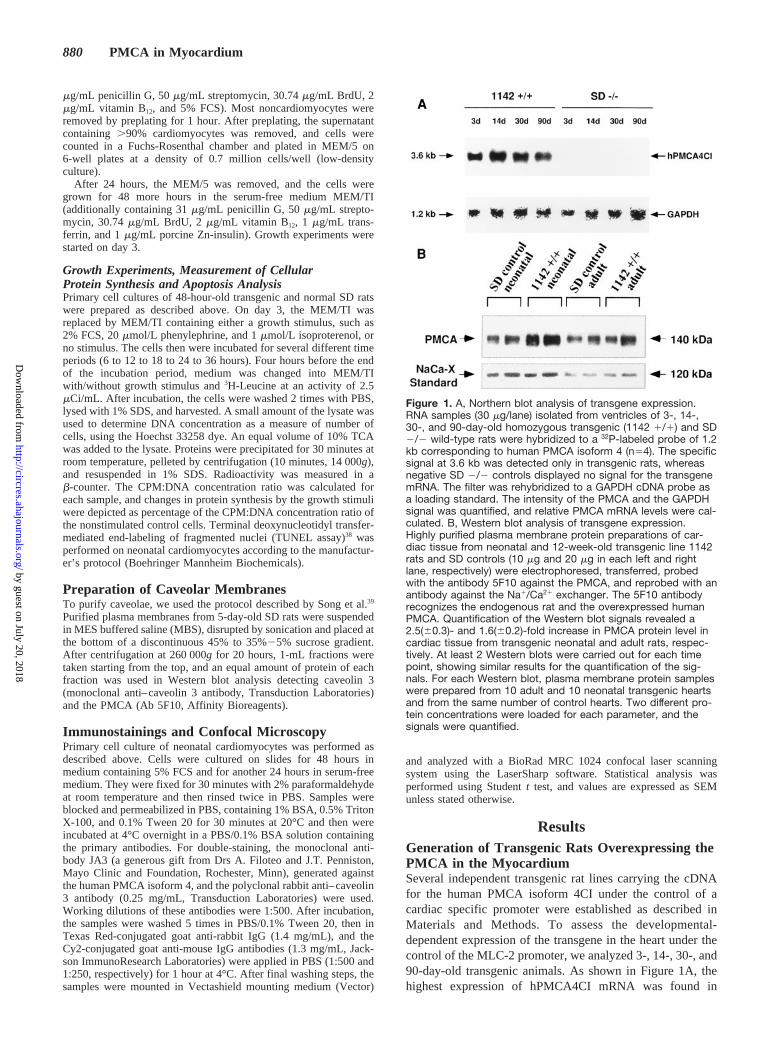
ResultsGeneration of Transgenic Rats Overexpressing thePMCA in the MyocardiumSeveral independent transgenic rat lines carrying the cDNAfor the human PMCA isoform 4CI under the control of acardiac specific promoter were established as described inMaterials and Methods. To assess the developmental-dependent expression of the transgene in the heart under thecontrol of the MLC-2 promoter, we analyzed 3-, 14-, 30-, and90-day-old transgenic animals. As shown in Figure 1A, thehighest expression of hPMCA4CI mRNA was found in
Figure 1. A, Northern blot analysis of transgene expression.RNA samples (30 mg/lane) isolated from ventricles of 3-, 14-,30-, and 90-day-old homozygous transgenic (1142 1/1) and SD2/2 wild-type rats were hybridized to a 32P-labeled probe of 1.2kb corresponding to human PMCA isoform 4 (n54). The specificsignal at 3.6 kb was detected only in transgenic rats, whereasnegative SD 2/2 controls displayed no signal for the transgenemRNA. The filter was rehybridized to a GAPDH cDNA probe asa loading standard. The intensity of the PMCA and the GAPDHsignal was quantified, and relative PMCA mRNA levels were cal-culated. B, Western blot analysis of transgene expression.Highly purified plasma membrane protein preparations of car-diac tissue from neonatal and 12-week-old transgenic line 1142rats and SD controls (10 mg and 20 mg in each left and rightlane, respectively) were electrophoresed, transferred, probedwith the antibody 5F10 against the PMCA, and reprobed with anantibody against the Na1/Ca21 exchanger. The 5F10 antibodyrecognizes the endogenous rat and the overexpressed humanPMCA. Quantification of the Western blot signals revealed a2.5(60.3)- and 1.6(60.2)-fold increase in PMCA protein level incardiac tissue from transgenic neonatal and adult rats, respec-tively. At least 2 Western blots were carried out for each timepoint, showing similar results for the quantification of the sig-nals. For each Western blot, plasma membrane protein sampleswere prepared from 10 adult and 10 neonatal transgenic heartsand from the same number of control hearts. Two different pro-tein concentrations were loaded for each parameter, and thesignals were quantified.
880 PMCA in Myocardium
by guest on July 20, 2018http://circres.ahajournals.org/
Dow
nloaded from

3- and 14-day-old animals. The level of mRNA encoding thetransgene decreased only slightly in adult animals.
Quantitative Immunoblotting and Measurement ofATPase Activity in Transgenics and ControlsTo determine the levels of transgene overexpression, highlypurified SL protein preparations from the myocardium oftransgenics and controls were subjected to Western blotanalysis. Quantitative immunoblotting revealed a 2.5-foldPMCA overexpression in transgenic neonatal cardiomyocytesand 1.6-fold overexpression of the calcium pump in adultcardiomyocytes compared with controls (Figure 1B).
To assess the function of the overexpressed PMCA, Ca21-dependent ATPase activity was measured in highly purifiedplasma membrane preparations of the myocardium. TheCa21-dependent ATPase activity in the hearts of adult ho-mozygous rats (line 1142) was 12.6mmol ADP/mg protein/hcompared with a level of 7.1 U in controls. These results fitwell with our protein data (see above) and suggest that thetransgene was entirely functional.
Expression of Endogenous Calcium-TransportingSystems in the Heart ofhPMCA4CI-Overexpressing RatsIn cardiac tissue from PMCA-transgenic rats, no compensa-tory up- or down-regulation of the Na1/Ca21 exchanger,SERCA2a, and endogenous rat PMCA 1 mRNA was detected(Figure 2). The transgenics showed the normal decrease inexpression of the Na1/Ca21 exchanger and increase in expres-sion of the SERCA2a and the endogenous PMCA1 duringdevelopment from neonatal to adult rats.
Furthermore, endogenous MLC-2 expression was not al-tered in transgenic animals (data not shown). This wasexcluded, because transgene expression was driven by the
MLC-2 promoter; hence, down-regulation of the endogenouspromoter activity might occur. This data showed that pheno-typic differences between transgenics and controls would bedue to transgene overexpression rather than compensatorychanges in other Ca21 transporters.
In Vivo Hemodynamic ExperimentsOne hypothesis was that the PMCA might play a role in thecardiac contraction/relaxation cycle. Physiological measure-ments assessing the contraction/relaxation parameters suchas left ventricular systolic pressure, mean aortic pressure,dP/dtmax, left ventricular end-diastolic pressure, RAP, andheart rate are summarized in Tables 1 and 2. Adult transgenicrats showed no significant differences in baseline or peakcardiac performance compared with SD wild-type rats.
Electrophysiological Studies andCalcium MeasurementsElectrophysiological studies and Fura-2 measurements onisolated adult cardiomyocytes were performed to determine ifthe increase in PMCA expression resulted in correspondingalterations of electrophysiological parameters and/or the cal-cium transient. As shown in Figure 3, there are no differencesin voltage dependence, activation, and inactivation behaviorof L-type Ca21 current between transgenic cells and controladult cardiomyocytes. Furthermore, transgenic and controlcardiomyocytes displayed an identical time course in fast[Ca21]i transients induced by calcium influx through L-typechannels in the presence of thapsigargin and ryanodine (SRblock; Figure 4A). The slow decline of [Ca21]i transients afterNa1 depletion was faster slightly but not significantly incardiomyocytes isolated from PMCA-transgenic animals(Figure 4B), attesting to the activity of the transgene butsuggesting that a decrease in [Ca21]i in the myofilamentcompartment was not a major effect of transgene overexpres-sion. When the SR was blocked by thapsigargin and ryano-dine, a marginally different time constant (t) of [Ca21]i
decline was seen:t5180.5 s (n514) in controls versust5214.7 s (n517) in transgenic cardiomyocytes (data notshown). Thet values of the monoexponential fit functions aregiven in the legend to Figure 4B. They are given without SD
Figure 2. Expression of endogenous Ca21-transporting systemsin cardiac tissue of PMCA-transgenic rats. RNA samples (30mg/lane) isolated from ventricle of 3-, 14-, 30-, and 90-day-oldhomozygous transgenic rats (1142 1/1) and SD 2/2 wild-typerats were hybridized to 32P-labeled probes corresponding toNa1/Ca21 exchanger (NaCa-X), SERCA2, endogenous rat PMCA1 (rPMCA1), and GAPDH (loading control). Quantification of thesignals revealed no differences in the mRNA expression level ofthe endogenous Ca21-transporting enzymes between transgenicand control rats (n53).
TABLE 1. Hemodynamic Measurements Before Thoracotomy
SD 2/2(12 Weeks)
1142 1/1(12 Weeks)
n 17 12
Body weight, g 319680 315675
Heart weight, g 1.6160.42 1.4260.37
Left ventricular systolic pressure,mm Hg
147610.1 14769.0
Mean arterial pressure, mm Hg 114612.0 11668.0
dP/dtmax, 31000 mm Hg/s 13.862.6 13.463.6
Left ventricular end-diastolicpressure, mm Hg
6.462.5 7.563.0
Right atrial pressure, mm Hg 0.161.3 20.860.9
Heart rate, min21 354625 336647
SD 2/2 indicates Sprague-Dawley control rats; 1142 1/1, PMCA-transgenic rats.
Hammes et al November 2, 1998 881
by guest on July 20, 2018http://circres.ahajournals.org/
Dow
nloaded from

because mean values of an exponential function constantwere fitted. The Fura-2 fluorescence ratios at rest wereslightly different: ratio 340:380 nm was 0.8960.17 (n514cells; 4 different animals) in controls versus 1.1460.36 nm(n517 cells; 4 different animals) in transgenic animals.Although both alterations could be indicative of compensa-tory mechanisms, they are so small that their biologicalrelevance is probably minor.
Cardiomyocyte GrowthOur second main hypothesis, derived from our16,21 and oth-ers’22–24 results, was that the PMCA is involved in long-termcellular processes such as growth and differentiation rather thanin beat-to-beat regulation of contraction/relaxation. As a modelsystem for cardiac hypertrophy, we used primary low-densitycultures of neonatal cardiomyocytes as a well-established mod-el.37 Transgenic compared with wild-type cardiomyocytes re-
Figure 3. Measurements of L-type calcium current. Characterization of L-type Ca21 current in patch-clamped adult cardiomyocytesfrom control (f; n533 cells; 4 animals) and transgenic animals (‚; n545 cells; 4 animals). A, Averaged current-voltage relations ofL-type Ca21 current. Current amplitudes were normalized to single cell capacitance. B, Voltage dependence of time to peak of L-typeCa21 current. C, Voltage dependence of d` and f`. Error bars5SD.
TABLE 2. Baseline Cardiac Performance and Peak Cardiac Performance DuringAcute Volume Loading
Baseline Performance Peak Performance
SD 2/2 1142 1/1 SD 2/2 1142 1/1
Mean arterial pressure, mm Hg 99615 94614 91615 8369
Heart rate, min21 356632 341631 348630 336619
Cardiac output, mL/min 97621 91629 166636 161638
Cardiac index, mL/min per kg 316662 286641 539683 514649
Stroke volume, mL 0.2760.06 0.2760.08 0.4860.10 0.4860.11
Stroke volume index, mL/kg 0.8960.14 0.8460.13 1.5560.18 1.5460.18
Total peripheral resistance,mm Hg/mL per min
1.0360.22 1.0960.34 0.5160.12 0.4960.12
Total peripheral resistance index,mm Hg/mL per min/kg
0.3260.10 0.3360.07 0.1660.05 0.1560.03
SD 2/2 indicates Sprague-Dawley control rats; 1142 1/1, PMCA-transgenic rats.
882 PMCA in Myocardium
by guest on July 20, 2018http://circres.ahajournals.org/
Dow
nloaded from

vealed a 1.6- and 2.3-fold higher protein synthesis rate onstimulation with 2% FCS for 24 hours and 36 hours, respectively(Figure 5A). After incubation with 20mmol/L phenylephrine,PMCA-overexpressing cells displayed a 2.0-fold higher level inprotein synthesis (Figure 5B). Transgenic cardiomyocytes dis-played a 1.4- and 2.0-fold higher peak in growth response onincubation with 1mmol/L isoproterenol for 12 hours and 24hours, respectively (Figure 5C).
Differences in growth were not due to a difference in theviability of transgenic and wild-type cardiomyocytes, as therewere no significant differences in the number of apoptoticcells in both cultures (Figure 6). Apoptotic cells were de-tected by the TUNEL assay.
Figure 4. Measurement of [Ca21]i in single cardiomyocytes. A,Fast [Ca21]i transients induced by voltage clamp pulses (dura-tion, 500 ms) from 240 to 20 mV, recorded after inhibition ofNa1/Ca21 exchange and SR. Top, Command voltage. Bottom,Averaged [Ca21]i transients recorded in cells from control (solidline; n513 cells; 4 different animals) and transgenic animals(dotted line; n517 cells; 4 different animals). Ratio values werenormalized to the mean of the first 500 ms (0) and the maximumvalues (1). B, Slow decline of [Ca21]i transients after Na1 depri-vation by equimolar substitution of LiCl for NaCl in the externalsolution. Mean values of single cell measurements in control(n514 cells; 4 different animals) and transgenic cardiomyocytes(transgenic; n517 cells; 4 different animals). To compare therecordings on different cells, the peak of [Ca21]i transient wasset to t50 s. Data were normalized to the maximum ratio values(1). Error bars5SD. The first 300 s of the averaged data werefitted by monoexponential functions with time constants oft571.4 s (control) and t568.6 s (transgenic).
Figure 5. Cardiomyocyte growth. Primary low density neonatalcardiomyocyte cultures from SD wild-type and transgenic ratswere incubated with different stimuli (A, 2% FCS; B, 20 mmol/Lphenylephrine; C, 1 mmol/L isoproterenol) for various periods.As a control parameter SD and transgenic cardiomyocytes werecultured in MEM/TI without additional factors. Protein synthesisrate was assessed by H3 leucin incorporation. Counts werestandardized to DNA concentration, and the relative protein syn-thesis rate in stimulated cells was standardized to the dataobtained from nonstimulated cells. For each parameter, 6 inde-pendent experiments (each in double) were performed. On aver-age, 20 neonatal animals from at least 2 litters (for transgenicsand controls each) were sacrificed for 1 experiment. Data arepresented as SEM. *P#0.05.
Hammes et al November 2, 1998 883
by guest on July 20, 2018http://circres.ahajournals.org/
Dow
nloaded from

Localization of the PMCA in CaveolaeTo begin to unravel the mechanisms for the altered growth intransgenic cardiomyocytes, we tested the hypothesis that thePMCA is localized to caveolae. In differential density gradi-ent centrifugation, the PMCA could be detected in the samemembrane fraction as caveolin 3 (Figure 7). Importantly, thisfraction contained only'3% of total membrane protein butclose to 100% of the PMCA as well as the bulk of caveolin3 protein. These results represent strong biochemical evi-dence for the localization of the PMCA in caveolae.
In addition, confocal microscopy studies demonstrated thatthe overexpressed hPMCA4CI (as well as the total PMCA-fraction; Figure 8g through 8i) and caveolin 3 were colocal-ized in cultured cardiac myocytes derived from transgenicanimals (Figure 8). Small color dots represent caveolae,whereas larger aggregates likely are Golgi complexes; shut-tling of caveolae between the plasma membrane and theGolgi complex has been shown repeatedly.40 Interestingly, in
analogy to several other proteins localized to caveolae,40 thereseem to be areas of the cell membrane where the PMCA islocalized outside caveolae (Figure 8; also see Discussion).
DiscussionThe present results demonstrate that: (1) overexpression ofthe SL calcium pump (PMCA) in the myocardium of trans-genic rats did not lead to measurable alterations in basal andstimulated contractile activity of the adult heart or otherhemodynamic parameters; (2) transgenic and control adultcardiomyocytes showed no differences in L-type calciumcurrent and fast calcium transients; (3) PMCA-overexpressing neonatal cardiomyocytes showed a greatlyaltered amplitude of growth in response to phenylephrine andisoproterenol (and FCS as an aggregate stimulus); and (4) atleast partial localization of the PMCA in caveolae of cardio-myocytes was demonstrated on the basis of differentialdensity- and confocal immunofluorescence experiments,compatible with the hypothesis that the pump might beresponsible for modulating the amplitude of caveolar signaltransduction.
It has been discussed whether the PMCA is a calciumpump simply maintaining low cytosolic calcium concentra-tions and preventing cells from Ca21 overload or whether ithas additional significant physiological roles.40–42 In thepresent work, we have investigated the following 2 hypoth-eses using transgenic overexpression in the rat heart muscleas a “gain of function” model.
First, we hypothesized that the PMCA might play a role inthe cardiac contraction/relaxation cycle. We reasoned that anincreased PMCA expression might influence the diastolicCa21-decline and hence cardiac relaxation. The second hy-pothesis—based on previous data—tentatively assigned thepump a potential role in regulation of long-term cellularprocesses in muscle cells, eg growth and differentiation.
The data presented here support an affirmation of thesecond hypothesis, whereas the first is made unlikely. Withregard to the role of the PMCA and other Ca21-transportingenzymes such as the sarcoplasmic reticulum Ca21-ATPase43,44
and the Na1/Ca21 exchanger45,46 in the cardiac contraction/relaxation cycle, most studies have investigated calciumtransients and concomitant contractions of isolated cardiacmyocytes. On the basis of these data, it has been calculatedthat in the rat heart, at each beat, only 1% to 2% of [Ca21]i
decline during relaxation is mediated by the PMCA and themitochondrial Ca21-uniporter together, whereas Na21/Ca21
exchange across the plasma membrane accounts for'25% ofcalcium removal; the sarcoplasmic reticulum pump is respon-sible for pumping the bulk flow of calcium ('75%) out of themyofilament compartment.47
These data have been disputed on the grounds that the useof isolated cells in these experiments may not be representa-tive of the situation in the intact myocardium. Furthermore,the sequential use of inhibitors of the various calciumtransporters may have interfered with other cellular functions,and no specific inhibitor of the PMCA is available.
In our transgenic “gain of function” model, overexpressionand activity of the transgene were ascertained, and compen-satory changes in the expression level of major endogenous
Figure 6. Percentage of apoptotic cardiomyocytes detected bythe TUNEL assay. After 12, 24, and 36 hours under basic cul-ture conditions in MEM/TI (described in Materials and Methods),neonatal cardiomyocytes isolated from PMCA-transgenic ani-mals did not show a different percentage of apoptotic cellscompared with wild-type controls. Cultures incubated with iso-proterenol, FCS, and phenylephrine in comparison to the growthexperiments (Figure 4) showed the same result for transgenicsand controls (data not shown).
Figure 7. Preparation of caveolar membranes on a density gra-dient. Displayed are equal amounts of protein, taken from the12 1-mL fractions of the density gradient column (fraction No. 1is at the top). The table shows the amount of protein in eachfraction as a percentage of total protein on the gradient. In frac-tion 5, in which the caveolin 3–positive caveolar membranes areenriched, the signal for the PMCA could be detected, whereasthe other fractions displayed only a faint or no signal for thePMCA. SL indicates SL protein was loaded as a control anddisplayed the signal for caveolin 3 and the PMCA.
884 PMCA in Myocardium
by guest on July 20, 2018http://circres.ahajournals.org/
Dow
nloaded from

Figure 8. Confocal microscopy studies showing colocalization of hPMCA4CI and caveolin 3. a through c, Double-immunolabeling ofhPMCA4CI (Cy2, green) and caveolin 3 (Texas Red, red) in isolated neonatal cardiomyocytes from PMCA-transgenic rats. In (a), greenindicates the localization of hPMCA4CI. Red staining in (b) shows distribution of caveolin 3. Colocalization of both stainings is indicatedin yellow in (c). Note that there is some PMCA staining outside the areas of caveolin staining, indicating that the PMCA may not belocalized exclusively to caveolae, a phenomenon often observed within the group of caveolar proteins.40 Larger patches represent theGolgi complex, a compartment with intense caveolar trafficking. d through f, Panels show stainings in cardiomyocytes isolated fromneonatal wild-type control rats. Stainings are as in (a) through (c). Some background staining by the hPMCA4CI antibody is seenbecause of faint cross-reactivity with rat PMCA. g through i, Confocal microscopy using the PMCA antibody 5F10, which is neitherspecies- nor isoform-specific. As in panels (a) through (f), where the Texas Red staining shows the distribution of PMCA, the Cy2(green) staining displays the caveolin 3 localization. Extensive, but not exclusive colocalization of PMCA and caveolin 3 is seen. A com-puterized image analysis revealed that '56% of the PMCA pixels are colocalized with the caveolin 3 pixels. j through k, Negative con-trols demonstrating the absence of nonspecific binding of the secondary antibodies. j, The polyclonal rabbit anti–caveolin 3 and theCy2-conjugated goat anti-mouse IgG antibodies were used for immunostaining. k, The broadly reactive monoclonal (and hence mouse)antibody 5F10 and the Texas Red–conjugated goat anti-rabbit IgG were utilized. l, Both secondary antibodies were used in parallel.Furthermore, the isoform specificity of the used primary antibodies has been shown extensively by us and others.21,61–63
Hammes et al November 2, 1998 885
by guest on July 20, 2018http://circres.ahajournals.org/
Dow
nloaded from

genes involved in cardiac calcium handling because of thePMCA overexpression were excluded. Thus, we believe thatdifferences in phenotype between transgenics and controlswere due to overexpression of the transgene rather than tocompensatory changes in other Ca21 transporters.
Functional alterations that may result from increasedPMCA expression first were investigated by in vivo hemo-dynamic measurements in adult transgenic rats and by deter-mining calcium transients in isolated adult cardiomyocytes.Hemodynamic parameters reflecting the contractility of theadult myocardium and pertinent fast calcium transients, aswell as a variety of other pivotal components of electrome-chanical coupling, were unchanged.
In this context, it is interesting to note that overexpressionof the SERCA2 ('20%) in transgenic mice indeed couldinduce a significantly faster decline of the calcium transientand resulted in enhanced cardiac contractility.48 Furthermore,transgenic mice overexpressing wild-type or a mutant form ofphospholamban, the regulator of the cardiac SERCA, toapproximately the same extent in the myocardium as in ouranimals showed alterations in contractile parameters pre-dicted by in vitro and single cell experiments.49,50 Overex-pressing the Na1/Ca21 exchanger in the myocardium oftransgenic mice did not significantly alter the resting calciumconcentration, the magnitude of triggered Ca21
i-transients, orthe Ca21 current density but did accelerate the rate of removalof Ca21 from the cytosol, when SR uptake was impaired bycaffeine.51
Because in our animals, overexpression of the PMCAwas at least as high or higher than the overexpression ofthese other proteins, which did lead to alterations incontractility, we conclude that the PMCA has little, if any,role in the beat-to-beat regulation of the contraction/relaxation cycle.
The second hypothesis tested in the present work tenta-tively assigned the PMCA a role in growth regulation. Thisconcept was based on our previous results, showing adramatic effect of PMCA overexpression on the differentia-tion of skeletal muscle cells21 and on results reported byothers demonstrating an influence of PMCA overexpressionon cellular growth.22,23,52,53 Furthermore, inhibition of thePMCA using the antisense approach showed an influence ofthe pump on differentiation of pheochromocytoma cells.24
Therefore, we focused our analysis on the growth ofneonatal cardiomyocytes in culture, which is an establishedmodel for the analysis of hypertrophic growth.37 There was asignificant difference in the response of transgenic andwild-type cardiac muscle cells to a variety of stimuli. PMCAoverexpressing cells on average showed a 2-fold higherprotein synthesis rate compared with controls.
These differences were not due to a difference in theviability of transgenic cardiomyocytes, as the percentage ofapoptotic cells was similar. Whereas neonatal PMCA over-expressing cardiomyocytes showed an increased growth, theadult myocardium of transgenic rats did not display signs ofhypertrophy. This could be because of compensatory mech-anisms (frequently seen in transgenic animals) preventinghypertrophy in the adult heart at least under normal condi-
tions. The effect of PMCA overexpression under pathologicalconditions awaits further testing.
Dissecting the mechanisms responsible for the alteration ingrowth was not the immediate goal of the present work, butas a step toward creating testable hypotheses, we investigatedwhether the PMCA is localized in caveolae of rat cardiomyo-cytes. Previously, there had been preliminary indications thatthe PMCA is localized in caveolae: Fujimoto54 publishedresults based on immunogold labeling of the PMCA, showingthe presence of the calcium pump in caveolae of variousmouse tissues, but only tentatively in the myocardium.Schnitzer et al55 reported the presence of the PMCA inpurified caveolar fractions of rat lung endothelial cells. Forthe first time, our results present biochemical evidence forlocalization of the PMCA in caveolae of the myocardium andshow colocalization of the PMCA and caveolin 3 based ondouble-immunostaining. Caveolin 3, but not caveolin 1 or 2,is expressed in the myocardium.40 It is of interest that ourimmunofluorescence studies show that there may be compart-ments of the cell where the PMCA is not colocalized withcaveolin, a phenomenon well known for other proteins.40 Onepossible interpretation is that there may be areas of the cellmembrane (“cholesterol-sphingolipid rafts”) that fulfill spe-cialized functions in signal transduction in the absence ofcaveolin.56 This possibility currently is under investigation inour laboratory.
Caveolae (which are abundant in striated muscle) havebeen implicated in signal transduction.40,57 Several proteinsrecently have been localized to caveolae. Among these areimportant signaling molecules such as Gsa, nitric oxidesynthase, ras, src-tyrosine kinase, and channels such as theIP3-sensitive Ca21 channel.40 In view of our results and theserecent findings about caveolae, we hypothesize that thecalcium pump modulates the amplitude of signaling throughmolecules targeted to caveolae either by direct interactionand/or a modification of subcellular Ca21 pools.58–60 It is notyet technically possible to measure calcium directly in 50- to100-nm structures. Therefore, indirect methods will have tobe used to address the latter question.
In conclusion, the data presented in this paper stronglysuggest that future concepts about the function of the PMCAin the myocardium (and possibly in other tissues) shouldinclude a role for PMCA in long-term cellular processes suchas growth and caveolar signal transduction. The assumptionof participation of the pump in the contraction/relaxationcycle, as well as the hypothesis that the PMCA represents amere “evolutionary remnant” in the myocardium, are bothmade unlikely by our data.
AcknowledgmentsThis work was supported by the German Research Foundation(Deutsche Forschungsgemeinschaft; TP B6, SFB 355). We wouldlike to thank Dr R. Busse (Frankfurt) for advice in the isolation ofcaveolae, Dr M. Paul (Berlin) for conceptual input in the generationof transgenic rats, Dr K. Chien (La Jolla, Calif), Dr B. Markham(Ann Arbor, Mich), Drs E. Strehler and J. Penniston (Rochester, NY)for kindly providing reagents, and Drs A. Maass and T. Pelzer(Wurzburg) for helpful discussions.
886 PMCA in Myocardium
by guest on July 20, 2018http://circres.ahajournals.org/
Dow
nloaded from
References1. Schatzmann HJ. ATP-dependent Ca21 extrusion from human red cells.
Experientia. 1966;22:364–368.2. Shull GE, Greeb J. Molecular cloning of two isoforms of the plasma
membrane Ca21-transporting ATPase from rat brain.J Biol Chem. 1988;263:8646–8650.
3. Strehler EE. Plasma membrane Ca21 pumps and Na1/Ca21 exchangers.Semin Cell Biol. 1990;1:283–295.
4. Carafoli E. The Ca21-pump of the plasma membrane.J Biol Chem.1992;267:2115–2118.
5. Verma AK, Filoteo AG, Stanford DR, Wieben ED, Penniston JT, StrehlerEE, Fischer R, Heim R, Vogel G, Mathews S, Page MA, James P, VorherrT, Krebs J, Carafoli E. Complete primary structure of a human plasmamembrane Ca21 pump.J Biol Chem. 1988;263:14152–14159.
6. Strehler EE, James P, Fischer R, Heim R, Vorherr T, Filoteo AG,Penniston JT, Carafoli E. Peptide sequence analysis and molecularcloning reveal two calcium pump isoforms in the human erythrocytemembrane.J Biol Chem. 1990;265:2835–2842.
7. Heim R, Iwata T, Zvaritch E, Adamo HP, Rutishauser B, Strehler EE,Guerini D, Carafoli E. Expression, purification, and properties of theplasma membrane Ca21 pump and of its N-terminally truncated 105-kDafragment.J Biol Chem. 1992;267:24476–24484.
8. Brown BJ, Hilfiker H, De Marco SJ, Zacharias DA, Greenwood TM,Guerini D, Strehler EE. Primary structure of human plasma membraneCa21-ATPase isoform 3.Biochim Biophys Acta. 1996;1283:10–13.
9. Greeb J, Shull GE. Molecular cloning of a third isoform of the calmod-ulin-sensitive plasma membrane Ca21-transporting ATPase that isexpressed predominantly in brain and skeletal muscle. J Biol Chem.1989;264:18569–18576.
10. Keeton TP, Shull GE. Primary structure of the rat plasma membraneCa21-ATPase isoform 4 and analysis of alternative splicing patterns atsplice site A.Biochem J.1995;306:779–785.
11. Brandt P, Neve RL, Kammesheidt A, Rhodas RE, Vanaman TC. Analysisof the tissue-specific distribution of mRNAs encoding the plasmamembrane calcium-pumping ATPases and characterization of an alterna-tively spliced form of PMCA 4 at the cDNA and genomic level.J BiolChem. 1992;267:4376–4385.
12. Keeton TP, Burk SE, Shull GE. Alternative splicing of exons encodingthe calmodulin-binding domains and the C termini of plasma membraneCa21-ATPase isoforms 1, 2, 3, and 4.J Biol Chem. 1993;268:2740–2748.
13. Stauffer TP, Hilfiker H, Carafoli E, Strehler EE. Quantitative analysis ofthe alternative splicing options of the human plasma membrane calciumpumps.J Biol Chem. 1993;268:25993–26003.
14. Stauffer TP, Guerini D, Carafoli E. Tissue distribution of the four geneproducts of the plasma membrane Ca21 pump.J Biol Chem. 1995;270:12184–12190.
15. Brandt P, Vanaman TC. Splicing of the muscle-specific plasmamembrane Ca21-ATPase isoform PMCA1c is associated with cell fusionin C2 myocytes.J Neurochem. 1994;62:799–802.
16. Hammes A, Oberdorf S, Strehler EE, Stauffer T, Carafoli E, Vetter H,Neyses L. Differentiation-specific isoform mRNA expression of the cal-modulin-dependent plasma membrane Ca21-ATPase.FASEB J. 1994;8:428–435.
17. Toescu EC, Petersen OH. Region-specific activity of the plasmamembrane Ca21-pump and delayed activation of Ca21 entry characterizethe polarized, agonist-evoked Ca21 signal in exocrine cells. J Biol Chem.1995;270:8528–8535.
18. Neyses L, Reinlib L, Carafoli E. Phosphorylation of the Ca21-pumpingATPase of heart sarcolemma and erythrocyte plasma membrane.J BiolChem. 1985;260:10283–10287.
19. Carafoli E, Stauffer T. The plasma membrane calcium pump: functionaldomains, regulation of activity, and tissue specificity of isoformexpression.J Neurobiol. 1994;25:312–324.
20. Bers DM, Bassani JWM, Bassani RA. Competition and redistributionamong calcium transport systems in rabbit cardiac myocytes.CardiovascRes. 1993;27:1772–1777.
21. Hammes A, Oberdorf-Maass S, Jenatschke S, Pelzer T, Maass A,Gollnick F, Meyer R, Afflerbach J, Neyses L. Expression of the plasmamembrane Ca21-ATPase in myogenic cells.J Biol Chem. 1996;271:30816–30822.
22. Liu BF, Guangmao C, Kuo TH. Suppression of rat aortic endothelial cellgrowth by overexpression of plasma membrane calcium pump.Mol BiolCell. 1995;6:14a. Abstract.
23. Husain M, See V, Jiang L, See V, Bein K, Simons M, Alper S, RosenbergRD. Regulation of vascular smooth muscle cell proliferation by plasmamembrane Ca21-ATPase.Am J Physiol. 1997;272:C1947–C1959.
24. Brandt PC, Sisken JE, Neve R, Vanaman TC. Blockade of plasmamembrane calcium pumping ATPase isoform 1 impairs nerve growthfactor-induced neurite extension in pheochromocytoma cells.Proc NatlAcad Sci U S A. 1996;93:13843–13848.
25. Henderson SA, Spencer M, Sen A, Kumar C, Siddiqui MAQ, Chien K.Structure, organization, and expression of the rat cardiac myosin lightchain-2 gene: identification of a 250-base pair fragment which conferscardiac-specific expression.J Biol Chem. 1989;264:18142–18148.
26. Gustafson TA, Markham BE, Bahl JJ, Morkin E. Thyroid hormoneregulates expression of a transfected alpha-myosin heavy-chain fusiongene in fetal heart cells.Proc Natl Acad Sci U S A. 1987;84:3122–3126.
27. Mullins JJ, Ganten D. Transgenic animals: new approaches to hyper-tension research.J Hypertens. 1990;8:35–37.
28. Tobwin H, Staehlin T, Gordon J. Electrophoretic transfer of proteins frompolyacrylamide gels to nitrocellulose sheets.Proc Natl Acad Sci U S A.1970;76:4350–4354.
29. Stegemann M, Meyer R, Haas HG, Robert-Nicoud M. The cell surface ofisolated cardiac myocytes: a light microscope study with use offluorochrome-coupled lectins.J Mol Cell Cardiol. 1990;22:787–803.
30. Linz KW, Meyer R. Modulation of L-type calcium current by internalpotassium in guinea pig ventricular myocytes.Cardiovasc Res. 1997;33:110–122.
31. Hamill OP, Marty A, Neher E, Sakmann B, Sigworth FJ. Improvedpatch-clamp techniques for high-resolution current recording from cellsand cell-free membrane patches. Pflugers Arch. 1981;391:85–100.
32. Eckert R, Tillotson DL. Calcium-mediated inactivation of the calciumconductance in cesium-loaded giant neurons ofAplysia californica.J Physiol Lond. 1981;314:265–280.
33. Levesque PC, Leblanc N, Hume JR. Release of calcium from guinea pigcardiac sarcoplasmatic reticulum induced by sodium-calcium exchange.Cardiovasc Res. 1994;28:370–378.
34. Grynkiewicz G, Poenie M, Tsien RY. A new generation of Ca21 indi-cators with greatly improved fluorescence properties.J Biol Chem. 1985;260:3440–3450.
35. Gollnick F, Meyer R, Stockem W. Visualization and measurement ofcalcium transients in Amoeba proteus by fura-2 fluorescence.Eur J CellBiol. 1991;55:262–271.
36. Pfeffer JM, Pfeffer MA, Braunwald E. Influence of chronic captopriltherapy on the infarcted left ventricle of the rat.Circ Res.1985;57:84–95.
37. Simpson P, Savion S. Differentiation of rat cardiomyocytes in single cellcultures with and without proliferating nonmyocardial cells.Circ Res.1982;50:101–116.
38. Gariveli Y, Sherman Y, Ben-Sasson SA. Identification of programmedcell death in situ via specific labeling of nuclear DNA fragmentation.J Cell Biol. 1992;119:493–501.
39. Song KS, Li S, Okamoto T, Quilliam LA, Sargiacomo M, Lisanti MP.Co-purification and direct interaction of ras with caveolin, an integralmembrane protein of caveolae microdomains.J Biol Chem. 1996;271:9690–9697.
40. Couet J, Li S, Okamoto T, Scherer PE, Lisanti M. Molecular and cellularbiology of caveolae: paradoxes and plasticities.Trends Cardiovasc Med.1997;7:103–110.
41. Monteith GR, Roufogalis BD. The plasma membrane calcium pump: aphysiological perspective on its regulation.Cell Calcium. 1995;18:459–470.
42. Brandt PC, Vanaman TC. The plasma membrane calcium pump: not justa another pretty ion translocase.Glycobiology. 1996;6:665–668.
43. Mac Lennan DH, Brandt CJ, Korczak B, Greene NM. Amino acidsequence of a Ca21 1 Mg21-dependent ATPase from rabbit musclesarcoplasmic reticulum, deduced from its complementary DNA sequence.Nature. 1985;316:696–700.
44. Arai M, Matsui H, Periasamy M. Sarcoplasmic gene expression in cardiachypertrophy and heart failure.Circ Res.1994;74:555–564.
45. Caroni P, Reinlib L, Carafoli E. Charge movements during the Na1-Ca21-exchange in heart sarcolemmal vesicles.Proc Natl Acad Sci U S A.1980;77:6354–6358.
46. Nicoll DA, Longoni S, Philipson KD. Molecular cloning and functionalexpression of the cardiac sarcolemmal Na1-Ca21 exchanger.Science.1990;250:562–565.
47. Bassani JWM, Bassani RA, Bers DM. Relaxation in rabbit and rat cardiaccells: species-dependent differences in cellular mechanisms.J Physiol.1994;476:279–293.
Hammes et al November 2, 1998 887
by guest on July 20, 2018http://circres.ahajournals.org/
Dow
nloaded from
48. He H, Giordano FJ, Hilal-Dandan R, Choi DJ, Rockman HA, Mc DonoughPM, Bluhm W, Meyer M, Sayen MR, Swanson E, Dillmann WH. Overex-pression of the rat sarcoplasmic reticulum Ca21-ATPase gene in the heart oftransgenic mice accelerates calcium transients and cardiac relaxation. J ClinInvest. 1997;100:380–389.
49. Kadambi VJ, Ponniah S, Harrer JM, Hoit BD, Dorn GW, Walsh RA,Kranias EG. Cardiac-specific overexpression of phospholamban alterscalcium kinetics and resultant cardiomyocyte mechanics in transgenicmice.J Clin Invest. 1996;97:533–539.
50. Chu G, Dorn GW, Luo W, Harrer JM, Kadambi VJ, Walsh RA, KraniasEG. Monomeric phospholamban overexpression in transgenic mousehearts.Circ Res.1997;81:485–492.
51. Adachi-Akahane S, Lu L, Li Z, Frank JS, Philipson KD, Morad M.Calcium signaling in transgenic mice overexpressing cardiac Na1-Ca21
exchanger.J Gen Physiol. 1997;109:717–729.52. Guerini D, Schroder S, Foletti D, Carafoli E. Isolation and character-
ization of a stable Chinese hamster ovary cell line overexpressing theplasma membrane Ca21-ATPase.J Biol Chem. 1995;270:14643–14650.
53. Liu BF, Xu X, Fridman R, Muallem S, Kuo TH. Consequences offunctional expression of the plasma membrane Ca21 pump isoform 1a.J Biol Chem. 1996;271:5536–5544.
54. Fujimoto T. The calcium pump of the plasma membrane is localized incaveolae.J Cell Biol. 1993;120:1147–1157.
55. Schnitzer JE, Oh P, Jacobsen BS, Dvorak AM. Caveolae from luminalplasmalemma of rat lung endothelium: microdomains enriched incaveolin, Ca21-ATPase, and inositol trisphosphate receptor.Proc NatlAcad Sci U S A. 1995;92:1759–1763.
56. Harder T, Simons K. Caveolae, DIGS, and the dynamics of sphingolipid-cholesterol microdomains.Curr Opin Cell Biol. 1997;9:534–542.
57. Anderson RGW. Caveolae: where incoming and outgoing messengersmeet.Proc Natl Acad Sci U S A. 1993;90:10909–10913.
58. Bootman MD, Berridge MJ. The elemental principles of calcium sig-naling.Cell. 1995;83:675–678.
59. Clapham DE. Calcium signaling.Cell 1995;80:259–268.60. Berridge MJ. The AM and FM of calcium signaling.Nature. 1997;386:
759–760.61. Borke JL, Minami J, Verma A, Penniston JT, Kumar R. Monoclonal
antibodies to human erythrocyte membrane Ca11-Mg11 adenosinetriphosphate pump recognize an epitope in the basolateral membrane ofhuman kidney distal tubule cells.J Clin Invest. 1987;80:1225–1231.
62. Caride AJ, Filoteo AG, Enyedi A, Verma AK, Penniston JT. Detection ofisoform 4 of the plasma membrane calcium pump in human tissues by usingisoform-specific monoclonal antibodies.Biochem J.1996;316:353–359.
63. Filoteo AG, Elwess NL, Enyedi A, Caride A, Aung HH, Penniston JT. Plasmamembrane Ca21 pump in rat brain.J Biol Chem. 1997;272:23741–23747.
888 PMCA in Myocardium
by guest on July 20, 2018http://circres.ahajournals.org/
Dow
nloaded from

Ganten, Roland Vetter, Kai Schuh, Claudia Benkwitz, Hans G. Zimmer and Ludwig NeysesUrsulaW. Linz, Rainer Meyer, Kai Hu, Hong Han, Peter Gaudron, Georg Ertl, Sigrid Hoffmann,
Annette Hammes, Silke Oberdorf-Maass, Tobias Rother, Katja Nething, Frank Gollnick, KlausOverexpression of the Sarcolemmal Calcium Pump in the Myocardium of Transgenic Rats
Print ISSN: 0009-7330. Online ISSN: 1524-4571 Copyright © 1998 American Heart Association, Inc. All rights reserved.is published by the American Heart Association, 7272 Greenville Avenue, Dallas, TX 75231Circulation Research
doi: 10.1161/01.RES.83.9.8771998;83:877-888Circ Res.
http://circres.ahajournals.org/content/83/9/877World Wide Web at:
The online version of this article, along with updated information and services, is located on the
http://circres.ahajournals.org//subscriptions/
is online at: Circulation Research Information about subscribing to Subscriptions:
http://www.lww.com/reprints Information about reprints can be found online at: Reprints:
document. Permissions and Rights Question and Answer about this process is available in the
located, click Request Permissions in the middle column of the Web page under Services. Further informationEditorial Office. Once the online version of the published article for which permission is being requested is
can be obtained via RightsLink, a service of the Copyright Clearance Center, not theCirculation Researchin Requests for permissions to reproduce figures, tables, or portions of articles originally publishedPermissions:
by guest on July 20, 2018http://circres.ahajournals.org/
Dow
nloaded from