Original Article - World Health...
6
Sudan Medical Monitor | July 2014 | Vol 9 | Issue 3 117 Address for correspondence: Ms. Mariam Elawad Almuk, Department of Radiology, Khartoum North Hospital, Ministry of Health, Khartoum, Sudan. E‑mail: [email protected] Evaluation of renal function using excretion urography and renal scintigraphy Mariam Elawad Almuk, Mohammad Elfadil Mohamed Garelnabi 1 , Abd‑Elmonem Saeed Ahamd 2 , Mazin Babikir Abdullah 2 Department of Radiology, Khartoum North Hospital, Ministry of Health, 1 College of Medical Radiologic Sciences, Sudan University of Sciences and Technology, Khartoum, Sudan, 2 College of Medical Radiologic Sciences, National University, National University, Sudan, USA Abstract Introduction: Despite the advances in radiologic techniques, no criterion standard exists for the noninvasive imaging evaluation of the urinary collecting system, with each modality having its own set of pitfalls that preclude optimal visualization of the entirety of the urinary system. Excretory urography has been used in general to assess the integrity of the urinary tract status. Also renal scintigraphy has been used for a long time to measure the relative renal function; where different radiopharmaceuticals such as Tc-99m DTPA were used. Objective: In this essence the main objective of this study was to evaluate the renal function using excretion urography (EU) and renal scintigraphy. This study consisted of 100 patients suffering from renal disorders; all patients underwent excretion uorography and renal scintigraphy 99mTc-DTPA. Result: The result of this study revealed that Tc-99m DTPA showed the same sensitivity as EU which is = 100% but Tc-99m DTPA has been accomplished in a shorter time (20 mints) in respect to EU (24 hours) for nonfunctioning kidney as well as patient will receive a substantial amount of radiation dose in EU. Conclusion: In conclusion in EU the amount of radiation to the patient increase in respect to the patient age; where exposure factor Kv and mAs increase linearly with the patient age in addition to the delayed film (nonfunctioning kidney) which results in more exposure, where entrance-surface air kerma also increases directly by 0.3 unit per each one Kvp and mAs. Key words: Excretion urography, pyelography, renal function, scintigraphy, Tc-99 m diethylenetriaminepentaacetic acid Access this article online Quick Response Code: Website: www.sudanmedicalmonitor.org DOI: *** marker for diagnosis and serial quantitative evaluation of renal obstruction. Also, they suggest the use of multiple techniques based on different principles for the complete evaluation of the renal scan. [1] Renal scintigraphy (RS) were used in initial evaluation of renal colic in order to determine the role of RS in patients with suspected acute urinary tract obstruction, a prospective study was performed comparing RS with emergency intravenous excretory urography (IVU) in 36 examinations. Their results showed that the sensitivity was = 93%. The results of their study support the use of RS combined with a plain film of the abdomen in the initial evaluation of renal colic. [2] In renal transplant stated that renal ischemia, whether caused by mechanical obstruction of the blood vessels or ureter or immunological rejection can be detected by qualitative and quantitative perfusion Original Article BACKGROUND Renal excretion was evaluated on the dynamic diethylenetriaminepentaacetic acid (DTPA) renal scan quantitatively by analyzing the Tc‑99m DTPA renogram using mean transit time (MTT) with deconvolution analysis and compared it to the perfusion index. MTT is a useful [Downloaded free from http://www.sudanmedicalmonitor.org on Monday, August 31, 2015, IP: 41.235.5.72]
Transcript of Original Article - World Health...
Sudan Medical Monitor | July 2014 | Vol 9 | Issue 3 117
Address for correspondence: Ms. Mariam Elawad Almuk, Department of Radiology, Khartoum North Hospital, Ministry of Health, Khartoum, Sudan. E‑mail: [email protected]
Evaluation of renal function using excretion urography and renal scintigraphy
Mariam Elawad Almuk, Mohammad Elfadil Mohamed
Garelnabi1, Abd‑Elmonem Saeed Ahamd2, Mazin Babikir
Abdullah2
Department of Radiology, Khartoum North Hospital, Ministry of Health,
1College of Medical Radiologic Sciences, Sudan University of
Sciences and Technology, Khartoum, Sudan, 2College of Medical
Radiologic Sciences, National University, National University,
Sudan, USA
AbstractIntroduction: Despite the advances in radiologic techniques, no criterion standard exists for the noninvasive imaging evaluation of the urinary collecting system, with each modality having its own set of pitfalls that preclude optimal visualization of the entirety of the urinary system. Excretory urography has been used in general to assess the integrity of the urinary tract status. Also renal scintigraphy has been used for a long time to measure the relative renal function; where different radiopharmaceuticals such as Tc-99m DTPA were used. Objective: In this essence the main objective of this study was to evaluate the renal function using excretion urography (EU) and renal scintigraphy. This study consisted of 100 patients suffering from renal disorders; all patients underwent excretion uorography and renal scintigraphy 99mTc-DTPA. Result: The result of this study revealed that Tc-99m DTPA showed the same sensitivity as EU which is = 100% but Tc-99m DTPA has been accomplished in a shorter time (20 mints) in respect to EU (24 hours) for nonfunctioning kidney as well as patient will receive a substantial amount of radiation dose in EU. Conclusion: In conclusion in EU the amount of radiation to the patient increase in respect to the patient age; where exposure factor Kv and mAs increase linearly with the patient age in addition to the delayed film (nonfunctioning kidney) which results in more exposure, where entrance-surface air kerma also increases directly by 0.3 unit per each one Kvp and mAs.
Key words: Excretion urography, pyelography, renal function, scintigraphy, Tc-99 m diethylenetriaminepentaacetic acid
Access this article onlineQuick Response Code:
Website: www.sudanmedicalmonitor.org
DOI: ***
marker for diagnosis and serial quantitative evaluation of renal obstruction. Also, they suggest the use of multiple techniques based on different principles for the complete evaluation of the renal scan.[1] Renal scintigraphy (RS) were used in initial evaluation of renal colic in order to determine the role of RS in patients with suspected acute urinary tract obstruction, a prospective study was performed comparing RS with emergency intravenous excretory urography (IVU) in 36 examinations. Their results showed that the sensitivity was = 93%. The results of their study support the use of RS combined with a plain film of the abdomen in the initial evaluation of renal colic.[2] In renal transplant stated that renal ischemia, whether caused by mechanical obstruction of the blood vessels or ureter or immunological rejection can be detected by qualitative and quantitative perfusion
Original Article
BACKGROUND
Renal excretion was evaluated on the dynamic diethylenetriaminepentaacetic acid (DTPA) renal scan quantitatively by analyzing the Tc‑99m DTPA renogram using mean transit time (MTT) with deconvolution analysis and compared it to the perfusion index. MTT is a useful
[Downloaded free from http://www.sudanmedicalmonitor.org on Monday, August 31, 2015, IP: 41.235.5.72]

Almuk, et al.: Evaluation of renal function using Excretion urography and renal scintigraphy
Sudan Medical Monitor | July 2014 | Vol 9 | Issue 3118
studies using 99mTc‑complexes such as pertechnetate, glucoheptonate and DTPA.[3] Klaipetch et al. were studied whether nonopacified kidney on excretory urography and DTPA indicate nonsalvageability. They reviewed 45 adult patients with chronic unilateral urinary tract obstruction, in whom IVU revealed nonopacified kidney on one side but normal excretion on the contralateral side Their results showed that most nonopacified kidneys on IVU were nonsalvageable, a quarter of them were found to be salvageable on RS.[4] In their experimental studies, which were made in thirteen patients with one side renal stone using radionuclide and excretion uorography, and with various animal models showed histological changes of renal tissue after extracorporeal shock wave lithotripsy (ESWL) treatment. Radio‑nuclide studies were made in 3 patients within 1‑week before and after ESWL treatment. Excretory parameter indicated urine flow improved in 4 patients and a delay in 3 patients. Pre‑ESWL studies by 99mTc‑DTPA detected decreased renal function in 4 patients while Post‑ESWL study with 99mTc‑DTPA demonstrated the increase of glomerular function in one patient and the decrease in 2 patients. This finding points the need to evaluate renal function before as well as after ESWL treatment in order to assess treatment success or complication occurrence.[5] American College of Radiology guidelines published in 2010, delineated the accepted indications for excretion urography (EU), so as to evaluate uriteral obstruction, assess urinary tract status posttrauma, congenital abnormalities, different types of lesions such as, hematuria, infection and masses. On the other hand, they discuss the risk factors pertaining to use of iodinated contrast, such as, allergy, asthma, cardiac status, renal insufficiency and miscellaneous. And also discusses the contraindications conditions for using compressing band, illustrated as following: Obstruction on the 5‑min image, abdominal masses, severe abdominal pain, abdominal surgery, trauma, urinary diversion, renal transplant.[6] Most adults with hematuria require urinary tract imaging. Intravenous pyelogram urography represents the standard leading techniques. A 69‑year‑old man a long history of smoking presented with gross hematuria, excretory urogram demonstrated a radiolucent filling defect in the distal left ureter. At the time of cystoscopy, bilateral retrograde pyelography is often employed to evaluate the upper tracts for pathology, and plain radiographs are obtained.[7] Patients with cervical cancer were evaluated using excretory urography, 100 patients with carcinoma of the cervix were studied to determine the excretory urography is needed for routine evaluation. Excretory urography gave more information in 10 patients. Twenty‑one hydroureters were identified by it. Excretory urography is currently sufficient in evaluation of stage I or IIA lesions.[8] Children patients with a first episode of urinary tract infection were examined by excretory urography and cystourethrography. The study
was made in 191 children underwent intravenous pyelography (IVP). Only three children (1.5%) had positive findings in the IVP that were of prognostic significance and were confined to that particular investigation alone.[9] Bronchospasm during excretory urography was investigated on 57 patients, whom mean pulmonary function was decreased during excretory urography. Patients with a history of allergy had significantly greater mean decreases in flow rates than those without. Most patients undergoing excretory urography have bronchospasm that is greater in magnitude in those with a history of allergy.[10] In 144 patients who had both dynamic and static renal imaging Yalçin et al. tried to find out can Tc 99m DTPA be used in adult patients in evaluation of relative renal function measurement as the reference Tc 99m dimercaptosuccinic acid (DMSA) method. The patients complained of differ diseases, 34 patients had hydronephrosis, 28 pyelonephritis, 53 renal calculi, 3 chronic renal failure, 2 acute renal failure, 1 benign renal neoplasia, 15 renal atrophy and 8 ureteropelvic junction stenosis. The result was Tc 99m DTPA is also a good method for the relative renal function evaluation when compared to Tc 99 m DMSA scan. Although Tc 99 m DMSA is the most reliable method for the calculation of relative renal function, Tc 99m DTPA can be another choice for the calculation of relative renal function without a complementary DMSA scan particularly in patients who require renogram curve and glomerular filtration rate (GFR) calculations.[11] Antoniou et al. they were studied prospectively 18 patients (14 male, 4 female) of median age 67 years (range: 55–81) with histological proven idiopathic pulmonary fibrosis (IPF). High resolution computed tomograpy (HRCT) scoring included the mean values of extent of disease. Mean values of these percentages represented the Total Interstitial Disease Score. The aim of this study was to assess the clearance of technetium‑99m‑DTPA and HRCT findings in the evaluation of patients with IPF. The result was 99 mTc‑DTPA lung scan is not well associated with HRCT abnormalities, pulmonary function tests, and the bronchoalveolar lavage fluid cellularity in patients with IPF. Further studies in large scale of patients are needed to define the role of this technique in pulmonary fibrosis.[12] Senthamizh et al. were studied the GFR estimation by COCKCROFT‑GAULT, Modification of Diet in Renal D i s e a s e ( M D R D ) a n d c h r o n i c k i d n e y disease‑epidemiology (CKD‑EPI) formula in comparison with DTPA renal scan – a comparative study among live related kidney donors in south India. They aimed to estimate GFR by prediction equations namely COCKCROFT‑GAULT, MDRD and CKD‑EPI, among live related kidney donors and to analyze how closely the GFR calculated by these equations correlate with that of DTPA renal scan., for undergoing 50 donors evaluation at the Department of Internal Medicine and the Department of Nephrology, Government Stanley Medical College and Hospital,
[Downloaded free from http://www.sudanmedicalmonitor.org on Monday, August 31, 2015, IP: 41.235.5.72]
Almuk, et al.: Evaluation of renal function using Excretion urography and renal scintigraphy
Sudan Medical Monitor | July 2014 | Vol 9 | Issue 3 119
Chennai, India. The result was that DTPA scan cannot be substituted by the GFR prediction equations in special situations like renal transplantation. Further studies are required to find newer ways of estimating GFR that can be applied in all clinical setting.[13] Cattell et al. evaluated EU in acute renal failure High‑dose EU has been carried out in 32 patients presenting with nonobstructive acute oliguric or nonoliguric renal failure An early, dense, persisting nephrogram has been observed in all patients with acute uncomplicated tubular necrosis and in patients with acute oliguric pyelonephritis. The study demonstrates that a careful analysis of the evolution of the nephrogram in patients with acute renal failure provides valuable information as to the nature of the parenchymal disease.[14]
MATERIALS AND METHODS
This study consisted of 100 patients underwent excretion uorography and RS 99mTc‑DTPA. EU depends upon the ability of the kidneys to concentrate and excrete circulating contrast medium. It is better‑termed EU rather than IVP because with modern methods the whole urinary tract, not just the Pelvo calycael system systems is demonstrated. Before the examination patients should take laxative and gas absorption tablets, so as to have intestinal tract free from gas and fecal masses, and should be fasting 6 h before the examination. The reason for this is to obtain a concentration of the contrast medium in the renal pelvis and there for visualization of radiographic detail. Control films of the renal tract are taken, to insure that the renal area is free from bowel gas and fecal masses. A volume of 20–40 ml of the contrast medium is administered to adult patients of average size. Radiographs were made at specified internal from the time of the completion of the injection of contrast medium.
Immediately after each film is exposed will be reviewed to determine according to the kidney function of the individual patient, the time intervals at which the most intense shadows will be obtained, a typical sequence would be: 5‑min after injection (in expiration technique), 10‑min after injection (in inspiration technique) and 20‑min after injection (in expiration technique). Exposure Factors: Usually for adult and stander size patient; KV = 70, mA = 200, time = 0.25 s with focus film distance of 40 inches.
In the renogram, the kidney curves are usually symmetrical and not identical. The peak of the Reno gram lies in the range of 3–5 min postinjection it will take 10–15 min for the activity to drop to the half its peak value, the clearance half time. The examination curried out as follows: Activity administrated (the close) 200–750 MBq, (5–20) mCi. The computer system setup for an acquisition of 20s frames for a period of 40‑min.
RESULT AND DISCUSSION
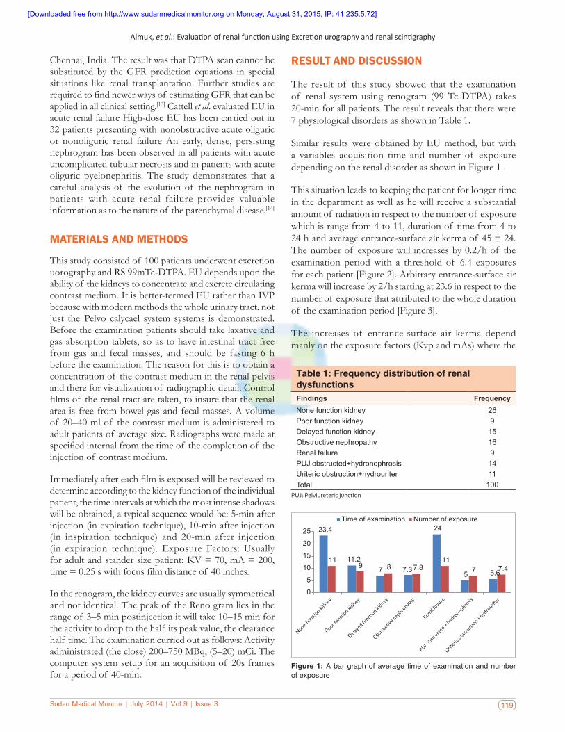
The result of this study showed that the examination of renal system using renogram (99 Tc‑DTPA) takes 20‑min for all patients. The result reveals that there were 7 physiological disorders as shown in Table 1.
Similar results were obtained by EU method, but with a variables acquisition time and number of exposure depending on the renal disorder as shown in Figure 1.
This situation leads to keeping the patient for longer time in the department as well as he will receive a substantial amount of radiation in respect to the number of exposure which is range from 4 to 11, duration of time from 4 to 24 h and average entrance‑surface air kerma of 45 ± 24. The number of exposure will increases by 0.2/h of the examination period with a threshold of 6.4 exposures for each patient [Figure 2]. Arbitrary entrance‑surface air kerma will increase by 2/h starting at 23.6 in respect to the number of exposure that attributed to the whole duration of the examination period [Figure 3].
The increases of entrance‑surface air kerma depend manly on the exposure factors (Kvp and mAs) where the
Table 1: Frequency distribution of renal dysfunctionsFindings FrequencyNone function kidney 26Poor function kidney 9Delayed function kidney 15Obstructive nephropathy 16Renal failure 9PUJ obstructed+hydronephrosis 14Uriteric obstruction+hydrouriter 11Total 100
PUJ: Pelviureteric junction
Figure 1: A bar graph of average time of examination and number of exposure
[Downloaded free from http://www.sudanmedicalmonitor.org on Monday, August 31, 2015, IP: 41.235.5.72]

Almuk, et al.: Evaluation of renal function using Excretion urography and renal scintigraphy
Sudan Medical Monitor | July 2014 | Vol 9 | Issue 3120
entrance‑surface air kerma increases by 0.3/Kvp and mAs as shown in Figures 4 and 5.
The increases of Kvp and mAs depend on the body characteristics of the patient and manly the age of the patient where the Kvp and mAs increases by 0.3/years, starting at 58 and 17 respectively as shown in Figures 5 and 6 respectively.
CONCLUSION
The result of this study demonstrated that excretion uorography and RS using 99mTc‑DTPA had the same sensitivity; while scintigraphy has the advantage over the excretion uorography by saving the patient time and resources as well. Excretion uorography has a variable time
Figure 2: Scatter plot show a direct linear relationship between the number of exposure and duration of excretion urography
Figure 3: Scatter plot show a direct linear relationship between the entrance-surface air kerma and duration of excretion urography
Figure 4: Scatter plot show a direct linear relationship between the entrance-surface air kerma and Kvp
Figure 5: Scatter plot depict a linear relationship between the Kvp and patient age
Figure 6: (a) Scatter plot depict a linear relationship between the Kvp and patient age. (b) Scatter plot depict a linear relationship between the mAs and patient age
a b
[Downloaded free from http://www.sudanmedicalmonitor.org on Monday, August 31, 2015, IP: 41.235.5.72]

Almuk, et al.: Evaluation of renal function using Excretion urography and renal scintigraphy
Sudan Medical Monitor | July 2014 | Vol 9 | Issue 3 121
of acquisition which includes several exposures which depends on personal characteristics age is one of them; if they were not well control, optimization of the end result will be very difficult, which might lead to repetition of the whole examination again.
REFERENCES
1. Kubota K, Atkins HL, Anaise D, Oster ZH, Pollack W. Quantitative evaluation of renal excretion on the dynamic DTPA renal scan. Clin Nucl Med 1989;14:8‑12.
2. EmbonOM,GrosharD,ShapiraC,KoritnyES,LidgiS,MijiritskyJ,et al. Renal scintigraphy in initial evaluation of renal colic. Urology 1992;39:566‑8.
3. Kirchnera PT, Rosenthalla L. Renal transplant evaluation transplant evaluation. Sci Direct 1982;12:370‑8.
4. Klaipetch A, Namwongprom S, Ekmahachai M, Lojanapiwat B. Excretory urography and renal scintigraphy for chronic obstructed kidney:Doesnonopacitymeannonsalvageability?SingaporeMedJ2013;54:267‑70.
5. Milena R, Mom CB, Marina V, Slobodan I, Vladisav S. Radionuclide evaluation of renal function in patients with renal stone treated by extracorporealshockwaveithotripsy.SciJFactaUnivSerMedBiol2000;7:102‑6.
6. American College of Radiology (ACR) guidelines, Exceratroy Uorography. 2010;51:1142‑1148.
7. Choyke PL. Radiologic evaluation of hematuria: Guidelines from the AmericanCollegeofRadiology’sappropriatenesscriteria.AmFam
Physician 2008;78:347‑52.8. Goldman SM, Fishman EK, Rosenshein NB, Gatewood OM,
Siegelman SS. Excretory urography and computed tomography in the initial evaluation of patients with cervical cancer: Are both examinationsnecessary?AJRAmJRoentgenol1984;143:991‑6.
9. Drachman R, Valevici M, Vardy PA. Excretory urography and cystourethrography in the evaluation of children with urinary tract infection. Clin Pediatr (Phila) 1984;23:265‑7.
10. Michael RL, Arthur TR, Sidney U, Charles EP. Evaluation of bronchospasm during excretory urography. Diagn Radiol 1977;124:5.
11. Yalçin H, Ozen A, Günay EC, Ozaslan IA, Ozer C. Can Tc 99m DTPA beUsedinAdultPatientsinEvaluationofRelativeRenalFunctionMeasurement as the Reference Tc 99m DMSA Method? Mol Imaging Radionucl Ther 2011;20:14‑8.
12. Antoniou KM, Malagari K, Tzanakis N, Perisinakis K, Symvoulakis EK, Karkavitsas N, et al. Clearance of technetium‑99m‑DTPA and HRCT findings in the evaluation of patients with idiopathic pulmonaryfibrosis.BMCPulmMed2006;6:4.
13. Senthamizh KS, Karthikeyan S, Sundaramurthy G. A study of glomerular filtration rate estimation by COCKCROFT‑GAULT,MDRD and CKD‑EPI formula in comparison with dtpa renal scan – A comparative study among live related kidney donors in southIndia.IntJBiolMedRes2013;4:3073‑7.
14. CattellWR,McIntoshCS,MoseleyIF,FryIK.Excretionurographyinacuterenalfailure.BrMedJ1973;2:575‑8.
How to cite this article: We will update details while making issue online***
Source of Support: Nil. Conflict of Interest: None declared.
Author Help: Online submission of the manuscripts
Articles can be submitted online from http://www.journalonweb.com. For online submission, the articles should be prepared in two files (first page file and article file). Images should be submitted separately.
1) First Page File: Prepare the title page, covering letter, acknowledgement etc. using a word processor program. All information related to your identity should
be included here. Use text/rtf/doc/pdf files. Do not zip the files.2) Article File: The main text of the article, beginning with the Abstract to References (including tables) should be in this file. Do not include any informa-
tion (such as acknowledgement, your names in page headers etc.) in this file. Use text/rtf/doc/pdf files. Do not zip the files. Limit the file size to 1 MB. Do not incorporate images in the file. If file size is large, graphs can be submitted separately as images, without their being incorporated in the article file. This will reduce the size of the file.
3) Images: Submit good quality color images. Each image should be less than 4096 kb (4 MB) in size. The size of the image can be reduced by decreas-
ing the actual height and width of the images (keep up to about 6 inches and up to about 1800 x 1200 pixels). JPEG is the most suitable file format. The image quality should be good enough to judge the scientific value of the image. For the purpose of printing, always retain a good quality, high resolution image. This high resolution image should be sent to the editorial office at the time of sending a revised article.
4) Legends: Legends for the figures/images should be included at the end of the article file.
[Downloaded free from http://www.sudanmedicalmonitor.org on Monday, August 31, 2015, IP: 41.235.5.72]

Sudan Medical Monitor | July 2014 | Vol 9 | Issue 3122
[Downloaded free from http://www.sudanmedicalmonitor.org on Monday, August 31, 2015, IP: 41.235.5.72]