Original Article Ultrastructural features of endometrial-myometrial interface … · 2016-08-09 ·...
9
Int J Clin Exp Pathol 2014;7(4):1469-1477 www.ijcep.com /ISSN:1936-2625/IJCEP1401069 Original Article Ultrastructural features of endometrial-myometrial interface and its alteration in adenomyosis Ying Zhang 1 , Li Zhou 1 , Tin C Li 2 , Hua Duan 1 , Pei Yu 1 , Hong Y Wang 3 1 Department of Gynecology Minimally Invasive Center, Beijing Obstetrics and Gynecology Hospital, Capital Medical University, Qi He Lou Street 17#, Dongcheng District, Beijing, China, Post code: 100006; 2 Department of Repro- ductive Medicine and Surgery, The Jessop Wing Sheffield Teaching Hospital, The Jessop Wing Tree Root Walk, Sheffield, United Kingdom, Post code: S10 2SF; 3 Department of Pathology, Fu Wai Hospital Cardiovascular Insti- tute, Chinese Academy of Medical Sciences Peking Union Medical College, Beijing, China, Post code: 100037 Received January 20, 2014; Accepted February 27, 2014; Epub March 15, 2014; Published April 1, 2014 Abstract: The endometrial-myometrial interface (EMI) is a specific functional region of uterus. However, our knowl- edge on EMI ultrastructure both in normal uterus and adenomyosis is far from enough to understand its pathology. In this study, used the samples of EMI and outer myometrium (OM) from the adenomyosis hysterectomy specimens and the subjects from the control uteri, we prospectively compared the ultrastructure of myocytes from EMI and OM, the ultrastructural changes of EMI between the proliferative and secretory phases, and the ultrastructural dif- ference of EMI between adenomyosis and the control group. In both adenomyosis and control group, there were differences in ultrastructure between myocytes from EMI and OM. Specifically, the myocytes from EMI were rich in organelles. In contrast, the myocytes from OM had abundant contractile structural components. In the proliferative phase, the myocytes from EMI in adenomyosis had significantly smaller cell and nucleus diameter than those from the control group, but in the secretory phase, the difference was not significant. In the control group, the various ultrastructural features of myocytes from EMI including the mean diameter of cell and nuclei and the myofilaments/ cytoplasm ratio exhibited cyclical changes, but in adenomyosis, the normal cyclical changes were absent. In conclu- sions, there are significant ultrastructural differences between the myocytes from EMI and OM. The myocytes in women with adenomyosis were significantly different to the control subjects, primarily because the normal cyclical changes were absent. Keywords: Adenomyosis, endometrial-myometrial interface, outer myometrium, ultrastructure Introduction The endometrial-myometrial interface (EMI), is also known as “junctional zone”, or “the inner myometrium”, first described in 1983 by Hricak et al [1]. EMI is different from the outer myome- trium (OM) in respect of the embryological ori- gin, structure, function and pathology. Embryo- logically, just like the endometrium, EMI is of Müllerian origin, while the OM is of non-Mülleri- an, mesenchymal origin [2, 3]. Structurally, EMI is the inner one third of myometrium composed of shorter muscle bundles. The OM is the outer two third of myometrium predominantly com- posed of longer muscle fibers. However, on light microscopic examination, the whole myo- metrium appears as a homogeneous structure comprised of smooth muscle cells, myocytes [2]. Tetlow et al. [4] used biopsy needle obtained EMI tissue from the uterus directed by ultra- sound; morphometric analysis of the specimen demonstrated a greater total nuclear area in EMI than the OM. Functionally, contractile activ- ity in the non-pregnant uterus, a type of peri- stalsis-like contraction, has been found to be present in EMI. The peristalsis is involved in sperm transport, regulation of implantation and hemostasis during menstruation [6-11]. Theref- ore, EMI plays a vital role in the various func- tions of the non-pregnant uterus, while the OM is mainly responsible for parturition. Patholo- gically, EMI is the most common region to be affected in adenomyosis and is characterized by irregularity and thickening [2, 3, 6, 8, 9, 12, 13]. Altered EMI contraction can be observed in adenomyosis including hyperperistalsis and
Transcript of Original Article Ultrastructural features of endometrial-myometrial interface … · 2016-08-09 ·...

Int J Clin Exp Pathol 2014;7(4):1469-1477www.ijcep.com /ISSN:1936-2625/IJCEP1401069
Original Article Ultrastructural features of endometrial-myometrial interface and its alteration in adenomyosis
Ying Zhang1, Li Zhou1, Tin C Li2, Hua Duan1, Pei Yu1, Hong Y Wang3
1Department of Gynecology Minimally Invasive Center, Beijing Obstetrics and Gynecology Hospital, Capital Medical University, Qi He Lou Street 17#, Dongcheng District, Beijing, China, Post code: 100006; 2Department of Repro-ductive Medicine and Surgery, The Jessop Wing Sheffield Teaching Hospital, The Jessop Wing Tree Root Walk, Sheffield, United Kingdom, Post code: S10 2SF; 3Department of Pathology, Fu Wai Hospital Cardiovascular Insti-tute, Chinese Academy of Medical Sciences Peking Union Medical College, Beijing, China, Post code: 100037
Received January 20, 2014; Accepted February 27, 2014; Epub March 15, 2014; Published April 1, 2014
Abstract: The endometrial-myometrial interface (EMI) is a specific functional region of uterus. However, our knowl-edge on EMI ultrastructure both in normal uterus and adenomyosis is far from enough to understand its pathology. In this study, used the samples of EMI and outer myometrium (OM) from the adenomyosis hysterectomy specimens and the subjects from the control uteri, we prospectively compared the ultrastructure of myocytes from EMI and OM, the ultrastructural changes of EMI between the proliferative and secretory phases, and the ultrastructural dif-ference of EMI between adenomyosis and the control group. In both adenomyosis and control group, there were differences in ultrastructure between myocytes from EMI and OM. Specifically, the myocytes from EMI were rich in organelles. In contrast, the myocytes from OM had abundant contractile structural components. In the proliferative phase, the myocytes from EMI in adenomyosis had significantly smaller cell and nucleus diameter than those from the control group, but in the secretory phase, the difference was not significant. In the control group, the various ultrastructural features of myocytes from EMI including the mean diameter of cell and nuclei and the myofilaments/cytoplasm ratio exhibited cyclical changes, but in adenomyosis, the normal cyclical changes were absent. In conclu-sions, there are significant ultrastructural differences between the myocytes from EMI and OM. The myocytes in women with adenomyosis were significantly different to the control subjects, primarily because the normal cyclical changes were absent.
Keywords: Adenomyosis, endometrial-myometrial interface, outer myometrium, ultrastructure
Introduction
The endometrial-myometrial interface (EMI), is also known as “junctional zone”, or “the inner myometrium”, first described in 1983 by Hricak et al [1]. EMI is different from the outer myome-trium (OM) in respect of the embryological ori-gin, structure, function and pathology. Embryo- logically, just like the endometrium, EMI is of Müllerian origin, while the OM is of non-Mülleri-an, mesenchymal origin [2, 3]. Structurally, EMI is the inner one third of myometrium composed of shorter muscle bundles. The OM is the outer two third of myometrium predominantly com-posed of longer muscle fibers. However, on light microscopic examination, the whole myo-metrium appears as a homogeneous structure comprised of smooth muscle cells, myocytes
[2]. Tetlow et al. [4] used biopsy needle obtained EMI tissue from the uterus directed by ultra-sound; morphometric analysis of the specimen demonstrated a greater total nuclear area in EMI than the OM. Functionally, contractile activ-ity in the non-pregnant uterus, a type of peri-stalsis-like contraction, has been found to be present in EMI. The peristalsis is involved in sperm transport, regulation of implantation and hemostasis during menstruation [6-11]. Theref- ore, EMI plays a vital role in the various func-tions of the non-pregnant uterus, while the OM is mainly responsible for parturition. Patholo- gically, EMI is the most common region to be affected in adenomyosis and is characterized by irregularity and thickening [2, 3, 6, 8, 9, 12, 13]. Altered EMI contraction can be observed in adenomyosis including hyperperistalsis and

Ultrastructural features of EMI
1470 Int J Clin Exp Pathol 2014;7(4):1469-1477
abnormal peristalsis which may be responsible for the associated clinical manifestations such as dysmenorrhea, metrorrhagia and infertility [3, 5, 6, 8, 9, 14, 15].
The structure and function of EMI has been receiving increasing attention from investiga-tors. Ultrasound and MR imaging are used for localization of EMI and diagnosis of the EMI-
Figure 1. Ultrastructures of endometrial-myometrial interface (EMI) and outer myometrium (OM) in ad-enomyotic and normal uteri. A. EMI of adenomyosis. B. EMI of normal uterus. In both cases, the myocytes are small with cytoplasmic processes. The cell ar-rangement is irregular. The nuclear envelops are smooth. Intercellular spaces are distended. There is no significant difference between the two groups. C. OM of adenomyosis. D. OM of normal uterus. In both cases, the cell arrangement is more regular. The nuclear envelope characterized with convolution and multi-folded. Again, there is no significant difference between the two groups. Scale bar equals to 2 μm. E. EMI myocytes representative picture. Myocytes are usually rich in organelles. There is abundant endo-plasmic reticulum (ER), mitochondria (MT) and Golgi (GI) in cytoplasm but devoid of contractile structural components. Scale bar equals to 500 nm.

Ultrastructural features of EMI
1471 Int J Clin Exp Pathol 2014;7(4):1469-1477
related disease such as adenomy-osis [3, 4, 16-19]. Videovagin- osonography, hysterosalpingoscin-tigraphy, High-resolution ultrasou- nd and cine MR imaging can all detect the contraction of EMI [3, 8, 20, 21]. Immunohistochemistry re- sults reveal that EMI is rich in estrogen and progesterone recep-tors which indicate that ovarian steroid hormones may regulate EMI function [22]. However, the re- search on ultrastructure of EMI is relatively scarce except for the stu- dy of Mehasseb et al. which found that there was no difference bet- ween EMI and OM [5]. In this study, we examined EMI and OM tissue in different menstrual phases obtain- ed from women with and without adenomyosis with a view to desc- ribing the ultrastructural changes in myocytes in women with aden- omyosis.
Materials and methods
Adenomyosis and control group
Ten hysterectomy specimens from pre-menopausal subjects with ade- nomyosis, 5 from the proliferative phase and 5 from the secretory phase, constituted the study gro- up. The diagnosis of adenomyosis was made before surgery based on the clinical symptoms and tran- svaginal ultrasonography, and the- n histologically confirmed postop-eratively. The clinical symptoms in- cluded dysmenorrhea and/or met- rorrhagia. The ultrasonographic di- agnostic criteria as described in previously published studies [23, 24] were as follows: an asymmet-ric or globular uterus; asymmetri-cal thickening of junctional zone in the anterior or posterior wall; sub-endometrial linear striations; het-erogeneous myometrial echotex-ture and myometrial cysts. In our study, we selected the cases with the thickened junctional zone in posterior wall. Another ten hyster-ectomy specimens from pre-meno-
Figure 2. A comparison of myocytes between endometrial-myometrial interface (EMI) and outer myometrium (OM) in the control group (A) and adenomyosis (B). In both groups, myocytes from EMI are significantly (P<0.05) smaller than those from OM in the secretory phase. The myo-filaments/cytoplasm ratio of myocytes from EMI is significantly (P<0.01) less than that from OM in both the proliferative and secretory phases.
Ultrastructural features of EMI
1472 Int J Clin Exp Pathol 2014;7(4):1469-1477
pausal subjects without adenomyosis but with early cervical neoplasm (CINIII and cervical cancer stage Ia), 5 from the proliferative phase and 5 from the secretory phase, constituted the control group. The mean±SD age of the study and control groups were 45.5±5.5 and 47.6±3.0 years respectively (no significant dif-ference). All patients had regular menstrual cycle (25-35 days), without myoma, previous uterus surgery and hormone use in the preced-ing three months. Patients with vascular and renal disease were excluded. This study was approved by our institutional ethics review board. Informed consent was obtained from each patient.
EMI specimens
After surgical removal, the uteri were immedi-ately opened with a Y-shaped incision. Multiple 5 mm3 samples were obtained from the EMI (underneath the endometrium) and the outer third of the myometrium. All samples were obtained from the posterior wall of the uterus near the fundus. Parallel full uterine samples were obtained for traditional histological exami-nation to confirm the presence or absence of adenomyosis and the stage of the menstrual cycle.
Transmission electron microscope assessed
Samples of EMI and OM were fixed in 2.5% buff-ered glutaraldehyde for 24 hours. The samples were then washed by 0.1 M phosphate buffer, post-fixed in buffered 1% osmium tetroxide for
30 minutes, rewashed and dehydrated in ace-tone series. They were embedded in Epon812 and then trimmed. One um semi-thin section was cut and stained by toluidine blue and basic fuchsin. Under optical microscope, EMI speci-mens were re-positioned and selected. Excess tissue was trimmed. Ultrathin 70-nm sections were cut from each sample, collected on 300 copper mesh grids, counter stained with 2% uranyl acetate and Reynolds’ lead citrate and examined using a JEM1010 transmission elec-tron microscope (Jeol, Tokyo, Japan).
Images analysis
Images were evaluated and the related indices were measured by two independent electron microscopy specialists who were blinded to the information of the specimen. For each speci-men examined under electron microscopy, 10 images were selected randomly in low magnifi-cation and high magnification, respectively. Using the Scion image measurement software, the mean diameter of cells and nuclei were measured through the nuclear center of the cells’ longitudinal axis in the low magnification pictures (2 μm, ×2500). The muscle fiber area and the cytoplasmic area were measured thr- ough the nuclear center section in high magni-fication pictures (500 nm, ×10000). The aver-age myofilaments/cytoplasm ratio was calcula- ted.
Statistical analysis
Statistical analysis was performed using SPSS for Windows 17.0. Data were expressed as the mean and standard errors of the mean. Com- parisons of the means were made using Stud- ent’s t test for non-paired samples. A P value of <0.05 was considered statistically significant.
Results
Comparison of ultrastructural features of myo-cytes from EMI and OM
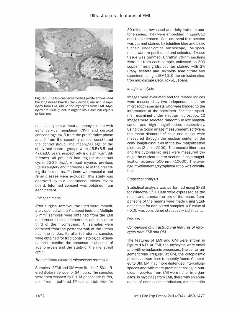
The features of EMI and OM were shown in Figure 1A-D. At EMI, the myocytes were small and with cytoplasmic processes. The cell arran- gement was irregular. At OM, the cytoplasmic processes were less frequently found. Compar- ed to OM, EMI had more distended intercellular spaces and with more prominent collagen bun-dles; myocytes from EMI were richer in organ-elles. In myocytes from EMI, there was an abun-dance of endoplasmic reticulum, mitochondria
Figure 3. The typical dense bodies (white arrows) and the long dense bands (black arrows) are rich in myo-cytes from OM, unlike the myocytes from EMI. Myo-cytes are usually lack in organelles. Scale bar equals to 500 nm.
Ultrastructural features of EMI
1473 Int J Clin Exp Pathol 2014;7(4):1469-1477
and Golgi (Figure 1E), the mitochondria exhib-ited unfolding of the internal cristae, and the rough endoplasmic reticulum and Golgi appara-tus were more prominent, denoting active pro-tein synthesis; however, the contraction related structures, such as cytoplasmic myofilaments, dense bodies and dense bands in cell mem-brane were less abundant, denoting diminished contractive capacity. In addition, myocytes fro- m EMI had smooth nuclear envelope, in con-trast to the convoluted nuclear envelope observ- ed in myocytes from OM.
The features of myocytes from EMI and OM in adenomyosis and control group were compared in Figure 2A, 2B. In proliferative phase, the mean cell diameter and the mean nucleus diameter of myocytes from EMI were not signifi-cantly different from those of OM both in the adenomyosis and the control groups. In the secretory phase, the mean cell diameter of myocytes from EMI was significantly (P<0.05)
significantly (P<0.01) higher than that from EMI (0.34±0.02) (Figure 2A, 2B).
Comparison of EMI ultrastructure between adenomyosis and control groups
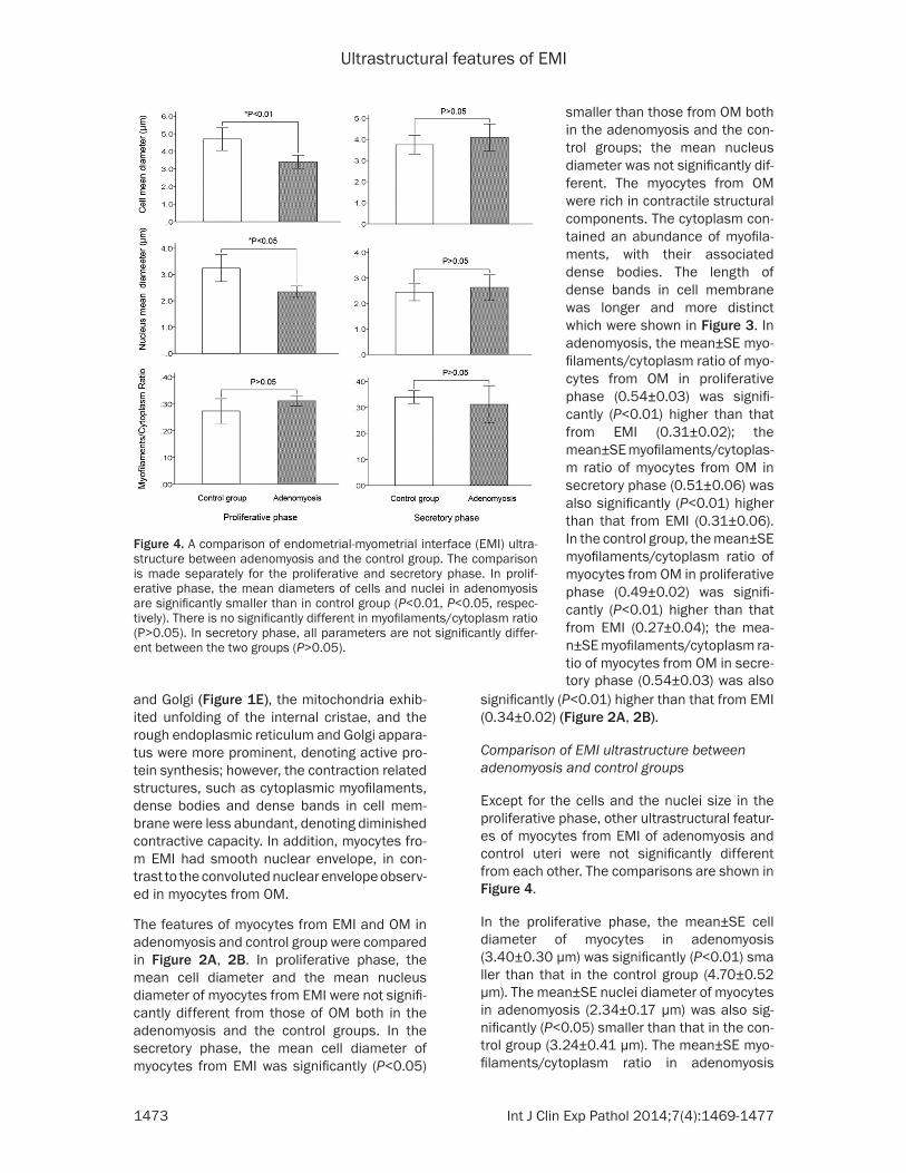
Except for the cells and the nuclei size in the proliferative phase, other ultrastructural featur- es of myocytes from EMI of adenomyosis and control uteri were not significantly different from each other. The comparisons are shown in Figure 4.
In the proliferative phase, the mean±SE cell diameter of myocytes in adenomyosis (3.40±0.30 μm) was significantly (P<0.01) sma ller than that in the control group (4.70±0.52 μm). The mean±SE nuclei diameter of myocytes in adenomyosis (2.34±0.17 μm) was also sig-nificantly (P<0.05) smaller than that in the con-trol group (3.24±0.41 μm). The mean±SE myo-filaments/cytoplasm ratio in adenomyosis
Figure 4. A comparison of endometrial-myometrial interface (EMI) ultra-structure between adenomyosis and the control group. The comparison is made separately for the proliferative and secretory phase. In prolif-erative phase, the mean diameters of cells and nuclei in adenomyosis are significantly smaller than in control group (P<0.01, P<0.05, respec-tively). There is no significantly different in myofilaments/cytoplasm ratio (P>0.05). In secretory phase, all parameters are not significantly differ-ent between the two groups (P>0.05).
smaller than those from OM both in the adenomyosis and the con-trol groups; the mean nucleus diameter was not significantly dif-ferent. The myocytes from OM were rich in contractile structural components. The cytoplasm con-tained an abundance of myofila-ments, with their associated dense bodies. The length of dense bands in cell membrane was longer and more distinct which were shown in Figure 3. In adenomyosis, the mean±SE myo-filaments/cytoplasm ratio of myo-cytes from OM in proliferative phase (0.54±0.03) was signifi-cantly (P<0.01) higher than that from EMI (0.31±0.02); the mean±SE myofilaments/cytoplas- m ratio of myocytes from OM in secretory phase (0.51±0.06) was also significantly (P<0.01) higher than that from EMI (0.31±0.06). In the control group, the mean±SE myofilaments/cytoplasm ratio of myocytes from OM in proliferative phase (0.49±0.02) was signifi-cantly (P<0.01) higher than that from EMI (0.27±0.04); the mea- n±SE myofilaments/cytoplasm ra- tio of myocytes from OM in secre-tory phase (0.54±0.03) was also
Ultrastructural features of EMI
1474 Int J Clin Exp Pathol 2014;7(4):1469-1477
(0.31±0.02) was not significantly (P=0.13) dif-ferent from that in the control group (0.27±0.04).
In the secretory phase, all parameters including mean cell diameter, mean nuclei diameter and the myofilaments/cytoplasm ratio in adenomy-osis were not significantly different from those in the control group. The amount and the mor-phology of the perinuclear cell organelles, such as endoplasmic reticulum, mitochondria and Golgi, were not different between the two groups in both the proliferative and secretory phases.
Comparison of EMI ultrastructure between proliferative and secretory phases
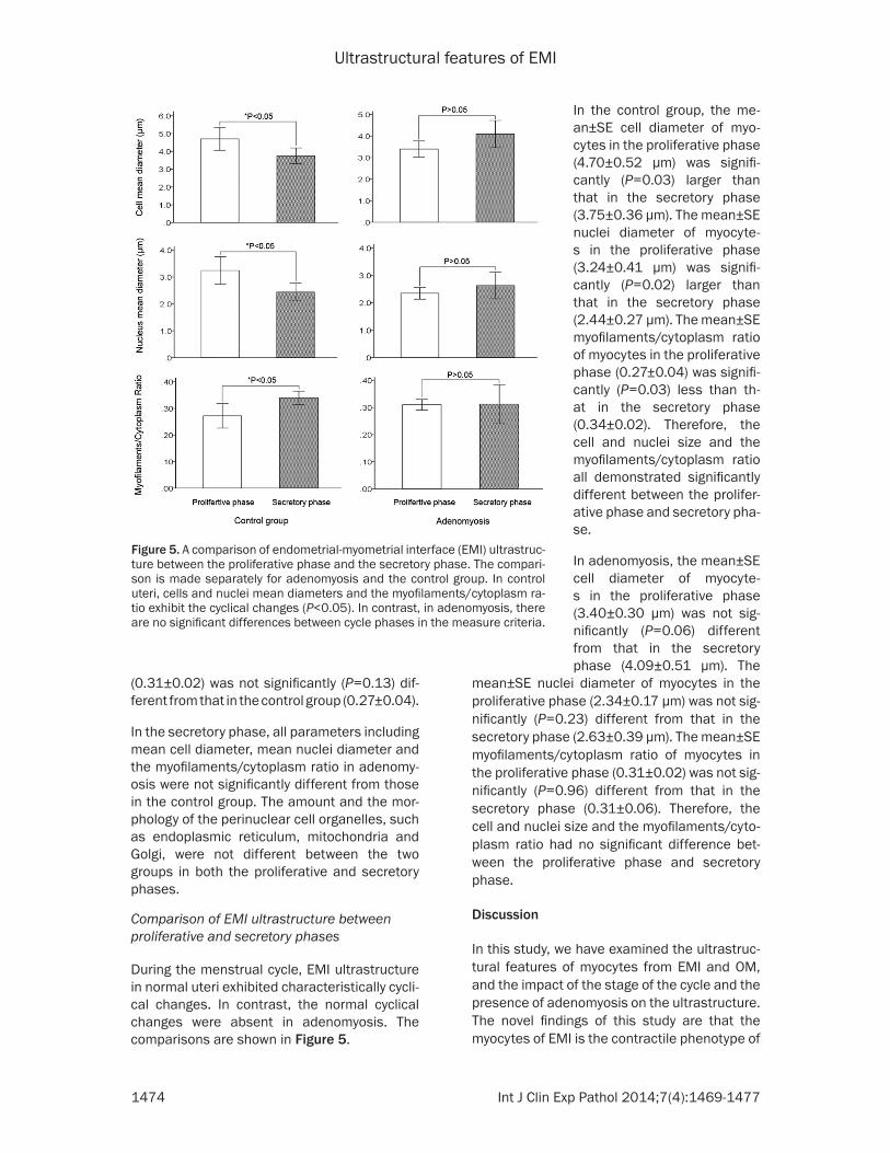
During the menstrual cycle, EMI ultrastructure in normal uteri exhibited characteristically cycli-cal changes. In contrast, the normal cyclical changes were absent in adenomyosis. The comparisons are shown in Figure 5.
mean±SE nuclei diameter of myocytes in the proliferative phase (2.34±0.17 μm) was not sig-nificantly (P=0.23) different from that in the secretory phase (2.63±0.39 μm). The mean±SE myofilaments/cytoplasm ratio of myocytes in the proliferative phase (0.31±0.02) was not sig-nificantly (P=0.96) different from that in the secretory phase (0.31±0.06). Therefore, the cell and nuclei size and the myofilaments/cyto-plasm ratio had no significant difference bet- ween the proliferative phase and secretory phase.
Discussion
In this study, we have examined the ultrastruc-tural features of myocytes from EMI and OM, and the impact of the stage of the cycle and the presence of adenomyosis on the ultrastructure. The novel findings of this study are that the myocytes of EMI is the contractile phenotype of
Figure 5. A comparison of endometrial-myometrial interface (EMI) ultrastruc-ture between the proliferative phase and the secretory phase. The compari-son is made separately for adenomyosis and the control group. In control uteri, cells and nuclei mean diameters and the myofilaments/cytoplasm ra-tio exhibit the cyclical changes (P<0.05). In contrast, in adenomyosis, there are no significant differences between cycle phases in the measure criteria.
In the control group, the me- an±SE cell diameter of myo-cytes in the proliferative phase (4.70±0.52 μm) was signifi-cantly (P=0.03) larger than that in the secretory phase (3.75±0.36 μm). The mean±SE nuclei diameter of myocyte- s in the proliferative phase (3.24±0.41 μm) was signifi-cantly (P=0.02) larger than that in the secretory phase (2.44±0.27 μm). The mean±SE myofilaments/cytoplasm ratio of myocytes in the proliferative phase (0.27±0.04) was signifi-cantly (P=0.03) less than th- at in the secretory phase (0.34±0.02). Therefore, the cell and nuclei size and the myofilaments/cytoplasm ratio all demonstrated significantly different between the prolifer-ative phase and secretory pha- se.
In adenomyosis, the mean±SE cell diameter of myocyte- s in the proliferative phase (3.40±0.30 μm) was not sig-nificantly (P=0.06) different from that in the secretory phase (4.09±0.51 μm). The

Ultrastructural features of EMI
1475 Int J Clin Exp Pathol 2014;7(4):1469-1477
smooth muscle cells; whilst myocytes from EMI belonging to the synthetic phenotype.
Myocyte ultrastructure of EMI and OM
In comparing the ultrastructure of myocytes from EMI and OM, we found a number of signifi-cant differences. Myocytes from OM had all the characteristics of cells involved in contractile function, including a higher content of myofila-ments in the cytoplasm, more prominent dense bodies and dense bands, and the convolutional nuclear membrane. The convoluted nuclear membrane is thought to be due to forces exert-ed on the internal structure of the myocytes by the contractile elements, which is one of the features of the contractile smooth muscle cells [25]. On the other hand, myocytes from EMI appear to have a different set of ultrastructure usually associated with protein synthesis, nam- ely an increased number of organelles, the larg-er nucleus/cytoplasm ratio and the less con-tractile components.
Previous studies on the vascular muscles showed that there are two spectrums of smooth muscle cells, contractile phenotype and syn-thetic phenotype. At the contractile extreme are smooth muscle cells with a fully functional contractile apparatus. The cells are character-ized by densely packed elements of the con-tractile machinery, (myofilaments, dense bod-ies, dense bands), but with minimal rough endoplasmic reticulum, Golgi, and free ribo-some [26, 27]. This type of cells is responsible for producing muscle force. At the synthetic phenotype extreme, the ultrastructure shows a cytoplasm devoid of contractile bundles with extensive rough endoplasmic reticulum, Golgi, and ribosome [28]. A synthetic phenotype is correlated with smooth muscle cells prolifera-tion and is reported to play an important role in regulating local microenvironment [29, 30].
Our observation also suggests that there are two subtypes in the uterus. Myocytes from OM are the contractile phenotype of smooth mus-cle cells; whilst myocytes from EMI belonging to the synthetic phenotype. The characteristics of myocytes from EMI and OM are consistent with their differential functions. The force during labor exerted by myocytes from OM is more powerful than the peristaltic waves produced by myocytes from EMI. Synthetic phenotype of myocytes from EMI is supposed to secret func-
tional proteins to regulate EMI morphogenesis and to participate in non-pregnant uterine func-tion, such as interacting with endometrium to regulate trophoblastic invasion in early preg-nancy [31-33].
Cyclical changes in myocyte ultrastructure
In comparing the ultrastructure of myocytes between adenomyosis and normal uteri, we found evidence of cyclical changes in the myo-cytes in control subjects but the cyclical chang-es appeared to have disappeared in subjects with adenomyosis. In the control group, the myocyte size, nuclear size, and the myofila-ments/cytoplasm ratio in the proliferative phase are all significantly different from those in the secretory phase. However, all the param-eters in adenomyosis group are not significant-ly different between the proliferative phase and secretory phase, which may provide an expla-nation for the loss of cyclical changes of peri-stalsis observed in the EMI.
In contrast to our observation that there was a cyclical change in the ultrastructural features of the myocytes from EMI, Mehasseb et al. [5] were unable to find any significant difference in the ultrastructure of myocytes between differ-ent phases of the menstrual cycle. The most likely explanation for the difference in the observations is in the sample size. The number of cases in Mehasseb’s study was rather small, with a total of only four adenomyosis cases and 6 control cases, and with only one adenomyo-sis case in the secretory phase. The small sam-ple size could have limited the power of the study to detect a significant cyclical difference. In contrast, our study involved a larger sample size, and the samples were carefully matched for the different phases of the menstrual cycle.
Noe et al. [22] found that myocytes in EMI is rich in estrogen and progesterone receptors which parallel those of the endometrium and exhibit a cyclic pattern, suggesting that the sex steroid hormones play a role in regulating the structure and function of the myocytes of EMI. It has been found that uterine contractions emanating from EMI vary throughout the men-strual cycle [3, 6, 7, 10], which in turn produce the cyclical peristaltic waves in normal sub-jects. Alternation in ER expression in the endo-metrium and inner myometrium [34] may account for the abnormal peristalsis observed in adenomyosis [9, 15, 35].

Ultrastructural features of EMI
1476 Int J Clin Exp Pathol 2014;7(4):1469-1477
In this study, we have detected subtle ultra-structural changes in the myocytes of women with adenomyosis in the proliferative phase, but there does not appear to be any obvious changes in the myofilaments. It seems that the contractile elements in myocytes of adenomyo-sis are not significantly deranged. The previous observation [6, 15, 35] that there are recog-nized hyperperistalsis in adenomyosis cannot be explained by changes in the ultrastructure of the myocytes. An earlier study by Mehasseb et al. [5] suggested that myocytes from EMI of adenomyosis displayed features of hypertrophy which in turn account for the thickening of the EMI observed in women with adenomyosis [3, 12, 13]. However, our finding that myocytes of EMI are smaller in adenomyosis (in proliferative phase) suggests that increase cell number is a more likely explanation for the thickening of the EMI. Morphometric study is required to confirm if it is indeed the case.
Future studies
To the best of knowledge, the observations in our study that (a) there are significant differ-ences in ultrastructure between myocytes from EMI and OM, and (b) the normal cyclical chang-es in ultrastructure observed in myocytes appear to have been lost in adenomyosis, are original. The latter observation identified the link between structural changes in myocytes of EMI and adenomyosis. However, in this study, we have not examined the functional aspects of myocytes from the EMI. Further studies are required, almost certainly involving expression of protein and mRNA from biopsy specimens obtained from the EMI. A specially designed hysteroscopically directed myometrial biopsy instrument, called spirotome, has been devel-oped by Prof Stephan Gordts (personal comm- unication).
Conclusion
To conclude, the myocytes from EMI and OM have distinctive ultrastructural features adapt-ed for different functions. We observed cyclical ultrastructural changes of EMI in control sub-ject but the changes appear to be lost in women with adenomyosis. The observations are con-sistent with functional disruption in the EMI of women with adenomyosis.
Acknowledgements
This study is support by State Key Clinical Specialty Construction Project of China (2010);
Fund of Capital Medical Development and Research (2007-3057).
Disclosure of conflict of interest
None of the authors have a conflict of interest.
Address correspondence to: Dr. Hua Duan, De- partment of Gynecology Minimally Invasive Center, Beijing Obstetrics and Gynecology Hospital, Capital Medical University, Qi He Lou Street 17#, Dongcheng District, Beijing, China, Post code: 100006. Tel: 086-13501019066; Fax: 086-01065235524; E- mail: [email protected]
References
[1] Hricak H, Alpers C, Crooks LE, Sheldon PE. Magnetic resonance imaging of the female pelvis: initial experience. AJR Am J Roentgenol 1983; 141: 1119-28.
[2] Fusi L, Cloke B, Brosens JJ. The uterine junc-tional zone. Best Pract Res Clin Obstet Gynae-col 2006; 20: 479-91.
[3] Brosens JJ, Barker FG, De Souza NM. Myome-trial zonal differentiation and uterine junction-al zone hyperplasia in the non-pregnant uter-us. Hum Reprod Update 1998; 4: 496-502.
[4] Tetlow RL, Richmond I, Manton DJ, Greenman J, Turnbull LW, Killick SR. Histological analysis of the uterine junctional zone as seen by trans-vaginal ultrasound. Ultrasound Obstet Gynecol 1999; 14: 188-93.
[5] Mehasseb MK, Bell SC, Pringle JH, Habiba MA. Uterine adenomyosis is associated with ultra-structural features of altered contractility in the inner myometrium. Fertil Steril 2010; 93: 2130-6.
[6] Kunz G, Leyendecker G. Uterine peristaltic ac-tivity during the menstrual cycle: characteriza-tion, regulation, function and dysfunction. Re-prod Biomed Online 2002; 4: 5-9.
[7] Daels J. Uterine contractility patterns of the outer and inner zones of the myometrium. Ob-stet Gynecol 1974; 44: 315-26.
[8] Naftalin J, Jurkovic D. The endometrial-myome-trial junction: a fresh look at a busy crossing. Ultrasound Obstet Gynecol 2009; 34: 1-11.
[9] Brosens I, Derwig I, Brosens J, Fusi L, Benag-iano G, Pijnenborg R. The enigmatic uterine junctional zone: the missing link between re-productive disorders and major obstetrical dis-orders? Hum Reprod 2010; 25: 569-74.
[10] Ijland MM, Evers JL, Dunselman GA, van Katwi-jk C, Lo CR, Hoogland HJ. Endometrial wavelike movements during the menstrual cycle. Fertil Steril 1996; 65: 746-749.
[11] Evers JL, Hoogland HJ. Velocity of endometrial wavelike activity in spontaneous cycles. Fertil Steril 1997; 68: 72-75.
Ultrastructural features of EMI
1477 Int J Clin Exp Pathol 2014;7(4):1469-1477
[12] Brosens JJ, De Souza NM, Barker FG. Uterine junctional zone: function and disease. Lancet 1995; 346: 558-60.
[13] Bazot M, Cortez A, Darai E, Rouger J, Chopier J, Antoine JM, Uzan S. Ultrasonography com-pared with magnetic resonance imaging for the diagnosis of adenomyosis: correlation with histopathology. Hum Reprod 2001; 16: 2427-33.
[14] Novellas S, Chassang M, Delotte J, Toullalan O, Chevallier A, Bouaziz J, Chevallier P. AJR Am J Roentgenol 2011; 196: 1206-13.
[15] Kissler S, Hamscho N, Zangos S, Wiegratz I, Schlichter S, Menzel C, Doebert N, Gruenwald F, Vogl TJ, Gaetje R, Rody A, Siebzehnruebl E, Kunz G, Leyendecker G, Kaufmann M. Utero-tubal transport disorder in adenomyosis and endometriosis-a cause for infertility. BJOG 2006; 113: 902-8.
[16] Naftalin J, Hoo W, Pateman K, Mavrelos D, Hol-land T, Jurkovic D. How common is adenomyo-sis? A prospective study of prevalence using transvaginal ultrasound in a gynaecology clin-ic. Hum Reprod 2012; 27: 3432-9.
[17] Thalluri V, Tremellen KP. Ultrasound diagnosed adenomyosis has a negative impact on suc-cessful implantation following GnRH antago-nist IVF treatment. Hum Reprod 2012; 27: 3487-92.
[18] Kishi Y, Suginami H, Kuramori R, Yabuta M, Suginami R, Taniguchi F. Four subtypes of ad-enomyosis assessed by magnetic resonance imaging and their specification. Am J Obstet Gynecol 2012; 207: 114, e1-7.
[19] Maheshwari A, Gurunath S, Fatima F, Bhat-tacharya S. Adenomyosis and subfertility: a systematic review of prevalence, diagnosis, treatment and fertility outcomes. Hum Reprod Update 2012; 18: 374-92.
[20] Kido A, Togashi K, Kataoka M, Wiegratz I, Schli-chter S, Menzel C, Doebert N, Gruenwald F, Vogl TJ, Gaetje R, Rody A, Siebzehnruebl E, Kunz G, Leyendecker G, Kaufmann M. The ef-fect of oral contraceptives on uterine contrac-tility and menstrual pain: an assessment with cine MR imaging. Hum Reprod 2007; 22: 2066-71.
[21] Kunz G, Beil D, Huppert P, Leyendecker G. Structural abnormalities of the uterine wall in women with endometriosis and infertility visu-alized by vaginal sonography and magnetic resonance imaging. Hum Reprod 2000; 15: 76-82.
[22] Noe M, Kunz G, Herbertz M, Mall G, Leyen-decker G. The cyclic pattern of the immunocy-tochemical expression of oestrogen and pro-gesterone receptors in human myometrial and endometrial layers: characterization of the en-dometrial-subendometrial unit. Hum Reprod 1999; 14: 190-7.
[23] Fedele L, Bianchi S, Dorta M, Arcaini L, Zanotti F, Carinelli S. Transvaginal ultrasonography in the diagnosis of diffuse adenomyosis. Fertil Steril 1992; 58: 94-7.
[24] Kepkep K, Tuncay YA, Göynümer G, Tutal E. Transvaginal sonography in the diagnosis of adenomyosis:which findings are most accu-rate? Ultrasound Obstet Gynecol 2007; 30: 341-345.
[25] Lane BP. Alterations in the cytologic detail of intestinal smooth muscle cells in various stag-es of contraction. J Cell Biol 1965; 27: 199-213.
[26] Rzucidlo EM, Martin KA, Powell RJ. Regulation of vascular smooth muscle cell differentiation. J Vasc Surg 2007; 45: A25-32.
[27] Beamish JA, He P, Kottke-Marchant K, March-ant RE. Molecular regulation of contractile smooth muscle cell phenotype: implications for vascular tissue engineering. Tissue Eng Part B Rev 2010; 16: 467-91.
[28] Hedin U, Thyberg J. Plasma fibronectin pro-motes modulation of arterial smooth-muscle cells from contractile to synthetic phenotype. Differentiation 1987; 33: 239-46.
[29] Rainger GE, Nash GB. Cellular pathology of atherosclerosis: smooth muscle cells prime co-cultured endothelial cells for enhanced leuko-cyte adhesion. Circ Res 2001; 88: 615-22.
[30] Warner SJC, Libby P. Human smooth muscle cells: target and source of tumour necrosis fac-tor. J Immunol 1989; 142: 100-109.
[31] Cameron IT, Davenport AP, van Papendorp C, Barker PJ, Huskisson NS, Gilmour RS, Brown MJ, Smith SK. Endothelin-like immunoreactivi-ty in human endometrium. J Reprod Fertil 1992; 95: 623-628.
[32] Bacon CR, Morrison JJ, O’Reilly G, Cameron IT, Davenport AP. ETA and ETB endothelin recep-tors in human myometrium characterized by the subtype selective ligands BQ123, BQ3020, FR139317 and PD151242. J Endocrinol 1995; 144: 127-134.
[33] Burrows TD, King A, Loke YW. Expression of in-tegrins by human trophoblast and differential adhesion to laminin or fibronectin. Hum Re-prod 1993; 8: 475-84.
[34] Mehasseb MK, Panchal R, Taylor AH, Brown L, Bell SC, Habiba M. Estrogen and progesterone receptor isoform distribution through the men-strual cycle in uteri with and without adeno-myosis. Fertil Steril 2011; 95: 2228-35, 2235.e1.
[35] Leyendecker G, Wildt L, Mall G. The pathophys-iology of endometriosis and adenomyosis: tis-sue injury and repair. Arch Gynecol Obstet 2009; 280: 529-38.