Original article Root and canal configurations of ...
5
93 Abstract: This cone beam computed tomography (CBCT) study aimed to describe the maxillary premolar anatomy of a South African subpopu- lation using two classification systems. A total of 601 premolars were evaluated. For each tooth, the root number and canal configurations were described using the classification systems devised by Vertucci (1984) and Ahmed et al. (2017). Correlations between root number and sex were determined using the chi-squared test (P = 0.05). Two roots were present in approximately half of all maxillary first premolars (54.1%, n = 171/316). The majority of maxillary second premolars displayed one root (78.2%, n = 223/285). Single-rooted maxillary second premolars were more common in females (P < 0.05). The Vertucci type IV configuration was most prevalent in the maxillary first premolars. In contrast, maxil- lary second premolars showed a greater tendency toward Vertucci’s type I configuration. The classification proposed by Ahmed et al. indicated the most prevalent maxillary first premolar configuration to be 2 MP B 1 P 1 . The most common configuration among the maxillary second premolars was 1 MP 1 . Diverse root and canal anatomical presentations were found in this subpopulation. Both classification systems adequately describe maxillary premolar anatomy; however, the system proposed by Ahmed et al. may more accurately describe complex teeth. Keywords; classification, cone beam computed tomography, maxillary premolars, root canal Introduction Endodontic treatment aims to clean, shape, and obturate entire root canal system to achieve the resolution of symptoms associated with irreversible inflammation of the pulp or infection of the pulpal or periapical tissues [1]. However, the root canal system may be complex [2] and/or colonized by a variety of micro-organisms [3]. Thus, a detailed understanding of tooth anatomy is vital to pursuing endodontic treatment because the inability to detect and treat any and all identified canals may result in treatment failure [4]. It is well known that root and canal configurations of the permanent human dentition may show considerable variation among them [5]. Maxillary premolars are no exception; the number of roots and canal configurations of these teeth have previously been described in different populations [2,6,7]. Various methods have been employed to study maxil- lary premolar anatomy. These include clearing and dye-staining [8,9], plastic casts [10], visual examination with magnification [9,11], and cone beam computed tomography (CBCT) [2,6,7]. The Vertucci classification system [5], introduced in 1984, has histori- cally been used for classifying canal configurations. Complexities of the canal systems of certain teeth (e.g., complex three-rooted maxillary premo- lars) may not allow for unambiguous description given their classification according to the Vertucci system [12]. An alternative system allowing for the detailed description of root and canal anatomy was therefore proposed by Ahmed and colleagues [12]. The two-dimensional nature of periapical radiographs may result in missed roots and canals [13]. Changing the horizontal tube angulation may improve visualization of the maxillary premolar anatomy [14]. The deploy- ment of this technique may, however, be limited in patients with a narrow palatal vault. Three-dimensional diagnostic imaging modalities such as CBCT allow for the greater detection of root and canal morphology prior to endodontic treatment [13]. CBCT is superior to periapical radiography in the successful detection of root canal anatomy [15-17]. When considering extracted teeth, Neelakantan demonstrated CBCT to be as accurate as the clearing and staining technique when determining maxillary premolar root and canal anatomy [16]. No previous South African anatomical studies of maxillary premolar teeth in living patients or extracted teeth using CBCT were located in the literature. The present study therefore aimed to determine the configura- tion of root and canal anatomy of the maxillary first and second premolars using CBCT in patients attending the Oral and Dental Hospital, University of Pretoria, South Africa. Both the Vertucci as well as an alternative clas- sification system proposed by Ahmed et al. [12] were used to classify these structures. Maxillary first and second premolars were organized by sex and side and compared with previously studied population groups. Materials and Methods Sample selection This cross-sectional retrospective study evaluated a total of 601 maxillary premolar images (316 first and 285 second maxillary premolars, respec- tively) obtained from 190 CBCT scans. The average age of the subjects was 36.9 years (range: 14-84 years). The images were used to determine the root number and canal configurations of maxillary premolars. Both left and right maxillary first and second premolars were included. Scans from both male (n = 81) and female (n = 109) subjects were evaluated. Partici- pant age at the time of the scans was recorded, but no information on race or ethnicity was collected. The existing CBCT database was used and no new scans were acquired for this study. Scans were assessed chronologi- cally back from the most recently acquired one until the necessary sample size was achieved. The study time period ranged from November 2017 to December 2018. Inclusion criteria Scans containing fully-formed maxillary premolars were included in this research. The scans needed to be of an acceptable enough quality that individual roots and canals might be able to be visualized. Only scans that included the entire pulp chamber and root canal system were considered. Exclusion criteria Teeth were excluded for the following reasons: premolars with open apices, incompletely visualized teeth, evidence of previous endodontic treatment, the presence of posts and crowns, surgical or pathological alterations made to tooth anatomy, or the existence of artifacts impeding proper visualiza- tion of tooth anatomy. Image acquisition All scans were acquired by a CBCT unit (Planmeca Pro-max 3D Max; Plan- meca Oy, Helsingfors, Finland) in the Division of Radiology, Department of Oral Pathology and Oral Biology, University of Pretoria, by an experi- Journal of Oral Science, Vol. 62, No. 1, 93-97, 2020 Original article Root and canal configurations of maxillary premolars in a South African subpopulation using cone beam computed tomography and two classification systems Glynn D. Buchanan 1) , Mohamed Y. Gamieldien 2) , Sheree Tredoux 1) , and Zunaid I. Vally 1) 1) Department of Odontology, School of Dentistry, University of Pretoria, Pretoria, South Africa 2) Department of Maxillofacial and Oral Surgery, School of Dentistry, University of Pretoria, Pretoria, South Africa (Received April 15, 2019; Accepted June 20, 2019) Correspondence to Dr. Glynn D. Buchanan, Department of Odontology, School of Dentistry, University of Pretoria, 31 Bophelo Road, Prinshof Campus, Riviera, Pretoria 0002, South Africa E-mail: [email protected] Color figures can be viewed in the online issue at J-STAGE. doi.org/10.2334/josnusd.19-0160 DN/JST.JSTAGE/josnusd/19-0160
Transcript of Original article Root and canal configurations of ...

93
Abstract: This cone beam computed tomography (CBCT) study aimed to describe the maxillary premolar anatomy of a South African subpopu-lation using two classification systems. A total of 601 premolars were evaluated. For each tooth, the root number and canal configurations were described using the classification systems devised by Vertucci (1984) and Ahmed et al. (2017). Correlations between root number and sex were determined using the chi-squared test (P = 0.05). Two roots were present in approximately half of all maxillary first premolars (54.1%, n = 171/316). The majority of maxillary second premolars displayed one root (78.2%, n = 223/285). Single-rooted maxillary second premolars were more common in females (P < 0.05). The Vertucci type IV configuration was most prevalent in the maxillary first premolars. In contrast, maxil-lary second premolars showed a greater tendency toward Vertucci’s type I configuration. The classification proposed by Ahmed et al. indicated the most prevalent maxillary first premolar configuration to be 2MP B1P1. The most common configuration among the maxillary second premolars was 1MP1. Diverse root and canal anatomical presentations were found in this subpopulation. Both classification systems adequately describe maxillary premolar anatomy; however, the system proposed by Ahmed et al. may more accurately describe complex teeth.
Keywords; classification, cone beam computed tomography, maxillary premolars, root canal
Introduction
Endodontic treatment aims to clean, shape, and obturate entire root canal system to achieve the resolution of symptoms associated with irreversible inflammation of the pulp or infection of the pulpal or periapical tissues [1]. However, the root canal system may be complex [2] and/or colonized by a variety of micro-organisms [3]. Thus, a detailed understanding of tooth anatomy is vital to pursuing endodontic treatment because the inability to detect and treat any and all identified canals may result in treatment failure [4].
It is well known that root and canal configurations of the permanent human dentition may show considerable variation among them [5]. Maxillary premolars are no exception; the number of roots and canal configurations of these teeth have previously been described in different populations [2,6,7]. Various methods have been employed to study maxil-lary premolar anatomy. These include clearing and dye-staining [8,9], plastic casts [10], visual examination with magnification [9,11], and cone beam computed tomography (CBCT) [2,6,7].
The Vertucci classification system [5], introduced in 1984, has histori-cally been used for classifying canal configurations. Complexities of the canal systems of certain teeth (e.g., complex three-rooted maxillary premo-lars) may not allow for unambiguous description given their classification according to the Vertucci system [12]. An alternative system allowing for the detailed description of root and canal anatomy was therefore proposed
by Ahmed and colleagues [12].The two-dimensional nature of periapical radiographs may result in
missed roots and canals [13]. Changing the horizontal tube angulation may improve visualization of the maxillary premolar anatomy [14]. The deploy-ment of this technique may, however, be limited in patients with a narrow palatal vault. Three-dimensional diagnostic imaging modalities such as CBCT allow for the greater detection of root and canal morphology prior to endodontic treatment [13]. CBCT is superior to periapical radiography in the successful detection of root canal anatomy [15-17]. When considering extracted teeth, Neelakantan demonstrated CBCT to be as accurate as the clearing and staining technique when determining maxillary premolar root and canal anatomy [16].
No previous South African anatomical studies of maxillary premolar teeth in living patients or extracted teeth using CBCT were located in the literature. The present study therefore aimed to determine the configura-tion of root and canal anatomy of the maxillary first and second premolars using CBCT in patients attending the Oral and Dental Hospital, University of Pretoria, South Africa. Both the Vertucci as well as an alternative clas-sification system proposed by Ahmed et al. [12] were used to classify these structures. Maxillary first and second premolars were organized by sex and side and compared with previously studied population groups.
Materials and Methods
Sample selectionThis cross-sectional retrospective study evaluated a total of 601 maxillary premolar images (316 first and 285 second maxillary premolars, respec-tively) obtained from 190 CBCT scans. The average age of the subjects was 36.9 years (range: 14-84 years). The images were used to determine the root number and canal configurations of maxillary premolars. Both left and right maxillary first and second premolars were included. Scans from both male (n = 81) and female (n = 109) subjects were evaluated. Partici-pant age at the time of the scans was recorded, but no information on race or ethnicity was collected. The existing CBCT database was used and no new scans were acquired for this study. Scans were assessed chronologi-cally back from the most recently acquired one until the necessary sample size was achieved. The study time period ranged from November 2017 to December 2018.
Inclusion criteriaScans containing fully-formed maxillary premolars were included in this research. The scans needed to be of an acceptable enough quality that individual roots and canals might be able to be visualized. Only scans that included the entire pulp chamber and root canal system were considered.
Exclusion criteriaTeeth were excluded for the following reasons: premolars with open apices, incompletely visualized teeth, evidence of previous endodontic treatment, the presence of posts and crowns, surgical or pathological alterations made to tooth anatomy, or the existence of artifacts impeding proper visualiza-tion of tooth anatomy.
Image acquisitionAll scans were acquired by a CBCT unit (Planmeca Pro-max 3D Max; Plan-meca Oy, Helsingfors, Finland) in the Division of Radiology, Department of Oral Pathology and Oral Biology, University of Pretoria, by an experi-
Journal of Oral Science, Vol. 62, No. 1, 93-97, 2020
Original article
Root and canal configurations of maxillary premolars in a South African subpopulation using cone beam computed tomography and two classification systemsGlynn D. Buchanan1), Mohamed Y. Gamieldien2), Sheree Tredoux1), and Zunaid I. Vally1)
1) Department of Odontology, School of Dentistry, University of Pretoria, Pretoria, South Africa2) Department of Maxillofacial and Oral Surgery, School of Dentistry, University of Pretoria, Pretoria, South Africa
(Received April 15, 2019; Accepted June 20, 2019)
Correspondence to Dr. Glynn D. Buchanan, Department of Odontology, School of Dentistry, University of Pretoria, 31 Bophelo Road, Prinshof Campus, Riviera, Pretoria 0002, South AfricaE-mail: [email protected]
Color figures can be viewed in the online issue at J-STAGE.doi.org/10.2334/josnusd.19-0160DN/JST.JSTAGE/josnusd/19-0160

94
enced radiographer. The principle of “as low as reasonably achievable”, as it relates to exposing patients to ionizing radiation, was strictly adhered to at the time of image acquisition. The images were viewed using the same manufacturer’s software (Planmeca Romexis). The scans were originally taken for a variety of reasons including for the diagnosis of maxillofacial trauma, the planning of implantology, and decisions about treatment for endodontic and orthodontic cases. All scans were retrospectively analyzed and no new scans were acquired for the purpose of this study.
The CBCT unit resolution ranged from 100 to 600 µm, with 300 to 750 basic frames. The anode current was 1 to 14 mA and the anode voltage was 54 to 90 kV. The focal spot was 0.6 × 0.6 mm in diameter. The unit was capable of producing scans with a voxel size of between 100 to 600 µm, with fields of view ranging between 5.0 × 5.7 cm and 23.0 × 27.5 cm in size.
Evaluation of scansThe evaluation of the scans followed the methodology previously described by Tian et al. and Abella et al. [2,6]. Each image was independently evalu-ated in the axial, coronal, and sagittal planes by two calibrated examiners, one with experience in endodontics and another with experience in oral
surgery, and then the findings were compared. In cases of disagreement, the images were discussed until a consensus was reached. Fifty scans were initially assessed by both examiners for the purpose of calibration. A maxi-mum allowed voxel size of 200 µm was selected. Scans exceeding this parameter were deemed to be of too low a quality for evaluation.
A single-rooted tooth was defined as follows: any that clearly displayed no bifurcation, or roots with a bifurcation in the apical-most portion of the root. Multiple-rooted teeth, whether two-or three-rooted, included teeth that demonstrated clearly bifurcated roots, whether partial or complete. This is in line with the methodology attributed to Pecora et al. [18].
In three rooted-teeth, if fusion was present along the entire root length or partial fusion with common canals, the tooth was classified according to the criteria set out by Zhang et al. [19], including the modifications sug-gested by Ahmed and Dummer [20]. Fused roots with communications were indicated using two slashes (//) [20].
Canal configuration was classified according to the criteria established by Vertucci (Fig. 1) as well as Ahmed et al. (Fig. 2) [5,12]. In cases of disagreement, an additional examiner with expertise in oral radiology was consulted for a final opinion.
The study data were captured using Microsoft Excel 2003 (Microsoft Corp., Redmond, WA, USA). Statistical analyses were performed using the SPSS version 23.0 software program (IBM Corp., Armonk, NY, USA). Root number and canal configurations, classified according to both the Ver-tucci and Ahmed et al. classification systems, respectively, were expressed as percentages of the total number of included teeth. The chi-squared test was used to compare categorical variables, with a significance level set at P < 0.05. Interobserver reliability was calculated using percentage agree-ments.
Ethical approval for this study was obtained from the Ethics Commit-tee of the Faculty of Health Sciences, University of Pretoria, South Africa (protocol no. 618/2018).
Results
Root configurationsThe numbers of roots of the maxillary first and second premolars according to sex in this study are described in Table 1.
Maxillary first premolarsThe majority of maxillary first premolars studied had two roots (n = 171/316; 54.1%). The remainder had either one (n = 139/316; 44%) or three roots (n = 6/316; 1.9%). No associations were found between the number of roots and left or right tooth positioning (P > 0.05).
The majority of males in this study (n = 82/142; 57.7%) demonstrated two roots, with the remainder displaying either one (n = 55/142; 38.7%) or three roots (n = 5/142; 3.5%). In comparison, approximately half (n = 89/174; 51%) of all females had two roots, while the remaining females had either one (n = 84/174; 48.3%) or three roots (n = 1/174; 0.6%). No statistical differences were found regarding sex and root number for maxil-lary first premolars (P > 0.05).
Maxillary second premolarsThe vast majority of maxillary second premolars had a single root (n = 223/285; 78.2%) and the remainder displayed either two (n = 58/285;
Table 1 Number of roots according to sex
Sex One root (%) Two roots (%) Three roots (%) Total
First premolars
Male 55 (38.7) 82 (57.7) 5 (3.5) 142
Female 84 (48.3) 89 (51.1) 1 (0.6) 174
Total 139 (44.0) 171 (54.1) 6 (1.9) 316
Second premolars
Male 86 (67.2) 38 (29.7) 4 (3.1) 128
Female 137 (87.3) 20 (12.7) 0 (0.0) 157
Total 223 (78.2) 58 (20.4) 4 (1.4) 285
Fig. 2 A diagram describing the new classification system for the description of root and canal anatomy as proposed by Ahmed et al. in 2017 (Reproduced with permission from John Wiley and Sons publisher-Saber et al., 2018). TN, tooth number; O, orifice; C, canal; F, foramen. Left super-script number represents the number of roots (left of TN). Root canal configurations are described per root (B, buccal; P, palatal).
Fig. 1 Canal configurations as originally described by Vertucci in 1984. The configurations are described as follows: (a) type I, (b) type II, (c) type III, (d) type IV, (e) type V, (f) type VI, (g) type VII, and (h) type VIII (Reproduced with permission from John Wiley and Sons publishers-Saber et al., 2018).
95
20.4%) or three roots (n = 4/285; 1.4%). No associations were found between the number of roots and left or right tooth positioning (P > 0.05).The majority of both males (n = 86/128; 67.2%) and females (n = 137/157; 87.3%) displayed a single-root configuration. The remaining males had either two (n = 38/128; 29.7%) or three roots (n = 4/128; 3.1%); however, while some female subjects displayed two-rooted maxillary second pre-molars (n = 20/157; 12.7%), no instances of three-rooted configurations were found. Further, females were significantly more likely to have single-rooted maxillary second premolars than males were (P < 0.05).
Canal configurationsThe root canal types according to the criteria described by Vertucci are summarized in Table 2, while those according to the classification system proposed by Ahmed et al. are summarized in Table 3.
Maxillary first premolarsThe most common canal configuration found in the maxillary first premo-lars was type IV (n = 227/316; 71.8%). Types I (n = 28/316; 8.9%) and II (n = 23/316; 7.3%) were also frequently seen, while types III (n = 15/316; 4.7%), V (n = 7/316; 2.2%), VI (n = 7/316; 2.2%), and VIII (n = 9/316; 2.8%) were less commonly identified. No type VII canals were found.
Of the maxillary first premolars, 20,9% (n = 66/316) had one apical foramen, 76.2% (n = 241/316) displayed two foramina, and the remainder had three apical foramina (n = 9/316; 2.8%). Approximately half of the single-rooted maxillary first premolars (n = 66/139; 47.5%) had one apical foramen and the remainder (n = 73/139; 52.5%) had two. Nearly all two-rooted maxillary first premolars (n = 168/171; 98.2%) had one foramen per root; however, a small number (n = 3/171; 1.8%) demonstrated two foramina in one root and a single foramen in the other. All three-rooted maxillary first premolars displayed a single foramen per root.
For the classification system proposed by Ahmed et al., the most common configuration found in single-rooted maxillary first premolars was 1MP1-2 (n = 61/139; 43.9%), followed by 1MP1 (n = 28/139; 20.1%) and 1MP2-1 (n = 23/139; 16.5%).
The most prevalent configuration displayed in two-rooted maxillary first premolars was 2MP B1P1 (n = 166/171; 97%). Three-rooted maxil-lary first premolars most commonly separated into three roots coronally and were described as a 3MP MB1DB1P1 configuration (n = 5/6; 83.3%). One maxillary first premolar displayed a complex root and canal anatomy with fusion of the mesiobuccal and distobuccal roots and was described as (RF1) 3MP MB//DB1-2-1 P1.
Maxillary second premolarsIn comparison, maxillary second premolars displayed all possible Vertucci canal configurations. The most prevalent configuration was type I (n = 107/285; 37.5%), followed by type IV (n = 96/285; 33.7%) and type II (n = 34/285; 11.9%). The remaining configurations—namely, type III (n = 15/285; 5.3%), type V (n = 21/285; 7.4%), type VI (n = 4/285; 1.4%), type VII (n = 2/285; 0.7%), and type VIII (n = 6/285; 2.1%)—were seen less frequently.
Of the maxillary second premolars, 54.7% (n = 156/285) had one apical foramen, 43.2% (n = 123/285) had two foramina, and 2.1% (n = 6/285) had three foramina. The majority of single-rooted maxillary second premolars had one foramen (n = 156/223; 69.9%) and the remainder had two foram-ina (n = 67/223; 30.1%) per root. Nearly all two-rooted second premolars
Table 2 Root canal types according to the Vertucci classification
Root number I (%) II (%) III (%) IV (%) V (%) VI (%) VII (%) VIII (%) TotalFirst premolars One 28 (20.1) 23 (16.5) 15 (10.8) 61 (43.9) 5 (3.6) 7 (5.0) - - 139
Two - - - 166 (97.0) 2 (1.2) - - 3 (1.8) 171Three - - - - - - - 6 (100.0) 6Total 28 (8.9) 23 (7.3) 15 (4.7) 227 (71.8) 7 (2.2) 7 (2.2) - 9 (2.8) 316
Second premolars One 107 (48.0) 34 (15.2) 15 (6.7) 41 (18.4) 20 (9.0) 4 (1.8) 2 (0.9) - 223Two - - - 55 (94.8) 1 (1.7) - - 2 (3.5) 58Three - - - - - - - 4 (100.0) 4Total 107 (37.5) 34 (11.9) 15 (5.3) 96 (33.7) 21 (7.4) 4 (1.4) 2 (0.7) 6 (2.1) 285
Table 3 Root and canal types according to the classification proposed by Ahmed et al. (2017)
Single-rooted classification 1MP1 1MP2-1 1MP1-2-1 1MP2 1MP1-2 1MP2-1-2 1MP1-2-1-2 TotalFirst premolars 28 (20.1) 23 (16.5) 15 (10.8) 61 (43.9) 5 (3.6) 7 (5.0) - 139Second premolars 107 (48.0) 34 (15.2) 15 (6.7) 41 (18.4) 20 (9.0) 4 (1.8) 2 (0.9) 223
Two-rooted classification 2MP 1B1P1 2MP B1P1 2MP B2P1
First premolars 2 (1.2) 166 (97) 3 (1.8) 171Second premolars 1 (1.7) 55 (94.8) 2 (3.5) 58
Three-rooted classification 3MP MB1DB1P1 3MP1(MB1DB1)P1 (RF1) 3MP MB//DB1-2-1 P1
First premolars 5 (83.3) 1 (16.7) 6Second premolars 3 (75.0) 1 (25.0) 4Total 601
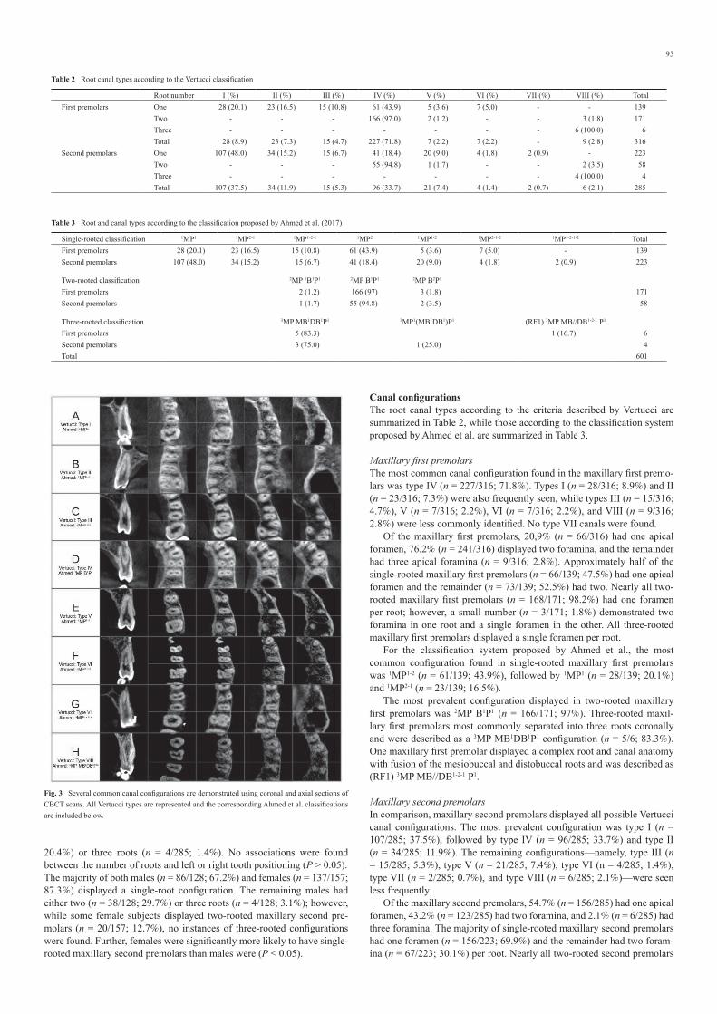
Fig. 3 Several common canal configurations are demonstrated using coronal and axial sections of CBCT scans. All Vertucci types are represented and the corresponding Ahmed et al. classifications are included below.

96
(n = 56/58; 96.5%) had one foramen per root, with a small number (n = 2/58; 3.5%) of two-rooted second premolars demonstrating two foramina in one root and a single foramen in the other. All three-rooted maxillary second premolars had one foramen per root.
For the classification system proposed by Ahmed et al., the most preva-lent classification in single-rooted maxillary second premolars was the 1MP1 configuration (n = 107/223; 48%) and the most common two-rooted classification was 2MP B1P1 (n = 55/58; 94.8%).
Three-rooted maxillary second premolars most commonly displayed the coronally separating 3MP MB1DB1P1 classification (n = 3/4; 75%). One maxillary second premolar displayed a common mesiobuccal and distobuccal root bifurcating in the apical third and was described as 3MP 1(MB1DB1)P1.
Interobserver agreement across the entire dataset, calculated as a percentage agreement, was 96.8% (n = 582/601). The overall agreement between the observers was considered to be high. Figure 3 demonstrates a selection of canal configurations found in the coronal and axial sections of CBCT scans.
Discussion
The complex nature of human tooth morphology requires dental practi-tioners performing endodontic treatment to have a good understanding of the common root and canal configurations as well as anatomical variations [6]. Per a literature review, no data appear to exist regarding the internal and external root and canal configurations of maxillary premolars in a South African population. This study is thus the first investigation to report on maxillary premolar morphology using CBCT from this geographical region.
The distributions of the root numbers of maxillary first premolars in the present study (single-rooted: 44%, two-rooted: 54.1% and three-rooted: 1.9%) were comparable with earlier findings of previous investigations [6,7,21-23]. Table 4 contrasts the root configurations in the present study to those found by previous investigations of different populations from a variety of geographical areas.
A lower frequency of single-rooted maxillary first premolars, as com-pared with the results of the present study, has been previously reported in American, Turkish, Polish, Saudi, Jordanian, and Indian populations [9-11,24-26]. The opposite was demonstrated in several populations from other geographical areas [2,27-29]. The differences in maxillary premolar root number may be related with ethnicity or geographical location.
The previously reported prevalence of three-rooted maxillary first premolars is highly variable, ranging from 0.8% to 9.2% [24,26]. The find-ing of the present study (1.9%) falls within the lower end of this reported
range. Cases of three-rooted maxillary first premolars have been reported in the endodontic literature and clinicians should be aware of this anatomi-cal variant [30], as the treatment of these cases may be challenging due to their complex anatomy [31].
Previous studies have demonstrated that the majority of maxillary second premolars have a single root. The reported prevalence ranged from 69.6% to 90.3% [6,11,18,32]. The findings of the present study are in agreement with these investigations. Three-rooted maxillary second premolars are uncommon, with just two other CBCT studies reporting a prevalence of three-rooted maxillary second premolars above 1% [6,7]. Variations in the root morphology of maxillary second premolars may be attributed to different sample sizes, populations, geographical areas, or methods of evaluation.
Maxillary first premolars displayed variable internal anatomy, with canal bifurcations noted at several levels along the root. All Vertucci canal configuration types except for types VII and VIII were noted in the single-rooted maxillary first premolars. The distribution of maxillary first premolar canal types in the present study was comparable with the findings of Awawdeh et al., who used the clearing technique [26]. The majority of maxillary first premolars in the present study demonstrated two canals, followed by one canal and three canals, respectively (Table 3). This finding was in agreement with those of previous studies using different evaluation techniques such as staining and clearing [5,11], visual examination with radiological technique [27], and plastic casts [10].
Maxillary second premolars displayed the greatest anatomical varia-tions with regard to internal canal configurations. Specifically, all possible Vertucci configurations were found in this tooth type. The majority of second premolars displayed either type I or type IV canal classifications, which are easily identifiable in a clinical setting. It is however impor-tant for clinicians to note the possibility of complex internal anatomical arrangements in maxillary second premolars, as these variations may be more difficult to identify and treat [6].
Overall, the vast majority of maxillary premolars evaluated in the present study could be readily classified using the Vertucci classification. Advantages of this system include its familiarity, ease of use, and the ability to readily compare the gathered results to those of previous investigations.
Ahmed et al. developed a new system for classifying root and canal morphology in 2017 in order to address the shortcomings of historical clas-sifications such as the Vertucci classification due to concerns that not all teeth could be adequately described by such [12]. While the authors of the present study are in agreement with this assertion, it must be considered that only a small number of teeth could not be adequately described using the Vertucci classification alone.
Teeth with complex anatomical arrangements, such as two-rooted
Table 4 Root forms of maxillary first and second premolars in different populations
Author (year) Population (sample size) One root (%) Two roots (%) Three roots (%)First premolars Ingle* (1965) Unavailable 43.0 55.0 2.0
Pineda & Kuttler (1972) [21] USA (n = 259) 43.0 54.6 2.4Carns & Skidmore (1973) [10] USA (n = 100) 22.0 72.0 6.0Walker (1987) [27] Chinese (n = 100) 60.0 40.0 -Pecora et al. (1991) [28] Brazilian (n = 240) 55.8 41.7 2.5Loh (1998) [29] Singaporean (n = 957) 49.4 50.6 -Kartal et al. (1998) [11] Turkish (n = 300) 37.3 61.3 1.3Chaparro et al. (1999) [22] Spanish (n = 150) 40.0 56.7 3.3Lipski et al. (2003) [24] Polish (n = 142) 15.5 75.4 9.2Atieh (2008) [25] Saudi (n = 246) 17.9 80.9 1.2Awawdeh (2008) [26] Jordanian (n = 600) 30.8 68.4 0.8Neelakantan (2011) [9] Indian (n = 350) 11.7 86.0 2.3Ozcan et al. (2012) [23] Turkish (n = 653) 45.2 55.7 1.1Tian et al. (2012) [2] Chinese (n = 300) 66.0 33.0 1.0Abella et al. (2015) [6] Spanish (n = 430) 46.0 51.4 2.6Saber et al. (2018) [7] Egyptian (n = 358) 45.8 53.1 1.1Present study South African (n = 316) 44.0 54.1 1.9
Second premolars Pecora et al. (1992) [18] Brazilian (n = 435) 90.3 9.7 -Kartal (1998) [11] Turkish (n = 300) 69.6 29.7 0.7Yang et al. (2014) [32] Chinese (n = 392) 86.5 13.5 -Abella et al. (2015) [6] Spanish (n = 374) 82.9 15.5 1.6Saber et al. (2018) [7] Egyptian (n = 342) 72.8 26.0 1.2Present study South African (n = 285) 78.2 20.4 1.4
* Ingle JI. Endodontics, 1st ed. Philadelphia, Lea & Febiger, 1965

97
first premolars containing three separate canals or three-rooted premolars with fusions of roots and/or canals may be better described by the system proposed by Ahmed et al. [7]. The present study included a small number of such teeth. The Ahmed et al. classification system allows for a single code to be established containing detailed information regarding the exact root number and canal configuration of any tooth. This may improve the consistency in reporting on teeth with complex anatomical arrangements, especially three-rooted premolars and teeth with three canals. For these cases, the newer system is a considerable improvement over the Vertucci classification, where all teeth displaying three canals were included in the type VIII configuration, regardless of the number of roots or canal con-figuration [5].
Scanning electron microscopy has previously demonstrated multiple apical foramina in maxillary premolars [33]. The present study is in agree-ment with this finding, especially regarding maxillary first premolars, which demonstrated two apical foramina for the majority of the time, even in single-rooted teeth. The majority of maxillary second premolars displayed a single apical foramen (Table 3).
Accurate determination of internal and external human maxillary premolar anatomy by CBCT scanning has been previously demonstrated [2,6,7,16]. Other techniques such as the staining and clearing technique and microfocus computed tomography have also been shown to accurately determine tooth morphology [2,16]. CBCT is, however, one of the only modalities that can be used to determine tooth morphology in living subjects. Differences in previously reported maxillary premolar anatomy described in other CBCT studies [2,6,7] may be attributed to the use of different image acquisition units and settings, varying sample sizes, and observer interpretation.
The results of the present study as well as those of previous investi-gations [2,6,7] suggest that CBCT may be a useful imaging modality for clinicians performing endodontic treatment to use in elucidating maxillary premolar anatomy. It is especially useful in the detection of complex tooth morphology or in instances where periapical radiographs do not provide a clear overview of a case prior to clinical treatment. However, due to the risks associated with radiation exposure and the relative ease seen with determining canal configurations in the majority of cases, CBCT cannot be recommended as a standard diagnostic imaging modality prior to end-odontic treatment.
The majority of maxillary first premolars in this South African sub-population demonstrated two roots and two canals, while maxillary second premolars most commonly displayed one root with a single canal. CBCT imaging, when clinically indicated, can be used to effectively determine complex internal and external root and canal anatomy in maxillary pre-molars. Maxillary premolar root and canal configurations can be described using both the Vertucci as well as the Ahmed et al. classification systems; however, the classification system proposed by Ahmed et al. may be better suited for describing complex anatomical arrangements seen in a small number of teeth.
AcknowledgmentsThe authors would like to thank Professor Ahmed Bhayat, Department of Community Dentistry, University of Pretoria, for statistical support.
Conflict of interestThe authors deny any conflict of interest exist related to this study.
References 1. European Society of Endodontology (2006) Quality guidelines for endodontic treatment:
consensus report of the European Society of Endodontology. Int Endod J 39, 921-930. 2. Tian YY, Guo B, Zhang R, Yu X, Wang H, Hu T et al. (2012) Root and canal morphology of
maxillary first premolars in a Chinese subpopulation evaluated using cone-beam computed
tomography. Int Endod J 45, 996-1003. 3. Pinheiro ET, Gomes BPFA, Ferraz CCR, Sousa ELR, Teixeira FB, Souza-Filho FJ (2003)
Microorganisms from canals of root-filled teeth with periapical lesions. Int Endod J 36, 1-11.
4. Fernandes NA, Herbst D, Postma TC, Bunn BK (2018) The prevalence of second canals in the mesiobuccal root of maxillary molars: a cone beam computed tomography study. Aust Endod J 45, 46-50.
5. Vertucci FJ (1984) Root canal anatomy of the human permanent teeth. Oral Surg Oral Med Oral Pathol 58, 589-599.
6. Abella F, Teixidó LM, Patel S, Sosa F, Duran-Sindreu F, Roig M (2015) Cone-beam Com-puted Tomography Analysis of the Root Canal Morphology of Maxillary First and Second Premolars in a Spanish Population. J Endod 41, 1241-1247.
7. Saber SEDM, Ahmed MHM, Obeid M, Ahmed HMA (2019) Root and canal morphology of maxillary premolar teeth in an Egyptian subpopulation using two classification systems: a cone beam computed tomography study. Int Endod J 52, 267-278.
8. Vertucci FJ, Gegauff A (1979) Root canal morphology of the maxillary first premolar. J Am Dent Assoc 99, 194-198.
9. Neelakantan P, Subbarao C, Ahuja R, Subbarao CV (2011) Root and canal morphology of Indian maxillary premolars by a modified root canal staining technique. Odontology 99, 18-21.
10. Carns EJ, Skidmore AE (1973) Configurations and deviations of root canals of maxillary first premolars. Oral Surg Oral Med Oral Pathol 36, 880-886.
11. Kartal N, Ozcelik B, Cimilli H (1998) Root canal morphology of maxillary premolars. J Endod 24, 417-419.
12. Ahmed HMA, Versiani MA, De-Deus G, Dummer PMH (2017) A new system for classify-ing root and root canal morphology. Int Endod J 50, 761-770.
13. Patel S, Dawood A, Whaites E, Pitt Ford T (2009) New dimensions in endodontic imaging: part 1. Conventional and alternative radiographic systems. Int Endod J 42, 447-462.
14. Martínez-Lozano MÁ, Forner-Navarro L, Sánchez-Cortés JL (1999) Analysis of radiologic factors in determining premolar root canal systems. Oral Surg Oral Med Oral Pathol Oral Radiol Endod 88, 719-722.
15. Matherne RP, Angelopoulos C, Kulild JC, Tira D (2008) Use of cone-beam computed tomography to identify root canal systems in vitro. J Endod 34, 87-89.
16. Neelakantan P, Subbarao C, Subbarao CV (2010) Comparative evaluation of modified canal staining and clearing technique, cone-beam computed tomography, peripheral quantitative computed tomography, spiral computed tomography, and plain and contrast medium-enhanced digital radiography in studying root canal morphology. J Endod 36, 1547-1551.
17. Domark JD, Hatton JF, Benison RP, Hildebolt CF (2013) An ex vivo comparison of digi-tal radiography and cone-beam and micro computed tomography in the detection of the number of canals in the mesiobuccal roots of maxillary molars. J Endod 39, 901-905.
18. Pecora JD, Sousa-Neto MD, Saquy PC, Woelfel JB (1992) In vitro study of root canal anatomy of maxillary second premolars. Braz Dent J 3, 81-85.
19. Zhang Q, Chen H, Fan B, Fan W, Gutmann JL (2014) Root and root canal morphology in maxillary second molar with fused root from a native Chinese population. J Endod 40, 871-875.
20. Ahmed HMA, Dummer PMH (2018) Advantages and applications of a new system for classifying roots and canal systems in research and clinical practice. Eur Endod J 3, 9-17.
21. Pineda F, Kuttler Y (1972) Mesiodistal and buccolingual roentgenographic investigation of 7,275 root canals. Oral Surg Oral Med Oral Pathol 33, 101-110.
22. Chaparro AJ, Segura JJ, Guerrero E, Jimenez-Rubio A, Murillo C, Feito JJ (1999) Number of roots and canals in maxillary first premolars: study of an Andalusian population. Endod Dent Traumatol 15, 65-67.
23. Özcan E, Çolak H, Hamidi MM (2012) Root and canal morphology of maxillary first premolars in a Turkish population. J Dent Sci 7, 390-394.
24. Lipski M, Wozniak K, Lagocka R, Tomasik M (2005) Root and canal morphology of the first human maxillary premolar. Durham Anthropol J 12, 2-3.
25. Atieh MA (2008) Root and canal morphology of maxillary first premolars in a Saudi popu-lation. J Contemp Dent Pract 9, 1-7.
26. Awawdeh L, Abdullah H, Al-Qudah A (2008) Root form and canal morphology of Jorda-nian maxillary first premolars. J Endod 34, 956-961.
27. Walker RT (1987) Root form and canal anatomy of maxillary first premolars in a southern Chinese population. Dent Traumatol 3, 130-134.
28. Pecora JD, Saquy PC, Sousa Neto MD, Woelfel JB (1991) Root form and canal anatomy of maxillary first premolars. Braz Dent J 2, 87-94.
29. Loh HS (1998) Root morphology of the maxillary first premolar in Singaporeans. Aust Dent J 43, 399-402.
30. Soares JA, Leonardo RT (2003) Root canal treatment of three-rooted maxillary first and second premolars—a case report. Int Endod J 36, 705-710.
31. Hartmann RC, Baldasso FER, Stürmer CP, Acauan MD, Scarparo RK, Morgental RD et al. (2013) Clinically relevant dimensions of 3-rooted maxillary premolars obtained via high-resolution computed tomography. J Endod 39, 1639-1645.
32. Yang L, Chen X, Tian C, Han T, Wang Y (2014) Use of cone-beam computed tomography to evaluate root canal morphology and locate root canal orifices of maxillary second pre-molars in a Chinese subpopulation. J Endod 40, 630-634.
33. Morfis A, Sylaras SN, Georgopoulou M, Kernani M, Prountzos F (1994) Study of the apices of human permanent teeth with the use of a scanning electron microscope. Oral Surg Oral Med Oral Pathol 77, 172-176.