ORIGINAL ARTICLE Open Access Biocompatibility and ...
13
ORIGINAL ARTICLE Open Access Biocompatibility and radioprotection by newly characterized melanin pigment and its production from Dietzia schimae NM3 in optimized whey medium by response surface methodology Sahar Eskandari and Zahra Etemadifar * Abstract Purpose: To characterize and optimize the productivity of melanin using an extremotolerant actinobacterium, Dietzia schimae NM3, for the first time. Methods: An extracellular brown pigment produced by D. schimae NM3 in the nutrient broth and cheese whey medium by adding L-tyrosine. The extracted melanin was analyzed by UV-visible, HPLC, and FTIR assays. The radical scavenging activity (by DPPH) and sun protection factor (SPF) of the extracted melanin were measured. The melanin cytotoxicity was assayed by MTT and chromate biosorption was measured by atomic absorption spectroscopy. Finally, melanin production by D. schimae NM3 was optimized by response surface methodology (RSM) using Box-Behnken design in the whey medium. Result: The purified melanin showed similar peak to the standard melanin (SIGMA) at 3.5 min in HPLC, and C=O bands, NH 2 , CH, C-N, and aromatic groups by FTIR. The radical scavenging activity (by DPPH) and SPF of the extracted melanin were obtained 188.9% and 20.22, respectively. Using MTT assay, the melanin revealed non-toxic effect on the normal human fibroblast (HFB) cell culture. The melanin yield 790 mg l -1 , and tyrosinase activity 3400 U ml -1 were obtained in the medium contained whey powder [5% (w v -1 )], L-tyrosine 2.5 g l -1 , CuSO 4 0.013 g l -1 , and pH 10.5, incubated at 32 °C for 3 days. The ANOVA results indicated significant P-value, model F-value, and probability, with insignificant lack of fit. After optimization with mono-factors, the nutrient broth came up with melanin yield as 1.2 g l -1 and tyrosinase activity as 4040 U ml -1 . Conclusion: This is the first report of melanin production by D. schimae NM3 and this natural melanin showed valuable biological properties such as high antioxidant activity and radioprotection (SPF) and the biocompatibility to human cell line. Keywords: Dietzia schimae NM3, Melanin, Antioxidant activity, L-tyrosine, Whey © The Author(s). 2021 Open Access This article is licensed under a Creative Commons Attribution 4.0 International License, which permits use, sharing, adaptation, distribution and reproduction in any medium or format, as long as you give appropriate credit to the original author(s) and the source, provide a link to the Creative Commons licence, and indicate if changes were made. The images or other third party material in this article are included in the article's Creative Commons licence, unless indicated otherwise in a credit line to the material. If material is not included in the article's Creative Commons licence and your intended use is not permitted by statutory regulation or exceeds the permitted use, you will need to obtain permission directly from the copyright holder. To view a copy of this licence, visit http://creativecommons.org/licenses/by/4.0/. * Correspondence: [email protected] Department of Cell and Molecular Biology & Microbiology, Faculty of Biological Science and Technology, University of Isfahan, Isfahan 8174673441, Iran Annals of Microbiology Eskandari and Etemadifar Annals of Microbiology (2021) 71:17 https://doi.org/10.1186/s13213-021-01628-6
Transcript of ORIGINAL ARTICLE Open Access Biocompatibility and ...
ORIGINAL ARTICLE Open Access
Biocompatibility and radioprotection bynewly characterized melanin pigment andits production from Dietzia schimae NM3 inoptimized whey medium by responsesurface methodologySahar Eskandari and Zahra Etemadifar*
Abstract
Purpose: To characterize and optimize the productivity of melanin using an extremotolerant actinobacterium,Dietzia schimae NM3, for the first time.
Methods: An extracellular brown pigment produced by D. schimae NM3 in the nutrient broth and cheese wheymedium by adding L-tyrosine. The extracted melanin was analyzed by UV-visible, HPLC, and FTIR assays. The radicalscavenging activity (by DPPH) and sun protection factor (SPF) of the extracted melanin were measured. Themelanin cytotoxicity was assayed by MTT and chromate biosorption was measured by atomic absorptionspectroscopy. Finally, melanin production by D. schimae NM3 was optimized by response surface methodology(RSM) using Box-Behnken design in the whey medium.
Result: The purified melanin showed similar peak to the standard melanin (SIGMA) at 3.5 min in HPLC, and C=Obands, NH2, CH, C-N, and aromatic groups by FTIR. The radical scavenging activity (by DPPH) and SPF of theextracted melanin were obtained 188.9% and 20.22, respectively. Using MTT assay, the melanin revealed non-toxiceffect on the normal human fibroblast (HFB) cell culture. The melanin yield 790 mg l−1, and tyrosinase activity 3400U ml−1 were obtained in the medium contained whey powder [5% (w v−1)], L-tyrosine 2.5 g l−1, CuSO4 0.013 g l−1,and pH 10.5, incubated at 32 °C for 3 days. The ANOVA results indicated significant P-value, model F-value, andprobability, with insignificant lack of fit. After optimization with mono-factors, the nutrient broth came up withmelanin yield as 1.2 g l−1 and tyrosinase activity as 4040 U ml−1.
Conclusion: This is the first report of melanin production by D. schimae NM3 and this natural melanin showedvaluable biological properties such as high antioxidant activity and radioprotection (SPF) and the biocompatibilityto human cell line.
Keywords: Dietzia schimae NM3, Melanin, Antioxidant activity, L-tyrosine, Whey
© The Author(s). 2021 Open Access This article is licensed under a Creative Commons Attribution 4.0 International License,which permits use, sharing, adaptation, distribution and reproduction in any medium or format, as long as you giveappropriate credit to the original author(s) and the source, provide a link to the Creative Commons licence, and indicate ifchanges were made. The images or other third party material in this article are included in the article's Creative Commonslicence, unless indicated otherwise in a credit line to the material. If material is not included in the article's Creative Commonslicence and your intended use is not permitted by statutory regulation or exceeds the permitted use, you will need to obtainpermission directly from the copyright holder. To view a copy of this licence, visit http://creativecommons.org/licenses/by/4.0/.
* Correspondence: [email protected] of Cell and Molecular Biology & Microbiology, Faculty ofBiological Science and Technology, University of Isfahan, Isfahan 8174673441,Iran
Annals of MicrobiologyEskandari and Etemadifar Annals of Microbiology (2021) 71:17 https://doi.org/10.1186/s13213-021-01628-6

IntroductionDietzia genus is an actinobacterium consuming manycompounds, also tyrosine is the sole carbon source forproducing melanin pigment (Koerner et al. 2009). Thisbacterium is in fact capable to biodegrade aniline, al-kane, and hydrocarbons in the soil polluted with crudeoil and aromatic compounds (SMT et al. 2013).Today, the consumers are concerned about the chem-
ical synthetic compounds of such pigments due to theirside effects, which can be related to their complex chem-ical structures compared to the natural ones. The micro-bial cells factories have been considered for their easyand fast production. The natural pigments from micro-bial sources are used in food, cosmetics, and the indus-tries without causing side effects or environmentalcontamination (Sun et al. 2016).Melanin pigment is a dark- or black-brown irregular
negatively charged hydrophilic complex polymer. It isproduced by oxidative polymerization of phenolic or in-dolic compounds in various organisms (Plonka and Gra-backa 2006). Melanins are resistant to heat, free radicals,radiation energy, and oxidizing agents. Generally speak-ing, melanin can protect microorganism against ionizingradiation (Cordero et al. 2017). Melanin possesses anti-UV radiation property by absorbing the electromagneticspectrum and preventing optical damage in the livingorganisms. As shown, melanin can synthesize in re-sponse to the antifungal drugs (Kurian and Bhat2014). Melanin granules are insoluble in water andcommon organic solvents (such as hexane, chloro-form, ethanol, methanol, or acetone) (Varga et al.2016). The melanin produced by Pseudomonas balear-ica has been characterized by spectroscopic andchemical properties (Zerrad et al. 2014). The carboxyl,phenolic, hydroxyl, and amine groups on melanin pig-ments provide numerous potential binding sites forinteracting with other molecules (Hoa et al. 2017).For this reason, melanins have some important prop-erties including antitumor, antimicrobial, and anti-venin activities. Moreover, these molecules canprotect the cells from DNA damage induced byhydrogen peroxide, indicating the antioxidant activityof melanin (El-Naggar and El-Ewasy 2017). A broadrange of bacteria such as Pseudomonads, Aeromonas,Azotobacter, Mycobacterium, Micrococcus, Bacillus,Legionella, and Streptomyces can produce melanin(Nosanchuk and Casadevall 2003). It is critical tooptimize the growth conditions for microorganisms,particularly the physiochemical and nutritional param-eters. An optimization method using “one factor at atime” is difficult to run and time-consuming (El-Nag-gar and El-Ewasy 2017). Response surface method-ology (RSM) is a set of mathematical methods withseveral independent variables (Surwase et al. 2012).
Recently, several studies have focused on the speciescapable to be used as different substances instead of hy-drocarbons so that to produce melanin at high purities.So far, there has been no report on melanin pigmentproduction by radioresistant Dietzia strains. The currentresearch mainly targets to produce and characterize mel-anin pigment from Dietzia schimae NM3 by L-tyrosinein the nutrient broth. In addition, in the present study,we proposed an inexpensive culture medium to opti-mally bioproduce the melanin by D. schimae NM3 usingeconomical cheese whey medium.
Materials and methodsMicrobial strain and inoculum preparationD. schimae NM3 (GeneBank accession no. KP207685)that was isolated and identified previously in our lab wasused for melanin pigment production in this study forthe first time. To prepare the work inoculum, one freshcolony of D. schimae NM3 was incubated in tryptoneglucose yeast extract (TGY) agar medium at 30 °C for 48h (Zamanian and Etemadifar 2017).
Melanin production and purificationThe nutrient broth containing 2 g l−1 L-tyrosine was in-oculated by 5% (v v−1) of the D. schimae culture (108
CFU ml−1) and incubated at 35 °C with 160 rpm shakingfor 4 days to observe dark melanin pigment (Wang et al.2015). The growth of D. schimae NM3 was assayed bymeasuring the optical density (OD600nm) spectrophoto-metrically (Milton Roy Spectronic 21D). Extracellularmelanin production was assayed by colorimetry (A400nm)after medium filtration by 0.45 μ membrane filter at dif-ferent intervals during the incubation period (El-Naggarand El-Ewasy 2017).
Melanin extractionIn order to extract melanin, the darkly pigmentedmedium was centrifuged at 3500 rpm for 15 min (by re-frigerating Eppendorf 5810R) and the bacterial cells (pel-leted) separated from the supernatant (El-Naggar andEl-Ewasy 2017). The supernatant was acidified by 3 moll−1 HCl to reach pH 3, left at room temperature for 24h, and then centrifuged at 3500 rpm for 15 min to separ-ate crude pigment granules (melanin). The pigmentgranules were washed by water, and the pellet was boiledin water bath for 5 min to avoid melanoidins formation,then was dried in the oven (Tarangini and Mishra 2013).To purify the melanin, it was dissolved in dimethyl sulf-oxide (DMSO) by shaking slowly in hot water until itwas completely dissolved, then centrifuged so that theimpurities got settled. The top liquid was kept in theplate and put in the oven to dry (Jakoby 1971).
Eskandari and Etemadifar Annals of Microbiology (2021) 71:17 Page 2 of 13

Tyrosinase assayThe fresh colonies of D. schimae NM3 were inoculatedin NB medium (pH 7.0) containing L-tyrosine and incu-bated at 35 °C on a rotary shaker at 160 rpm. The cellswere harvested by centrifugation at 5000 rpm for 5 minat 4 °C and disrupted by ultrasonic (280 kHz) for tentimes at 1-min intervals on ice. For tyrosinase assay, 0.1ml of crude intracellular enzymes, 1.0 ml of 0.5 mol l−1
phosphate buffer (pH 6.5), 1.0 ml of 0.001 mol l−1 L-tyrosine, and 0.9 ml of reagent water were added to atest tube. After 4–5 min waiting for the mixture to getdeoxygenated and the temperature to get balanced (doneinside ice), the absorbance was recorded at 280 nm usingUV-Vis spectrophotometer. The enzyme activity was cal-culated by Eq. (1) (Ingle and Khobragade 2013; Royet al. 2014).
Units of Enzyme U=mlð Þ¼ ΔA280 nm= min Test −ΔA280nm= min Blank df½ �
0 : 001½ � 0 : 1½ �ð1Þ
Analytical methodsUV-visible spectroscopyThe purified melanin was dissolved in 0.1 mol l−1 NaOH,sonicated for 5 min, and its UV-visible absorbance was re-corded at the spectrum range 200–400 nm using SpecordPlus UV-VIS spectrophotometer (Wang et al. 2015).
FTIR analysisFor FTIR analysis, the melanin-containing KBr pelletswere prepared following the method developed by Sunet al. (2016). The melanin was analyzed with FTIR (Ni-colet AVATAR360) at wave numbers 4000–500 cm−1.The FTIR instrument’s resolution was 4.0 cm−1, the op-tical path difference (OPD) velocity as 0.20 cm s−1, andthe data collection interval of 1.0 s. The actual spectrawere measured as the absorbance ratio. The backgroundspectra were measured by the clean KBr pellet contain-ing no sample material (Sun et al. 2016).
HPLC analysisThe pure melanin and the standard melanin (Sigma) weredissolved in 0.5 mol l−1 DMSO solution prior to HPLC ana-lysis. The quantitative method was done as stated by Sunet al., and the detection wavelength was set at 280 nm andthe column temperature was set at 25 °C (Sun et al. 2016).
In vitro cytotoxicity assay (MTT Assay)Human fibroblast cell line (HFB) was used for in vitrostudies. The cells were cultured in T-flask containingRPMI 1640 medium supplemented with 10% fetal bovineserum (FBS) and 1% antibiotic (penicillin/streptomycin)
solution and incubated in a humidified incubator at 37 °Cunder 5% CO2 atmosphere. The cells were harvested using0.05% trypsin and centrifuged for 4 min at 1000 rpm.Then, the cells were dispersed and inoculated in a 96-wellplate at a density of 104 cells per well and incubated for 24h under the same conditions to allow the cells to adhere.Following the cells being exposed to microbial melanin
at different concentrations (31, 62.5, 125, 250, and 500μg ml−1) for 72 h, the medium was completely discarded,and 100 μL of (MTT) solution (0.5 mg ml−1 in medium)was added to each well and the plates were re-incubatedat 37 °C for 4 h. After that, the absorbance was mea-sured at 570 nm. The cell viability was determined asthe ratio of absorbance values from each treatment andthe control. Upon being exposed to different concentra-tions of melanin, the cell morphology was investigatedby optical microscope (Arun et al. 2015). The percentageof the killed cells was calculated by Eq. (2).
killed cells% ¼ 100 - the mean control OD=mean sample ODð Þ�100ð2Þ
Sun protection factor measurementSun protection factor (SPF) value was assayed in ethanolsolution containing 100 mg pigment by measuring theabsorbance in the spectra of the 280–400 nm range. TheNM3 strain extracted melanin’s SPF value was deter-mined by Mansur mathematical equation (Eq. 3) (Huanget al. 2011).
SPF ¼ CF � λ� I λ½ � � Abs λ½ � ð3Þ
DPPH-based radical scavenging assayRadical scavenging activity was assayed according to themodified method of Brand-Williams et al. The crude ex-tracted microbial melanin and the ascorbic acid asstandard antioxidant were supplied at different concen-trations (2.5, 5, 10 mg l−1). Then, 1 ml of each concen-tration of melanin and ascorbic acid were added to 3 mlout of 0.1 m mol DPPH (2,2-diphenyl-1-picrylhydrazyl)solution. After that, the tubes were incubated in the darkat 37 °C for 30 min, and the absorbance was monitoredat 516 nm by spectrophotometer (Milton Roy Spectronic21D). The scavenging effect was measured using Eq. 4(Zerrad et al. 2014).
DPPH inhibition %ð Þ ¼ control absorbance − test absorbanceð Þ � 100
ð4Þ
Metal biosorption activityMelanin solution was supplied at different concentra-tions (0, 25, 50, 100 mg l−1). The metal solution of
Eskandari and Etemadifar Annals of Microbiology (2021) 71:17 Page 3 of 13

K2Cr2O4 (2 m mol) was prepared and mixed with mel-anin, and the activated carbon was used as the standardsorbent in this test. The initial pH 4.0 was kept fixed forall tubes test; then, the solution was shaken fast at roomtemperature for 1 h. The solution was filtered with 0.45μ membrane filter and then analyzed by atomic absorp-tion spectroscopy to measure metal biosorption (Chenet al. 2009).All the results indicated as means ± standard deviation
(SD) of three independent experiments.
Melanin production in whey mediumD. schimae culture suspension (108 CFU ml−1) was inoc-ulated as 5% (v v−1) into the whey medium contained 5%whey powder, and 0.25% L-tyrosine and incubated at 35°C with 160 rpm shaking for 96 h. After different timesintervals the bacterial growth (A600nm), sugarutilization (by DNS method), L-tyrosine consumption(A492nm), and melanin production (A400nm) were de-termined spectrophotometrically (Ganesh Kumar et al.2013).
Optimizing melanin pigment production using Box-Behnken design-based RSMAt first, the optimization was performed with one factorat a time (temperature, light, shaking rate, culturemedium, pH, L-tyrosine, ion catalysts, salts) to enhancemelanin production. As the results indicate, four import-ant factors were selected for RSM optimization of themelanin production by Dietzia. The 27 designed runsare shown in Table 2. According to the results of onefactor at a time optimization in the previous experi-ments by RSM statistical method in actinobacteria,four factors (L-tyrosine, pH, CuSO4, and temperature)were selected as the most effective factors involved inthe melanin production and the bacterial growth(Dholakiya et al. 2017). Expert design software11.1.2.0 was applied for the runs’ design type (Box-Behnken) and design model (quadratic) (El-Batal et al.2017).The model’s statistical analysis was represented as
ANOVA (Surwase et al. 2013). The multivariate modelis presented as (Eq. 5).
Y ¼ β 0þXk
i¼1BiXiþ
Xk
i¼1BiXi
þXk
i¼1
Xk
i¼1Bijx1x1þ ϵ ð5Þ
Finally, the optimized factors of whey medium wereused in the nutrient broth. After the incubation period,melanin yield and tyrosinase activity were measured.
ResultsD. schimae NM3 strain is not capable to transform L-tyrosine into melanin in the presence of high carbohy-drate and nitrogen nutrients available in the enrichedmedia such as TGY and TSA. In the nutrient agar withL-tyrosine as a substrate, the dark pigmented melaninwas observed after 3–4 days (Fig. S1). Bacterial growthand melanin production are depicted in Fig. S2. Thebacteria were growing using L-tyrosine by producingvarious mediator metabolites transformed into melaninat the stationary phase (52–64 h). L-tyrosine begins todecrease after 18 h and the next peak indicates the nextmetabolite’s formation. These reactions continue untilthe reduction of tyrosine peak and the melanin peakformation.It is worth mentioning that the melanin production
was at its highest level (450 mg l−1) after 52–64 h. Thetyrosinase activity was obtained as 2010 U ml−1 at thistime. After melanin production optimization using someimportant factors, i.e., pH 9, temperature (32 °C), andtyrosine (2 g l−1), the nutrient broth got to 1.2 g l−1
level.
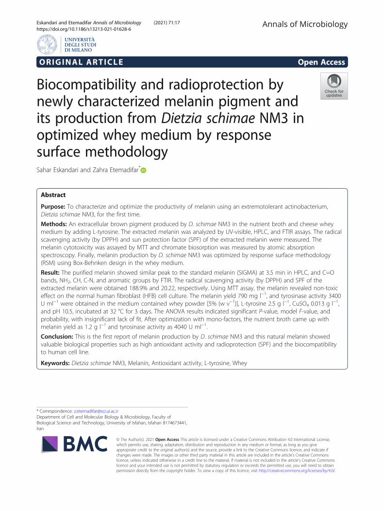
Melanin characterizationThe photographs were taken by an optical microscope(Fig. 1a, b) to represent melanin layer in the cell wall(melanin ghost) after acid (KOH) treatment (MHS et al.2013). The scan electron microscopy image of melaninwith big structure in the cell wall is illustrated in Fig. 1cand d.D. schimae NM3 extracted melanin revealed absorb-
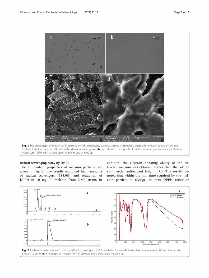
ance in the spectrum of 200–300 nm range by UV-visible spectrophotometry (Fig. S3), which was likestandard melanin. The standard melanin’s HPLC chro-matogram included a single symmetrical elution peak,and the retention times were 3.5 min, which were likethe NM3 melanin’s peak pattern (Fig. 2a, b).To perceive the melanin’s structure and its functional
groups, FTIR analysis was performed on the purifiedmelanin from D. schimae and the standard melanin. Thesimilar spectral pattern suggested that both melanin pig-ments had identical structure (Fig. 2c). To determine thestructure and the present excess bands in the melaninproduct of D. schimae, IR Pal V.2.0 was applied and thencompared with the standard melanin (Table S2). How-ever, the purified melanin from NM3 and the standardmelanin demonstrated some peaks which may be causedby some fragments produced by the destruction in theacid precipitation step.
Sunscreen protection factor (SPF)The UV-Abs-derived results at different wavelengths forSPF determination are listed at Table 1. The D. schimaederived melanin’s SPF value was measured as 20.22.
Eskandari and Etemadifar Annals of Microbiology (2021) 71:17 Page 4 of 13
Radical scavenging assay by DPPHThe antioxidant properties of melanin particles aregiven in Fig. 3. The results exhibited high amountsof radical scavengers (188.9%) and reduction ofDPPH in 10 mg l−1 melanin from NM3 strain. In
addition, the electron donating ability of the ex-tracted melanin was obtained higher than that of thecommercial antioxidant (vitamin C). The results de-noted that within the min time required by the mel-anin particle to divulge, its max DPPH reduction
Fig. 1 The photograph of melanin of D. schimae by light microscopy without staining in stationary phase after melanin extraction by acidtreatment (a), the bacterial cell walls with external melanin ghosts (b), and electron micrograph of purified melanin granules by scan electronmicroscopy (SEM), with magnification × 500 (c) and × 5000 (d)
Fig. 2 Analysis of melanin from D. schimae NM3. Chromatogram (HPLC) analysis of strain NM3 extracted natural melanin (a) and the standardmelanin (SIGMA) (b); FTIR graph of melanin from D. schimae and the standard melanin (c)
Eskandari and Etemadifar Annals of Microbiology (2021) 71:17 Page 5 of 13

and the scavenging activity depend on pigment’sdose.
Biosorption of Cr6+
The adsorption efficiency of Cr6+ by melanin was exam-ined by dose dependence. Based on the results in Fig. 4,melanin could remove Cr6+ with adsorption efficiency33.3%, 22.9%, and 17.7% in the presence of melanin con-centrations at 25, 50, and 100 mg ml−1, respectively,while under the same conditions, the activated carbon atthe concentrations of 25, 50, and 100 mg ml−1 have theadsorption efficiencies of 24%, 8%, and 0%, respectively.In general, the adsorption capacity of melanin washigher than that of the activated carbon.
MTT assayThe results of cell cultures in the presence of purifiedmelanin from D. schimae, in comparison with the con-trol cell without melanin, were shown in Fig. 5. As the
results exhibited, the melanin (at the concentrations upto 250 μg ml−1) lacked toxicity on the normal cells,which indicates the bacterial melanin’s biocompatibility.These results could also confirm that no significant tox-icity was observed due to the impurities. No tangiblechanges were seen in the morphology of the cells ex-posed to melanin compared with the control cells (Fig.S4).
Melanin production in whey mediumDietzia NM3 produced melanin pigment in wheymedium contained L-tyrosine (Fig. 6). The obtained re-sults showed this bacterium utilized whey sugar andgrew to OD=3; simultaneously, it produced melanin byL-tyrosine consumption. After melanin production inwhey medium, we performed the optimization experi-ment. Without whey, any growth did not occur in themedium, and in the whey medium without L-tyrosine,pigment was not produced.
RSM-based optimization of melanin production by D.schimae NM3D. schimae NM3 was inoculated in whey medium withL-tyrosine as substrate. The dark brown pigment mel-anin was diffused after 3 days in whey broth medium asan inexpensive medium. In large-scale fermentation (1 l)of whey medium as an inexpensive medium, the maxmelanin production was gained as 790 mg l−1. Throughthis method, it is merely viable to gain the proper con-centration for each factor separately. The resulted modelcan be observed in Table 2, in which the melanin yield(Y) is a function of the independent variables’ value. Thistable has three replicates at the central point. The resultsfor predicting the response are given as the final equa-tion from Box-Behnken experiment (Eq. 6).
Table 1 SPF result with purified melanin from D. schimae NM3strain
λ Abs
290 0.005595
295 0.02737
300 0.08737
305 0.087523
310 0.043804
315 0.021814
320 0.004464
SPF = CF × λ × I[λ] × Abs[λ]
SPF = 10 × 1 × 2.022 = 20.22
EE erythema effect spectrum, I solar intensity spectrum, Abs the absorbance ofsunscreen product (OD), CFs correction factor (= 10)
Fig. 3 Radical scavenging activity (RSA%) of the melanin derived from D. schimae NM3 at different concentrations (error bars indicatestandard deviations)
Eskandari and Etemadifar Annals of Microbiology (2021) 71:17 Page 6 of 13

Yield ¼ þ780 − 39:17 Aþ 65 Bþ 55:83 Cþ 86:67 Dþ 30 AB
þ47:50 ACþ 30 ADþ 42:50 BCþ 47:50 BDþ 32:50 CD
− 343:75 A2 − 200 B2 − 173:75 C2 − 227:50 D2
ð6Þ
As spotted in Eq. (6), among the abovementioned fac-tors, the pH is negative. Their negative effect on the re-sponse function suggests that higher values of thisparameter lead to lower melanin yield. CuSO4 possessesthe max effect out of these four parameters.Equation (6) was statistically evaluated through F-test
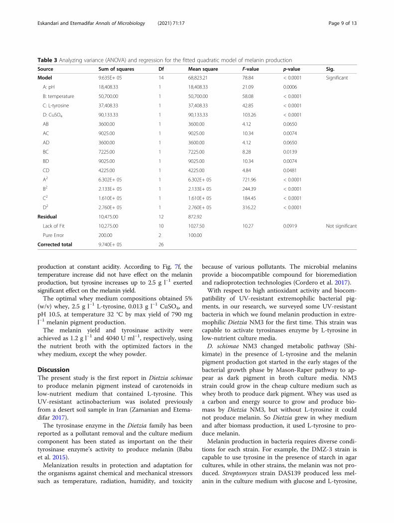
and the ANOVA of surface response quadratic modeldepicted in Table 2. F-value is 78.84, being typically veryhigh, which indicates the model Box-Behnken as fullymeaningful. P-value < 0.0001 refers to the model beingsignificant.
Lack of fit (0.0919) estimation can be a good reasonfor the accuracy of the model data. As the model ana-lysis derived results indicated (Table 3), Std.Dev wasachieved as 29.55, which confirms the model’s accuracy.This parameter value for melanin yield of 790 mg l−1
level denotes the accuracy of the model. The closer R-squared to 1, the better relationship exists between thelab (adjusted R2 = 0.97) and the estimated results (pre-dicted R2 = 0.938).The 3-D graphs show the overall impact of the factors’
relationship. In Fig. 7a, two factors of temperature at 32°C and cooper 0.013 g l−1 are constant, and two factorsof acidity and tyrosine are measured by their interaction.The blue color means the lowest and red means thehighest product. The interactions between L-tyrosineand pH improved the melanin yield by increasing thepH from 9 to 11 and decreasing L-tyrosine to 2.2–2.8 gl−1.
Fig. 4 NM3 strain extracted melanin’s effect on biosorption of Cr+6; activated carbon used as standard (error bars indicate standard deviations)
Fig. 5 Normal human fibroblast cells (HFB) related cell viability % after being exposed to different concentrations of purified melanin from D.schimae NM3
Eskandari and Etemadifar Annals of Microbiology (2021) 71:17 Page 7 of 13

The interactions between copper and temperature aregiven in Fig. 7b. Two factors of acidity at 10.5 and L-tyrosine 2.5 g l−1 are constant, and two factors oftemperature and cooper were measured by their interac-tions. It was indicated that by the temperature rise to 32°C and Cu to 0.013 g l−1, the melanin productionincreased.The interactions between copper and pH (Fig. 7c) indi-
cated the melanin production increase in the pH range ofabout 9 to 11 with the copper concentration decrease to0.013 g l−1. The interactions between L-tyrosine and CuSO4
(Fig. 7d) signal the melanin production increase which de-pends on L-tyrosine 2.5 g l−1 than copper. The pH andtemperature (Fig. 7e) denoted that the temperature andacidity factors exert an equal effect on the melanin produc-tion. The interaction of two factors, i.e., temperature and L-tyrosine (Fig. 7f) exhibited the factors’ effect on the melanin
Fig. 6 Melanin production in whey medium contain whey powder5% (w v−1), and L-tyrosine (0.25%) after 96-h incubation at 35 °Cwith 160 rpm shaking
Table 2 Experimental design for melanin production optimization by D. schimae NM3 (layout and results)
Std Run Factor 1 Factor 2 Factor 3 Factor 4 Response 1
A: pH B: temperature, °C C: L-tyrosine, g l−1 D: CuSO4, g l−1 Melanin yield, mg l−1
2 1 12.0 29 2.5 0.013 140
24 2 10.5 35 2.5 0.020 550
17 3 9.0 32 1 0.013 280
10 4 12.0 32 2.5 0.005 80
26 5 10.5 32 2.5 0.013 790
11 6 9.0 32 2.5 0.020 300
5 7 10.5 32 1 0.005 280
6 8 10.5 32 4 0.005 300
21 9 10.5 29 2.5 0.005 220
15 10 10.5 29 4 0.013 350
18 11 12.0 32 1 0.013 80
19 12 9.0 32 4 0.013 320
23 13 10.5 29 2.5 0.020 320
16 14 10.5 35 4 0.013 600
4 15 12.0 35 2.5 0.013 290
3 16 9.0 35 2.5 0.013 280
20 17 12.0 32 4 0.013 310
27 18 10.5 32 2.5 0.013 770
13 19 10.5 29 1 0.013 320
22 20 10.5 35 2.5 0.005 260
14 21 10.5 35 1 0.013 400
9 22 9.0 32 2.5 0.005 220
25 23 10.5 32 2.5 0.013 780
7 24 10.5 32 1 0.020 400
1 25 9.0 29 2.5 0.013 250
12 26 12.0 32 2.5 0.020 280
8 27 10.5 32 4 0.020 550
Eskandari and Etemadifar Annals of Microbiology (2021) 71:17 Page 8 of 13
production at constant acidity. According to Fig. 7f, thetemperature increase did not have effect on the melaninproduction, but tyrosine increases up to 2.5 g l−1 exertedsignificant effect on the melanin yield.The optimal whey medium compositions obtained 5%
(w/v) whey, 2.5 g l−1 L-tyrosine, 0.013 g l−1 CuSO4, andpH 10.5, at temperature 32 °C by max yield of 790 mgl−1 melanin pigment production.The melanin yield and tyrosinase activity were
achieved as 1.2 g l−1 and 4040 U ml−1, respectively, usingthe nutrient broth with the optimized factors in thewhey medium, except the whey powder.
DiscussionThe present study is the first report in Dietzia schimaeto produce melanin pigment instead of carotenoids inlow-nutrient medium that contained L-tyrosine. ThisUV-resistant actinobacterium was isolated previouslyfrom a desert soil sample in Iran (Zamanian and Etema-difar 2017).The tyrosinase enzyme in the Dietzia family has been
reported as a pollutant removal and the culture mediumcomponent has been stated as important on the theirtyrosinase enzyme’s activity to produce melanin (Babuet al. 2015).Melanization results in protection and adaptation for
the organisms against chemical and mechanical stressorssuch as temperature, radiation, humidity, and toxicity
because of various pollutants. The microbial melaninsprovide a biocompatible compound for bioremediationand radioprotection technologies (Cordero et al. 2017).With respect to high antioxidant activity and biocom-
patibility of UV-resistant extremophilic bacterial pig-ments, in our research, we surveyed some UV-resistantbacteria in which we found melanin production in extre-mophilic Dietzia NM3 for the first time. This strain wascapable to activate tyrosinases enzyme by L-tyrosine inlow-nutrient culture media.D. schimae NM3 changed metabolic pathway (Shi-
kimate) in the presence of L-tyrosine and the melaninpigment production got started in the early stages of thebacterial growth phase by Mason-Raper pathway to ap-pear as dark pigment in broth culture media. NM3strain could grow in the cheap culture medium such aswhey broth to produce dark pigment. Whey was used asa carbon and energy source to grow and produce bio-mass by Dietzia NM3, but without L-tyrosine it couldnot produce melanin. So Dietzia grew in whey mediumand after biomass production, it used L-tyrosine to pro-duce melanin.Melanin production in bacteria requires diverse condi-
tions for each strain. For example, the DMZ-3 strain iscapable to use tyrosine in the presence of starch in agarcultures, while in other strains, the melanin was not pro-duced. Streptomyces strain DAS139 produced less mel-anin in the culture medium with glucose and L-tyrosine,
Table 3 Analyzing variance (ANOVA) and regression for the fitted quadratic model of melanin production
Source Sum of squares Df Mean square F-value p-value Sig.
Model 9.635E+ 05 14 68,823.21 78.84 < 0.0001 Significant
A: pH 18,408.33 1 18,408.33 21.09 0.0006
B: temperature 50,700.00 1 50,700.00 58.08 < 0.0001
C: L-tyrosine 37,408.33 1 37,408.33 42.85 < 0.0001
D: CuSO4 90,133.33 1 90,133.33 103.26 < 0.0001
AB 3600.00 1 3600.00 4.12 0.0650
AC 9025.00 1 9025.00 10.34 0.0074
AD 3600.00 1 3600.00 4.12 0.0650
BC 7225.00 1 7225.00 8.28 0.0139
BD 9025.00 1 9025.00 10.34 0.0074
CD 4225.00 1 4225.00 4.84 0.0481
A2 6.302E+ 05 1 6.302E+ 05 721.96 < 0.0001
B2 2.133E+ 05 1 2.133E+ 05 244.39 < 0.0001
C2 1.610E+ 05 1 1.610E+ 05 184.45 < 0.0001
D2 2.760E+ 05 1 2.760E+ 05 316.22 < 0.0001
Residual 10,475.00 12 872.92
Lack of Fit 10,275.00 10 1027.50 10.27 0.0919 Not significant
Pure Error 200.00 2 100.00
Corrected total 9.740E+ 05 26
Eskandari and Etemadifar Annals of Microbiology (2021) 71:17 Page 9 of 13

while it produces high melanin in the culture mediumcontaining glycerol and starch (Dastager et al. 2006;Madhusudhan et al. 2014).The antioxidant level of NM3 strain melanin (188.9%)
was higher than that of the ascorbic acid as the standard.
In other bacteria, the antioxidant activity was 30% and44.7%, much lower than the melanin from NM3; how-ever, the antioxidant activity was reported to account formore than 60% in other research cases (El-Naggar andEl-Ewasy 2017; Mishra and Singh 2015). Melanin can
Fig. 7 3-D response surface curve representing interactions among pH and L-tyrosine (a), temperature and CuSO4 (b), pH and CuSO4 (c), L-tyrosine and CuSO4 (d), pH and temperature (e), and temperature and L-tyrosine (f) of the melanin production by D. schimae NM3 inwhey medium
Eskandari and Etemadifar Annals of Microbiology (2021) 71:17 Page 10 of 13

potentially stabilize harmful unpaired electrons such asthose from ROS and show high antioxidant (DPPH) andsun protection factor (SPF) (Geis et al. 1984). Some fac-tors capable to change the SPF values of sunscreens in-clude different solvents, the concentration, and the typeof emulsion used in the formulation (Dutra et al. 2004).Melanins have been used commercially in cosmetics’materials against ultraviolet damage, and also in food-stuff (Brenner and Hearing 2008). Melanin possesseshigh photo stability and strong UV-absorption than themetabolites of Porphyra-334, palythine andmycosporine-like amino acids reported before (Solano2020). Melanin has physiological and photoprotectiveproperties that could absorb wide broad UV-visiblespectrum. Melanin is resistant to degradation and havethe ability to remove up to 90% of the absorbed energyfrom the sunlight radiation such as heat (Solano 2014).At present, the main commercial application of melaninis as a dye in lenses of sunglasses. In this case, the nat-ural origin of the pigment and the capacity to reducehigh energy visible light are highlighted as an advantage(https://espeyewear.com/) (Martínez et al. 2019).Metal biosorption potential in some bacterial melanin
is up to 97% at 120 min (Chen et al. 2009). In this study,the saturated biosorption was seen up to 50% within 60min, the reason behind which is the functional groupssurrounding melanin’s structure. After 1 h, due to bio-sorption, Vibrio parahaemolyticus melanin reducedmetal from 8.0 mg ml−1 to 0.35 mg ml−1 (Abbas et al.2014; Hoa et al. 2017).The melanin from Streptomyces DMZ-3 strain at con-
centration of 64 μg mL−1 inhibited cancer cells growingafter 24 h (Madhusudhan et al. 2014). Melanin concen-tration of 100 μg mL−1 from Streptomyces glaucescensNEAE-H has been shown to inhibit the growth of fibro-blast cancer cells as 81.3% (El-Naggar and El-Ewasy2017). This high inhibitory activity is due to the largestructure of its melanin and may be due in part to thesolvent. The extracted melanin produced by DietziaNM3 strain could dissolve in distilled water, afteraddition of small amount of DMSO was dissolved in dis-tilled water. This melanin in concentration of 500 μgmL−1 showed low inhibition effect (16%) on fibroblastnormal cells growth. However, at concentrations lowerthan 250 μg mL−1, it was not toxic to normal cells andwas biocompatible. One of the advantages of this mel-anin is biocompatibility even in high concentrations.Box-Behnken design for a bioproduct optimization has
fewer design points and less expensive than central com-posite designs with the same number of factors. Box-Behnken designs do not have axial points. Thus, all de-sign points fall within their safe operating zone. Box-Behnken designs factors are not set at their high levelsat the same time (Stamenković et al. 2018). Hence, Box-
Behnken design is easy to predict the lower and upperlimits at 3 level point, and this method was used for themelanin optimization in this study.The optimized melanin production by Auricularia
auricula demonstrated 519.54 mg l−1 yield (Kurian andBhat 2014), the optimized melanin production by Strep-tomyces glaucescens NEAE-H was obtained as 316.50 μgml−1 by Placket–Burman’s design (El-Naggar and El-Ewasy 2017). In this study, according to the RSM results,the max yield of melanin by Dietzia NM3 was gained as790 mg l−1 under the optimal whey medium and cultureconditions. These results confirmed that the developedmodel being valid in the present study. According to theresults, the model’s quality was adequately good andmight describe the real relationship among the mediumcomponents.Compared to other studies, the HPLC peak pattern of
melanin from D. schimae NM3 showed proper extrac-tion with appropriate column chromatography and simi-larity with the synthesized standard melanin in terms ofthe functional group and chemical elements (Sun et al.2016). In some studies, the brown melanin synthesizedwith L-tyrosine has a different peak time than the mel-anin naturally produced by microorganisms in the cul-ture medium. For instance, the melanin produced byCryptococcus neoformans exhibited a peak time within 9min (Frases et al. 2007).According to the classification method, various ab-
sorption peaks showing bands and groups in the FTIRspectra represent the traits of D. schimae NM3 extractedmelanin being of eumelanin type (Sun et al. 2016).Eumelanin structure in FTIR revealed the groups -OHand -NH2, C=C, –COO-, C=O in the 1600-3419 regionin Auricularia auricular (Sun et al. 2016) and Bacillusthuringiensis (Sansinenea et al. 2015). Dietzia showedlow carbon structure with -OH and -NH2 and the func-tional groups and peaks, representing them similar tothe standard (Kurian and Bhat 2014).
Study impactDietzia is a drought- and ultraviolet-resistant actinobac-terium, whose melanin pigment was not reported. In thisstudy, the potential of this bacterium to produce a darkbrown melanin pigment using L-tyrosine was exhibited.Also, for the growth of Dietzia to produce the brownmelanin, a simple and cheap culture medium of whey(recycled materials of dairy factories) was used in thisstudy. To produce melanin by activating the tyrosinaseenzyme, it is imperative to optimize various physio-logical and dietary conditions employed for productionby RSM procedure. The melanin exhibited various bio-logical properties such as the ability to scavenge freeradicals and to protect against the sun ultraviolet rays.These properties make this pigment a good choice to be
Eskandari and Etemadifar Annals of Microbiology (2021) 71:17 Page 11 of 13

used in cosmetics and pharmaceutical applications. Themelanin from Dietzia NM3 also possesses the capabilityto remove chromium ions by adsorption, which could bean option to remove metals without any environmentalpollution or the risk of damaging the skin surfaces of theliving organisms.
Supplementary InformationThe online version contains supplementary material available at https://doi.org/10.1186/s13213-021-01628-6.
Additional file 1: Table S1. Normalized product function forcalculating SPF. Table S2. FTIR analysis of the extracted melaninstructure. Figure S1. Colonies of Dietzia schimae NM3 on NA mediumwith L-tyrosine (A), and dark pigmentation on NB medium with L- tyro-sine (B), after 72 hours. Figure S2. Melanin production of Dietzia schimaeNM3 in broth media (4 days incubation). Growth of bacteria (A600nm), L-tyrosine reduction (A492nm) and melanin production (A400nm). Figure S3.The UV-VIS spectra of synthetic melanin (SIGMA) solution as standard(B1), and melanin extracted from Dietzia schimae NM3 (D1) solution in5% NaOH pH 9. Figure S4. HFB cell line after 72 hours treatment by mel-anin (125, and 500 μg/ml) from Dietzia schimae NM3, and without mel-anin (blank) (×40).
AcknowledgementWe sincerely appreciate the University of Isfahan for financially supportingthis research which covers the part of Ph.D. thesis. We are also grateful to Dr.Abbasi for assisting us in the cell culture.
Authors’ contributionsSE performed and analyzed the experimental research and wrote themanuscript. ZE planned the study and edited the manuscript. The authorsread and approved the final manuscript.
FundingNo funding available.
Availability of data and materialsAll data generated and analyzed during this study are included in this article.
Declarations
Ethics approval and consent to participateNot applicable
Consent for publicationNot applicable
Competing interestsThe authors declare that they have no competing interests.
Received: 4 December 2020 Accepted: 16 March 2021
ReferencesAbbas SH, Ismail IM, Mostafa TM, Sulaymon AH (2014) Biosorption of heavy
metals: a review. J Chem Sci Technol 3:74–102Arun G, Eyini M, Gunasekaran P (2015) Characterization and biological activities of
extracellular melanin produced by Schizophyllum commune (Fries). Indian JExp Biol 53(6):380–387
Babu SS, Mohandass C, Vijayaraj A, Dhale MA (2015) Detoxification and colorremoval of Congo Red by a novel Dietzia sp.(DTS26)–a microcosm approach.Ecotoxicol Environ Saf 114:52–60
Brenner M, Hearing VJ (2008) The protective role of melanin against UV damagein human skin. Photochem Photobiol 84(3):539–549. https://doi.org/10.1111/j.1751-1097.2007.00226.x
Chen S, Xue C, Wang J, Feng H, Wang Y, Ma Q, Wang D (2009) Adsorption ofPb(II) and Cd(II) by squid Ommastrephes bartrami melanin. Bioinorg ChemAppl 2009:901563. https://doi.org/10.1155/2009/901563
Cordero RJ, Vij R, Casadevall A (2017) Microbial melanins for radioprotection andbioremediation. Microb Biotechnol 10:1186
Dastager S et al (2006) Seperation, identification and analysis of pigment(melanin) production in Streptomyces. Afr J Biotechnol 5:1131–1134
Dholakiya RN, Kumar MA, Mody KH (2017) Production and characterization ofmelanin from Streptomyces cavourensis strain RD8 using response surfaceoptimization. Environ Pollution Protect 2:168–178
Dutra EA, ERM K-H, MIRM S (2004) Determination of sun protection factor (SPF) ofsunscreens by ultraviolet spectrophotometry. Revista Brasileira de CiênciasFarmacêuticas 381:385–340. https://doi.org/10.1590/S1516-9332200400030001
El-Batal AI, El-Sayyad GS, El-Ghamery A, Gobara M (2017) Response surfacemethodology optimization of melanin production by Streptomyces cyaneusand synthesis of copper oxide nanoparticles using gamma radiation. J ClustSci 37(1):63
El-Naggar NE-A, El-Ewasy SM (2017) Bioproduction, characterization, anticancerand antioxidant activities of extracellular melanin pigment produced bynewly isolated microbial cell factories Streptomyces glaucescens. NEAE-H SciRep 7:42129
Frases S, Salazar A, Dadachova E, Casadevall A (2007) Cryptococcus neoformanscan utilize the bacterial melanin precursor homogentisic acid for fungalmelanogenesis. Appl Environ Microbiol 73(2):615–621. https://doi.org/10.1128/AEM.01947-06
Ganesh Kumar C, Sahu N, Narender Reddy G, RBN P, Nagesh N, Kamal A (2013)Production of melanin pigment from Pseudomonas stutzeri isolated from redseaweed H ypnea musciformis. Lett Appl Microbiol 57:295–302
Geis PA, Wheeler MH, Szaniszlo PJ (1984) Pentaketide metabolites of melaninsynthesis in the dematiaceous fungus Wangiella dermatitidis. Arch Microbiol137(4):324–328. https://doi.org/10.1007/BF00410729
Hoa PT, Thuy LB, Thang ND (2017) Natural melanin as a potential biomaterial forelimination of heavy metals and bacteria from aqueous solution. Nat SciTechnol 32:2588–1140
Huang S, Pan Y, Gan D, Ouyang X, Tang S, Ekunwe SI, Wang H (2011) Antioxidantactivities and UV-protective properties of melanin from the berry ofCinnamomum burmannii and Osmanthus fragrans. Med Chem Res 20:475–481
Ingle SS, Khobragade C (2013) Production and purification of the tyrosinaseenzyme from soil bacteria Helix. 6:436–440
Jakoby WB (1971) [23] Crystallization as a purification technique. In: Methods inenzymology, 22. Elsevier, pp 248-252
Koerner RJ, Goodfellow M, Jones AL (2009) The genus Dietzia: a new home forsome known and emerging opportunist pathogens FEMS. Immunol MedMicrobiol 55(3):296–305. https://doi.org/10.1111/j.1574-695X.2008.00513.x
Kurian N, Bhat SG (2014) Bacterial melanins. Microbial Bioprod 1:97–110Madhusudhan DN, Mazhari BB, Dastager SG, Agsar D (2014) Production and cytotoxicity
of extracellular insoluble and droplets of soluble melanin by Streptomyces lusitanusDMZ-3. Biomed Res Int 2014:306895. https://doi.org/10.1155/2014/306895
Martínez LM, Martinez A, Gosset G (2019) Production of melanins withrecombinant microorganisms. Front Bioeng Biotechnol 7:285. https://doi.org/10.3389/fbioe.2019.00285
MHS R, Ramesh T, Subramanian J, Kalaiselvam M (2013) Production andcharacterization of melanin pigment from halophilic black yeast Hortaeawerneckii. Int J Pharma Res Rev 2:9–17
Mishra S, Singh HB (2015) Silver nanoparticles mediated altered gene expressionof melanin biosynthesis genes in Bipolaris sorokiniana. Microbiol Res 172:16–18. https://doi.org/10.1016/j.micres.2015.01.006
Nosanchuk JD, Casadevall A (2003) The contribution of melanin to microbialpathogenesis. Cell Microbiol 5(4):203–223. https://doi.org/10.1046/j.1462-5814.2003.00268.x
Plonka PM, Grabacka M (2006) Melanin synthesis in microorganisms-biotechnological and medical aspects. Acta Biochim Pol 53(3):429–443.https://doi.org/10.18388/abp.2006_3314
Roy S, Das I, Munjal M, Karthik L, Kumar G, Kumar S, KVB R (2014) Isolation andcharacterization of tyrosinase produced by marine actinobacteria and itsapplication in the removal of phenol from aqueous environment. Front Biol9:306–316
Sansinenea E, Salazar F, Ramirez M, Ortiz A (2015) An ultra-violet tolerant wild-type strain of melanin-producing Bacillus thuringiensis. Jundishapur JMicrobiol 8(6):e20910. https://doi.org/10.5812/jjm.20910v2
Eskandari and Etemadifar Annals of Microbiology (2021) 71:17 Page 12 of 13

SMT G, Razavi SH, Mousavi SM (2013) Comparison of antioxidant and free radicalscavenging activities of biocolorant synthesized by Dietzia natronolimnaeaHS-1 cells grown in batch, fed-batch and continuous cultures. Ind Crop Prod49:10–16
Solano F (2014) Melanins: skin pigments and much more—types, structuralmodels, biological functions, and formation routes. New J Sci 18:2014
Solano F (2020) Photoprotection and skin pigmentation: melanin-relatedmolecules and some other new agents obtained from natural sources.Molecules 25:1537
Stamenković OS, Kostić MD, Radosavljević DB, Veljković VB (2018) Comparison ofbox-behnken, face central composite and full factorial designs inoptimization of hempseed oil extraction by n-hexane: a case study. PeriodicaPolytechnica Chem Eng 62:359–367
Sun S, Zhang X, Sun S, Zhang L, Shan S, Zhu H (2016) Production of naturalmelanin by Auricularia auricula and study on its molecular structure. FoodChem 190:801–807. https://doi.org/10.1016/j.foodchem.2015.06.042
Surwase SN, Jadhav SB, Phugare SS, Jadhav JP (2013) Optimization of melaninproduction by Brevundimonas sp. SGJ using response surface methodology3. Biotech 3:187–194
Surwase SN, Patil SA, Jadhav SB, Jadhav JP (2012) Optimization of l-DOPAproduction by Brevundimonas sp. SGJ using response surface methodology.Microb Biotechnol 5(6):731–737. https://doi.org/10.1111/j.1751-7915.2012.00363.x
Tarangini K, Mishra S (2013) Production, characterization and analysis of melaninfrom isolated marine Pseudomonas sp. using vegetable waste. Res J Eng Sci2278:9472
Varga M, Berkesi O, Darula Z, May NV, Palágyi A (2016) Structural characterizationof allomelanin from black oat. Phytochemistry 130:313–320. https://doi.org/10.1016/j.phytochem.2016.07.002
Wang H, Qiao Y, Chai B, Qiu C, Chen X (2015) Identification and molecularcharacterization of the homogentisate pathway responsible for pyomelaninproduction, the major melanin constituents in Aeromonas media. WS PLoSOne 10:e0120923. https://doi.org/10.1371/journal.pone.0120923
Zamanian SN, Etemadifar Z (2017) Radical scavengering of pigments from novelstrains of Dietzia schimae and Microbacterium esteraromaticum. Prog Biol Sci6:159–170
Zerrad A, Anissi J, Ghanam J, Sendide K, El Hassouni M (2014) Antioxidant andantimicrobial activities of melanin produced by a Pseudomonas Balearicastrain. J Biotechnol Lett 5:87 ISSN:0976-7045
Publisher’s NoteSpringer Nature remains neutral with regard to jurisdictional claims inpublished maps and institutional affiliations.
Eskandari and Etemadifar Annals of Microbiology (2021) 71:17 Page 13 of 13