ORIGINAL ARTICLE Left ventricle radial contraction pattern ... · pia de resincronizare cardiac ă...
7
319 Romanian Journal of Cardiology | Vol. 28, No. 3, 2018 ORIGINAL ARTICLE Left ventricle radial contraction pattern is altered by right ventricular pacing in patients with heart failure and baseline intraventricular dyssynchrony Radu-Gabriel Vatasescu 1 , Alexandra Vasile 1 , Corneliu Iorgulescu 1 , Dana Constantinescu 2 , Cristina Caldararu 3 , Dragos Cozma 4 , Maria Dorobantu 1 Contact address: Radu Vatasescu, MD Pacing and Clinical Electrophysiology Lab. Department of Cardiology, Emergency Clinical Hospital, 014451, Bucharest, Romania. E-mail: [email protected] 1 Department of Cardiology, Emergency Clinical Hospital, Bucharest, Romania 2 „Monza” Cardiovascular Center, Bucharest, Romania 3 Sanador Hospital, Bucharest, Romania 4 Institute of Cardiovascular Diseases, Timisoara, Romania Abstract: Aims – Baseline mechanical intraventricular dyssynchrony showed only a weak correlation with response to CRT in HF patients with wide QRS. We aimed to evaluate the effects of RV pacing on baseline intraventricular dyssyn- chrony in patients submitted to CRT. Methods – In 40 consecutive HF patients (LBBB, sinus rhythm, normal PR interval, 22 ischemic etiology, 65.5±10.7 years, 21 women, NYHA class 3.3 ± 0.5, LV ejection fraction 20.1±4.1%), speckle tracking radial strain was performed during sinus rhythm (ODO mode) and during RV pacing (DDD with optimum AV interval) one week after biventricular device implantation. RV lead was placed on interventricular septum (RVS, n=30) and RV apex (RVA, n=10). Patients had significant baseline intraventricular dyssynchrony, (i.e. ≥130 ms time difference in peak septal wall to infero-lateral wall strain). Maximum LV delay area (MDA) was defined as the segment with the latest systolic peak from the 6 regional color-coded time-strain curves. Midventricular global radial strain (mGRS) was determined averaging the segmental radial strain values. Results – Overall, RV pacing did not significantly increased intraventricular dyssynchrony (350±98 ms vs. 322±90 ms during SR, p=0.08). However, RVA pacing significantly increased LV dyssynchrony (367±58 ms vs. 312±60 ms during SR, p<0.001). mGRS was significantly reduced during RV pacing (13.3±8.5% vs. 18.3±7.4% during SR, p<0.001). The location of MDA shifted during RV pacing in 31 out of 40 patients (77%). Conclusions – In HF patients with wide QRS submitted to CRT, RV pacing alters the pattern of intraventricular dyssynchrony and impairs LV strain. Keywords: cardiac resynchronization therapy, LBBB, intraventricular dyssynchrony, RV pacing, LV strain Rezumat: Obiective – Asincronismul mecanic intraventricular iniţial prezintă doar o slabă corelaţie cu răspunsul la tera- pia de resincronizare cardiacă la pacienţii cu ICC şi QRS larg. Obiectivul studiului a fost evaluarea efectelor de stimulare de VD asupra asincronismului intraventricular la pacienţii trataţi cu terapie de resincronizare cardiacă. Metoda – La 40 de paci- enţi consecutivi cu insuficienţă cardiacă (ritm sinusal, BRS, interval PR normal, 21 au fost de sex feminin, 22 ischemici, vârsta 65,5±0,5 ani, FEVS 20,1±4,1%) şi terapie de resincronizare cardiacă la o săptămână post-implant a fost efectuată ecocardio- grafie speckle tracking cu evaluarea deformării radiale în ritm sinusal (mod ODO) vs stimulare VD (mod DDD cu interval AV optim). Poziţionarea sondei de VD a fost în 30 din cazuri septală, iar în 10 apicală. Toţi pacienţii aveau în condiţii bazale asincronism intraventricular semnificativ (timpul între vârful de contracţie septal şi cel al peretelui inferolateral de peste 130ms în incidenţa parasternal ax scurt la nivelul muşchilor papilari). Aria cu întârziere maximă a ventriculului stâng a fost definită prin identificarea segmentului cu cea mai mare întârziere dintre cele 6 segmente studiate în aceeaşi incidenţă. De- formarea radială midventriculară globală a fost determinată făcând o medie a deformării radiale pe fiecare segment studiat. Rezultate: Stimularea septală de VD în modul DDD nu a crescut semnificativ disincronia intraventriculară (350±98 ms vs. 322±90 ms, p= 0,08), spre deosebire de stimularea apicală a VD în modul DDD care s-a dovedit a creşte semnificativ disin- cronia de contracţie a VS (367±58 ms vs. 312±60, p=<0,001). Stimularea VD a redus semnificativ deformarea midventricula- ră radială globală (13,3±8,5% vs 18,3±7,4%, p<0,001). Localizarea ariei de întârziere maximă a contracţiei de VS s-a schimbat în timpul stimulării VD la 31 din 40 de pacienţi (77%). Concluzii – La pacienţii cu insuficienţă cardiacă şi QRS larg referiţi pentru TRC, stimularea de VD alterează pattern-ul de disincronie intraventriculară şi alterează deformarea sistolică a VS. Cuvinte cheie: terapie de resincronizare cardiacă, BRS, asincronism intraventricular, stimularea de VD, deformarea de VS
Transcript of ORIGINAL ARTICLE Left ventricle radial contraction pattern ... · pia de resincronizare cardiac ă...

319
Romanian Journal of Cardiology | Vol. 28, No. 3, 2018
ORIGINAL ARTICLE
Left ventricle radial contraction pattern is altered by right ventricular pacing in patients with heart failure and baseline intraventricular dyssynchronyRadu-Gabriel Vatasescu1, Alexandra Vasile1, Corneliu Iorgulescu1, Dana Constantinescu2, Cristina Caldararu3, Dragos Cozma4, Maria Dorobantu1
Contact address:Radu Vatasescu, MDPacing and Clinical Electrophysiology Lab. Department of Cardiology, Emergency Clinical Hospital, 014451, Bucharest, Romania.E-mail: [email protected]
1 Department of Cardiology, Emergency Clinical Hospital, Bucharest, Romania
2 „Monza” Cardiovascular Center, Bucharest, Romania3 Sanador Hospital, Bucharest, Romania4 Institute of Cardiovascular Diseases, Timisoara, Romania
Abstract: Aims – Baseline mechanical intraventricular dyssynchrony showed only a weak correlation with response to CRT in HF patients with wide QRS. We aimed to evaluate the effects of RV pacing on baseline intraventricular dyssyn-chrony in patients submitted to CRT. Methods – In 40 consecutive HF patients (LBBB, sinus rhythm, normal PR interval, 22 ischemic etiology, 65.5±10.7 years, 21 women, NYHA class 3.3 ± 0.5, LV ejection fraction 20.1±4.1%), speckle tracking radial strain was performed during sinus rhythm (ODO mode) and during RV pacing (DDD with optimum AV interval) one week after biventricular device implantation. RV lead was placed on interventricular septum (RVS, n=30) and RV apex (RVA, n=10). Patients had signifi cant baseline intraventricular dyssynchrony, (i.e. ≥130 ms time difference in peak septal wall to infero-lateral wall strain). Maximum LV delay area (MDA) was defi ned as the segment with the latest systolic peak from the 6 regional color-coded time-strain curves. Midventricular global radial strain (mGRS) was determined averaging the segmental radial strain values. Results – Overall, RV pacing did not signifi cantly increased intraventricular dyssynchrony (350±98 ms vs. 322±90 ms during SR, p=0.08). However, RVA pacing signifi cantly increased LV dyssynchrony (367±58 ms vs. 312±60 ms during SR, p<0.001). mGRS was signifi cantly reduced during RV pacing (13.3±8.5% vs. 18.3±7.4% during SR, p<0.001). The location of MDA shifted during RV pacing in 31 out of 40 patients (77%). Conclusions – In HF patients with wide QRS submitted to CRT, RV pacing alters the pattern of intraventricular dyssynchrony and impairs LV strain.Keywords: cardiac resynchronization therapy, LBBB, intraventricular dyssynchrony, RV pacing, LV strain
Rezumat: Obiective – Asincronismul mecanic intraventricular iniţial prezintă doar o slabă corelaţie cu răspunsul la tera-pia de resincronizare cardiacă la pacienţii cu ICC şi QRS larg. Obiectivul studiului a fost evaluarea efectelor de stimulare de VD asupra asincronismului intraventricular la pacienţii trataţi cu terapie de resincronizare cardiacă. Metoda – La 40 de paci-enţi consecutivi cu insufi cienţă cardiacă (ritm sinusal, BRS, interval PR normal, 21 au fost de sex feminin, 22 ischemici, vârsta 65,5±0,5 ani, FEVS 20,1±4,1%) şi terapie de resincronizare cardiacă la o săptămână post-implant a fost efectuată ecocardio-grafi e speckle tracking cu evaluarea deformării radiale în ritm sinusal (mod ODO) vs stimulare VD (mod DDD cu interval AV optim). Poziţionarea sondei de VD a fost în 30 din cazuri septală, iar în 10 apicală. Toţi pacienţii aveau în condiţii bazale asincronism intraventricular semnifi cativ (timpul între vârful de contracţie septal şi cel al peretelui inferolateral de peste 130ms în incidenţa parasternal ax scurt la nivelul muşchilor papilari). Aria cu întârziere maximă a ventriculului stâng a fost defi nită prin identifi carea segmentului cu cea mai mare întârziere dintre cele 6 segmente studiate în aceeaşi incidenţă. De-formarea radială midventriculară globală a fost determinată făcând o medie a deformării radiale pe fi ecare segment studiat.Rezultate: Stimularea septală de VD în modul DDD nu a crescut semnifi cativ disincronia intraventriculară (350±98 ms vs. 322±90 ms, p= 0,08), spre deosebire de stimularea apicală a VD în modul DDD care s-a dovedit a creşte semnifi cativ disin-cronia de contracţie a VS (367±58 ms vs. 312±60, p=<0,001). Stimularea VD a redus semnifi cativ deformarea midventricula-ră radială globală (13,3±8,5% vs 18,3±7,4%, p<0,001). Localizarea ariei de întârziere maximă a contracţiei de VS s-a schimbat în timpul stimulării VD la 31 din 40 de pacienţi (77%). Concluzii – La pacienţii cu insufi cienţă cardiacă şi QRS larg referiţi pentru TRC, stimularea de VD alterează pattern-ul de disincronie intraventriculară şi alterează deformarea sistolică a VS. Cuvinte cheie: terapie de resincronizare cardiacă, BRS, asincronism intraventricular, stimularea de VD, deformarea de VS

Radu-Gabriel Vatasescu et al.RV pacing changes dyssynchrony pattern in heart failure
Romanian Journal of CardiologyVol. 28, No. 3, 2018
320
WHATS NEW?In patients with CHF due to LVD, LBBB and normal PR interval, during CRT with standard “optimized” AVI interval: RV pacing changes LV dyssynchrony pattern
(shifts the maximum delay area) RV pacing augments LV dyssynchrony (signifi -
cantly at least for RVA leads) RV pacing further impairs LV strain (suggesting a
deleterious effect on LV systolic function)
INTRODUCTIONCardiac resynchronization therapy (CRT) improves quality of life (QoL), reduces hospitalizations and total mortality in patients with left ventricle (LV) systolic dysfunction, wide QRS and moderate to severe chro-nic heart failure (CHF) despite optimal medical the-rapy1. Clinical response to CRT is observed in 60%1 to 70%2 of the patients, while structural response (LV reverse remodeling) is present in only 56% of the pati-ents2. Noteworthy, CRT improves long-term survival only in patients with signifi cant LV reverse remodeling (a ≥10% reduction in LV end systolic volume)3. Pati-ent selection guided by echocardiographic detection of mechanical intraventricular dyssynchrony seemed appealing, with some data showing a superior effect of CRT in patients with a concordance between ma-ximum delay area and LV lead position4. However, a prospective trial failed to prove that anyone of the echocardiographic parameters available for identifi -cation of baseline intraventricular dyssynchrony has a good correlation with clinical or structural response to CRT2. Possible explanations could be the weak re-producibility of these parameters5 and complex torsi-on movement of the asynchronous failing LV6. An al-ternative explanation could reside in the biventricular pacing confi guration used to deliver CRT in the majo-rity of centers, constantly introducing right ventricle (RV) pacing, an issue that has never been explored.
It is currently not know if RV pacing during CRT does not change the magnitude and the distribution of intraventricular dyssynchrony, an issue that was addressed with the present investigation.
METHODSPatients: Between January 2010 and February 2012, we selected 40 consecutive patients with CRT and complete echocardiographic windows (including an analyzable mid-ventricular short axis view). Eligibility for CRT was chronic moderate to severe heart failu-
re [New York Heart Association (NYHA) functional class III or IV] on optimal pharmacological therapy, moderate to severe LV systolic dysfunction [LV ejecti-on fraction (LVEF) £ 35%] and left bundle branch block (LBBB) with QRS complex ≥120 ms. Ischemic heart disease was considered the etiology of LV systolic dys-function in the presence of signifi cant coronary artery stenosis (³50% in one or more of the major epicardi-al coronary arteries) and/or a history of myocardial infarction and/or previous coronary revascularization. The study protocol was approved by the institution ethic committee and written informed consent was obtained in all patients.
Cardiac resynchronization therapy device implantation: The right atrial lead was positioned conventionally into the right atrial appendage (RAA). After coronary sinus (CS) cannulation and occlusive retrograde CS venogram, LV lead (Attain BP 4194, Medtronic Inc., Minneapolis, MN, USA) was inserted in a lateral or postero-lateral vein. Right ventricular lead was placed on the interventricular septum in 30 patients (guided by the earliest detected RV electro-gram relative to the beginning of intrinsic QRS and the narrowest paced QRS)7. In 10 patients the RV lead was implanted at RV apex (RVA) (one operator implanting exclusively RVA leads). All leads were connected to a dual chamber biventricular implantable pacemaker or cardioverter-defi brillator (Insync III or Insync Maximo, Medtronic Inc.).ECG measurements: QRS duration was deter-mined during intrinsic rhythm and during DDD RV pacing using 12-leads recordings at a 50 mm/s speed.
Echocardiographic evaluation: All patients under -went standard transthoracic 2D and color Doppler echocardiography one week after implantation of a CRT device with a commercially available system (Vingmed Vivid 7, General Electric-Vingmed, Milwau-kee, Wisconsin, USA). Using a 3.5 MHz transducer (16 cm depth), images were obtained in the paras-ternal (long- and short-axis) and apical (2-, 3-, and 4-chamber) views. LV volumes [end-diastolic volume (LVEDV), end-systolic volume (LVESV)] and LVEF were calculated from the conventional apical 2- and 4-chamber images, using the biplane Simpson’s for-mula. Digital routine gray-scale 2D cine-loops from 3 consecutive beats (with gain settings adjusted to op-timize endocardial defi nition) were obtained at end-expiratory apnea from mid-LV short-axis view at the papillary muscle level. After a 5 minutes equilibrium

Romanian Journal of CardiologyVol. 28, No. 3, 2018
321
Radu-Gabriel Vatasescu et al.RV pacing changes dyssynchrony pattern in heart failure
ced observer was given data sets with no access to information regarding all prior measurements. Intra- and inter-observer variability were calculated as an absolute difference between two measurements over the mean of those measurements and presented as the mean percentage error.
Statistical analysis: The measured values are expre-ssed as mean ± SD. Data showing Gaussian distributi-on were compared using paired and Student’s t-tests (comparing data in the subgroups). Dichotomous va-riables were compared using x2 test. Non-parametric data were compared using Wilcoxon test. The level of signifi cance was set at 0.05.
RESULTSPatients: Baseline characteristics of the 40 patients included in this study are summarized in Table 1. Mean age was 65.5±10.7 years (21 women), with moderate to severe CHF (mean NYHA functional class 3.3 ± 0.5), with severe LV systolic dysfunction (LVD, mean baseline LVEF 20.1±4.1%). The etiology of LVD was ischemic in 22 patients. All patients were in sinus rhythm and QRS morphology was left bundle bran-ch block (LBBB) in all patients. Mean heart rate was 70±14 bpm during intrinsic rhythm and 71±13 bpm during DDD RV pacing (p=NS).
LV dyssynchrony: There was no difference between QRS duration during intrinsic rhythm (180±18 ms) and QRS duration during RV pacing (179±35 ms, p=NS). Radial dyssynchrony assessed by 2D mid-ventricular speckle-tracking radial strain had a inter- and intra-observer variability of 12+8 and respectively 8+5%. Overall RV pacing has not signifi cantly increased the quantity of intraventricular dyssynchrony (350±98 ms vs. 322±90 ms during SR, p=0.08) (Table 2). In the group with RVA lead LV dyssynchrony signifi cantly in-creased from 312±60 ms in SR to 367±58 ms during RVA pacing (p<0.001).
The LV breakthrough area: The area with the ear-liest systolic peak during SR was antero-septal in 30 patients, anterior in 6 patients and inferior in 4 pati-
phase, images were acquired during intrinsic rhythm (CRT-off, ODO) or during RV pacing (DDD 30, with the standard optimum AV delay, i.e. the shortest possible AV delay without mitral infl ow truncation)8. Sector width was optimized to allow for complete myocardial visualization while maximizing frame rate (mean 63±14 Hz). Offl ine analysis of radial strain was then performed on digitally stored images (EchoPAC 7.0.0 GE Vingmed Ultrasound). Using a point-and-click approach a circular endocardial region of interest was traced counterclockwise beginning at 9 o’clock at end-systole, with special care taken to adjust tracking of all endocardial segments. A second larger concentric circle was then automatically generated and manually adjusted near the epicardium or manually traced. The region of interest was individually fi ne-tuned using vi-sual assessment during cineloop playback to ensure that segments were tracked appropriately. The mid-LV image was divided into six standard segments and time-strain curves were generated from each seg-ment. LV breakthrough area and LV maximum delay area were defi ned as the segments with the earliest and respectively latest systolic peak from the 6 re-gional color-coded time-strain curves, while radial dyssynchrony was determined as the time differences in peak strain between the earliest and latest segment, with a cutoff value of ≥130 ms4. Midventricular global radial strain (mGRS) was calculated averaging the 6 segmental peak systolic strain values of the LV mid-ventricular short-axis view9.Reproducibility analysis: Intra- and inter-observer variability of echocardiographic measurements were evaluated in 14 randomly selected patients. To test intra-observer variability, the same primary opera-tor analyzed selected data sets twice at least 3 weeks apart. Operator was blinded to the result of the previ-ous measurements during second evaluation. For the inter-observer variability testing, a second experien-
Table 2. LV dyssynchrony and radial shortening during sinus rhythm and during RV pacing (n=30)Parameter Intrinsic RV pacing P valueQRS duration (ms) 180±18 179±35 NSLV dyssynchrony (ms) 322±90 350±98 0.08 Global radial strain (%) 18.3±7.4 13.3±8.5 <0.001
Table 1. Baseline patient characteristics (n=30)Sex (female/male) 21/19Age (years) 65.5±10.7Etiology (ischemic/idiopathic) 22/18NYHA functional class 3.3±0.5LV End Diastolic Volume (ml) 235±71LV End Systolic Volume (ml) 182±63LV ejection fraction % 20.1±4.1Sinus rhythm 40 (100%)PR interval (ms) 171±25QRS width (ms) 180±18LBBB morphology n (%) 40 (100%)NYHA=New York Heart Association, LV=left ventricular, LBBB = left bundle branch block
Radu-Gabriel Vatasescu et al.RV pacing changes dyssynchrony pattern in heart failure
Romanian Journal of CardiologyVol. 28, No. 3, 2018
322
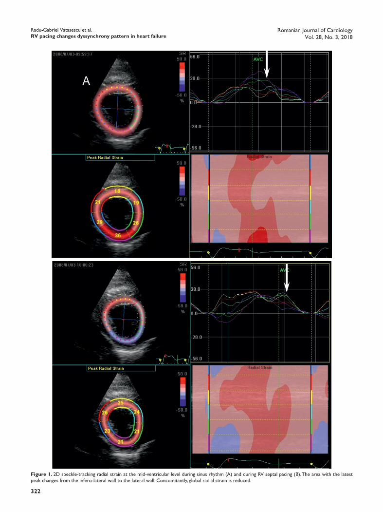
Figure 1. 2D speckle-tracking radial strain at the mid-ventricular level during sinus rhythm (A) and during RV septal pacing (B). The area with the latest peak changes from the infero-lateral wall to the lateral wall. Concomitantly, global radial strain is reduced.

Romanian Journal of CardiologyVol. 28, No. 3, 2018
323
Radu-Gabriel Vatasescu et al.RV pacing changes dyssynchrony pattern in heart failure
Area of LV breakthrough and area of maxi-mum delay: Changes in the location of the area of maximum delay during RVA pacing in patients with LVD and LBBB have been described during LV endo-cardial mapping10,11 as well as at the level of the LV epi-cardium12,13. If this changes in electrical activation are translated into changes in the contraction pattern is currently not known. Present study showed that in pa-tients with LVD and LBBB, although DDD RV pacing with optimum AV delay does not signifi cantly change the area of earliest systolic peak, it does change the location of maximum LV delay at midventricular level in more than 75% of the patients. This might explain the weak correlation between echocardiographic pa-rameters available for identifi cation of baseline intra-ventricular dyssynchrony and clinical or structural res-ponse to CRT2. An indirect support for the effects of RV pacing on dyssynchrony pattern comes from stu-dies of epicardial CRT. Placing the LV lead at sites of maximum electrical delay assessed during RVA pacing signifi cantly increased the percentage of responders15.
Effects of RV pacing on LV dyssynchrony: RV pacing increases the risk of HF and death in patients with systolic LV dysfunction (LVD)15,16 as well as in pati-ents with normal baseline LV systolic function17,18. The risk is higher in patients with baseline wider QRS19,20 as well as in patients with wider paced QRS21,22. The underlying mechanism is induction of intraventricu-lar dyssynchrony, with consecutive impairment of LV systolic function, an effect observed acutely in pati-ents with normal baseline systolic function23-25 as well as in patients with systolic LVD26,27, In patients with systolic LVD, intraventricular dyssynchrony induced by RV pacing is further augmented in the presence of a wide QRS27-29, especially in the presence of LBBB29. In the present study RV pacing overall did not signi-fi cantly increase intraventricular dyssynchrony in pa-tients with systolic LVD and LBBB. However, in the small subgroup of patients with RVA pacing there was a signifi cant increase in LV dyssynchrony. This can be explained by the fact that the vast majority of patients in the present study had RVS pacing, which is probably less dyssynchronous than RVA pacing8,30 or in some patients is able to partially capture distal part of the His fascicle and/or LBB31. Another possible explanati-on is that the DDD pacing with optimized AV interval used in this study may still allow some degree of fusi-on with intrinsic activation in patients with normal AV conduction, therefore blurring the deleterious effects of RV pacing32.
ents. The location of breakthrough area during DDD RV pacing remained unchanged in 35 out of 40 pati-ents. The mean time interval from beginning of QRS to the earliest systolic peak during SR and during RV pacing was similar (234±75 ms vs. 220±94 ms, p=NS).
Maximum LV delay area: Concomitantly the lo-cation of the maximum delay area shifted in 31 out of 40 patients (77%) (Figure 1). Baseline maximum de-lay area was located on the lateral wall in 9 patients (22.5%), on the infero-lateral wall in 20 patients (50%) and on the inferior wall in 11 patients (27.5%). During RV pacing maximum delay area was located in the in-ferior wall in 31 patients (77.5%), on the infero-lateral wall in 5 patients (12.5%) and on the lateral wall in 4 patients (10%).
LV radial deformation: The mean midventricular peak systolic global radial strain was signifi cantly redu-ced during RV pacing (13.3±8.5% vs. 18.3±7.4% during SR, p<0.001) (Figure 2).
DISCUSSIONSThis study shows that in patients with moderate to severe CHF, LV systolic dysfunction, LBBB and normal PR interval, CRT with standard optimized AV delay8 introduces RV pacing. RV pacing produces an overall a non-signifi cant increase in LV dyssynchrony, changes the dyssynchrony pattern and further impairs LV glo-bal radial strain. Specifi cally, RVA pacing signifi cantly worsened LV dyssynchrony. This change of LV mecha-nic dyssynchrony pattern induced by RV pacing during CRT may explain why echocardiographic indices of in-tra-ventricular dyssynchrony as assessed during sinus rhythm are not well correlated with CRT response.
Figure 2. Acute effects of RV pacing on LV mid-ventricular global radial strain.

Radu-Gabriel Vatasescu et al.RV pacing changes dyssynchrony pattern in heart failure
Romanian Journal of CardiologyVol. 28, No. 3, 2018
324
References1. McAlister FA; Ezekowitz J; Hooton N; Vandermeer B; Spooner C;
Dryden DM; et al. Cardiac resynchronization therapy for patients with left ventricular systolic dysfunction: a systematic review. JAMA. 2007 Jun 13;297(22):2502-14.
2. Chung ES,. Leon AR, Tavazzi L, Sun J-P, Nihoyannopoulos P, Merlino J, et al. Results of the Predictors of Response to CRT (PROSPECT) Trial. Circulation 2008;117:2608-2616.
3. Yu CM; Bleeker GB; Fung JW; Schalij MJ; Zhang Q; van der Wall EE; et al. Left ventricular reverse remodeling but not clinical improve-ment predicts long-term survival after cardiac resynchronization the rapy. Circulation 2005;112:1580-6.
4. Suffoletto MS, Dohi K, MD; Cannesson M, Saba S, Gorcsan J. Nov-el Speckle-Tracking Radial Strain From Routine Black-and-White Echocardiographic Images to Quantify Dyssynchrony and Predict Response to Cardiac Resynchronization Therapy. Circulation 2006; 113:960-968
5. Vesely MR, Li S, Kop WJ, Reese A, Marshall J, Shorofsky SR, et al. Test-retest reliability of assessment for intraventricular dyssynchro-ny by tissue Doppler imaging echocardiography. Am J Cardiol. 2008; 101:645-50.
6. Anderson LJ, Miyazaki C, Sutherland GR, Oh JK. Patient Selection and Echocardiographic Assessment of Dyssynchrony in Cardiac Re-synchronization Therapy. Circulation 2008;117:2009-2023.
7. Victor F, Mabo P, Mansour H, Pavin D, Kabalu G, De Place C, et al. A Randomized Comparison of Permanent Septal Versus Apical Right Ventricular Pacing: Short-Term Results. J Cardiovasc Electrophysiol 2006;17: 238-242.
8. Barold SS, Ilercil A, Herweg B. Echocardiographic optimization of the atrioventricular and interventricular intervals during cardiac re-synchronization. Europace. 2008; Suppl 3:iii88-95.
9. Delgado V, Ypenburg C, Zhang Q, Mollema SA, Fung JW, Schalij MJ, et al. Changes in global left ventricular function by multidirectional strain assessment in heart failure patients undergoing cardiac resyn-chronization therapy. J Am Soc Echocardiogr 2009;22:688-94.
10. Vatasescu R, Berruezo A, Mont L, Tamborero D, Sitges M, Silva E, et al. Midterm ‘super-response’ to cardiac resynchronization therapy by biventricular pacing with fusion: insights from electro-anatomical mapping. Europace. 2009 Dec;11(12):1675-82.
11. Vassallo JA, Cassidy DM, Miller JM, Buxton AE, Marchlinski FE, Jo-sephson ME. Left ventricular endocardial activation during right ven-tricular pacing: effect of underlying heart disease. J Am Coll Cardiol 1986;7:1228-33.
12. Pastore CA, Tobias N, Samesima N, Filho MM, Pedrosa A, Nish-ioka S, et al. Body surface potential mapping investigating the ven-tricular activation patterns in the cardiac resynchronization of pa-tients with left bundle-branch block and heart failure. J Electrocardiol 2006;39:93-102.
13. Jia P, Ramanathan C, Ghanem RN, Ryu K, Varma N, Rudy Y. Electro-cardiographic imaging of cardiac resynchronization therapy in heart failure: Observation of variable electrophysiologic responses. Heart Rhythm 2006;3:296-310.
14. Edgerton JR, Edgerton ZJ, Mack MJ, Hoffman S, Dewey TM, Herbert MA, Ventricular Epicardial Lead Placement for Resynchronization by Determination of Paced Depolarization Intervals: Technique and Ra-tionale. Ann Thorac Surg 2007;83:89-92.
15. Wilkoff BL, Cook JR, Epstein AE, Greene HL, Hallstrom AP, Hsia H, et al; on behalf of the Dual Chamber and VVI Implantable De-fi brillator Trial Investigators. Dual chamber pacing or ventricular backup pacing in patients with an implantable defi brillator: the Dual Chamber and VVI Implantable Defi brillator (DAVID) trial. JAMA 2002;288:3115-23.
16. Steinberg JS, Fischer A, Wang P, Schuger C, Daubert J, McNitt S, et al; MADIT II Investigators. The clinical implications of cumulative right ventricular pacing in the multicenter automatic defi brillator tri-al II. J Cardiovasc Electrophysiol 2005;16:359-65.
17. Sweeney MO, Hellkamp AS, Ellenbogen KA, Greenspon AJ, Freed-man RA, Lee KL, et al; MOde Selection Trial Investigators. Ad-verse effect of ventricular pacing on heart failure and atrial fi brilla-
LV radial deformation: Intraventricular dyssyn-chrony induced and/or augmented by RV pacing alters LV systolic function acutely24,25,28,29 as well as chroni-cally18,26, and this effect is largest in patients with systo-lic LVD and LBBB29. Present investigation showed that midventricular GRS was signifi cantly reduced during RV pacing, suggesting an acute reduction in LV systolic function since GRS has been reported to be correla-ted with LVEF9,33. This also might explain the superior response in HF patients with limited RV pacing during CRT10,34,35.
LIMITATIONSThis is an acute study and present fi ndings may not apply to a chronic RV pacing. However, current data showed that baseline dyssynchrony induced by RV pacing signifi cantly impacts LV function on long term21,23, suggesting that the effect is persistent. The results may be limited as well by the relatively small number of patients in this study as well as intra- and interobserved variability in measuring radial strain. Although the latter is in range with other studies (or even smaller)36, these could explain the lack of statis-tical signifi cance for the difference in the magnitude of intraventricular dyssynchrony. Moreover, the proto-col used for RVA pacing (DDD with optimized AVI i.e. shortest AVI without mitral infl ow truncation), may allow fusion with intrinsic rhythm in a signifi cant pro-portion of patients26, possibly obscuring the changes in LV activation. However, in the vast majority of the patients the present study showed a shift in the LV dyssynchrony pattern. If we consider also that the AVI used refl ects common practice in CRT optimization in many centers, this suggest that present fi ndings might have a signifi cant impact in clinical practice, warranting attention and further research.
CONCLUSIONSIn patients with systolic LVD and LBBB, RV pacing changes the location of maximum LV delay area and, especially for RVA leads, augments intraventricular dyssynchrony, and supplementary impairs LV stra-in. This might explain the weak correlation between baseline mechanical intraventricular dyssynchrony as assessed during intrinsic rhythm and the response to CRT.
Confl ict of interest: none declared.

Romanian Journal of CardiologyVol. 28, No. 3, 2018
325
Radu-Gabriel Vatasescu et al.RV pacing changes dyssynchrony pattern in heart failure
Versus Idiopathic Left Bundle Branch Block With and Without Left Ventricular Dysfunction. Am J Cardiol 2004;93:1243-1246.
28. Pastore G, Noventa F, Piovesana P, Cazzin R, Aggio S, Verlato R, et al. Left ventricular dyssynchrony resulting from right ventricular apical pacing: relevance of baseline assessment. Pacing Clin Electro-physiol 2008;31:1456-62.
29. Schmidt M, Rittger H, Marschang H, Sinha AM, Daccarett M, Brach-mann Jet al. Left ventricular dyssynchrony from right ventricular pacing depends on intraventricular conduction pattern in intrinsic rhythm. Eur J Echocardiogr 2009;10:776-83.
30. Yu CC, Liu YB, Lin MS, Wang JY, Lin JL, Lin LC. Septal pacing pre-serving better left ventricular mechanical performance and contrac-tile synchronism than apical pacing in patients implanted with an atrioventricular sequential dual chamber pacemaker. Int J Cardiol 2007;118:97-106.
31. Sharma PS, Dandamudi G, Herweg B, Wilson D, Singh R, Na-perkowski A, Koneru JN, Ellenbogen KA, Vijayaraman P. Permanent His-bundle pacing as an alternative to biventricular pacing for car-diac resynchronization therapy: A multicenter experience. Heart Rhythm. 2018 Mar;15(3):413-420.
32. Tops LF, Suffoletto MS, Bleeker GB, Boersma E, van der Wall EE, Gorcsan J 3rd, et al. Speckle-tracking radial strain reveals left ven-tricular dyssynchrony in patients with permanent right ventricular pacing. J Am Coll Cardiol 2007;50:1180-8.
33. Altman M, Bergerot C, Aussoleil A, Davidsen ES, Sibellas F, Ovize M, Bonnefoy-Cudraz E, Thibault H, Derumeaux G. Assessment of left ventricular systolic function by deformation imaging derived from speckle tracking: a comparison between 2D and 3D echo modalities. Eur Heart J Cardiovasc Imaging. 2014 Mar;15(3):316-23
34. Van Gelder BM, Bracke FA, Meijer A, Pijls NHJ. The Hemodynamic Effect of Intrinsic Conduction during Left Ventricular Pacing as Com-pared to Biventricular Pacing. J Am Coll Cardiol 2005; 46:2305-10.
35. Lee KL, Burnes JE, Mullen TJ, Hettrick DA, Tse HF, Lau CP. Avoid-ance of right ventricular pacing in cardiac resynchronization therapy improves right ventricular hemodynamics in heart failure patients. J Cardiovasc Electrophysiol 2007;18:497-504.
36. Tanaka H, Nesser HJ, Buck T, Oyenuga O, Jánosi RA, Winter S, et al. Dyssynchrony by speckle-tracking echocardiography and response to cardiac resynchronization therapy: results of the Speckle Tracking and Resynchronization (STAR) study. Eur Heart J. 2010;31(14):1690-700.
tion among patients with normal baseline QRS duration in a clinical trial of pacemaker therapy for sinus node dysfunction. Circulation 2003;107:2932-7.
18. Vatasescu R, Shalganov T, Paprika D, Kornyei L, Prodan Z, Bodor G, et al. Evolution of left ventricular function in pediatric patients with permanent right ventricular pacing for isolated congenital heart block: a medium term follow-up. Europace. 2007; 9:228-32.
19. Sweeney M.O., Hellkamp A.S., Lee K.L., Lamas G.A. Association of Prolonged QRS Duration With Death in a Clinical Trial of Pacemak-er Therapy for Sinus Node Dysfunction. Circulation. 2005;111:2418-2423.
20. Hayes J.J., Sharma A.D., Love J.C. Herre JM, Leonen AO, Kudenchuk PJ; DAVID Investigators. Abnormal Conduction Increases Risk of Adverse Outcomes From Right Ventricular Pacing. J Am Coll Car-diol 2006;48:1628-33.
21. Shukla H.H., Hellkamp A.S., James E.A. Flaker GC, Lee KL, Swee-ney MO, et al; Mode Selection Trial (MOST) Investigators. Heart failure hospitalization is more common in pacemaker patients with sinus node dysfunction and a prolonged paced QRS duration. Heart Rhythm 2005;2:245-251.
22. Miyoshi F, Kobayashi Y, Itou H Onuki T, Matsuyama T, Watanabe N, et al. Prolonged Paced QRS Duration as a Predictor for Congestive Heart Failure in Patients with Right Ventricular Apical Pacing. PACE 2005;28:1182-1188.
23. Gomes JA, Damato AN, Akhtar M, Dhatt MS, Calon AH, Reddy CP, et al. Ventricular septal motion and left ventricular dimensions dur-ing abnormal ventricular activation. Am J Cardiol 1977;39:641-50.
24. Delgado V, Tops LF, Trines SA, Zeppenfeld K, Marsan NA, BertiniM, et al. Acute effects of right ventricular apical pacing on left ventri-cular synchrony and mechanics. Circ Arrhythmia Electrophysiol 2009;2:135-45.
25. Liu WH, Chen MC, Chen YL, Guo BF, Pan KL, Yang CH, et al. Rightventricular apical pacing acutely impairs left ventricular function and induces mechanical dyssynchrony in patients with sick sinus syn-drome: a real-time three-dimensional echocardiographic study. J Am Soc Echocardiogr 2008; 21:224-9.
26. Tops LF, Schalij MJ, Holman ER, van Erven L, van der Wall EE, Bax JJ. Right ventricular pacing can induce ventricular dyssynchrony in pa-tients with atrial fi brillation after atrioventricular node ablation. J Am Coll Cardiol 2006;48:1642-8.
27. Kang S-J., Song J-K., Yang H.S. Song JM, Kang DH, Rhee KS, et al. Systolic and Diastolic Regional Myocardial Motion of Pacing-Induced


![Studiu de caz INNOSOC - Sociallabsociallab.fer.hr/wordpress/wp-content/uploads/2017/... · Studiu de caz INNOSOC ~ o v µso v ]îìíóVÀ ]µv Æ ]v Titlul studiului de caz: Promovarea](https://static.fdocuments.in/doc/165x107/5e25004e1aeac624731d8ecb/studiu-de-caz-innosoc-studiu-de-caz-innosoc-o-v-so-v-v-v-.jpg)