
ORIGINAL ARTICLE High-Glucose Environment...
9
High-Glucose Environment Enhanced Oxidative Stress and Increased Interleukin-8 Secretion From Keratinocytes New Insights Into Impaired Diabetic Wound Healing Cheng-Che E. Lan, 1,2,3 Ching-Shuang Wu, 4 Shu-Mei Huang, 1,2,5 I-Hui Wu, 1,2 and Gwo-Shing Chen 1,2 Impaired wound healing frequently occurs in patients with diabetes. Interleukin (IL)-8 production by keratinocyte is re- sponsible for recruiting neutrophils during healing. Intense in- flammation is associated with diabetic wounds, while reduction of neutrophil infiltration is associated with enhanced healing. We hypothesized that increased neutrophil recruitment by keratino- cytes may contribute to the delayed healing of diabetic wounds. Using cultured human keratinocytes and a diabetic rat model, the current study shows that a high-glucose environment enhanced IL-8 production via epidermal growth factor receptor (EGFR)– extracellular signal–regulated kinase (ERK) pathway in a reactive oxygen species (ROS)-dependent manner in keratinocytes. In ad- dition, diabetic rat skin showed enhanced EGFR, ERK, and IL-8 expression compared with control rats. The dermal neutrophil infiltration of the wound, as represented by expression of myelo- peroxidase level, was also significantly higher in diabetic rats. Treating diabetic rats with dapsone, an agent known to inhibit neutrophil function, was associated with improved healing. In conclusion, IL-8 production and neutrophil infiltration are in- creased in a high-glucose environment due to elevated ROS level and contributed to impaired wound healing in diabetic skin. Tar- geting these dysfunctions may present novel therapeutic approaches. Diabetes 62:2530–2538, 2013 W ound healing is a dynamic, interactive pro- cess involving coagulation, inflammation, tis- sue formation, and tissue remodeling (1–3). Impaired skin wound healing is a common cause of morbidity and mortality among patients with di- abetes (4). Dysregulation of glucose homeostasis and ele- vated glucose levels are the central etiologies of diabetes (5). With the increasing prevalence of diabetes around the globe (6), understanding the mechanisms responsible for poor diabetic wound healing is an important public health issue. Normal wound healing is a carefully orchestrated pro- cess in which proper induction of inflammatory cytokines leads to neutrophil recruitment that is essential for fending off potential infections to the disrupted barrier (7). In ad- dition to their roles in controlling microbial invasion, neutrophils had been thought to play a limited role during the healing process of uncontaminated wound (8). How- ever, persistence of inflammation and neutrophil infil- tration are characteristics associated with impaired wound healing among patients with diabetes (9–11). Treatments that enhance diabetic wound healing are often associated with reducing inflammatory cytokines in the diabetic wound environment (12,13). Intriguingly, a study by Dovi, He, and DiPietro (14) demonstrated that reepithelialization after wounding is accelerated by neutrophil depletion. These findings suggested that inflammation, in particular neutrophils, has a direct impact on wound healing. An important yet still unanswered question during diabetic wound healing is how a high-glucose environment affects chemotactic activity of keratinocytes to neutrophils. Ker- atinocytes, the immune competent cells forming the outer layer of the skin, are important providers of neutrophil chemotactic cytokines including chemokine (C-X-C motif) ligand 1 (CXCL-1) and interleukin (IL)-8, both of which are identified as the major chemotactic cytokines in human wound fluid (15). It has been shown that epidermal wound generates prominent chemotactic activity toward neutrophils in the injured skin and that IL-8 production derived from keratinocytes is critically involved in this process (15,16). Besides recruiting neutrophils, IL-8 has also been recognized to activate neutrophil function in- cluding increasing superoxide and hydrogen peroxide formation (17,18). These evidences indicated that epi- dermal keratinocytes, through IL-8 secretion, serve as the mediator between skin wound and neutrophil re- cruitment/activation during the early phase of wound healing. As aforementioned, a common denominator frequently observed among poor diabetic healing wounds is the presence of prolonged inflammation consisting of neu- trophils and macrophages (9–11). Intriguingly, neu- trophils derived from diabetic patients have been shown to display reduced migration in response to IL-8 che- motaxis (19). Therefore, we hypothesized that a strong chemotactic factor is present at the diabetic wound site that allows for effective recruitment of the chemotactic response–defective neutrophils. With use of cultured normal human keratinocytes and a diabetic rat model, the current study was launched to explore the effect of a high-glucose environment on epidermal keratinocytes in the context of neutrophil recruitment and propose a novel therapeutic option for treating acute diabetic wounds. From the 1 Department of Dermatology, Kaohsiung Medical University Hos- pital, Kaohsiung, Taiwan; the 2 Department of Dermatology, College of Medicine, Kaohsiung, Taiwan; the 3 Department of Dermatology, Kaohsiung Municipal Ta-Tung Hospital, Kaohsiung Medical University, Kaohsiung, Tai- wan; the 4 Department of Medical Laboratory Science and Biotechnology, Kaohsiung Medical University, Kaohsiung, Taiwan; and the 5 Department of Biological Sciences, National Sun Yat-sen University, Kaohsiung, Taiwan. Corresponding author: Gwo-Shing Chen, [email protected]. Received 11 December 2012 and accepted 14 February 2013. DOI: 10.2337/db12-1714 Ó 2013 by the American Diabetes Association. Readers may use this article as long as the work is properly cited, the use is educational and not for profit, and the work is not altered. See http://creativecommons.org/licenses/by -nc-nd/3.0/ for details. 2530 DIABETES, VOL. 62, JULY 2013 diabetes.diabetesjournals.org ORIGINAL ARTICLE
-
Upload
nguyenkhue -
Category
Documents
-
view
219 -
download
0
Transcript of ORIGINAL ARTICLE High-Glucose Environment...
High-Glucose Environment Enhanced Oxidative Stressand Increased Interleukin-8 SecretionFrom KeratinocytesNew Insights Into Impaired Diabetic Wound HealingCheng-Che E. Lan,
1,2,3Ching-Shuang Wu,
4Shu-Mei Huang,
1,2,5I-Hui Wu,
1,2and Gwo-Shing Chen
1,2
Impaired wound healing frequently occurs in patients withdiabetes. Interleukin (IL)-8 production by keratinocyte is re-sponsible for recruiting neutrophils during healing. Intense in-flammation is associated with diabetic wounds, while reductionof neutrophil infiltration is associated with enhanced healing. Wehypothesized that increased neutrophil recruitment by keratino-cytes may contribute to the delayed healing of diabetic wounds.Using cultured human keratinocytes and a diabetic rat model, thecurrent study shows that a high-glucose environment enhancedIL-8 production via epidermal growth factor receptor (EGFR)–extracellular signal–regulated kinase (ERK) pathway in a reactiveoxygen species (ROS)-dependent manner in keratinocytes. In ad-dition, diabetic rat skin showed enhanced EGFR, ERK, and IL-8expression compared with control rats. The dermal neutrophilinfiltration of the wound, as represented by expression of myelo-peroxidase level, was also significantly higher in diabetic rats.Treating diabetic rats with dapsone, an agent known to inhibitneutrophil function, was associated with improved healing. Inconclusion, IL-8 production and neutrophil infiltration are in-creased in a high-glucose environment due to elevated ROS leveland contributed to impaired wound healing in diabetic skin. Tar-geting these dysfunctions may present novel therapeutic approaches.Diabetes 62:2530–2538, 2013
Wound healing is a dynamic, interactive pro-cess involving coagulation, inflammation, tis-sue formation, and tissue remodeling (1–3).Impaired skin wound healing is a common
cause of morbidity and mortality among patients with di-abetes (4). Dysregulation of glucose homeostasis and ele-vated glucose levels are the central etiologies of diabetes(5). With the increasing prevalence of diabetes around theglobe (6), understanding the mechanisms responsible forpoor diabetic wound healing is an important public healthissue.
Normal wound healing is a carefully orchestrated pro-cess in which proper induction of inflammatory cytokinesleads to neutrophil recruitment that is essential for fending
off potential infections to the disrupted barrier (7). In ad-dition to their roles in controlling microbial invasion,neutrophils had been thought to play a limited role duringthe healing process of uncontaminated wound (8). How-ever, persistence of inflammation and neutrophil infil-tration are characteristics associated with impaired woundhealing among patients with diabetes (9–11). Treatmentsthat enhance diabetic wound healing are often associatedwith reducing inflammatory cytokines in the diabeticwound environment (12,13). Intriguingly, a study by Dovi,He, and DiPietro (14) demonstrated that reepithelializationafter wounding is accelerated by neutrophil depletion.These findings suggested that inflammation, in particularneutrophils, has a direct impact on wound healing. Animportant yet still unanswered question during diabeticwound healing is how a high-glucose environment affectschemotactic activity of keratinocytes to neutrophils. Ker-atinocytes, the immune competent cells forming the outerlayer of the skin, are important providers of neutrophilchemotactic cytokines including chemokine (C-X-C motif)ligand 1 (CXCL-1) and interleukin (IL)-8, both of which areidentified as the major chemotactic cytokines in humanwound fluid (15). It has been shown that epidermalwound generates prominent chemotactic activity towardneutrophils in the injured skin and that IL-8 productionderived from keratinocytes is critically involved in thisprocess (15,16). Besides recruiting neutrophils, IL-8 hasalso been recognized to activate neutrophil function in-cluding increasing superoxide and hydrogen peroxideformation (17,18). These evidences indicated that epi-dermal keratinocytes, through IL-8 secretion, serve asthe mediator between skin wound and neutrophil re-cruitment/activation during the early phase of woundhealing.
As aforementioned, a common denominator frequentlyobserved among poor diabetic healing wounds is thepresence of prolonged inflammation consisting of neu-trophils and macrophages (9–11). Intriguingly, neu-trophils derived from diabetic patients have been shownto display reduced migration in response to IL-8 che-motaxis (19). Therefore, we hypothesized that a strongchemotactic factor is present at the diabetic wound sitethat allows for effective recruitment of the chemotacticresponse–defective neutrophils. With use of culturednormal human keratinocytes and a diabetic rat model,the current study was launched to explore the effect ofa high-glucose environment on epidermal keratinocytesin the context of neutrophil recruitment and proposea novel therapeutic option for treating acute diabeticwounds.
From the 1Department of Dermatology, Kaohsiung Medical University Hos-pital, Kaohsiung, Taiwan; the 2Department of Dermatology, College ofMedicine, Kaohsiung, Taiwan; the 3Department of Dermatology, KaohsiungMunicipal Ta-Tung Hospital, Kaohsiung Medical University, Kaohsiung, Tai-wan; the 4Department of Medical Laboratory Science and Biotechnology,Kaohsiung Medical University, Kaohsiung, Taiwan; and the 5Department ofBiological Sciences, National Sun Yat-sen University, Kaohsiung, Taiwan.
Corresponding author: Gwo-Shing Chen, [email protected] 11 December 2012 and accepted 14 February 2013.DOI: 10.2337/db12-1714� 2013 by the American Diabetes Association. Readers may use this article as
long as the work is properly cited, the use is educational and not for profit,and the work is not altered. See http://creativecommons.org/licenses/by-nc-nd/3.0/ for details.
2530 DIABETES, VOL. 62, JULY 2013 diabetes.diabetesjournals.org
ORIGINAL ARTICLE

RESEARCH DESIGN AND METHODS
Keratinocyte culture and treatment. Keratinocytes were cultivated aspreviously described (20). The treatments of keratinocytes included 1) culti-vation with 6 mmol/L D-glucose, 26 mmol/L D-glucose, or 20 mmol/L mannitolfor 7 days; 2) cultivation with or without advanced glycation end production(AGE)-modified BSA (BioVision, Mountain View, CA) for 2 days; 3) cultivationwith 6 or 26 mmol/L D-glucose for 7 days with addition of 50 mmol/L L-ascorbicacid (Sigma) on the seventh day; and 4) cultivation with 6 mmol/L D-glucosefor 7 days, followed by epidermal growth factor receptor (EGFR) small in-terfering RNA (siRNA) transfection.Real-time quantitative PCR detection for IL-8 mRNA. Total RNA wasextracted using the TRIzol method (Gibco BRL, Gaithersburg, MD) and pro-cessed as recommended by the manufacturer. Five micrograms of RNA werereverse transcribed to cDNA as the PCR template. The primer sequences usedare listed in Table 1. Amplification and detection were performed with an ABIPrism 7500 sequence detection system (Applied Biosystems, Hammonton, NJ).The fold of gene change was calculated as 2^-DDCT.Measurement of IL-8 and CXCL-1 in cultured keratinocytes. Thesupernatants derived from keratinocytes cultivated at indicated conditionswere collected and stored at 220°C. The concentrations of IL-8 and CXCL-1were determined using a commercially available ELISA kit (R&D Systems,Minneapolis, MN) according to the manufacturer’s instructions. In blockingexperiments, the keratinocytes were treated with 10 mmol/L mitogen-activatedprotein kinase (MAPK) kinase (MEK)1/2 inhibitor (U0126; Calbiochem) andincubated for 24 h.Western blotting analysis. Total cellular proteins from cultured keratinocyteswere extracted with radioimmunoprecipitation assay buffer (0.1% SDS, 50 mmol/L Tris-HCl, 150 mmol/L NaCl, 0.5% sodium deoxycholate, and 1% NP-40, pH 7.5)containing protease inhibitor cocktail (Roche, Mannheim, Germany). One hun-dredmicrogramsof proteinswere loaded into 8%SDS-PAGEand transferred ontoa polyvinylidene fluoridemembrane. After blocking and washing, the membraneswere incubated with first antibodies against phosphorylated (p-) extracellularsignal–regulated kinases (ERKs), total ERK, (Cell Signaling, Beverly, MA), anda-tubulin (Santa Cruz Biotechnology, Santa Cruz, CA). The membranes werethen incubated with horseradish peroxidase (HRP)-labeled secondary antibody(Millipore, Billerica, MA) and developed with the ImmobilonTW WesternChemiluminescent HRP Substrate (Millipore). The blots were analyzed bya digital imaging system (Alpha Imager 2000; Alpha Innotech, San Leandro, CA).Flow cytometry analysis of membrane and intracellular p-EGFR. ForEGFR detection, the cultured keratinocytes were harvested, washed with PBS,and incubated with p-EGFR antibody (Cell Signaling) dissolved in 1% BSA/PBSsolution at room temperature for 30 min. After washing, the cells were thenincubated with Alexa Fluor 488 secondary antibody (Invitrogen, Carlsbad, CA)for 30 min. Subsequently, the keratinocytes were washed again with PBS andfixed with 2% paraformaldehyde at 37°C for 10 min. After centrifugation, thecell pellets were resuspended in 90% methanol for permeabilization at 4°C for15 min. After centrifugation and discarding of the supernatants, the cells wereagain incubated with p-EGFR and Alexa Fluor 488 secondary antibody. Theexpressions of p-EGFR were analyzed on a FACScan (Becton Dickinson,Mountain View, CA) at FL1 channel with CELLQuest Pro Software.siRNA experiment. The treated keratinocytes were transfected with Lip-ofectamine 2000 (Invitrogen) according to themanufacturer’s instructions.Westernblotting was performed to confirm the success of transfection. The supernatantswere collected and subjected to IL-8 ELISA assay as previously described.
Lipid peroxidation (malondialdehyde) assay.Malondialdehyde (MDA) and4-hydroxynonenal are natural products of lipid peroxidation. Measurement ofMDA is recognized as an important indicator of cell oxidative damage. Thecommercially available MDA assay kit (BioVision) was used to detect the MDAlevel according to the manufacturer’s instructions. The fluorescent intensity ofeach sample was determined with a microplate reader (FLx800; Bio-TEK,Winooksi, VT). The absorbance was recorded at 532 and 553 nm for excitationand emission, respectively, and the results were analyzed with Kcjuniorsoftware.Streptozotocin-induced diabetes.MaleWistar rats with initial weight of 200–300 g were acclimated for 1 week. Diabetic condition was induced by in-travenous injection of streptozotocin with 50 mg/kg dosage. After 24 h, theblood glucose levels were determined among rats that received injection.Glucose concentration .250 mg/dL in heparinized tail vein blood (measuredby glucometer) was considered as successful induction. All treated and con-trol rats were maintained with ad libitum access to water and diet. For in-dicated experiments, the diabetic rats were fed with 30 mg/kg dapsone (UChiu Pharmaceutical, Taoyuan, Taiwan) by oral gavage twice a day 2 daysbefore wounding and throughout the healing process.Wound-healing model. After 1 week of diabetes induction, the normal anddiabetic rats were used for wound-healing studies. Prior to injury, rats wereanesthetized by injection of a Zoletil 50 solution (50 mg/kg body wt i.p.). Aftershaving of the dorsal hair and cleansing of the skin with 70% ethanol, full-thickness excisional wounds were created using 6-mm biopsy stamps (Stiefel,Offenbach, Germany). Each wound region was digitally photographed, andwound sizes at indicated time points were recorded. In addition, 1 and 3 daysafter wounding, the perilesional rat skin was harvested with 8-mm punch forfurther analysis. The streptozotocin-induced diabetic rat model used in thisstudy reflects healing of acute wound under hyperglycemic conditions. Sincethe gist of the study is to investigate the effect of a high-glucose environment onkeratinocyte during the initial stage of acute wound healing in terms of neu-trophil recruitment, this animal model fulfills our purpose.Analysis of tissue myeloperoxidase. The measurement of myeloperoxidase(MPO) levels in tissue has been shown to reflect neutrophil content. MPOcontent in wound tissue was determined as previously described by Dovi, He,and DiPietro (14). Briefly, the wounds were homogenized in 2 mL 20 mmol/Lphosphate buffer, pH 7.4. The homogenates were then centrifuged at 12,000gfor 45 min, and the supernatants were decanted. The pellets were resuspendedin 1 mL 50 mmol/L phosphate buffer containing 10 mmol/L EDTA and 0.5%hexadecyltrimethylammonium bromide. After a freeze-thaw cycle, the sampleswere briefly sonicated and incubated at 60°C for 2 h. The samples werecentrifuged at 500g for 10 min, and the supernatants were transferred to 1.5-mL tubes. For analysis, a standard curve ranging from 0 to 3.0 units/mL MPO(Sigma) was generated. Aliquots of samples (50 mL) or standards were placedin 12 3 75 mm glass tubes with 500 mL assay buffer (0.1 mol/L phosphatebuffer, pH 5.4; 1% hexadecyltrimethylammonium bromide; and 0.43 mg/mL3,39,5,59-tetramethylbenzidine). The reactions began by the addition of 50 mL15 mmol/L H2O2, followed by incubation at 37°C for 15 min and finished withaddition of 1.0 mL cold 0.2 mol/L sodium acetate, pH 3.0. The absorbance ofeach sample and standard were read at 655 nm within 10 min.Immunohistochemical staining. Three-micron paraffin sections weredeparaffinized in xylene and rehydrated in graded alcohol dilutions. Endoge-nous peroxidase activity was blocked by incubation with 3% H2O2 for 5 min.Antigen retrieval was performed by pressure cooking for 10 min (121°C, 1.2kg/cm2) in 0.01 mol/L citrate buffer (pH 6.0). The slides were then incubatedwith mouse anti–p-EGFR and p-ERK antibodies (1:400 dilution; Cell Signaling)at room temperature for 60 min. Antibody reactions were detected with bio-tinylated link anti-mouse antibody (Biocare Medical, Concord, CA) for 20 min atroom temperature followed by incubation with Trekavidin-HRP (Biocare Medical)for 20 min. The color was developed using DAB substrate-chromogen solution(Biocare Medical). The slides were then counterstained with hematoxylin-eosin.Real-time quantitative PCR analysis of rat skin IL-8 mRNA. The methodshave previously been described (21). The primer sequence used is listed in Table 1.Statistical analysis. SPSS system for Windows, version 12.0 (SPSS, Chicago,IL), was used for statistical analysis. For each experiment, at least three in-dependent experiments were performed. The results are expressed as means 6SD. Student t test was used for statistical evaluation between control and ex-perimental groups in the study. In addition, one-way ANOVA followed by posthoc Fisher’s least-significant-difference test was used for comparison of woundhealing between different animal groups. A P value ,0.05 is considered to bestatistically significant.
RESULTS
High-glucose environment increased IL-8 expres-sion at both mRNA and protein levels in culturedkeratinocytes, and inhibition tests suggested that ERK
TABLE 1Primer sequence of genes investigated in the study
Gene Sequence (primer)
IL-8 59-CGTGGCTCTCTTGGCAGCCTTCCTGAT-39(forward)
59-TCAAAAACTTCTCCACAACCCTCTGCA-39(reverse)
GAPDH 59-CCACCCATGGCAAATTCC-39 (forward)59-GGGATTTCCATTGATGACA-39 (reverse)
EGFR siRNA A, 59-UUAACUUUCUCACCUUCUGGGAUCC-3959-GGAUCC CAGAAGGUGAGAAAGUUAA-39B, 59-AAAUCUGUGAUCUUGACAUGCUGCG-3959-CGCAGCAUGUCAAGAUCACAGAUUU-39
b-actin (rat) 59-tctgtgtggattggtggctct-39 (forward)59-gactcatcgtactcctgcttgct-39 (reverse)
IL-8 (rat) 59-tcaacgggcagaatcaaagag-39 (forward)59-ctcagacagcgaggcacatc-39 (reverse)
C.-C.E. LAN AND ASSOCIATES
diabetes.diabetesjournals.org DIABETES, VOL. 62, JULY 2013 2531
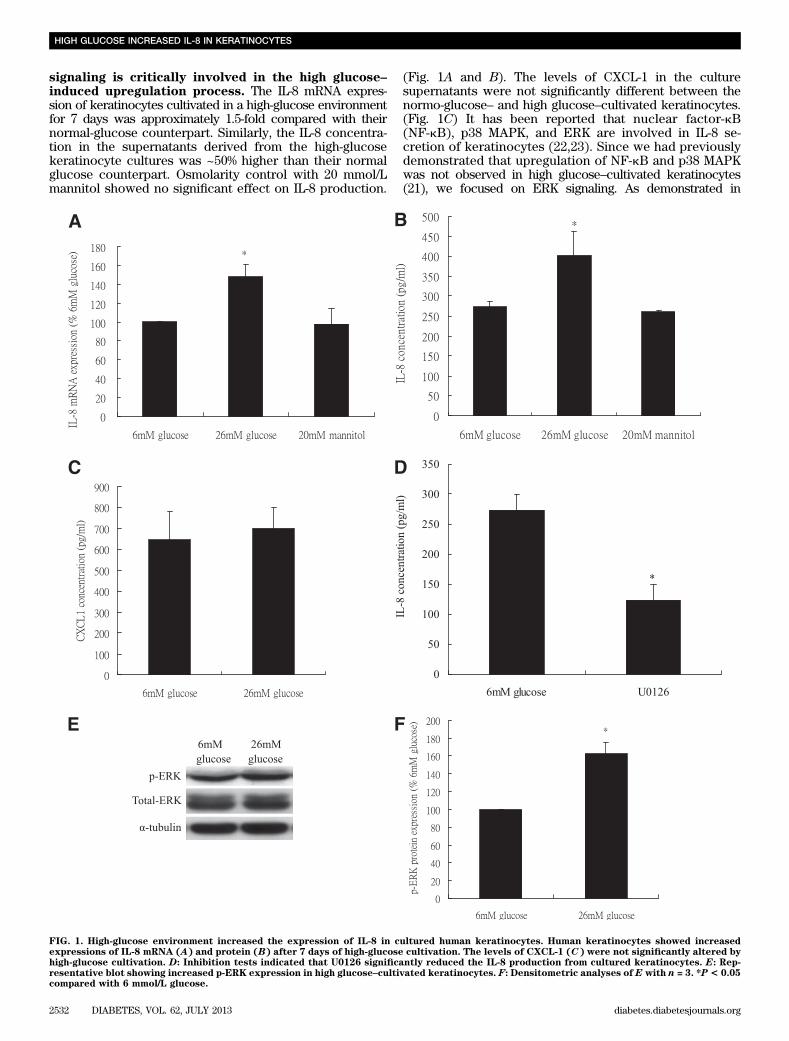
signaling is critically involved in the high glucose–induced upregulation process. The IL-8 mRNA expres-sion of keratinocytes cultivated in a high-glucose environmentfor 7 days was approximately 1.5-fold compared with theirnormal-glucose counterpart. Similarly, the IL-8 concentra-tion in the supernatants derived from the high-glucosekeratinocyte cultures was ~50% higher than their normalglucose counterpart. Osmolarity control with 20 mmol/Lmannitol showed no significant effect on IL-8 production.
(Fig. 1A and B). The levels of CXCL-1 in the culturesupernatants were not significantly different between thenormo-glucose– and high glucose–cultivated keratinocytes.(Fig. 1C) It has been reported that nuclear factor-kB(NF-kB), p38 MAPK, and ERK are involved in IL-8 se-cretion of keratinocytes (22,23). Since we had previouslydemonstrated that upregulation of NF-kB and p38 MAPKwas not observed in high glucose–cultivated keratinocytes(21), we focused on ERK signaling. As demonstrated in
FIG. 1. High-glucose environment increased the expression of IL-8 in cultured human keratinocytes. Human keratinocytes showed increasedexpressions of IL-8 mRNA (A) and protein (B) after 7 days of high-glucose cultivation. The levels of CXCL-1 (C) were not significantly altered byhigh-glucose cultivation. D: Inhibition tests indicated that U0126 significantly reduced the IL-8 production from cultured keratinocytes. E: Rep-resentative blot showing increased p-ERK expression in high glucose–cultivated keratinocytes. F: Densitometric analyses of E with n = 3. *P< 0.05compared with 6 mmol/L glucose.
HIGH GLUCOSE INCREASED IL-8 IN KERATINOCYTES
2532 DIABETES, VOL. 62, JULY 2013 diabetes.diabetesjournals.org
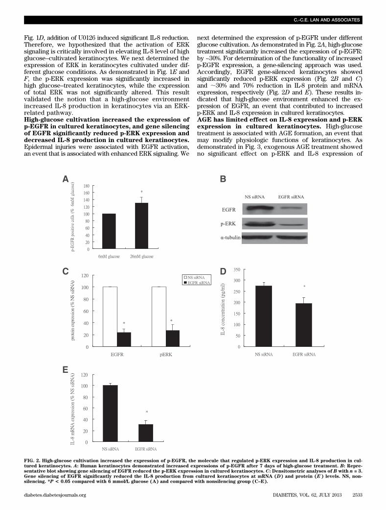
Fig. 1D, addition of U0126 induced significant IL-8 reduction.Therefore, we hypothesized that the activation of ERKsignaling is critically involved in elevating IL-8 level of highglucose–cultivated keratinocytes. We next determined theexpression of ERK in keratinocytes cultivated under dif-ferent glucose conditions. As demonstrated in Fig. 1E andF, the p-ERK expression was significantly increased inhigh glucose–treated keratinocytes, while the expressionof total ERK was not significantly altered. This resultvalidated the notion that a high-glucose environmentincreased IL-8 production in keratinocytes via an ERK-related pathway.High-glucose cultivation increased the expression ofp-EGFR in cultured keratinocytes, and gene silencingof EGFR significantly reduced p-ERK expression anddecreased IL-8 production in cultured keratinocytes.Epidermal injuries were associated with EGFR activation,an event that is associated with enhanced ERK signaling. We
next determined the expression of p-EGFR under differentglucose cultivation. As demonstrated in Fig. 2A, high-glucosetreatment significantly increased the expression of p-EGFR:by ~30%. For determination of the functionality of increasedp-EGFR expression, a gene-silencing approach was used.Accordingly, EGFR gene-silenced keratinocytes showedsignificantly reduced p-ERK expression (Fig. 2B and C)and ;30% and 70% reduction in IL-8 protein and mRNAexpression, respectively (Fig. 2D and E). These results in-dicated that high-glucose environment enhanced the ex-pression of EGFR, an event that contributed to increasedp-ERK and IL-8 expression in cultured keratinocytes.AGE has limited effect on IL-8 expression and p-ERKexpression in cultured keratinocytes. High-glucosetreatment is associated with AGE formation, an event thatmay modify physiologic functions of keratinocytes. Asdemonstrated in Fig. 3, exogenous AGE treatment showedno significant effect on p-ERK and IL-8 expression of
FIG. 2. High-glucose cultivation increased the expression of p-EGFR, the molecule that regulated p-ERK expression and IL-8 production in cul-tured keratinocytes. A: Human keratinocytes demonstrated increased expressions of p-EGFR after 7 days of high-glucose treatment. B: Repre-sentative blot showing gene silencing of EGFR reduced the p-ERK expression in cultured keratinocytes. C: Densitometric analyses of B with n = 3.Gene silencing of EGFR significantly reduced the IL-8 production from cultured keratinocytes at mRNA (D) and protein (E) levels. NS, non-silencing. *P < 0.05 compared with 6 mmol/L glucose (A) and compared with nonsilencing group (C–E).
C.-C.E. LAN AND ASSOCIATES
diabetes.diabetesjournals.org DIABETES, VOL. 62, JULY 2013 2533
cultured keratinocytes. These results indicated that AGEplays a limited role in the excessive IL-8 level observed inour experimental conditions.Lipid peroxidation product was increased in highglucose–cultivated keratinocytes, and ascorbic acidtreatment reduced p-EGFR and p-ERK expressionsas well as normalized IL-8 secretion in high glucose–cultivated keratinocytes. Increased oxidative stress hasbeen associated with a high-glucose environment. In sup-port of this notion, the level of MDA, a surrogate markerfor estimation of damage induced by reactive oxygen spe-cies (ROS), was significantly increased in high glucose–cul-tivated keratinocytes by approximately threefold comparedwith control (Fig. 4A). Treating high glucose–cultivatedkeratinocytes with ascorbic acid normalized the expressionof p-EGFR and p-ERK (Fig. 4B–D). In addition, the IL-8concentration in culture supernatants derived from highglucose–cultivated keratinocytes was 146.14 6 25.21% ofthe control without ascorbic acid treatment, while afterexogenous ascorbic acid treatment the supernatants fromhigh-glucose cultures contained 107.84 6 18.37% of IL-8compared with control. No significant difference in IL-8concentration was found between normal and high glucose–cultivated keratinocytes after addition of ascorbic acid intothe culture media. These results indicated that increasedoxidative stress is associated with enhanced IL-8 production
in high glucose–treated keratinocytes. The perilesional skinof the diabetic rats demonstrated significantly higher IL-8expression compared with that of control rats afterwounding. In addition, the level of neutrophil infiltration, asreflected by levels of MPO, was significantly higher in di-abetic wounds compared with control.
For validation of our in vitro results in vivo, a diabeticrat model was used. Accordingly, diabetic rats showedimpaired wound healing. The length of time required toachieve complete wound healing was 9.0 6 0.4 and 11.5 60.8 days, respectively, for control and diabetic rats (n =12). Representative figures of healing process are shown inFig. 5A, and the planimetry analysis is shown in Fig. 5B.The rates of healing for control and diabetic rats were12.8 and 6.9% healing/day, respectively, from day 0 to day9. It should be noted that in our wound model, a fibrinblot (crust) invariably formed after wounding. For thecontrol rats, most crust resolved after day 3 postwound-ing, and wound closure proceeded smoothly afterward.For the diabetic rats, most crusts did not resolve untilafter 5 days. Therefore, the delayed resolution of crustmay be considered part of impaired wound healing in ourexperiment.
In terms of IL-8 expression, wounding increased IL-8 ex-pression in both control and diabetic rat skin. The IL-8 in-crease in diabetic rat skin was significantly higher comparedwith control skin. More specifically, the ratio of IL-8 increasein diabetic perilesional skin was 1.75 6 0.08-fold and 1.32 60.02-fold of control perilesional rat skin 1 and 3 days afterwounding, respectively. Immunohistologically, the epidermisof diabetic rats showed more intense staining of p-EGFR andp-ERK compared with control (Fig. 5C). The dermal neutro-phil infiltration, as reflected by MPO level, was significantlyhigher in diabetic rats compared with control rats (Fig. 5D).Systemic dapsone treatment reduced MPO level ofthe wound and improved healing of diabetic rat skin.Since we hypothesized that increased neutrophil in-filtration contributes to impaired healing in diabetic rats,systemic dapsone, an agent recognized to impair neutro-phil function, was administered to diabetic rats. The timerequired for diabetic rats to achieve complete wound clo-sure was significantly shortened after systemic dapsonetreatment. More specifically, the time required for com-plete wound closure in diabetic rats reduced from 11.5 60.8 days without dapsone treatment to 8.9 6 0.5 days afterdapsone treatment (Fig. 5A), and the rate of healing fromday 0 to day 9 improved from 6.9 to 11.6% healing/day. Thefibrin clot also resolved at earlier time (3 days) post-wounding. In addition, the level of dermal MPO in controland diabetic rats with dapsone treatment 1 day afterwounding was 5.55 6 0.17-fold and 6.09 6 0.13-fold, re-spectively, compared with respective dermis before wound-ing. No difference was found between these two groups.Therefore, systemic dapsone treatment shortened the dura-tion required for wound healing and reduced the level ofMPO during wound healing in diabetic rats.
DISCUSSION
Although neutrophils derived from diabetic patients demon-strated defective chemotactic response to IL-8, the diabeticwounds were characterized by excessive and prolongedneutrophil infiltration. This intriguing phenomenon led us tohypothesize that a high-glucose environment may elicit ker-atinocytes to increase secretion of IL-8, an event which inturn results in excessive neutrophil recruitment.
FIG. 3. AGE has limited effect on p-ERK and IL-8 expression in culturedkeratinocytes. A: Treating cultured human keratinocytes with AGE-modified BSA did not significantly affect the p-ERK expression asdemonstrated by Western blotting analyses. B: Densitometry analysesof A, n = 3. C: IL-8 production from cultured keratinocytes was notsignificantly altered by AGE treatment.
HIGH GLUCOSE INCREASED IL-8 IN KERATINOCYTES
2534 DIABETES, VOL. 62, JULY 2013 diabetes.diabetesjournals.org

Previously, it was reported that the injury-induced im-mune responses were mediated by activation of EGFR. In-triguingly, IL-8 production from keratinocytes in responseto skin injury also occurred in an EGFR-dependent manner(16). In this study, we demonstrated that high-glucose en-vironment renders keratinocytes prone to produce higherlevels of IL-8 due to enhanced EGFR-ERK signaling. Theseresults corroborated with a previous study showing thatEGFR-ERK cascade regulates the production of IL-8 in lungcancer cells (24). It should be noted that our experimentalcondition did not significantly alter the physiologic status(viability, growth, and differentiation) of culture keratino-cytes as demonstrated in our previous study (21).
Different factors may initiate the process resulting inelevated IL-8 levels and enhanced neutrophil recruitmentto diabetic wound. One potential event responsible forincreased levels of IL-8 involves formation of AGE. Morespecifically, AGE has been shown to stimulate proin-flammatory chemokine production in endothelial cellsvia activation of both MAPK and NF-kB pathways (25). Inour experimental conditions, however, AGE did not appearto play a significant role, as neither IL-8 nor p-ERK levelswere significantly altered by AGE treatment. Therefore,a pathway other than AGE formation was evaluated. Pre-vious studies demonstrated that high blood glucose inducesoxidative stress that leads to generation of ROS, an eventthat participates in the development of diabetes complica-tions and propagates excessive inflammatory cascade (26).It has been shown that ROS signaling may be involved inIL-8 production in different cells (27) and that increased ROSlevels may activate EGFR cascade (28,29). In accordancewith these reports, our results demonstrate that keratinocytes
cultivated in a high-glucose environment showed in-creased level of MDA, an end product of lipid perox-idation resulting from excess ROS. Moreover, IL-8production and EGFR-ERK expression of high glucose–cultivated keratinocytes were significantly reduced byascorbic acid, a major water-soluble antioxidant knownto counteract the effects of ROS on human skin (30).Therefore, increased oxidative stress may be the initi-ating event responsible for increased IL-8 production inkeratinocytes cultivated under a high-glucose environ-ment.
For examination of our hypothesis in vivo, a diabetic ratmodel for acute wound healing during hyperglycemiccondition was used. As expected, the skin wounds of di-abetic rats required longer time to heal compared withcontrol. Moreover, although skin wounding increased IL-8expression of the perilesional skin from both diabetic andcontrol rats, the increase in diabetic rats was significantlyhigher. Since neutrophil derived from diabetic patientsshowed reduced migration in response to IL-8 chemotaxis(19), the enhanced IL-8 expression from diabetic rat skinprovided a reasonable explanation for increased neutro-phil infiltration to the diabetic wounds. In addition, ouranimal diabetic model also showed increased p-EGFRand p-ERK expression in the epidermis and elevated MPOexpression in the dermis compared with control. Theseresults indicated that the EGFR-ERK activation and neu-trophil infiltration are indeed more intense in the epider-mis and dermis, respectively, of the diabetic rat woundscompared with control. The results from our in vitroexperiments suggest that antioxidants may reduce IL-8expression from high glucose–cultivated keratinocytes and
FIG. 4. Lipid peroxidation product MDA is increased in high glucose–cultivated keratinocytes, and ascorbic acid normalized the expressions ofp-EGFR and p-ERK in high glucose–cultivated keratinocytes. A: Human keratinocytes demonstrated increased expressions of MDA after 7 days ofhigh-glucose treatment. *P< 0.05 compared with 6 mmol/L glucose. Addition of ascorbic normalized the expression of p-EGFR (B) and p-ERK (C).D: Densitometric analyses of C with n = 3.
C.-C.E. LAN AND ASSOCIATES
diabetes.diabetesjournals.org DIABETES, VOL. 62, JULY 2013 2535

therefore improve diabetic wound healing via normaliza-tion of overzealous inflammatory reactions. In support ofthis hypothesis, a recent report has shown that supple-mentation of dietary antioxidants selectively regulates theinflammatory responses and promotes wound healing indiabetic mice (31).
The mechanism regarding neutrophil-mediated delayedhealing remains elusive. In a previous study using neu-trophil-depleted mice, both wild-type and diabetic miceshowed accelerated wound closure (14). In the samestudy, it was shown that neutrophils retard wound closureby impeding reepithelialization but not the overall dermal
FIG. 5. Diabetic rats showed impaired wound healing and increased dermal MPO expression compared with control. A: At 9 days after wounding,control rats showed healing with complete reepithelialization. On the other hand, diabetic rats showed incomplete wound healing. Diabetic ratsreceiving dapsone treatment also showed adequate healing 9 days after wounding. B: Wound closure in different groups as expressed by percent ofremaining wound area compared with initial wound. *P < 0.05 compared with control; #P < 0.05 compared with diabetic rat treated with dapsone.C: Expression of p-EGFR and p-ERK in control and diabetic rat skin. Left panel: original magnification 3400. Right panel: original magnification31,000. D: The increase in dermal MPO expression of diabetic rats was significantly higher than control rats at 1 and 3 days after wounding. *P <0.05 compared with control rat.
HIGH GLUCOSE INCREASED IL-8 IN KERATINOCYTES
2536 DIABETES, VOL. 62, JULY 2013 diabetes.diabetesjournals.org

repair. Therefore, it was proposed that excessive pro-tease secretion by neutrophils may inhibit keratinocytemigration and proliferation via induction of keratinocytedetachment. In our experiment, crust of the diabetic ratshowed delayed resolution compared with control. It isknown that neutrophils are capable of secreting variouscytokines, including tumor necrosis factor-a and vascular
endothelial growth factor that are known to induce vascularpermeability and promote fibrin deposition (32–34). Thismay provide a reasonable explanation for the delayed res-olution of fibrin clot observed in our diabetic rats. To ex-plore the functional role of increased neutrophil infiltrationon diabetic wound healing in our study, dapsone wasgiven to the diabetic rat during the wound-healing process.
FIG. 5. Continued.
C.-C.E. LAN AND ASSOCIATES
diabetes.diabetesjournals.org DIABETES, VOL. 62, JULY 2013 2537
Dapsone is an antimicrobial agent with anti-inflammatoryproperties and is known to inhibit the function of neu-trophils (35). Accordingly, our results demonstrated thatsystemic dapsone treatment improved wound healing (clotswere resolved 3 days after wounding) and reduced theneutrophil infiltration to the wound site of the diabetic ratskin. These results further supported the notion that ex-cessive neutrophil infiltration contributed to the impairedwound healing in diabetic animals. Corroborating with pre-vious studies on promoting diabetic wound repair (12,13),reduction of inflammatory cytokines in the diabetic woundenvironment may be an important approach to improvinghealing in diabetic patients.
In summary, excessive neutrophil infiltration contributedto the impaired healing process in acute wounds associatedwith hyperglycemic environment. In addition, oxidativestress created by the high-glucose environment contributedto elevated p-EGFR expression that subsequently resultedin enhanced ERK signaling and increased IL-8 production inepidermal keratinocytes. As impaired wound healing in di-abetic patients is still an important clinical condition thatfrequently imposes therapeutic challenges to physicians andposes serious complications for patients, potential therapiestargeting oxidative stress–dependent EGFR-ERK signaling–induced IL-8 secretion in keratinocytes may be a potentialtherapeutic strategy for ameliorating delayed healing ofacute diabetic wound.
ACKNOWLEDGMENTS
This study was supported by National Science Council,Taiwan (grants NSC98-2314-B-037-016-MY3 and NSC101-2314-B-037-020-MY3).
No potential conflicts of interest relevant to this articlewere reported.
C.-C.E.L. wrote the manuscript and researched data.C.-S.W., S.-M.H., and I.-H.W. researched data and contrib-uted to discussion. G.-S.C. contributed to discussion andreviewed and edited the manuscript. G.-S.C. is the guar-antor of this work and, as such, had full access to data inthe study and takes responsibility for the integrity of thedata and the accuracy of the data analysis.
REFERENCES
1. Mutsaers SE, Bishop JE, McGrouther G, Laurent GJ. Mechanisms of tissuerepair: from wound healing to fibrosis. Int J Biochem Cell Biol 1997;29:5–17
2. Singer AJ, Clark RA. Cutaneous wound healing. N Engl J Med 1999;341:738–746
3. Falanga V. Wound healing and its impairment in the diabetic foot. Lancet2005;366:1736–1743
4. Goodson WH 3rd, Hung TK. Studies of wound healing in experimentaldiabetes mellitus. J Surg Res 1977;22:221–227
5. Wertheimer E, Spravchikov N, Trebicz M, et al. The regulation of skinproliferation and differentiation in the IR null mouse: implications for skincomplications of diabetes. Endocrinology 2001;142:1234–1241
6. Danaei G, Finucane MM, Lu Y, et al.; Global Burden of Metabolic RiskFactors of Chronic Diseases Collaborating Group (Blood Glucose). Na-tional, regional, and global trends in fasting plasma glucose and diabetesprevalence since 1980: systematic analysis of health examination surveysand epidemiological studies with 370 country-years and 2$7 million par-ticipants. Lancet 2011;378:31–40
7. Martin P, Leibovich SJ. Inflammatory cells during wound repair: the good,the bad and the ugly. Trends Cell Biol 2005;15:599–607
8. Simpson DM, Ross R. The neutrophilic leukocyte in wound repair a studywith antineutrophil serum. J Clin Invest 1972;51:2009–2023
9. Eming SA, Krieg T, Davidson JM. Inflammation in wound repair: molecularand cellular mechanisms. J Invest Dermatol 2007;127:514–525
10. Moore K. Cell biology of chronic wounds: the role of inflammation.J Wound Care 1999;8:345–348
11. Pierce GF. Inflammation in nonhealing diabetic wounds: the space-timecontinuum does matter. Am J Pathol 2001;159:399–403
12. Hamed S, Ullmann Y, Egozi D, et al. Fibronectin potentiates topicalerythropoietin-induced wound repair in diabetic mice. J Invest Dermatol2011;131:1365–1374
13. Schürmann C, Linke A, Engelmann-Pilger K, et al. The dipeptidyl pepti-dase-4 inhibitor linagliptin attenuates inflammation and accelerates epi-thelialization in wounds of diabetic ob/ob mice. J Pharmacol Exp Ther2012;342:71–80
14. Dovi JV, He LK, DiPietro LA. Accelerated wound closure in neutrophil-depleted mice. J Leukoc Biol 2003;73:448–455
15. Rennekampff HO, Hansbrough JF, Kiessig V, Doré C, Sticherling M,Schröder JM. Bioactive interleukin-8 is expressed in wounds and enhanceswound healing. J Surg Res 2000;93:41–54
16. Roupé KM, Nybo M, Sjöbring U, Alberius P, Schmidtchen A, Sørensen OE.Injury is a major inducer of epidermal innate immune responses duringwound healing. J Invest Dermatol 2010;130:1167–1177
17. Baggiolini M, Walz A, Kunkel SL. Neutrophil-activating peptide-1/interleukin 8, a novel cytokine that activates neutrophils. J Clin Invest1989;84:1045–1049
18. Peveri P, Walz A, Dewald B, Baggiolini M. A novel neutrophil-activating factorproduced by humanmononuclear phagocytes. J Exp Med 1988;167:1547–1559
19. Chanchamroen S, Kewcharoenwong C, Susaengrat W, Ato M,Lertmemongkolchai G. Human polymorphonuclear neutrophil re-sponses to Burkholderia pseudomallei in healthy and diabetic sub-jects. Infect Immun 2009;77:456–463
20. Lan CC, Liu IH, Fang AH, Wen CH, Wu CS. Hyperglycaemic conditionsdecrease cultured keratinocyte mobility: implications for impaired woundhealing in patients with diabetes. Br J Dermatol 2008;159:1103–1115
21. Lan CC, Wu CS, Huang SM, et al. High-glucose environment inhibitsp38MAPK signaling and reduces human b-defensin-3 expression [cor-rected] in keratinocytes. Mol Med 2011;17:771–779
22. Huang SM, McCance DJ. Down regulation of the interleukin-8 promoter byhuman papillomavirus type 16 E6 and E7 through effects on CREB bindingprotein/p300 and P/CAF. J Virol 2002;76:8710–8721
23. Frankart A, Coquette A, Schroeder KR, Poumay Y. Studies of cell signaling ina reconstructed human epidermis exposed to sensitizers: IL-8 synthesis andrelease depend on EGFR activation. Arch Dermatol Res 2012;304:289–303
24. Zhang Y, Wang L, Zhang M, Jin M, Bai C, Wang X. Potential mechanism ofinterleukin-8 production from lung cancer cells: an involvement of EGF-EGFR-PI3K-Akt-Erk pathway. J Cell Physiol 2012;227:35–43
25. Liu J, Zhao S, Tang J, et al. Advanced glycation end products and lipopoly-saccharide synergistically stimulate proinflammatory cytokine/chemokineproduction in endothelial cells via activation of both mitogen-activatedprotein kinases and nuclear factor-kappaB. FEBS J 2009;276:4598–4606
26. Brownlee M. Biochemistry and molecular cell biology of diabetic compli-cations. Nature 2001;414:813–820
27. Hwang YS, Jeong M, Park JS, et al. Interleukin-1beta stimulates IL-8 ex-pression through MAP kinase and ROS signaling in human gastric carci-noma cells. Oncogene 2004;23:6603–6611
28. Nakai K, Yoneda K, Igarashi J, Moriue T, Kosaka H, Kubota Y. AngiotensinII enhances EGF receptor expression levels via ROS formation in HaCaTcells. J Dermatol Sci 2008;51:181–189
29. von Montfort C, Fernau NS, Beier JI, Sies H, Klotz LO. Extracellular gen-eration of hydrogen peroxide is responsible for activation of EGF receptorby ultraviolet A radiation. Free Radic Biol Med 2006;41:1478–1487
30. Jin GH, Liu Y, Jin SZ, Liu XD, Liu SZ. UVB induced oxidative stress inhuman keratinocytes and protective effect of antioxidant agents. RadiatEnviron Biophys 2007;46:61–68
31. Park NY, Lim Y. Short term supplementation of dietary antioxidants se-lectively regulates the inflammatory responses during early cutaneouswound healing in diabetic mice. Nutr Metab (Lond) 2011;8:80
32. Keck PJ, Hauser SD, Krivi G, et al. Vascular permeability factor, an en-dothelial cell mitogen related to PDGF. Science 1989;246:1309–1312
33. Nawroth P, Handley D, Matsueda G, et al. Tumor necrosis factor/cachectin-induced intravascular fibrin formation in meth A fibrosarcomas. J Exp Med1988;168:637–647
34. Kasama T, Miwa Y, Isozaki T, Odai T, Adachi M, Kunkel SL. Neutrophil-derived cytokines: potential therapeutic targets in inflammation. Curr DrugTargets Inflamm Allergy 2005;4:273–279
35. Wozel G, Lehmann B. Dapsone inhibits the generation of 5-lipoxygenaseproducts in human polymorphonuclear leukocytes. Skin Pharmacol 1995;8:196–202
HIGH GLUCOSE INCREASED IL-8 IN KERATINOCYTES
2538 DIABETES, VOL. 62, JULY 2013 diabetes.diabetesjournals.org


![The University of the State of New York REGENTS HIGH ...Living Environment–Jan. ’20 [6] 27 The diagram below illustrates the movement of glucose across a cell membrane. Glucose](https://static.fdocuments.in/doc/165x107/5ed1286e6480da46ab6a6099/the-university-of-the-state-of-new-york-regents-high-living-environmentajan.jpg)


