Original Article Hepatopancreatic intoxication of … · Int J Clin Exp Med 2015;8(5):7297-7305...
9
Int J Clin Exp Med 2015;8(5):7297-7305 www.ijcem.com /ISSN:1940-5901/IJCEM0007213 Original Article Hepatopancreatic intoxication of lambda cyhalothrin insecticide on albino rats Manal EA Elhalwagy 1 , Sherif H Abd-Alrahman 2,3 , AA Nahas 4 , Reem M Ziada 4 , Aziza H Mohamady 5,6 1 Department of Biochemistry, Faculty of Science for Girls, King Abdulaziz University, P.O Box, 51459, Jeddah, -21453, KSA; 2 Biochemistry Department, College of Science, King Saud University, PO Box, 2455, Riyadh 11451, Saudi Arabia; 3 Department of Pesticide Residue and Environmental Pollution, Pesticide Central Laboratory, Agriculture Research Center, Giza 12618, Egypt; 4 Department of Mammalian Toxicology, Pesticide Central Laboratory, Agriculture Research Center, Giza 12618, Egypt; 5 Bioassay Department, Pesticide Central Laboratory, Agriculture Research Center, Giza 12618, Egypt; 6 Chemistry Department, Faculty of Science, Hail University, Hail, Kingdom of Saudi Arabia Received February 19, 2015; Accepted April 20, 2015; Epub May 15, 2015; Published May 30, 2015 Abstract: Background: Despite the known adverse effects of lambda cyhalothrin insecticide, little is known about its hepatopancreatic intoxication effects. The present study was carried out to elucidate sub-chronic effect of Karat 2.5% EC formulation of lambda cyhalothrin on male albino rats. Methods: To explore the effects of exposure to lambda cyhalothrin on rats and its mechanism, low (1/40 of LD 50 , 5 mg/kg/day) and high dose (1/4 of LD 50 , 50 mg/kg/day) lambda cyhalothrin were applied to rats via drinking water for 3 months. Blood samples were collected monthly, and the animals were dissected for liver and pancreas’s examination at the end of the experiment. Lambda cyhalothrin administration was associated with the elevation in lipid peroxidation marker, malondialdehyde (MDA), reduction in SH-protein a major marker for antioxidant, as well as basel paraoxonase (PON) in both treated groups throughout the experimental periods. Results: In addition, significant elevations in liver enzymes alanin amino trans- ferase, (ALT), and aspartate amino transferase (AST), as well as plasma acetylcholinesterase (AChE) and glucose level. While, significant reduction in insulin level through the experimental periods. Results of histopathological and histochemical studies showed that lambda cyhalothrin exposure induces liver and pancreatic tissues damage and depletion in glycogen content was pronounced in liver of both treated groups. Conclusions: In conclusion subchronic intoxication with lambda cyhalothrin formulation induced remarkable changes in the examined parameters. Keywords: Pyrethroids, oxidative stress, liver enzymes, pancreatic intoxication Introduction Environmental factors such as life style, drugs, and pollutants play important role in the pro- gression and/or precipitation of diseases like diabetes, hypertension, obesity, and cardiovas- cular disorders. Indiscriminate use of pesti- cides resulted in pollution of water, air, soil, and food. Nowadays, the overall pattern of pesti- cides use has been changed considerably in comparison with the past while the hazards of using such chemicals have been accentuated by the sharp rise in their use in agriculture, industry, and by householders [1-3]. Pyrethroid insecticides have been used in agricultural and home formulations for more than 30 years and account for approximately one-fourth of the worldwide insecticide market [4]. Pyrethroids may be classified into two large groups [5, 6]; type I pyrethroids (e.g. allethrin, permethrin) lack a cyano moiety. Type II pyrethroids (e.g. deltamethrin, fenvalerate and cyhalothrin) have a cyano group in the α-position. In recent years, the use of synthetic pyrethroids has increased due to their obvious advantages. Lambda- cyhalothrin is one of the newer synthetic pyre- throid insecticides with effective immediate and persistent activity against a large variety of arthropods harmful both to human and ani- mal health and to vegetal production. These types of halogenated and lipophilic compounds are generally recognized as potent neurotoxi- cants, characterized by high insecticidal prop- erties and low mammalian toxicity [7]. Exposure to Lambda-cyhalothrin poses both acute and chronic risks. Acute effects include skin and eye irritation, non-cardiogenic pulmonary ede- ma, cardiovascular toxicity, coma, convulsions
Transcript of Original Article Hepatopancreatic intoxication of … · Int J Clin Exp Med 2015;8(5):7297-7305...

Int J Clin Exp Med 2015;8(5):7297-7305www.ijcem.com /ISSN:1940-5901/IJCEM0007213
Original ArticleHepatopancreatic intoxication of lambda cyhalothrin insecticide on albino rats
Manal EA Elhalwagy1, Sherif H Abd-Alrahman2,3, AA Nahas4, Reem M Ziada4, Aziza H Mohamady5,6
1Department of Biochemistry, Faculty of Science for Girls, King Abdulaziz University, P.O Box, 51459, Jeddah, -21453, KSA; 2Biochemistry Department, College of Science, King Saud University, PO Box, 2455, Riyadh 11451, Saudi Arabia; 3Department of Pesticide Residue and Environmental Pollution, Pesticide Central Laboratory, Agriculture Research Center, Giza 12618, Egypt; 4Department of Mammalian Toxicology, Pesticide Central Laboratory, Agriculture Research Center, Giza 12618, Egypt; 5Bioassay Department, Pesticide Central Laboratory, Agriculture Research Center, Giza 12618, Egypt; 6Chemistry Department, Faculty of Science, Hail University, Hail, Kingdom of Saudi Arabia
Received February 19, 2015; Accepted April 20, 2015; Epub May 15, 2015; Published May 30, 2015
Abstract: Background: Despite the known adverse effects of lambda cyhalothrin insecticide, little is known about its hepatopancreatic intoxication effects. The present study was carried out to elucidate sub-chronic effect of Karat 2.5% EC formulation of lambda cyhalothrin on male albino rats. Methods: To explore the effects of exposure to lambda cyhalothrin on rats and its mechanism, low (1/40 of LD50, 5 mg/kg/day) and high dose (1/4 of LD50, 50 mg/kg/day) lambda cyhalothrin were applied to rats via drinking water for 3 months. Blood samples were collected monthly, and the animals were dissected for liver and pancreas’s examination at the end of the experiment. Lambda cyhalothrin administration was associated with the elevation in lipid peroxidation marker, malondialdehyde (MDA), reduction in SH-protein a major marker for antioxidant, as well as basel paraoxonase (PON) in both treated groups throughout the experimental periods. Results: In addition, significant elevations in liver enzymes alanin amino trans-ferase, (ALT), and aspartate amino transferase (AST), as well as plasma acetylcholinesterase (AChE) and glucose level. While, significant reduction in insulin level through the experimental periods. Results of histopathological and histochemical studies showed that lambda cyhalothrin exposure induces liver and pancreatic tissues damage and depletion in glycogen content was pronounced in liver of both treated groups. Conclusions: In conclusion subchronic intoxication with lambda cyhalothrin formulation induced remarkable changes in the examined parameters.
Keywords: Pyrethroids, oxidative stress, liver enzymes, pancreatic intoxication
Introduction
Environmental factors such as life style, drugs, and pollutants play important role in the pro-gression and/or precipitation of diseases like diabetes, hypertension, obesity, and cardiovas-cular disorders. Indiscriminate use of pesti-cides resulted in pollution of water, air, soil, and food. Nowadays, the overall pattern of pesti-cides use has been changed considerably in comparison with the past while the hazards of using such chemicals have been accentuated by the sharp rise in their use in agriculture, industry, and by householders [1-3]. Pyrethroid insecticides have been used in agricultural and home formulations for more than 30 years and account for approximately one-fourth of the worldwide insecticide market [4]. Pyrethroids may be classified into two large groups [5, 6];
type I pyrethroids (e.g. allethrin, permethrin) lack a cyano moiety. Type II pyrethroids (e.g. deltamethrin, fenvalerate and cyhalothrin) have a cyano group in the α-position. In recent years, the use of synthetic pyrethroids has increased due to their obvious advantages. Lambda-cyhalothrin is one of the newer synthetic pyre-throid insecticides with effective immediate and persistent activity against a large variety of arthropods harmful both to human and ani-mal health and to vegetal production. These types of halogenated and lipophilic compounds are generally recognized as potent neurotoxi-cants, characterized by high insecticidal prop-erties and low mammalian toxicity [7]. Exposure to Lambda-cyhalothrin poses both acute and chronic risks. Acute effects include skin and eye irritation, non-cardiogenic pulmonary ede- ma, cardiovascular toxicity, coma, convulsions

Hepatopancreatic intoxication of lambda cyhalothrin
7298 Int J Clin Exp Med 2015;8(5):7297-7305
and severe muscle fasciculation [8]. Chronic effects in rats include decreased body weights, organ weight changes (liver, kidney, brain, heart and lung), reduced brain size [9], cell damage (neoplastic and histopathological lesions), tum- ors [10] and endocrine toxicity [11]. The aim of the present study is to evaluate, subchronic effect of karate formulation of lambda cyhalo-thrin well known insecticide on liver and pan-creas of albino rats.
Materials and methods
Animals and insecticide exposure protocol
A total of 24 male Rattus Norvogious rats aged 16-18 weeks and weighing 20±20 gm were used in this study. All procedures involving animals were performed in accordance with the guidelines of the standard procedures laid down by OECD 1981 [12] subchronic oral toxic-ity rodent 90 days study, the protocol of this study was been approved by department of Mammalian Toxicology, Pesticide Central Labo- ratory, Agriculture Research Center, Egypt. In the present study, the subchronic effects of lambda cyhalothrin on liver and pancreas of male rats have been determined by exposing the animals to different doses (5 and 50 mg/kg/day) equivalent to 2.5% and 25%, respec-tively, of its oral LD50, 200 mg/kg (Tomlin, 1994). Rats were given low [5 mg/kg/day, Low dose group (LDG) n=8] and high [50 mg/kg/day, High dose group (HDG), n=8] doses of lambda cyhalothrin in drinking water for 3 months (subchronic effects). Lambda cyhalo-thrin (Karat 2.5% EC formulation) was pur-chased from El-Naser Company (El-Naser Co. Ltd, Egypt). Control animals (n=8) were supple-mented with free access of drinking water. Blood samples has been collected from the retro-orbital plexus vein according to Schermer [13], on heparinized tubes, after 1st, 2nd and 3rd month of treatment periods. Plasma was sepa-rated by centrifugation of the blood samples at 3600 rpm for 15 minutes, and kept at -20ºC for subsequent use. At the end of the experiment rats were sacrificed by dislocation before tis-sue sampling for histopathological and histo-chemical studies.
Histopathology and histochemical examina-tions
Liver and pancreas organs were dissected and the tissue samples were waxed in Bouin’s solu-
tion for 14-18 h, processed in a series of grad-ed ethanol and embedded in paraffin. Paraffin sections were cut with at 5 µm thickness and stained with haematoxylin and eosin for light microscopy examination. Other sections from liver and pancreas were stained with Periodic Acid Schiff (PAS) stain according to Bancroft and Stevens [14]. The sections were viewed and photographed on an Olympus light micro-scope (Olympus BX51, Tokyo, Japan) with attachment photograph machine (Olympus C-5050, Olympus Optical Co. Ltd., Japan).
Biochemical assay
Three ml from the incubation solution of potas-sium phosphate buffer (pH 7.9 and 75 mmol/l) and DTNB (0.25 mmol/L) was added to 10 µL of plasma samples. Acetylcholinesterase activity was induced by the addition of 10 µL acetylthio-choline iodide (3 mmol/L) and absorbance at 412 nm was read by spectrophotometer [15] Whereas, total thiol groups of plasma were evaluated spectrophotometrically at 412 nm using DTNB reagent [16]. Lipid peroxidation of plasma was determined by reaction of TBA with MDA the end product of lipid peroxidation according to Ohkawa et al. [17], the pink color produced by these reactions was measured spectrophotometrically at 532 nm to measure Malondialdehyde (MDA) level. Plasma transam-inases (AST and ALT) activities were deter-mined according to Reitman and Frankel [18]. Plasma glucose level was determined using the commercial diagnostic kit of stanbio Co., Spain. Total plasma insulin level was determined using radio immunoassay kit of DPC. Co. American, the Coat-A-Count Insulin procedure is a solid phase radioimmunoassay, where in I125 labeled insulin competes for a fixed time with insulin in plasma or serum sample for sites on insulin specific antibody, counting the tubes on gamma counter then yields a number, which converts by way of calibration curve to a mea-sure of insulin present in plasma samples. Paraoxonase was determined in plasma by the method established by Eckerson et al. [19].
Statistical analysis
Data obtained from the biochemical analysis of different groups are represented in tables as Mean ± Standard error (mean ± SE). The signifi-cance difference between groups was calcu-lated by one-way analysis of variance (ANOVA) at P<0.05 using the SPSS-PC computer soft-ware package version 10.

Hepatopancreatic intoxication of lambda cyhalothrin
7299 Int J Clin Exp Med 2015;8(5):7297-7305
Results
Biochemical results
As demonstrated in Table 1 an elevation in lipid peroxidation biomarker (MDA) was recorded in each of HD and LD treated groups significant versus control and HD groups, respectively at P<0.05 through time intervals 1 month to 3 months. Regarding to the total plasma thiol pro-tein, a significant reduction in the level of thiol protein was recorded in all intoxicated groups throughout the experimental periods as depict-ed in Table 1. On the other hands, the present-ed data showed reduction in the activity of plasma paraoxonase (PON) among treated groups all through the experimental periods, significant versus control at P<0.05. It must be noted here that there was a significant eleva-tion in PON activity versus control in LL group at P<0.05 at the 3rd month of treatment. Moreover, Table 2 revealed that intoxication with both
doses of karate HD and LDinduced gradual elevation in plasma AChE activity through all the experimental periods. Significant elevation was in LD group versus control and HD group at 2nd month was noticed. As regards to the results of the liver biomarker enzymes, alanine amino-transferase (ALT) and aspartate aminotransfer-ase (AST), chronic intoxication with both doses of karate induced gradual increase in each of ALT and AST enzymes, significant versus con-trol and other groups throughout the experi-mental periods. A gradual elevation in plasma glucose in HD and LD groups level was record-ed during the experimental periods, concomi-tant to these findings, reduction in plasma insu-lin level in all treated groups versus control group was recorded significant at P<0.05.
Histopathological results
Liver examination: Control group showed normal liver section showing the central vein
Table 1. Effect of subchronic intoxication with karate on some oxidative stress markers in plasma of male albino rats
Groups MDA (µ mol/dL) SH-proteins (µ mol/dl) Paraoxonase (µu/ml)1st Month Control 15.48 + 1.12 75.15 + 1.96 82.13 + 8.50
high dose (HL) 20.91 ± 4.26a 40.21 + 3.95a 67.86 ± 3.13low dose (LL) 23.78 + 1.54b 61.61 + 1.73b 68.64 ± 3.13
2nd Month Control 16.36 + 1.50 74.50 + 6.22 77.16 ± 26.72high dose (HL) 18.925 + 1.20 24.15 + 2.11a 79.31 ± 6.94a
low dose (LL) 21.63 + 2.09 57.02 + 7.92a,b 48.93 ± 6.67a
3rd Month Control 15.49 + 1.56 76.99 + 5.61 78.55 ± 11.23high dose (HL) 24.37 + 1.36 30.77 + 2.53a 49.99 ± 36.03low dose (LL) 33.37 + 4.41a,b 53.38 + 5.78a,b 95.70 ± 14.48a
Results were expressed as mean + SE of 8 rats. asignificance difference versus control at P<0.05; bsignificance difference versus HL at P<0.05; csignificance difference versus LL at P<0.05.
Table 2. Effect of subchronic intoxication with Karate on some liver and pancreatic markers in plasma of male albino rats
Groups AChE (U/ml) ALT (U/ml) AST (U/ml) Glucose (mg/dl) Insulin (µIu/ml)1st Month Control 538.14 + 22.65 30.00 + 1.11 55.63 + 1.96 108.99 + 4.60 17.66 + 4.89
high dose (HL) 580.38 + 52.45 55.23 + 2.36 86.36 + 3.25a 145.94 + 9.44 4.27 + 0.35a
low dose (LL) 585.62 + 40.42 45.32 + 1.99 95.36 + 6.35a,b 190.96 + 14.95 6.26 + 0.75a
2nd Month Control 557.33 + 19.80 28.56 + 1.25 58.36 + 5.66 109.59 + 7.44 18.86 + 4.29high dose (HL) 586.99 + 22.67 63.23 + 2.11 100.25 + 4.25a 149.96 + 14.51 4.15 + 0.74a
low dose (LL) 650.83 + 16.52a,b 44.56 + 3.55a,b 111.24 + 5.69a 128.35 + 11.63 3.69 + 0.28a
3rd Month Control 572.94 + 37.02 29.65 + 3.11 60.53 + 2.54 113.15 + 9.33 17.32 + 0.62high dose (HL) 639.38 + 33.57 88.23 + 5.63a 99.88 + 2.87a 180.24 + 9.65 11.14 + 0.89low dose (LL) 581.28 + 36.41 74.52 + 4.52a,b 104.58 + 5.49a,b 372.99 + 25.33 9.46 + 2.82a
Results were expressed as mean + SE of 8 rats. asignificance difference versus control at P<0.05; bsignificance difference versus HL at P<0.05; csignificance difference versus LL at P<0.05.

Hepatopancreatic intoxication of lambda cyhalothrin
7300 Int J Clin Exp Med 2015;8(5):7297-7305
and the cords of hepatocytes are radiating from it. The hepatocytes are polygonal in shape and contain vesicular nuclei and acidophilic cyto-plasm, the liver cells cords entrap liver blood sinusoids between them, these sinusoids are lined by sinusoid lining cells (Figure 1A). Group HD showed dilatation and congestion of the central vein, liver architecture is disturbed. Degenerative changes were seen in the hepa-tocytes in the form of vacuolation of their cyto-plasm, absence or faint pyknotic nuclei. Widening of the liver sinusoids with mononucle-ar cellular infiltration was reported (Figure 1B) Group LD showed slight widening and conges-tion of the central vein, degenerative changes of the hepatocytes, disturbed liver architecture and widening of the liver sinusoids were noted (Figure 1C).
Pancreas examination: Control group showed the acini of the pancreas with their pyramidal cells and each cell has a round nucleus with the characteristic basal basophilia and apical acidophilia. The acini are divided by thick connective tissue septa into groups. Islets of Langerhans were seen as group of round cells
scattered between the pancreatic acini and are rich in blood supply (Figure 2A). Group HD showed disturbed acinar architecture with shrinkage of acini. No defined cell boundaries and absence of nuclei in some cells, few hem-orrhagic spots were seen (Figure 2B) Group LD also showed disturbed acini to a lesser extent compared to the high dose. Only few cells showed vacuolation of their cytoplasm and no changes were noticed in the endocrine part of the Pancreas (Figure 2C).
Histochemical results
Liver examination: The control liver stained with PAS showed normal liver architecture and cells with red coloration of the wall of the cen-tral vein representing the glycogen in the blood vessel wall and few spots in the liver cells (Figure 3A). PAS stained section showed deple-tion of the glycogen from the liver cells and vacuolation of their cytoplasm was noticed in HD treated group (Figure 3B). In PAS stained section minimal deposition of glycogen was noticed in the liver cells of LD treated group (Figure 3C).
Figure 1. A. Photomicrograph of section of control liver section showing the central vein and the cords of hepato-cytes are radiating from it. B. A photomicrograph of liver of group HL showing marked dilatation and congestion of the central vein (CV), vacuolated cytoplasm and degenerated shrunken cells (big arrows) mononuclear cellular infil-tration (arrows). C. Group LL showing vacuolation of cytoplasm of the liver cells (small arrows), some cells appeared shrunken with absent nuclei (big arrows). H&E. ×250.

Hepatopancreatic intoxication of lambda cyhalothrin
7301 Int J Clin Exp Med 2015;8(5):7297-7305
Figure 2. A. Normal pancreas showing closely packed acini with basal basophilia and apical acidophilia. B. A photo-micrograph of section of pancreas of group HL showing increased cellularity of Islets of langerhans (L). C. Pancreas section of group LL showing No changes from the control. H&E. ×250.
Figure 3. A. control liver stained PAS with showed normal liver architecture and cells with red coloration of the wall of the central vein representing the glycogen in the blood vessel wall and few spots in the liver cells (arrows). B. PAS stained sections of group HL accumulation of glycogen was noticed in the cytoplasm of the liver cells (G). C. PAS stained section of group LL slight deposition of glycogen (G). ×250.
Hepatopancreatic intoxication of lambda cyhalothrin
7302 Int J Clin Exp Med 2015;8(5):7297-7305
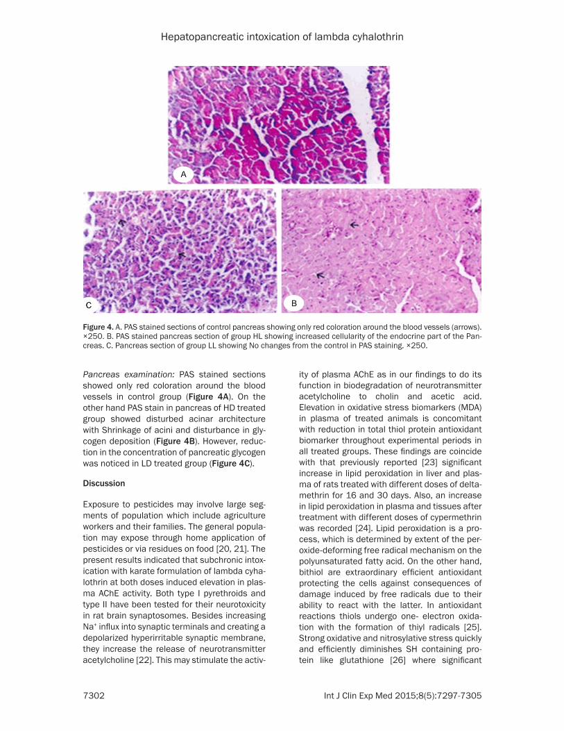
Pancreas examination: PAS stained sections showed only red coloration around the blood vessels in control group (Figure 4A). On the other hand PAS stain in pancreas of HD treated group showed disturbed acinar architecture with Shrinkage of acini and disturbance in gly-cogen deposition (Figure 4B). However, reduc-tion in the concentration of pancreatic glycogen was noticed in LD treated group (Figure 4C).
Discussion
Exposure to pesticides may involve large seg-ments of population which include agriculture workers and their families. The general popula-tion may expose through home application of pesticides or via residues on food [20, 21]. The present results indicated that subchronic intox-ication with karate formulation of lambda cyha-lothrin at both doses induced elevation in plas-ma AChE activity. Both type I pyrethroids and type II have been tested for their neurotoxicity in rat brain synaptosomes. Besides increasing Na+ influx into synaptic terminals and creating a depolarized hyperirritable synaptic membrane, they increase the release of neurotransmitter acetylcholine [22]. This may stimulate the activ-
ity of plasma AChE as in our findings to do its function in biodegradation of neurotransmitter acetylcholine to cholin and acetic acid. Elevation in oxidative stress biomarkers (MDA) in plasma of treated animals is concomitant with reduction in total thiol protein antioxidant biomarker throughout experimental periods in all treated groups. These findings are coincide with that previously reported [23] significant increase in lipid peroxidation in liver and plas-ma of rats treated with different doses of delta-methrin for 16 and 30 days. Also, an increase in lipid peroxidation in plasma and tissues after treatment with different doses of cypermethrin was recorded [24]. Lipid peroxidation is a pro-cess, which is determined by extent of the per-oxide-deforming free radical mechanism on the polyunsaturated fatty acid. On the other hand, bithiol are extraordinary efficient antioxidant protecting the cells against consequences of damage induced by free radicals due to their ability to react with the latter. In antioxidant reactions thiols undergo one- electron oxida-tion with the formation of thiyl radicals [25].Strong oxidative and nitrosylative stress quickly and efficiently diminishes SH containing pro-tein like glutathione [26] where significant
Figure 4. A. PAS stained sections of control pancreas showing only red coloration around the blood vessels (arrows). ×250. B. PAS stained pancreas section of group HL showing increased cellularity of the endocrine part of the Pan-creas. C. Pancreas section of group LL showing No changes from the control in PAS staining. ×250.

Hepatopancreatic intoxication of lambda cyhalothrin
7303 Int J Clin Exp Med 2015;8(5):7297-7305
decrease in sulfhydryl content in blood of ani-mals treated with different doses of lambda cyhalothrin. The extent of liver damage appears to be considerable as evidenced by increase in plasma levels of ALT and AST as shown in our results where elevation in liver marker enzymes were pronounced when animals intoxicated with LD and HD, these results were confirmed by histopathological studies where degenera-tion in hepatocytes and vaculation in cyto-plasm. In consistent with the present results [27] the treatment with cypermethrin induced elevation in liver enzymes activity which in gen-eral related to intensity of cellular damage. The damage occurred probably through oxidative stress mechanism and decrease in the activity of free radical scavengers [28]. Elevation in blood glucose level was recorded all through the experimental periods these results run par-allel with that previously reported [29, 30] pro-longed administration of cyhalothrin, deltame-thrin and cismethrin for rabbits and rats respec-tively induced significant increase in blood and brain glucose level. Concurrent to the glucose results, hypoinsulinemia was recorded in all treated groups all through the experimental periods accompanied with depletion in glyco-gen content in liver and pancreas as shown in histochemical examination for liver and pan-creas. Disturbed acinar architecture, no defined cell boundaries and absence of nuclei in some cells as well as increased of cellularity was recorded in pancreas tissues. The B cells secrete insulin in response to elevated glucose levels and also respond to other substances such as glucagons and acetylcholine [31]. Oxidative stress and decreased of content of free radical scavengers are probably the conse-quence of pyrethroids toxicity [23, 28] that may damage cell wall and change the depolarization of cell wall. Depletion of glycogen in the liver cells was noticed in both rats treated with the high dose. Subchronic exposure, which resem-bles human exposure, may induce diabetes associated with stimulation of hepatic gluco-neogenesis and glycogenolysis in favor of glu-cose release into the blood [32]. Potential roles for paraoxonase in the detoxification of lipid peroxidation and also as an antioxidant have been suggested [33]. Low serum paraoxonase activity may lead to increase in reactive oxygen species, occurrence of oxidative stress and oxi-dative stress can cause hyperglycemia as men-tioned above. The activity of this enzyme has
been found to be markedly lower in patients with type I and type II diabetes mellitus than controls [34].
Conclusions
In conclusion chronic intoxication with lambda cyhalothrin induced elevation of plasma AChE and hyperglycemia induced due to oxidative stress that induce disturbance in liver function and pancreatic function.
Acknowledgements
This project was supported by King Saud University, Deanship of Scientific Research, College of Science Research Center.
Disclosure of conflict of interest
None.
Address correspondence to: Dr. Sherif H Abd-Alrahman, Biochemistry Department, College of Science, King Saud University, PO Box, 2455, Riyadh 11451, Saudi Arabia. Tel: 00966561776615; E-mail: [email protected]
References
[1] Giovanni Annuzzia, Bozzetto L, Patti L, Santan-gelo C, Giacco R, Di Marino L, De Natale C, Ma-sella R, Riccardi G, Rivellese AA. Type 2 diabe-tes mellitus is characterized by reduced postprandial adiponectin response: a possible link with diabetic postprandial dyslipidemia. Metabolism 2009; 59: 567-74.
[2] Gray H, Wreghitt T, Stratton IM, Alexander GJ, Turner RC, O’Rahilly S. High prevalence of hep-atitis C infection in Afro-Caribbean patients with type 2 diabetes and abnormal liver func-tion tests. Diabet Med 1995; 12: 244-249.
[3] Gloyn AL, van de Bunt M, Stratton IM, Lonie L, Tucker L, Ellard S, Holman RR. Prevalence of GCK mutations in individuals screened for fasting hyperglycaemia. Diabetologia 2009; 52: 172-174.
[4] Hill NR, Hindmarsh PC, Stevens RJ, Stratton IM, Levy JC, Matthews DR. A method for as-sessing quality of control from glucose profiles. Diabet Med 2007; 24: 753-758.
[5] Hussain T, Al-Daghri NM, Al-Attas OS, Draz HM, Abd Al-Rahman SH, Yakout SM. Plasma neuro-peptide Y levels relate cigarette smoking and smoking cessation to body weight regulation. Regul Pept 2012; 176: 22-27.
[6] Hites RA. Polybrominated diphenyl ethers in the environment and in people: a meta-analy-

Hepatopancreatic intoxication of lambda cyhalothrin
7304 Int J Clin Exp Med 2015;8(5):7297-7305
sis of concentrations. Environ Sci Technol 2004; 38: 945-956.
[7] International FH. Pres Erving fertility; 2003. pp. 3-23.
[8] Iqbal MJ, Bollaert M, Chickris N, James B, Hig-ginbotham DA, Peterson R, Murphy L. Ginseng modifies the diabetic phenotype and genes as-sociated with diabetes in the male ZDF rat. Phytomedicine 2007; 14: 681-689.
[9] Kohner EM, Stratton IM, Aldington SJ, Turner RC, Matthews DR. Microaneurysms in the de-velopment of diabetic retinopathy (UKPDS 42). UK Prospective Diabetes Study Group. Diabe-tologia 1999; 42: 1107-1112.
[10] Kothari V, Stevens RJ, Adler AI, Stratton IM, Manley SE, Neil HA, Holman RR. UKPDS 60: risk of stroke in type 2 diabetes estimated by the UK Prospective Diabetes Study risk engine. Stroke 2002; 33: 1776-1781.
[11] Lee DH, Lee IK, Song K, Steffes M, Toscano W, Baker BA, Jacobs DR Jr. A strong dose-re-sponse relation between serum concentra-tions of persistent organic pollutants and dia-betes: results from the National Health and Examination Survey 1999-2002. Diabetes Care 2006; 29: 1638-1644.
[12] OCED. Guidelines for testing of chemicals, No. 203. Organization for Economic Cooperation and Development. 1992.
[13] Levy JC, Cull CA, Stratton IM, Holman RR, Turn-er RC. [The UKPDS study on glycemic control and arterial hypertension in type II diabetes: objectives, structure and preliminary results]. Journ Annu Diabetol Hotel Dieu 1993; 123-137.
[14] Lewis CA, Smith PG, Stratton IM, Darby SC, Doll R. Estimated radiation doses to different or-gans among patients treated for ankylosing spondylitis with a single course of X rays. Br J Radiol 1988; 61: 212-220.
[15] Li Z, Sandau CD, Sandau CD, Romanoff LC, Caudill SP, Sjodin A, Needham LL, Patterson DG Jr. Concentration and profile of 22 urinary polycyclic aromatic hydrocarbon metabolites in the US population. Environ Res 2008; 107: 320-331.
[16] Hu ML, Dillard CJ. Plasma SH and GSH mea-surement. Method of enzymology 1994; 233: 385-387.
[17] Mehta S, Smith K, Feuz M. The global burden of diseas e from household use of solid fuels: a source of indoor pollution. Epidemiology 2002; 13: 64.
[18] Reitman S, Frankel S. A colorimetric method for the determination of serum glutamic oxal-acetic and glutamic pyruvic transaminases. Am J Clin Pathol 1957; 28: 56-63.
[19] Miller RL, Garfinkel R, Horton M, Camann D, Perera FP, Whyatt RM, Kinney PL. Polycyclic
aromatic hydrocarbons, environmental tobac-co smoke, and respiratory symptoms in an in-ner-city birth cohort. Chest 2004; 126: 1071-1078.
[20] Mohammad WK. “Soil Pollution Hazardous to Environment”: A case study on the chemical composition and correlation to automobile traffic of the roadside soil of Jeddah city, Saudi Arabia. J Hazard Mater 2009; 168: 1280-1283.
[21] Mosmann TR, Cherwinski H, Bond MW, Giedlin MA, Coffman RL. Two types of murine helper T cell clone. I. Definition according to profiles of lymphokine activities and secreted proteins. J Immunol 1986; 136: 2348-2357.
[22] Mosmann TR, Coffman RL. TH1 and TH2 cells: different patterns of lymphokine secretion lead to different functional properties. Annu Rev Immunol 1989; 7: 145-173.
[23] Needham LL, Calafat AM, Barr DB. Uses and issues of biomonitoring. Int J Hyg Environ Health 2007; 210: 229-238.
[24] Atessahin A, Karahan I and Ririncci I. Effects of Phenobarbital on Serum and Liver Paraox-onase and Arylesterase Activities in Rats. Turk J Vet Anim Sci 2004; 28: 363-367.
[25] Wardman P. Editor reaction of thiyl radicals. In: Biothiols in health and disease; EPC, editor. New York: Marcel Dekker; 1995.
[26] Chun OK, Kang HG. Estimation of risks of pes-ticide exposure, by food intake, to Koreans. Food Chem Toxicol 2003; 41: 1063-1076.
[27] Muthuviveganandavel V, Muthuraman P, Muthu S, Srikumar K. Toxic effects of carben-dazim at low dose levels in male rats. J Toxicol Sci 2008; 33: 25-30.
[28] Manna S, Bhattacharyya D, Mandal TK, Das S. Repeated dose toxicity of alfa-cypermethrin in rats. J Vet Sci 2004; 5: 241-245.
[29] Ray DE. The contrasting actions of two pyre-throids (deltamethrin and cismethrin) in the rat. Neurobehav Toxicol Teratol 1982; 4: 801-804.
[30] Shakoori AR, Aslam F, Sabir M, Ali SS. Effect of prolonged administration of insecticide (Cyha-lothrin/Karate) on the blood and liver of rab-bits. Folia Biol (Krakow) 1992; 40: 91-99.
[31] Meyer A, Seidler FJ, Cousins MM, Slotkin TA. Developmental neurotoxicity elicited by gesta-tional exposure to chlorpyrifos: when is adeny-lyl cyclase a target? Environ Health Perspect 2003; 111: 1871-1876.
[32] Abdollahi M, Donyavi M, Pournourmohammadi S, Saadat M. Hyperglycemia associated with increased hepatic glycogen phosphorylase and phosphoenolpyruvate carboxykinase in rats following subchronic exposure to malathi-on. Comp Biochem Physiol C Toxicol Pharma-col 2004; 137: 343-347.

Hepatopancreatic intoxication of lambda cyhalothrin
7305 Int J Clin Exp Med 2015;8(5):7297-7305
[33] Mackness MI, Harty D, Bhatnagar D, Winocour PH, Arrol S, Ishola M, Durrington PN. Serum paraoxonase activity in familial hypercholester-olaemia and insulin-dependent diabetes mel-litus. Atherosclerosis 1991; 86: 193-199.
[34] Mackness B, Durrington PN, Boulton AJ, Hine D, Mackness MI. Serum paraoxonase activity in patients with type 1 diabetes compared to healthy controls. Eur J Clin Invest 2002; 32: 259-264.