Original Article Evaluation of Temporomandibular …twin block remains the most popular and...
7
Journal of Contemporary Orthodontics, July-September 2018;2(3):39-47 Original Article ABSTRACT Introduction: In today’s scenario, it is very common for an orthodontist to see a Skeletal Class II Malocclusion due to retrognathic mandible in growing patients. Although various approaches for the treatment have been devised, twin block remains the most acceptable and popular among the functional appliances. Amongst the various approaches made to- wards the evaluation and assessment of the changes occurring in the temporomandibular joint caused by the functional appliance, MRI has been the choice of approach for the same owing to its diversified advantages. Aim and objectives: To evaluate and compare hard and soft tissue changes in the Tempo- romandibular Joint caused by Clarks Twin Block appliance before and after the correction of class II molar relation by using MRI. Result: The results of the study suggested that the forward position of condyle within the glenoid fossa and retrusion of articular disc in relation to condylar head was statistically sig- nificant after twin block therapy. At stage 1 (Pre-treatment), 8 of the 10 cases had anterior condylar position, and 2 patients had a posterior condylar position. Whereas, stage II (Post treatment) revealed 9 of the 10 patients showing anterior condylar position and 1 showed concentric condylar position. Conclusion: The position of condyle in glenoid fossa and articular disc retrusion in relation to condylar head was statistically significant after twin block therapy. Forward relocation of the C-GF complex seems to be one of the mechanisms of action of functional appliances. Key message: MRI study on functional appliance therapy supports and provides evidence for anterior remodelling of the condyle-glenoid fossa complex at the end of 6 months treat- ment with Twin block appliance. Key words: Twin Block, MRI, Skeletal Class II malocclusion. INTRODUCTION Skeletal Class II malocclusion due to mandibular retrogna- thism in growing individual is not an uncommon thing to be seen in our daily practice and it needs an urgent attention of the clinician as utilization of growth for skeletal correction is utmost important. It has been indicated that 11% of prevalence of class II malocclusion in US population, which accounts for 20-30% of all orthodontic patients. 1 Retrusive chin position is exhibited by 75% of Skeletal class II cases. Once, skeletal class II is established, it persists during the growing years. 4–6 The effects and stability Class II treatment in growing stage, with functional appliances has been surrounded by much con- troversy and uncertainty. Various advances have been made for management of skeletal class II malocclusion correction, twin block remains the most popular and acceptable functional appliance till date. 7 Functional appliance enhance the forward growth of mandible by displacing condyles downward and forward within the glenoid fossa. They are shown to modify the neuromuscular environment of the dentition. 8,9 Before the advent of MRI, numerous methods, such as cephalogram, orthopantomogram, tomograms, bone scintig- Evaluation of Temporomandibular Joint Changes with Functional Appliance—An MRI Study 1 Siddharth Dixit, 2 K Sridhar, 3 Upendra Jain, 4 Amit Prakash, 5 Sashibhushan Ekka, 6 Chandrika Dubey 1,6 Senior Lecturer, 2 Former Professor, 3 Head and Professor, 4,5 Reader 1-6 Department of Orthodontics and Dentofacial Orthopedics 1-5 Peoples College of Dental Sciences and Research Centre, Bhanpur, Bhopal, Madhya Pradesh, India 6 Peoples Dental Academy and Research Centre, Bhanpur, Bhopal, Madhya Pradesh, India To cite: Dixit S, Sridhar K, Jain U, Prakash A, Ekka S, Dubey C. Evaluation of Temporomandibular Joint Changes with Functional Appliance—An MRI Study. J Contemp Orthod 2018;2(3): 39-47. Received on: 20-07-2018 Accepted on: 23-08-2018 Source of Support: Nil Conflict of Interest: None 07
Transcript of Original Article Evaluation of Temporomandibular …twin block remains the most popular and...

Journal of Contemporary Orthodontics, July-September 2018;2(3):39-47
Original Article
ABSTRACTIntroduction: In today’s scenario, it is very common for an orthodontist to see a Skeletal Class II Malocclusion due to retrognathic mandible in growing patients. Although various approaches for the treatment have been devised, twin block remains the most acceptable and popular among the functional appliances. Amongst the various approaches made to-wards the evaluation and assessment of the changes occurring in the temporomandibular joint caused by the functional appliance, MRI has been the choice of approach for the same owing to its diversified advantages.Aim and objectives: To evaluate and compare hard and soft tissue changes in the Tempo-romandibular Joint caused by Clarks Twin Block appliance before and after the correction of class II molar relation by using MRI.Result: The results of the study suggested that the forward position of condyle within the glenoid fossa and retrusion of articular disc in relation to condylar head was statistically sig-nificant after twin block therapy. At stage 1 (Pre-treatment), 8 of the 10 cases had anterior condylar position, and 2 patients had a posterior condylar position. Whereas, stage II (Post treatment) revealed 9 of the 10 patients showing anterior condylar position and 1 showed concentric condylar position.Conclusion: The position of condyle in glenoid fossa and articular disc retrusion in relation to condylar head was statistically significant after twin block therapy. Forward relocation of the C-GF complex seems to be one of the mechanisms of action of functional appliances.Key message: MRI study on functional appliance therapy supports and provides evidence for anterior remodelling of the condyle-glenoid fossa complex at the end of 6 months treat-ment with Twin block appliance.Key words: Twin Block, MRI, Skeletal Class II malocclusion.
INTRODUCTIONSkeletal Class II malocclusion due to mandibular retrogna-thism in growing individual is not an uncommon thing to be seen in our daily practice and it needs an urgent attention of the clinician as utilization of growth for skeletal correction is utmost important. It has been indicated that 11% of prevalence of class II malocclusion in US population, which accounts for 20-30% of all orthodontic patients.1 Retrusive chin position is exhibited by 75% of Skeletal class II cases. Once, skeletal class II is established, it persists during the growing years.4–6
The effects and stability Class II treatment in growing stage, with functional appliances has been surrounded by much con-troversy and uncertainty. Various advances have been made for management of skeletal class II malocclusion correction, twin block remains the most popular and acceptable functional appliance till date.7 Functional appliance enhance the forward growth of mandible by displacing condyles downward and forward within the glenoid fossa. They are shown to modify the neuromuscular environment of the dentition.8,9
Before the advent of MRI, numerous methods, such as cephalogram, orthopantomogram, tomograms, bone scintig-
Evaluation of Temporomandibular Joint Changes with Functional Appliance—An MRI Study1Siddharth Dixit, 2K Sridhar, 3Upendra Jain, 4Amit Prakash, 5Sashibhushan Ekka, 6Chandrika Dubey1,6Senior Lecturer, 2Former Professor, 3Head and Professor, 4,5Reader1-6Department of Orthodontics and Dentofacial Orthopedics1-5Peoples College of Dental Sciences and Research Centre, Bhanpur, Bhopal, Madhya Pradesh, India6Peoples Dental Academy and Research Centre, Bhanpur, Bhopal, Madhya Pradesh, India
To cite: Dixit S, Sridhar K, Jain U, Prakash A, Ekka S, Dubey C. Evaluation of Temporomandibular Joint Changes with Functional Appliance—An MRI Study. J Contemp Orthod 2018;2(3): 39-47.
Received on: 20-07-2018
Accepted on: 23-08-2018
Source of Support: Nil
Conflict of Interest: None
07

08
Siddharth Dixit et al
Figure 1: Sagittal view of the condylar neck and head showing transfer of the FH plane from Lateral Cephalogram to MRI scan
raphy with radiologic markers like 99mTc-MDP, arthroscopy, arthrography and CT scanning have been employed to evaluate and assess the progress and changes made by growth modula-tion appliance, but advantages of MRI superseded all the other methods because of the following reasons (1) it is non-invasive, (2) radiation free, (3) it has superior resolution, (4) it gives accurate assessment of bone as well as soft tissues,10,11 (5) it is considered a mode of choice for imaging internal derange-ments of TMJ.12
Hence, the present study was conducted to appraise the changes occurred in TMJ after the correction of class II maloc-clusion by twin block appliance.
AIM AND OBJECTIVESThe aim of this study was to evaluate and to compare hard and soft tissue changes in the Temporomandibular Joint caused by Clarks Twin Block appliance before and after the correction of class II molar relation by using MRI.
MATERIAL AND METHODSThe target population for the present study comprised of those individuals with Class II malocclusion with overjet of more than 7 mm. The inclusion criteria:(1) Patients with age between 12-16 years. (2) Moderate to severe skeletal class II malocclusion with convex profile. (3) Retrognathic mandible confirmed clinically and cephalometrically. (4) Positive visual treatment objective (VTO). (5) Mandibular plane angle (FMA between 18-28?). The exclusion criteria: (1) Class II surgical cases. (2) increased FMA more than 28?.(3) prognathic man-dible. (4) combination of prognathic maxilla and retrognathic mandible (Severe class II cases). All records, including MRI scans, were collected at two stages: 1) Stage I (pre-treatment) After a detailed case his-tory, clinical examination, radiographic evaluation with lateral Cephalogram and confirming the treatment criteria, an MRI was taken. The patients were then treated with Twin block appliance followed by fixed appliance therapy. 2) Stage II (after 6 months of Twin block functional appliance therapy). A Lateral Cephalogram and an MRI was taken and evaluated for the changes post Twin block appliance therapy.
EVALUATION OF MRIEach MRI scan was carefully evaluated visually. Both the T1 and T2 weighted sequences in the coronal and sagittal planes were evaluated for documenting the changes in the morphology of the CH, articular disc and glenoid fossa. Further evaluation of sagittal concentricity, eminence angle, sagittal disc position,
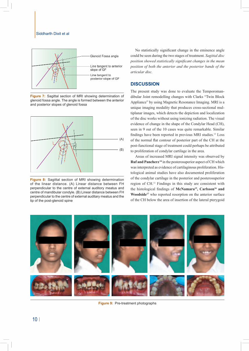
glenoid fossa angle and linear metric measurements, which were measured on the sagittal section passing through the central part of the condyle, and for the coronal disc position in the coronal section. The section displayed maximum length of posterior border of condyle and ramus, whereas coronal position exhibited widest part of condyle. Any pathologi-cal findings, such as spur⁄osteophyte formation or abnormal resorption areas on the CH and perforation or loss of contour of the articular disc, were carefully looked for and recorded. The eminence angle and the sagittal disc position were measured in relation to two reference lines: the posterior con-dylar line (PC line) and the Frankfurt Horizontal plane (FH Plane). The PC line was drawn directly on the MRI scan, while the FH plane was transferred from the lateral cephalogram to the MRI scan.7 Sagittal condylar concentricity was evaluated,8 a positive value indicated an anterior position of the condyle, while a negative value indicated a posterior position of condyle and zero value was referred to as concentric. The eminence angle, the sagittal disc position and the coronal disc position were evaluated by the method described by Chintakanon et al. The eminence angle was measured as the angle formed by a line tangential to the posterior slope of the articular eminence relative to PC line and FH plane. The sagittal disc position was evaluated using PC line and FH plane. The intersecting point between a line parallel to the PC line passing through the condylar centre and the roof of the fossa was constructed and was referred to as the 12 o’clock position in the glenoid fossa (Figures 1, 2A, 2B, 3, 4, 5A, 5B, 6, 7, 8).
RESULTThe results of the study indicated that forward condylar posi-tion within the glenoid fossa and articular disc retrusion with respect to the condylar head was statistically significant after twin block therapy. Forward relocation of the C-GF complex seems to be one of the mechanisms of action of functional appliances.

Evaluation of Temporomandibular Joint Changes with Functional Appliance—An MRI Study
Journal of Contemporary Orthodontics, July-September 2018;2(3):39-47
Figures 2A and B: (A) Determination of the long axis of the condylar neck on Cephalogram. Three concentric circles drawn in the condylar head and neck region confined within the outer cortical margins. The first circle is drawn in the condylar head. Two more successive circles are drawn in the neck region such that the circumference of each of them passes through the centre of the previously drawn circle. A line joining the circle centres in the condylar neck region is considered as the long axis of the condylar neck. (B) Determination of the long axis of the condylar neck on MRI
Figure 3: Sagittal section of MRI demonstrating evaluation of sagittal concentricity. Formula: (P)A/(P + A) · 100 = % of displacement, where A represents narrowest anterior interarticular joint space and B represents narrowest posterior interarticular joint space
Figure 4: Sagittal section of MRI showing determination of eminence angle. A = Angle between the line tangent to posterior slope of articular eminence and posterior condylar (PC) line; B = Angle between the line tangent to posterior slope of articular eminence and Frankfurt horizontal (FH) line
Figures 5A and B: Sagittal section of MRI showing determination of sagittal disc position: (A) Determination of disc position relative to the 12 o’clock position using the PC line. Angle of anterior band of disc to PC line (X). Angle of posterior band of disc to PC line (Y). (B) Determination of disc position relative to the 12 o’clock position using the FH plane. Angle of anterior band of disc to FH plane (X). Angle of posterior band of disc to FH plane (Y)
A
B
Figure 6: Coronal section of MRI showing determination of coronal disc position. Negative value represents the lateral side; Positive value represents the medial side
Photographs (Figure 9), Lateral Cephalogram (Figure 11) and anatomy of condyle (Figure 13). At stage II (Post treatment), 9 of the 10 patients showed an anterior condylar position and 1 showed concentric condylar position.As seen in Pre-treatment Extraoral and Intraoral Photographs (Figure 10). Lateral Cephalogram (Figure 12) and anatomy of condyle (Figure 14).
At stage 1 (Pre-treatment), 8 of the 10 cases had anterior condylar position, and 2 patients had a posterior condylar position. As seen in Pre-treatment Extraoral and Intraoral
09
Siddharth Dixit et al
Figure 7: Sagittal section of MRI showing determination of glenoid fossa angle. The angle is formed between the anterior and posterior slopes of glenoid fossa
Figure 8: Sagittal section of MRI showing determination of the linear distance. (A) Linear distance between FH perpendicular to the centre of external auditory meatus and centre of mandibular condyle. (B) Linear distance between FH perpendicular to the centre of external auditary meatus and the tip of the post-glenoid spine
Figure 9: Pre-treatment photographs
No statistically significant change in the eminence angle could be seen during the two stages of treatment. Sagittal disc position showed statistically significant changes in the mean position of both the anterior and the posterior bands of the articular disc.
DISCUSSIONThe present study was done to evaluate the Temporoman-dibular Joint remodelling changes with Clarks “Twin Block Appliance” by using Magnetic Resonance Imaging. MRI is a unique imaging modality that produces cross-sectional mul-tiplanar images, which detects the depiction and localization of the disc works without using ionizing radiation. The visual evidence of change in the shape of the Condylar Head (CH), seen in 9 out of the 10 cases was quite remarkable. Similar findings have been reported in previous MRI studies.13 Loss of the normal flat contour of posterior part of the CH at the post-functional stage of treatment could perhaps be attributed to proliferation of condylar cartilage in the area. Areas of increased MRI signal intensity was observed by Ruf and Pancherz 14 in the posterosuperior aspect of CH which was interpreted as evidence of cartilaginous proliferation. His-tological animal studies have also documented proliferation of the condylar cartilage in the posterior and posterosuperior region of CH.11 Findings in this study are consistent with the histological findings of McNamara15, Carlsson16 and Woodside17 who reported resorption on the anterior surface of the CH below the area of insertion of the lateral pterygoid
10

Evaluation of Temporomandibular Joint Changes with Functional Appliance—An MRI Study
Journal of Contemporary Orthodontics, July-September 2018;2(3):39-47
Figure 10: Post-treatment Photographs
Figure 11: Pre-treatment CEPH
Figure 12: Post-treatment CEPH
When posterior band of the disc is situated between 11 and 1 o’clock with respect to the superior most aspect of the CH., The articular disc is considered to be in a normal. However, the normal 12 o’clock position varies with the reference line taken. In the present study, the posterior band of the disc was found to be situated between 11 and 12 o’clock positions at pre-treatment, in respect to the PC line, and between 12 and 1 o’clock positions in respect to the FH plane. During the total course of treatment, the position of the articular disc remained between the 11 and 1 o’clock positions, with respect to both the reference lines. Thus, it could be argued that functional
muscle after functional mandibular advancement in monkeys. The loss of notching could perhaps be attributed to the reduc-tion in functional stimulation of that region due to reduction in tension generated by the lateral pterygoid muscle (LPM) which attaches to the area. In this study, anterior position of the condyle in the glenoid fossa was a common finding before stage I. these finding collaborate with the findings with Pullinger and Chintakanon18 whose study also showed anterior position of condyle within the glenoid fossa in Class II Div 1 Cases. These findings are similar to those of Ruf and Pancherz14 and Kinzinger19 in one study, Kinzinger20 concluded that patients treated with a rigid, fixed functional orthopaedic appliance did not achieve occlusion and joints returned to a physiological condyle-fossa relationship post treatment.
11

Siddharth Dixit et al
appliance therapy does not cause pathological displacement of the articular disc. These findings are supported by Pancherz13 and Chintakanon.21 Ruf and Pancherz 14 also documented posterior displacement of the disc within physiologic limits after functional appliance therapy.
Insertion of a functional appliance leads to production of stretch in the retrodiscaltisues which cause either stretching or posterior displacement of the disc resulting in the anterior repositioning of condyle in glenoid fossa. In our study, we found that the change in the position of the anterior band of the disc followed that of the posterior band. Thus, it could be argued that the articular disc was displaced during therapy and did not lose its morphology due to the tension produced by the stretched retrodiscal tissues. This finding also suggests that functional appliance therapy does not lead to alteration of the shape or morphology of the articular disc. When evaluated in the coronal section, the position of the disc did not change with therapy. In our study, 8 patients had a centrally positioned disc over the condyle, Two patients had a laterally displaced disc. The present study infers that the internal anatomic arrangements of the TMJ and the condyle glenoid fossa relationship, which are significantly altered during the phase of functional appliance therapy, move to acquire a relationship which is close to the pre-treatment relationship, or, in other words, a normalization of altered relationship occurs during the second phase of therapy which is aimed to maintain and stabilize the changes produced by the functional appliances. This study highlights with greater evidence, on the basis of MRI, that the Condyle-Glenoid Fossa complex assumes an anterior position in the craniofacial com-plex while the internal anatomic arrangement within the TMJ complex normalizes to pre-treatment position after functional appliance therapy. A major limitation of the study design was the small sample size. Nevertheless, the findings are obvious and significant. These findings should be confirmed in future studies on a bigger sample size. Another area of concern was the lack of controls in our study design, which could not be addressed due to ethical constraints. It is proposed that future studies on functional appliances should concentrate more on glenoid fossa and temporomandibular joint remodelling, and specifically, positional change of the glenoid fossa. The study also highlights the need for the development of precise proto-cols for evaluation of the MRI of TMJ. Future research should focus to develop some simpler but more accurate radiologic markers or methods, such as 3-D CBCT, which would allow easier superimposition of cephalometric images and MRI scans.
SUMMARY AND CONCLUSIONClass II malocclusion in growing individual is not an un-common thing to be seen in our daily practice. Functional appliances are designed to enhance the forward growth of
Figure 13: Pre-treatment anatomy of the condyle and the TMJ complex Note the prominent notch on the anteriorsurface, and the flat contour on the postero-superior surface of the condylar head
Figure 14: Changes in condylar head marphalogy and the TMJ after functional appliance therapy. Note the increase in the superior joint space with loss of the anterior natch, and the flat cantaur an the posterosuperior surface of the candylar head
12

Evaluation of Temporomandibular Joint Changes with Functional Appliance—An MRI Study
Journal of Contemporary Orthodontics, July-September 2018;2(3):39-47
the mandible by encouraginga functional displacement of the mandibular condyles downward and forward in the glenoid fossa. The results of the study suggested that forward condylar position within the glenoid fossa and articular disc retrusion with respect to the condylar head was statistically significant after twin block therapy. Forward relocation of the C-GF com-plex seems to be one of the mechanisms of action of functional appliances. As of this study which was done to evaluate the Temporomandibular Joint remodelling changes with Clarks “Twin Block Appliance” by using Magnetic Resonance Imag-ing we can conclude that–1. Condylar Position Shifted Anteriorly,2. Articular Disc Shifted Posteriorly,3. Eminence Angle Had No Significant Change And4. Coronal Disc Position Remained Unaffected. Hence, to summarize, we can say that the current MRIs-tudy on functional appliance therapy supports and provides evidencefor anterior remodelling of the condyle-glenoidfossa complex at the end of 6 months treatment with Twin block appliance.
BIBLIOGRAPHY 1. Proffit WR, Fields HW, Moray LJ. Prevalence of malocclusion
and orthodontic treatment need in the United States: estimated from the NHANES III survey. Int J Adult OrthodOrthognath Surg. 1998;13(2):97-106.
2. Buckhardt DR, McNamara JA Jr, Baccetti T. Maxillary molar distalization or mandibular enhancement: a cephalometric comparison of comprehensive orthodontic treatment including the pendulum and the Herbst appliances. Am J OrthodDentofa-cialOrthop. 2003; 123(2):108-16.
3. Renfroe EW. A study of the facial pattern associated with class I, class II div 1 and class II div 2 malocclusions. Angle Orthod. 1948;18:70-5.
4. Baccetti T, Franchi L, McNamara JA Jr, et al. Early dentofacial features of class II malocclusion: a longitudinal study from the deciduous trough the mixed dentition. Am J OrthodDentofa-cialOrthop. 1997;111(5):502-9.
5. Lulla P, Gianelly AA. The mandibular plane and mandibular rotation. Am J Orthod. 1976; 70(5):567-71.
6. Chung CH, Wong WW. Craniofacial growth in untreated skeletal class II patients: a longitudinal study. Am J Orthod-DentofacialOrthop. 2002;122(6):619-26.
7. TR Shyagali, DP Bhayya. Cephalometric evaluation of treat-ment effect of twin block appliance in class II div 1 malocclu-sion. J Int Oral health. 2010;2(4):57-64.
8. Graber TM, Rakosi T, Petrovic AG. Dentofacialorthopedics with functional appliances. St Louis: CV Mosby; 1985. pp. 271-85.
9. Graber TM. Physiologic principles of functional appliances. St Louis: CV Mosby; 1985. pp. 84-86.
10. Harms SE, Wilk RM, Wolford LM, et al. The temporoman-dibular joint: magnetic resonance imaging using surface coils. Radiology. 1985;157(1):133-6.
11. Katzberg RW, Schenck J, Roberts D, et al. Magnetic resonance imaging of the temporo- mandibular joint meniscus. Oral Surg Oral Med Oral Pathol. 1985;59:332-5.
12. Helms CA. The Temporomandibular Joint. In: Higgins CB, Hricak H, Helms CA, eds. MRI of the Body. 3rdedition. Phila-delphia: Lippincott Publishers; 1997. pp. 1257-66.
13. Pancherz H, Ruf S, Faubert CT. Mandibular articular disk positionchanges during Herbst treatment: A prospective longitudinal MRI study. Am J OrthodDentofacOrthop. 1999:116(2):20-14.
14. Ruf S, Pancherz H. Temporomandibular joint growth adapta-tion in Herbst treatment: a prospective magnetic resonance imaging and cephalometric roentgenographic study. Eur J Orthod. 1998;20(4):375-88.
15. McNamara JA Jr. Neuromuscular and skeletal adaptations to altered function in the orofacial region. Am J Orthod. 1973;64(6):578-606.
16. McNamara JA, Carlson DS. Quantitative analysis of tempo-romandibular joint adaptations to protrusive function. Am J Orthod. 1979;76(6):593-611.
17. Woodside DG, Metaxas A, Altuna G. The influence of func-tional appliance therapy on glenoid fossa remodeling. Am J OrthodDentofacOrthop. 1987;92(3):181-98.
18. Wadhawan N, Kumar S, Kharbanda OP, et al. Temporoman-dibular joint adaptations following two-phase therapy: an MRI study. OrthodCraniofac Res. 2008;11(4):235-50.
19. Kinzinger G, Kober C, Diedrich P. Topography and mor-phology of the mandibular condyle during fixed functional orthopaedic treatment – a magnetic resonance imaging study. J OrofacOrthop. 2007;68(2):124-47.
20. Kinzinger GS, Roth A, Gulden N, et al. Disc-condyle Rela-tionships during Class II Treatment with the Functional Man-dibular Advancer (FMA) (Part II). Dentomaxillofac Radiol. 2006;35:347-56.
21. Chintakanon K, Sampson W, Wilkinson T, et al. A prospective study of Twin-block appliance therapy assessed by magnetic resonance imaging. Am J Orthod Dentofacial Orthop. 2000; 118(5):494-504.
13