Original Article Effect of cavity design and material type ...
9
Dental Research Journal 1 © 2021 Dental Research Journal | Published by Wolters Kluwer - Medknow 1 Original Article Effect of cavity design and material type on fracture resistance and failure pattern of molars restored by computer‑aided design/ computer‑aided manufacturing inlays/onlays Roqaia Mohammad Alassar 1 , Amira Mohammad Samy 2 , Fatma Mahmoud Abdel‑Rahman 3 Departments of 1 Crowns and Bridges and 3 Dental Materials, Faculty of Dental Medicine for Girls, Al‑Azhar University, Nasr City, 2 Department of Conservative Dentistry, Faculty of Oral and Dental Medicine, Modern University for Technology and Information, Cairo, Egypt ABSTRACT Background: The maximum conservation of tooth structure and the use of restorative materials with elastic modulus close to the dental structure may promote greater longevity of the tooth/restoration complex. This study was conducted to evaluate the effect of cavity design and material type on fracture resistance and failure pattern of molars restored by computer‑aided design/computer‑aided manufacturing (CAD/CAM) inlays/onlays. Materials and Methods: In this in vitro study, 55 human maxillary molars were embedded in resin blocks and divided into control group (CG) and five main groups: Group 1: Inlay, Group 2: Conventional onlay/mesiobuccal (MB), Group 3: Conservative onlay/MB, Group 4: Conventional onlay/MB and distobuccal (DB), and Group 5: Conservative onlay/MB and DB. Then, each group was divided into two subgroups: (A) CeraSmart (CS) and (B) Katana Zirconia (KZ). Restorations were cemented by RelyX Ultimate and then thermocycled. The universal testing machine was used to measure fracture loads. Failure was determined using a magnifying lens. Data were statistically analyzed using ANOVA followed by Tukey’s post hoc test (P < 0.05). Results: Group 5 showed the highest significant fracture load, whereas the least significant value was recorded in Group 2. KZ recorded higher significant fracture loads than CS in all tested groups. Groups 1, 2, and 3 restored by CS showed lower fracture load than CG, but the difference was insignificant with Group 1. CS restorations showed restorable failure, while unrestorable pattern was predominant in KZ restorations (P < 0.05). Conclusion: KZ inlays and onlays can be used safely in terms of fracture resistance as both have values exceed the physiologic requirements. CS inlays and onlays/MB and DB are of fracture resistance comparable to intact teeth.The use of conservative onlay design with more cusp coverage guarantees better resistance of CS restorations. Being force absorbing material, the predominant failure of teeth restored by CS was restorable. Key Words: Ceramics, computer‑aided design, onlays Received: 03-May-2019 Revised: 05-Aug-2019 Accepted: 03-May-2020 Published: 17-Mar-2021 Address for correspondence: Dr. Roqaia Mohammad Alassar, Department of Crowns and Bridges, Faculty of Dental Medicine for Girls, Al-Azhar University, Nasr City, Cairo, Egypt. E-mail: roqaiaalassar.26@ azhar.edu.eg Access this article online Website: www.drj.ir www.drjjournal.net www.ncbi.nlm.nih.gov/pmc/journals/1480 How to cite this article: Alassar RM, Samy AM, Abdel‑Rahman FM. Effect of cavity design and material type on fracture resistance and failure pattern of molars restored by computer‑aided design/computer‑aided manufacturing inlays/onlays. Dent Res J 2021;18:14. This is an open access journal, and articles are distributed under the terms of the Creative Commons Attribution‑NonCommercial‑ShareAlike 4.0 License, which allows others to remix, tweak, and build upon the work non‑commercially, as long as appropriate credit is given and the new creations are licensed under the identical terms. For reprints contact: [email protected] [Downloaded free from http://www.drjjournal.net on Saturday, April 10, 2021, IP: 193.176.84.3]
Transcript of Original Article Effect of cavity design and material type ...

Dental Research Journal
1© 2021 Dental Research Journal | Published by Wolters Kluwer - Medknow 1
Original ArticleEffect of cavity design and material type on fracture resistance and failure pattern of molars restored by computer‑aided design/computer‑aided manufacturing inlays/onlaysRoqaia Mohammad Alassar1, Amira Mohammad Samy2, Fatma Mahmoud Abdel‑Rahman3
Departments of 1Crowns and Bridges and 3Dental Materials, Faculty of Dental Medicine for Girls, Al‑Azhar University, Nasr City, 2Department of Conservative Dentistry, Faculty of Oral and Dental Medicine, Modern University for Technology and Information, Cairo, Egypt
ABSTRACT
Background: The maximum conservation of tooth structure and the use of restorative materials with elastic modulus close to the dental structure may promote greater longevity of the tooth/restoration complex. This study was conducted to evaluate the effect of cavity design and material type on fracture resistance and failure pattern of molars restored by computer‑aided design/computer‑aided manufacturing (CAD/CAM) inlays/onlays.Materials and Methods: In this in vitro study, 55 human maxillary molars were embedded in resin blocks and divided into control group (CG) and five main groups: Group 1: Inlay, Group 2: Conventional onlay/mesiobuccal (MB), Group 3: Conservative onlay/MB, Group 4: Conventional onlay/MB and distobuccal (DB), and Group 5: Conservative onlay/MB and DB. Then, each group was divided into two subgroups: (A) CeraSmart (CS) and (B) Katana Zirconia (KZ). Restorations were cemented by RelyX Ultimate and then thermocycled. The universal testing machine was used to measure fracture loads. Failure was determined using a magnifying lens. Data were statistically analyzed using ANOVA followed by Tukey’s post hoc test (P < 0.05).Results: Group 5 showed the highest significant fracture load, whereas the least significant value was recorded in Group 2. KZ recorded higher significant fracture loads than CS in all tested groups. Groups 1, 2, and 3 restored by CS showed lower fracture load than CG, but the difference was insignificant with Group 1. CS restorations showed restorable failure, while unrestorable pattern was predominant in KZ restorations (P < 0.05).Conclusion: KZ inlays and onlays can be used safely in terms of fracture resistance as both have values exceed the physiologic requirements. CS inlays and onlays/MB and DB are of fracture resistance comparable to intact teeth. The use of conservative onlay design with more cusp coverage guarantees better resistance of CS restorations. Being force absorbing material, the predominant failure of teeth restored by CS was restorable.
Key Words: Ceramics, computer‑aided design, onlays
Received: 03-May-2019Revised: 05-Aug-2019Accepted: 03-May-2020Published: 17-Mar-2021
Address for correspondence: Dr. Roqaia Mohammad Alassar, Department of Crowns and Bridges, Faculty of Dental Medicine for Girls, Al-Azhar University, Nasr City, Cairo, Egypt. E-mail: [email protected]
Access this article online
Website: www.drj.irwww.drjjournal.netwww.ncbi.nlm.nih.gov/pmc/journals/1480
How to cite this article: Alassar RM, Samy AM, Abdel‑Rahman FM. Effect of cavity design and material type on fracture resistance and failure pattern of molars restored by computer‑aided design/computer‑aided manufacturing inlays/onlays. Dent Res J 2021;18:14.
This is an open access journal, and articles are distributed under the terms of the Creative Commons Attribution‑NonCommercial‑ShareAlike 4.0 License, which allows others to remix, tweak, and build upon the work non‑commercially, as long as appropriate credit is given and the new creations are licensed under the identical terms.
For reprints contact: [email protected]
[Downloaded free from http://www.drjjournal.net on Saturday, April 10, 2021, IP: 193.176.84.3]

Alassar, et al.: Fracture resistance of CAD/CAM inlays and onlays
2 Dental Research Journal / 2021
INTRODUCTION
Computer‑aided design/computer‑aided manufacturing (CAD/CAM) esthetic restorations have more advantages than direct composite restorations; simply because of a wide range of ceramic varieties of higher fracture strength that can be selected. In addition, improved dental laboratory processing, quick fabrication, and the accuracy of restorations encourage many practitioners to shift from direct composite to CAD/CAM restorations.[1,2] For unsatisfactory restorations or extensive carious lesions in molars, the use of ceramics with adhesive techniques achieves the concept of conservation and offers more esthetic restorations.[3]
On restoring posterior teeth, which cavity design and material type guarantee high fracture resistance along with favorable failure pattern; conventional or conservative design, and rigid or force‑absorbing flexible material? The question asked by many practitioners.
It is advisable to consider the mechanical properties of the restorative materials, followed by cavity designing.[4‑6] Particularly, modulus of elasticity has to be considered as high elastic modulus materials tend to accumulate stresses; while materials of low elastic modulus absorb stresses.[7] Therefore, Katana Zirconia (KZ) ML HT and CeraSmart (CS) as two ceramic materials of extremely different Elastic Modulus were suggested to study the difference in both material behavior under load.
Multi‑layered High translucency KZ (KZML HT, Kuraray Noritake Dental Inc.,) discs can be used for fabricating full contour crowns, bridges, veneers, frameworks, inlays, and onlays.[8] KatanaHT provides esthetic appearance, superior strength (1100 MPa), high modulus of elasticity (210 GPa), excellent mechanical performance, and easy milling properties with higher precision compared to other ceramic.[8,9]
CS (GC, Alsip, USA) force absorbing flexible nano ceramic CAD/CAM block combines the best characteristics of high strength ceramic and a unique esthetic of composite. It is composed of 71 wt. % silica (20 nm) and barium glass (300 nm) nanoparticles.[10] Full homogeneous and even distribution of nanoceramic network leads to unique elastic modulus similar to that of dentin (18 ± 2 GPa).[11] According to the manufacturer, uniform scuttle (very short interparticle distance) of silanated and bonded
particles is the factor of acceptable marginal accuracy and high flexural strength (220 MPa).[10‑12]
Cavity design in molars covering one or more than one cusp seems to be the most controversial point. According to the number of cusps involved in the preparation, the restorations can be classified as inlays (all cusps are intact), onlays (one or more cusps are involved), or overlays (all cusps are involved).[13] In addition, the cavity width and depth may influence cusp deflection, and consequently tooth resistance to fracture.[14]
The fracture resistance of molars restored with lithium‑disilicate and zirconia inlays/onlays was evaluated.[13] It was concluded that cuspal coverage decreased fracture resistance of the tooth/restoration complex. Molars restored with zirconia inlays/onlays showed similar fracture resistance to intact teeth. In general, the failure patterns in lithium‑disilicate samples were limited to the restoration itself. In contrast, the failure of zirconia samples involved both the tooth and the restoration.[13]
In addition, the effect of cavity design and ceramic type on the fracture resistance of CAD/CAM onlays in molars was studied. The conservative onlays exhibited increased fracture resistance and more favorable failure modes. It was concluded that molars restored with lithium disilicate CAD/CAM ceramic onlays exhibited higher fracture resistance than molars restored with leucite CAD/CAM ceramic onlays.[15]
The objective of this in vitro study was to compare the effect of CS and KZML HT on fracture resistance and the failure pattern of molars received different inlay/onlay cavity designs. The null hypothesis tested was that cavity design and material type have no influence on fracture resistance and failure pattern of molars restored by CS and KZML HT inlays/onlays.
MATERIALS AND METHODS
To conduct the present in vitro study, 55 freshly extracted caries‑free human maxillary 1st molars were selected in accordance with guidelines from Research Ethics Committee approval of Faculty of Dental Medicine for Girls, Al Azhar University. The teeth were rinsed thoroughly under running water, cleaned, and stored in 0.1% thymol sol until use. The teeth were embedded in epoxy resin (East Coast Resin, USA) blocks up to 1 mm below the cementoenamel junction (CEJ).
[Downloaded free from http://www.drjjournal.net on Saturday, April 10, 2021, IP: 193.176.84.3]

Figure 1: Cavity designs investigated in the study.
Alassar, et al.: Fracture resistance of CAD/CAM inlays and onlays
3Dental Research Journal / 2021 3
Samples groupingThe teeth were randomly divided into intact teeth as a CG (n = 5) and five main groups (n = 10) according to cavity design: Group 1: Inlay design (I), Group 2: Conventional onlay with mesiobuccal (MB)‑cusp coverage (Conv O MB), Group 3: Conservative onlay with MB‑cusp coverage (Cons O MB), Group 4: Conventional onlay with MB and distobuccal (DB)‑cusp coverage (Conv O MB and DB), and Group 5: Conservative onlay with MB and DB‑cusp coverage (Cons O MB and DB). Then, each main group was further divided into two subgroups (n = 5) according to material type; a) CS, and b) KZML HT.
Cavity preparationsAll samples received standardized mesio‑occluso‑distal (MOD) inlay preparations, in accordance with general principles for esthetic inlay restorations.[16] For conservative onlay designs, MB and DB cusps were prepared with the shoulder finish line [Figure 1].
Cavity preparation guidelinesComputer Numerical Control (CNC milling machine, USA) with two diamond stones selected from the Inlay/Onlay preparation Kit (Zhengzhou Smile Dental Equipment Co., China) was used to standardize all preparations. The occlusal cavity occupied buccolingually (4 ± 1 mm) and mesiodistally (7 ± 1 mm). The depth was 2 mm measured from central groove. Proximal cavities were extended with flared buccal and lingual walls (5 mm). The proximal box was 4 mm long and
1.5 mm deep. Occlusal divergence angle was set at 10°–12°. Cavosurface margins were finished in butt joints (90°) with no bevels. Internal line and point angles were rounded. MB and DB cusps were 2 mm occlusally reduced with butt joint for conventional onlay design, and with 1 mm cusp shoulder for conservative design. Prepared dentin was sealed with an adhesive system (Single bond, 3M, USA) to prevent contamination.
Restorations constructionCAD/CAM Roland system (DWX‑51D Roland DG Co., Japan) was used for the construction of all restorations in this study. Five CS and five KZ restorations were constructed from CS blocks (CSTM universal GC, Europe) and KZHT monolithic multi‑layered disc (Kuraray Noritake Dental Inc., Japan), respectively, according to the following procedure [Figure 2].
(1) Each sample was sprayed with light‑reflecting powder (Occlutec, Scan spray. Renfert GmBh. USA), and secured on the tray of smart optics‑three‑dimensional‑scanner (scan Box, Germany) for scanning. (2) Data were transferred to the computer connected to milling machine to start designing. The fully anatomical inlay/onlay design was formed according to the manufacturers’ directions and software recommendations. (3) Milling of CS blocks and KZ disc was then activated. After milling, KZ restorations were sintered in Tabeo sintering furnace (TABEO, Germany) at 1550°C for 8 h. Then,
[Downloaded free from http://www.drjjournal.net on Saturday, April 10, 2021, IP: 193.176.84.3]

Figure 3: Silanization of intaglio surfaces of restorations before cementation.
Figure 2: Restorations construction; (a) Three‑dimensional digital image, (b) Designing, (c) CeraSmart blocks fixed in Roland Machine, and (d) Katana Zirconia disc in Roland Machine.
dc
ba
Alassar, et al.: Fracture resistance of CAD/CAM inlays and onlays
4 Dental Research Journal / 2021
each restoration was examined using magnifying lens (10X, Optics Co., Ltd., China) and checked for complete seating on its corresponding model. (4) CS restorations were finished and polished using coarse and fine silicone points (GC America Inc., USA) and diapolisher paste (GC America Inc., USA), with low speed handpiece, according to the manufacturer recommendations. On the other hand, a silicon diamond point (Kuraray Dental Inc., Japan) and pearl surface Z (Kuraray Noritake Dental Inc., Japan) were used for finishing and polishing of KZ restorations.
Cementation of restorationsAll restorations were cemented using RelyX Ultimate resin cement (3M, ESPE, Germany) after surface conditioning of tooth structure and intaglio surfaces of constructed inlays and onlays, in accordance with their respective manufacturers’ instructions.
The prepared cavities were etched for 15 s with Blue Etch (36% phosphoric acid, StalowaWola, Polska)
then rinsed, dried, and bonded with double coat of Single bond (3M, ESPE, Germany).
CS intaglio surfaces were etched with 5% hydrofluoric acid gel (Ivoclar, Germany) for 60 s then washed in an ultrasonic cleaner (Senden, Germany) and dried. A ceramic primer‑containing silane coupling agent (Bisco, USA) was applied and allowed to dry for 60 s.
KZ intaglio surfaces were sandblasted using alumina particles (50 μm), then cleaned in an ultrasonic cleaner and dried. A ceramic primer (Bisco, USA) was applied and allowed to react for 20 s [Figure 3].
RelyX Ultimate, the dual‑cure resin cement (3M, ESPE, Germany) was applied on the prepared cavities. Then, each restoration was bonded to its corresponding cavity with finger pressure; excess cement was removed immediately with a microbrush. A load applicator of 3 Kg then used to apply constant load for full seating of restorations, then each surface was light cured for 20 s according to the manufacturer’s instructions.
After cementation, samples were stored for 24 h in distilled water at 37°C, then thermocycled in automatic thermal cycling machine (Ropota, automated thermocycling, Turkey) for 5000 cycles in two water baths at 5 and 55°C.
Testing proceduresAll samples were individually mounted on a computer controlled materials testing machine (Model 3345; Instron Industrial Products, Norwood, MA, USA) with a load cell of 5 kN and data were recorded using computer software (Instron® Bluehill Lite Software). Samples were secured to the lower fixed compartment of the testing machine by tightening screws. Fracture test was done by compressive mode of load applied occlusally using a metallic rod with round tip (3.8 mm diameter) attached to the upper movable
[Downloaded free from http://www.drjjournal.net on Saturday, April 10, 2021, IP: 193.176.84.3]
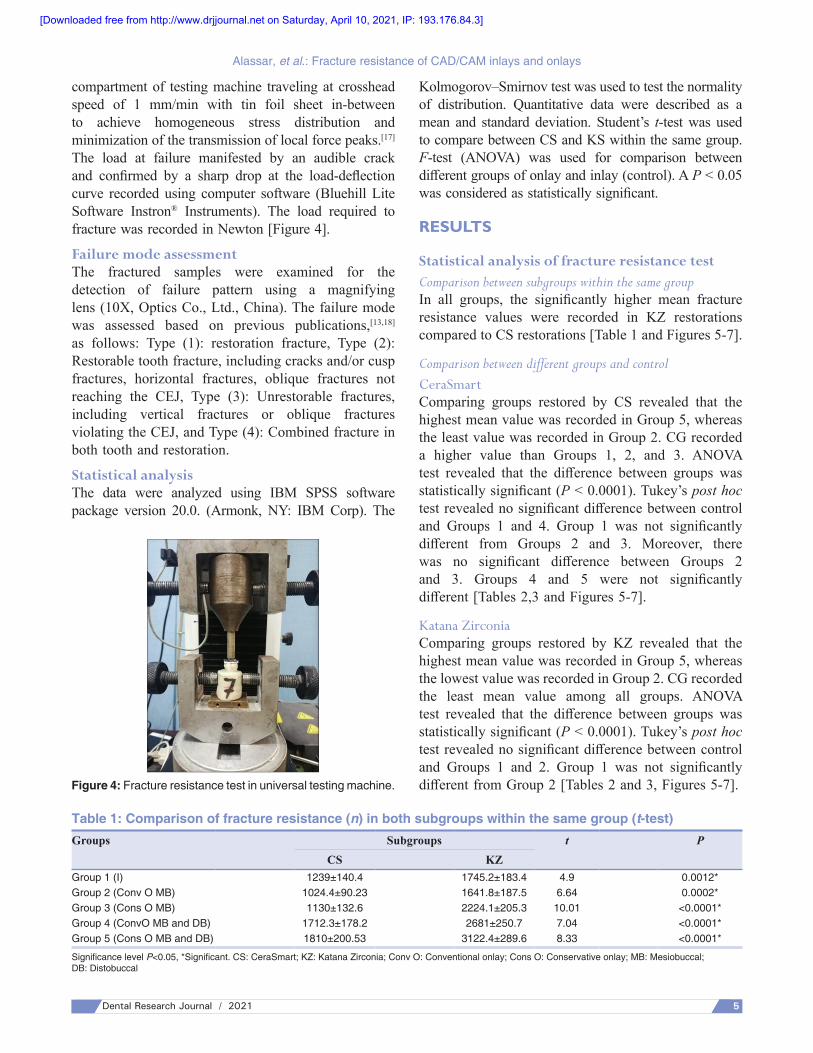
Figure 4: Fracture resistance test in universal testing machine.
Alassar, et al.: Fracture resistance of CAD/CAM inlays and onlays
5Dental Research Journal / 2021 5
compartment of testing machine traveling at crosshead speed of 1 mm/min with tin foil sheet in‑between to achieve homogeneous stress distribution and minimization of the transmission of local force peaks.[17] The load at failure manifested by an audible crack and confirmed by a sharp drop at the load‑deflection curve recorded using computer software (Bluehill Lite Software Instron® Instruments). The load required to fracture was recorded in Newton [Figure 4].
Failure mode assessmentThe fractured samples were examined for the detection of failure pattern using a magnifying lens (10X, Optics Co., Ltd., China). The failure mode was assessed based on previous publications,[13,18] as follows: Type (1): restoration fracture, Type (2): Restorable tooth fracture, including cracks and/or cusp fractures, horizontal fractures, oblique fractures not reaching the CEJ, Type (3): Unrestorable fractures, including vertical fractures or oblique fractures violating the CEJ, and Type (4): Combined fracture in both tooth and restoration.
Statistical analysisThe data were analyzed using IBM SPSS software package version 20.0. (Armonk, NY: IBM Corp). The
Kolmogorov–Smirnov test was used to test the normality of distribution. Quantitative data were described as a mean and standard deviation. Student’s t‑test was used to compare between CS and KS within the same group. F‑test (ANOVA) was used for comparison between different groups of onlay and inlay (control). A P < 0.05 was considered as statistically significant.
RESULTS
Statistical analysis of fracture resistance testComparison between subgroups within the same groupIn all groups, the significantly higher mean fracture resistance values were recorded in KZ restorations compared to CS restorations [Table 1 and Figures 5‑7].
Comparison between different groups and controlCeraSmartComparing groups restored by CS revealed that the highest mean value was recorded in Group 5, whereas the least value was recorded in Group 2. CG recorded a higher value than Groups 1, 2, and 3. ANOVA test revealed that the difference between groups was statistically significant (P < 0.0001). Tukey’s post hoc test revealed no significant difference between control and Groups 1 and 4. Group 1 was not significantly different from Groups 2 and 3. Moreover, there was no significant difference between Groups 2 and 3. Groups 4 and 5 were not significantly different [Tables 2,3 and Figures 5‑7].
Katana ZirconiaComparing groups restored by KZ revealed that the highest mean value was recorded in Group 5, whereas the lowest value was recorded in Group 2. CG recorded the least mean value among all groups. ANOVA test revealed that the difference between groups was statistically significant (P < 0.0001). Tukey’s post hoc test revealed no significant difference between control and Groups 1 and 2. Group 1 was not significantly different from Group 2 [Tables 2 and 3, Figures 5‑7].
Table 1: Comparison of fracture resistance (n) in both subgroups within the same group (t‑test)Groups Subgroups t P
CS KZGroup 1 (I) 1239±140.4 1745.2±183.4 4.9 0.0012*Group 2 (Conv O MB) 1024.4±90.23 1641.8±187.5 6.64 0.0002*Group 3 (Cons O MB) 1130±132.6 2224.1±205.3 10.01 <0.0001*Group 4 (ConvO MB and DB) 1712.3±178.2 2681±250.7 7.04 <0.0001*Group 5 (Cons O MB and DB) 1810±200.53 3122.4±289.6 8.33 <0.0001*
Significance level P<0.05, *Significant. CS: CeraSmart; KZ: Katana Zirconia; Conv O: Conventional onlay; Cons O: Conservative onlay; MB: Mesiobuccal; DB: Distobuccal
[Downloaded free from http://www.drjjournal.net on Saturday, April 10, 2021, IP: 193.176.84.3]

Figure 6: Bar chart showing mean maximum load (n) in Katana Zirconia group.
Figure 5: Bar chart showing mean maximum load (n) in CeraSmart group.
Figure 7: Bar chart showing mean maximum load (n) in CeraSmart and Katana Zirconia subgroups.
Alassar, et al.: Fracture resistance of CAD/CAM inlays and onlays
6 Dental Research Journal / 2021
Failure pattern analysisThe most common failure pattern in CS restorations was restorable, whereas unrestorable failure pattern was the predominant in KZ restorations [Table 4].
DISCUSSION
On restoring posterior teeth, the clinician always thinks how to gain maximum strength where extensive
masticatory forces are applied. The use of some types of ceramics remains limited in the posterior region.[19] Gaining strength can be achieved by proper material selection and optimum cavity design, as well.
Inlay and onlay designs greatly support the treatment philosophy of minimal intervention as up to 70% of the coronal tooth structure is removed when teeth are prepared for a full‑coverage crown, whereas less than half of this percentage is required to be removed for an onlay.[20]
The results of this study revealed that cavity design and material type had a significant effect on fracture resistance of molars restored by CAD/CAM inlays and onlays. Therefore, the null hypothesis was rejected.
Regarding cavity design, there were no significant differences in fracture resistance values between the inlay design and intact teeth. These results are in accordance with Harsha et al.,[8] Saridag et al.,[13] and Cubas et al.[21] studies. This might be attributed to the little quantity of tooth structure removed during inlay preparation.[13] Furthermore, according to various in vitro studies,[13,21,22] adhesive cementation of inlay in place, regained stability of the prepared tooth and explained the high fracture resistance values of KZ inlay restoration. Moreover, it was found the elastic modulus of the resin cement may also affect the fracture strength values of the teeth restored with ceramic inlays and onlays.[21] Adhesive cements with higher elastic modulus increased the fracture strength values of inlay and onlay restorations.[21]
In this study, the teeth received conservative onlay designs with MB‑or MB and DB‑cusp coverage showed higher fracture resistance compared to those received conventional designs with MB‑or MB and DB‑cusp coverage, respectively. The shoulder margin in the conservative design seemed to have the effect of ferrule resulted in better stress distribution.[23,24] This agreed with Oyar and Durkan[24] who concluded that cavity designs with shoulder margins showed the highest fracture resistance, whereas butt joint designs had the lowest fracture resistance. However, the difference was material related as it was significant between KZ restorations and insignificant between those restored by CS.
In both subgroups (CS and KZ), as the preparation amount was increased, (i.e., from inlay to onlay with MB‑cusp design) the fracture resistance was decreased. These results coincided with Saridag et al. study,[13] in
[Downloaded free from http://www.drjjournal.net on Saturday, April 10, 2021, IP: 193.176.84.3]

Alassar, et al.: Fracture resistance of CAD/CAM inlays and onlays
7Dental Research Journal / 2021 7
which cusp coverage decreased the fracture resistance of molars restored by lithium‑disilicate onlays. However, as the preparation amount was increased to involve the DB cusp, (i.e., from onlay with MB‑cusp coverage to onlay with MB and DB‑cusp design) the fracture resistance was increased significantly in KZ
subgroups and insignificantly with CS restorations. This may be explained by increasing restoration’s surface area that related directly to mechanical properties of the restorative material used.[13]
These findings were supported by Harsha et al.[8] who confirmed that increase cuspal coverage had shown a significant increase in the fracture resistance than the sound teeth. On the contrary, Cubas et al.,[21] Stappert et al.,[25] and Yoon et al.,[26] reported that cuspal coverage had no influence on improving the fracture resistance.
Regarding the material type, teeth restored by KZ recorded significantly higher fracture resistance compared to intact teeth and those restored by CS. This was expected because zirconia, the strongest and toughest of all dental ceramics, has a flexural strength of 800–1200 MPa that meets the mechanical requirements for high stress‑bearing posterior restorations.[27,28] Due to its strength and esthetic properties, KZML HT was selected in the present study. The fracture resistance values of KZ obtained in the present study (1745.2 N‑3122.4 N) indicates that KZ inlay and onlay restorations are able to withstand the high masticatory forces associated with bruxism and other parafunctions.
Although the teeth restored by CS showed the lowest fracture resistance, conservative onlay design with MB and DB‑cusp coverage recorded higher fracture resistance than intact teeth. Therefore, according to the present study, it was recommended to use CS with conservative design and more cusp coverage to gain higher fracture resistance. However, the difference was insignificant.
In the current study, all samples recorded fracture load values above 1024 N that exceeded the maximum biting force of 700–900 N for posterior single teeth reported in the literature.[29,30] It was difficult to compare these values to previous studies due to great deviations caused by different types of ceramics used, different test methods and different preparation designs.[15]
On examination of fractured samples, it was clearly observed that the most common failure mode of the teeth restored by KZ was unfavorable fracture (i.e., un‑restorable), whereas restorable failure was the predominant in CS subgroups. This may be related to the high Elastic Modulus of zirconia.[31] Zirconia is considered a stiff material that transmits stresses to the underlying
Table 2: Comparison of fracture resistance (n) in inlay and different groups of onlay design within the same material (ANOVA test)Groups Subgroups
CS KZControl 1435b±150.9 1435e±150.9Group 1 (I) 1239b,c±140.4 1745.2d,e±183.4Group 2 (Conv O MB) 1024.4c,d±90.23 1641.8d,e±187.5Group 3 (Cons O MB) 1130c,d±132.6 2224.1c±205.3Group 4 (Conv O MB and DB) 1712.3b,c±178.2 2681b±250.7Group 5 (Cons O MB and DB) 1810a,b±200.53 3122.4a±289.6F 21.68 46.34P <0.0001* <0.0001*
Significance level P<0.05, *Significant. Tukey’s post hoc test: Within the same comparison, means sharing the same superscript letter are not significantly different. CS: CeraSmart; KZ: Katana Zirconia; Conv O: Conventional onlay; Cons O: Conservative onlay; MB: Mesiobuccal; DB: Distobuccal
Table 3: Detailed outcome of Tukey’s post hoc testSubgroups Group 1 Group 2 Group 3 Group 4 Group 5CS
Control 0.3568 NS 0.0034* 0.0436* 0.0800 NS 0.0083*Group 1 ‑ 0.2655 NS 0.8651 NS 0.0007* 0.0001*Group 2 ‑ 0.8796 NS 0.0000* 0.0000*Group 3 ‑ 0.0000* 0.0000*Group 4 ‑ 0.9098 NS
KZControl 0.2451 NS 0.6600 NS 0.0001* 0.0000* 0.0000*Group 1 ‑ 0.9722 NS 0.0200* 0.0000* 0.0000*Group 2 ‑ 0.0033* 0.0000* 0.0000*Group 3 ‑ 0.0289* 0.0000*Group 4 ‑ 0.0371*
Significance level P<0.05, *Significant. NS: Nonsignificant; CS: CeraSmart; KZ: Katana Zirconia
Table 4: Failure pattern after fracture resistance test (n)Groups Subgroups Type (1) Type (2) Type (3) Type (4)Group 1 CS 0 3 1 1
KZ 0 0 5 0Group 2 CS 0 3 2 0
KZ 1 1 3 0Group 3 CS 1 3 0 1
KZ 0 0 5 0Group 4 CS 2 3 0 0
KZ 2 0 3 0Group 5 CS 0 2 2 1
KZ 0 0 5 0
CS: CeraSmart; KZ: Katana Zirconia
[Downloaded free from http://www.drjjournal.net on Saturday, April 10, 2021, IP: 193.176.84.3]

Alassar, et al.: Fracture resistance of CAD/CAM inlays and onlays
8 Dental Research Journal / 2021
tooth structure that leads to unfavorable failure. In contrast, the unique composition of CS allows the material to have modulus of elasticity similar to that of dentin (18 ± 2 GPa),[31] absorbs forces and equally distributes stresses, leading to favorable failure.[12]
In addition to small sample size, the limitation of the study is that no mechanical loading was applied as part of the artificial aging process, which would have provided insight into its negative effects on mechanical properties.
CONCLUSION
Within the limitations of this study, the following could be concluded:1. Fracture resistance and failure pattern of teeth
restored by CAD/CAM inlays and onlays are greatly affected by cavity design and material type
2. CS inlays and onlays with MB and DB‑cusp coverage are all of fracture resistance comparable to intact teeth
3. Regardless cavity designs, the teeth restored by KZML HT have the best fracture resistance
4. Conservative onlay design guarantees higher fracture resistance
5. The more cusp coverage, the higher fracture resistance of teeth restored by CS and KZML HT onlays, and
6. Being force absorbing flexible material, the predominant failure pattern of teeth restored by CS was restorable.
AcknowledgmentsThe authors would like to thank Prof. Dr. Mona H. Mandour, Professor of Crowns and Bridges, Faculty of Dental Medicine for Girls, Al‑Azhar University, for comments that greatly improved the manuscript.
Financial support and sponsorshipNil.
Conflicts of interestThe authors report no conflicts of interest. The authors alone are responsible for the content and writing of this article.
REFERENCES
1. Giordano R, McLaren EA. Ceramics overview: Classification by microstructure and processing methods. Compend Contin Educ Dent 2010;31:682‑4, 686, 688.
2. Yüksel E, Zaimoğlu A. Influence of marginal fit and cement types on microleakage of all‑ceramic crown
systems. Braz Oral Res 2011;25:261‑6.3. Veneziani M. Posterior indirect adhesive restorations: Updated
indications and the morphology driven Preparation technique. Int J Esthet Dent 2017;12:204‑30.
4. Carvalho MA, Lazari PC, Gresnigt M, Del Bel Cury AA, Magne P. Current options concerning the endodontically‑treated teeth restoration with the adhesive approach. Braz Oral Res 2018;32:e74.
5. Ţălu S, Alb SF, Pârvu AE, Dudea D, Lainović T, Gasparik C, et al. Factors influencing the choice of dental material and procedure for crown restoration of posterior teeth – Design of a “decision guide”. HVM Int J Bioflu×2016;8:141‑7.
6. Frankenberger R, Hehn J, Hajtó J, Krämer N, Naumann M, Koch A, et al. Effect of proximal box elevation with resin composite on marginal quality of ceramic inlays in vitro. Clin Oral Investig 2013;17:177‑83.
7. O¨zkir SE. Effect of restoration material on stress distribution on partial crowns: A 3D finite element analysis. J Dent Sci 2018;13:311‑7.
8. Harsha MS, Praffulla M, Babu MR, Leneena G, Krishna TS, Divya G. The effect of cavity design on fracture resistance and failure pattern in monolithic zirconia partial coverage restorations – An in vitro study. J Clin Diagn Res 2017;11:ZC45‑8.
9. Mehl C, Ludwig K, Steiner M, Kern M. Fracture strength of prefabricated all‑ceramic posterior inlay‑retained fixed dental prostheses. Dent Mater 2010;26:67‑75.
10. Lauvahutanon S, Takahashi H, Oki M, Arksornnukit M, Kanehira M, Finger WJ. In vitro evaluation of the wear resistance of composite resin blocks for CAD/CAM. Dent Mater J 2015;34:495‑502.
11. Gulec L, Ulusoy N, Cengiz E. Indirect resin composite restorations fabricated with chair‑side CAD/CAM systems. Cumhuriyet Dent J 2016;19:247‑55.
12. Awada A, Nathanson D. Mechanical properties of resin‑ceramic CAD/CAM restorative materials. J Prosthet Dent 2015;114(4):587‑93.
13. Saridag S, Sevimay M, Pekkan G. Fracture resistance of teeth restored with all‑ceramic inlays and onlays: An in vitro study. Oper Dent 2013;38:626‑34.
14. Al‑Thobity AM, Altaher A, Alharbi A, Elnour M. An in vitro comparison of the fracture resistance of standard and modified mesio‑occluso‑distal cavity designs restored with resin composite restoration. Int J Health Sci (Qassim) 2018;12:26‑30.
15. Vianna AL, Prado CJ, Bicalho AA, Pereira RA, Neves FD, Soares CJ. Effect of cavity preparation design and ceramic type on the stress distribution, strain and fracture resistance of CAD/CAM onlays in molars. J Appl Oral Sci 2018;26:e20180004.
16. Rocca GT, Rizcalla N, Krejci I, Dietschi D. Evidence‑based concepts and procedures for bonded inlays and onlays. Part II. Guidelines for cavity preparation and restoration fabrication. Int J Esthet Dent 2015;10:392‑413.
17. Schultheis S, Strub JR, Gerds TA, Guess PC. Monolithic and bi‑layer CAD/CAM lithium‑disilicate versus metal‑ceramic fixed dental prostheses: Comparison of fracture loads and failure modes after fatigue. Clin Oral Investig 2013;17:1407‑13.
18. Salameh Z, Ounsi HF, Aboushelib MN, Al‑Hamdan R, Sadig W, Ferrari M. Effect of different onlay systems on fracture resistance
[Downloaded free from http://www.drjjournal.net on Saturday, April 10, 2021, IP: 193.176.84.3]

Alassar, et al.: Fracture resistance of CAD/CAM inlays and onlays
9Dental Research Journal / 2021 9
and failure pattern of endodontically treated mandibular molars restored with and without glass fiber posts. Am J Dent 2010;23:81‑6.
19. Silva LH, Lima E, Miranda RB, Favero SS, Lohbauer U, Cesar PF. Dental ceramics: A review of new materials and processing methods. Braz Oral Res 2017;31:e58.
20. Edelhoff D, Ahlers MO. Occlusal onlays as a modern treatment concept for the reconstruction of severely worn occlusal surfaces. Quintessence Int 2018;49:521‑33.
21. Cubas GB, Habekost L, Camacho GB, Pereira‑Cenci T. Fracture resistance of premolars restored with inlay and onlay ceramic restorations and luted with two different agents. J Prosthodont Res 2011;55:53‑9.
22. Yu W, Guo K, Zhang B, Weng W. Fracture resistance of endodontically treated premolars restored with lithium disilicate CAD/CAM crowns or onlays and luted with two luting agents. Dent Mater J 2014;33:349‑54.
23. Schmidlin PR, Stawarczyk B, DeAbreu D, Bindl A, Ender A, Ichim IP, et al. Fracture resistance of endodontically treated teeth without ferrule using a novel H‑shaped short post. Quintessence Int 2015;46:97‑108.
24. Oyar P, Durkan R. Effect of cavity design on the fracture resistance of zirconia onlay ceramics. Niger J Clin Pract 2018;21:687‑91.
25. Stappert CF, Att W, Gerds T, Strub JR. Fracture resistance of different partial‑coverage ceramic molar restorations: An in vitro investigation. J Am Dent Assoc 2006;137:514‑22.
26. Yoon HI, Sohn PJ, Jin S, Elani H, Lee SJ. Fracture resistance of CAD/CAM‑fabricated lithium disilicate MOD inlays and onlays with various cavity preparation designs. J Prosthodont 2019;28:e524‑9.
27. Homaei E, Farhangdoost K, Tsoi JK, Matinlinna JP, Pow EH. Static and fatigue mechanical behavior of three dental CAD/CAM ceramics. J Mech Behav Biomed Mater 2016;59:304‑13.
28. Al‑Amleh B, Lyons K, Swain M. Clinical trials in zirconia: A systematic review. J Oral Rehabil 2010;37:641‑52.
29. Flanagan D. Bite force and dental implant treatment: A short review. Med Devices (Auckl) 2017;10:141‑8.
30. Koc D, Dogan A, Bek B. Bite force and influential factors on bite force measurements: A literature review. Eur J Dent 2010;4:223‑32.
31. Al‑shibri S, Elguindy J. Fracture resistance of endodontically treated teeth restored with lithium disilicate crowns retained with fiber posts compared to lithium disilicate and cerasmart endocrowns. In vitro study. Dent J 2017;7:12.
[Downloaded free from http://www.drjjournal.net on Saturday, April 10, 2021, IP: 193.176.84.3]