
WT1 modulates apoptosis by transcriptionally upregulating the bcl2 protoncogene.pdf

Int J Clin Exp Pathol 2017;10(11):11288-11299www.ijcep.com /ISSN:1936-2625/IJCEP0062810
Original Article CCL4 promotes the cell proliferation, invasion and migration of endometrial carcinoma by targeting the VEGF-A signal pathway
Fu Hua1,2, Ye Tian1,3
1Department of Radiotherapy & Oncology, The Second Affiliated Hospital of Soochow University, Suzhou 215004, China; 2Department of Gynecology, Huai’an First People’s Hospital, Nanjing Medical University, Huai’an 223300, China; 3Institute of Radiotherapy & Oncology, Soochow University, Suzhou 215004, China
Received May 15, 2017; Accepted October 12, 2017; Epub November 1, 2017; Published November 15, 2017
Abstract: Chemokine (C-C motif) ligand 4 (CCL4) and vascular endothelial growth factor-A (VEGF-A) are involved in the progression and metastasis of some tumors, including ovarian cancer, colon cancer and prostate cancer. However, the roles of CCL4 and VEGF-A in human endometrial cancer (EC) are still unclear. Here, we demonstrated that the production of CCL4 and VEGF-A was significantly higher in EC tissues than in normal tissues, and their expression profiles were associated with the clinical stage of EC. In addition, we found that CCL4 promoted the angiogenesis and invasive ability of EC tumors by increasing the production of VEGF-A. We further confirmed the effect of CCL4 in the growth of EC tumors by silencing the expression of CCL4 in EC cell lines. Finally, we found that CCL4 upregulated VEGF-A expression by activating STAT3, and it enhanced the progression and metastasis of EC. Our study showed that CCL4 promoted tumor growth by upregulating VEGF-A expression, which affected the STAT3 signal pathway in the EC cells.
Keywords: Endometrial cancer, CCL4, VEGF-A, STAT3
Introduction
Endometrial carcinoma (EC) is thought to be the most common gynecologic malignancy in the world, with a mean age at diagnosis of 60 years [1]. Epidemiological data have shown that due in large part to the obvious clinical symp-toms, approximately 73% of EC patients have stage I at diagnosis, which has a low recurrence rate, and the five year survival is approximately 85% [2, 3]. However, approximately 10% of patients are diagnosed at stage II, which has a very poor prognosis [2, 4]. Advanced stage (III-IV) EC is less common, and it is often associat-ed with metastasis. Previous studies have indi-cated that advanced stage EC can metastasize to the ovaries, lymph nodes and even outside the abdomen, which is fatal [5]. Currently avail-able treatments, such as surgery, radiation and chemotherapy, are not very effective for EC, especially in patients with advanced stage dis-ease [5, 6]. It is very important to understand the underlying mechanism of EC so that we can
explore more effective strategies for the detec-tion, prevention and treatment of this disease.
Cancer cells need oxygen and nutrients for their survival, and they are hence located near blood vessels; without blood vessel support, tumors could not survive or metastasize to other organs [7, 8]. Numerous studies have shown that angio- genesis is regulated by pro- and anti-angiogenic agents, such as transforming growth factor β (TGF-β), matrix metalloproteinases (MMPs) and vascular endothelial growth factor-A (VEGF-A). Among these agents, VEGF-A is thought to be the major inducer of angiogenesis, and evi-dence has shown that VEGF-A is involved in the growth of various tumors [9, 10]. While the roles of VEGF-A in EC remain unclear, it is impor-tant to understand the effects of VEGF-A in EC.
Previous studies have demonstrated that many factors can stimulate cancer cells to release VEGF-A, such as chemokines, a kind of chemo-tactic cytokine that is produced by stimulation

CCL4/VEGF-A axis in endometrial carcinoma
11289 Int J Clin Exp Pathol 2017;10(11):11288-11299
like cytokines and growth factors [11, 12]. The activation of chemokines results in the tran-scription regulation of a target gene, which is involved in the cancer cell proliferation, inva-sion, and metastasis [13]. Chemokine (CC motif) ligand 4 (CCL4) is a member of the che-mokine family, and it is associated with the leu-kocyte traffic, angiogenesis, and metastasis of various tumors, such as ovarian cancer, colon cancer and prostate cancer [14-16]. Recently, several studies have demonstrated that CCL3 promotes the metastasis of some cancers through the VEGF-A signal pathway, although the roles of CCL4 in VEGF-A and angiogenesis in endometrial cancer have not been clarified [10, 17, 18]. In this article, we showed that CCL4 promoted tumor angiogenesis by upregu-lating VEGF-A expression via the phosphoryla-tion of STAT3 in EC.
Materials and methods
Acquisition of tissue specimens
We collected 22 EC tissue specimens from pa- tients who were diagnosed with endometrial cancer and treated at Huai’an First People’s Hospital between Jun 2014 and Jun 2016. In- formed consent was obtained from all patients.
Cell culture
The human endometrial cancer cell lines AN3CA, HEC-1B, and the endothelial progenitor cells (EPCs) were obtained from ATCC (Rockville, MD, USA). AN3CA and HEC-1B were cultured in Dulbecco’s modified Eagle medium (DMEM, Gibco, Gaithersburg, MD), which was supple-mented with 10% fetal bovine serum (FBS) (HyClone, Logan, USA). While EPCs were main-tained in RPMI-1640 (Gibco), they were also supplemented with 10% FBS. All cells were cul-tured in a humidified atmosphere containing 5% CO2 at 37°C.
Construction of a stable expression CCL4 shRNA cell line
The CCL4 shRNA and control shRNA were designed and obtained from GenePharma (Shanghai, China). HEC-1B and AN3CA cells (5 × 10⁴ cells/well) were seeded in 24-well plates and incubated overnight, and then, they were transduced with CCL4-shRNA or control shRNA
lentiviral supplemented with 8 mg/mL poly-brene (Sigma-Aldrich, the Netherlands). Finally, the media was removed, and the resuspended cells were maintained in fresh medium contain-ing 2 μg/ml puromycin to select for stable transfectants.
Detection of cell growth phenotypes
The effect of CCL4-shRNA on the proliferation of EC cells was evaluated by MTT, colony forma-tion and apoptosis assays. The stable expres-sion CCL4 AN3CA and HEC-1B cells were plated in 96-well culture plates (3 × 10³ per well) and incubated for 12, 24, 36 and 48 hours. Then, the MTT (0.5 mg/ml; Sigma-Aldrich, USA) was added to each well (20 μl/well). After 4 hours of additional incubation, the MTT solution was dis-carded, 200 ml of DMSO (Sigma, USA) was added, and the plates were shaken gently. The absorbance was measured on an ELISA reader at a wavelength of 490 nm. For the colony for-mation assay, cells were counted and seeded in 12-well plates (in triplicate) at 100 cells per well. Fresh culture medium was replaced every 3 days. The number of viable cell colonies was determined after 14 days, and colonies were fixed with methanol, stained with crystal violet, photographed and counted. For the cell apop-tosis assay, cell apoptotic rate was determined by using Annexin V-FITC and a PI staining flow cytometry kit (KeyGEN BioTECH, China) accord-ing to manufacturer’s instructions. Briefly, the cells in the different groups were harvested and washed twice with PBS. Next, the cells were resuspended in 500 ml of binding buffer included in the kit. Then, 5 ml Annexin V and 5 ml propidium iodide (PI) were added to the cells and incubated at room temperature for 15 min-utes in the dark. The cells’ apoptotic rate was then tested by flow cytometry within 1 h. Each experiment was performed in triplicate.
Quantitative real-time PCR
The total RNA of the EC cells was prepared using a TRIzol kit (Invitrogen, Carlsbad, CA, USA) following the manufacturer’s instructions, and then, it was reverse transcribed into cDNA using an oligo primer using the RevertAid First Strand cDNA Synthesis kit (Thermo Fisher). A quantitative real-time polymerase chain reac-tion (q-PCR) assay was carried out using SYBR Premix Ex Taq (Takara, Japan). The sequence of all the primers used in the current study were

CCL4/VEGF-A axis in endometrial carcinoma
11290 Int J Clin Exp Pathol 2017;10(11):11288-11299
designed and purchased from Sangon, China, and the sequences of these primers are listed in Table 1.
Western blot
Total proteins of AN3CA and HEC-1B cells were extracted using RIPA buffer (0.1% SDS, 1% Triton X-100, 1 mM MgCl2, 10 mM Tris-HCl, pH 7.4) including a protease inhibitor in 4°C for at least 30 mins. The total protein concentration was estimated using the BCA method (Thermo Fisher Scientific, Rockford, IL, USA) according to the manufacturer’s instructions. The pro-teins (50 μg) were separated by SDS-PAGE, and then transferred to nitrocellulose membranes (Millipore, Billerica, MA, USA). Nonspecific bind-ing sites of membranes were blocked by 5% bovine serum albumin (BSA) for 2 h at room temperature, and primary antibodies were incu-bated overnight at 4°C. Horseradish peroxi-dase-conjugated secondary antibodies were applied for 2 h. Proteins were then detected by enhanced chemiluminescent reagents. Actin was used as an internal control. The following antibodies were used: anti-pSTAT3 (1:200, Ab- cam, Cambridge, UK), anti-STAT3 (1:200, Ab- cam, Cambrige, UK), and anti-actin (1:2000, CST Inc., CST, Danvers, Massachusetts, USA).
Immunohistochemical (IHC) staining
The samples of normal or EC tissues were pre-pared and analyzed using a Histostain-Plus kit (MRBiotech, Emeryville, USA), as previously described [19]. Briefly, the paraffin-embedded specimens were stained with CCL4, CCR5 and VEGF-A primary antibodies at a dilution of 1:300 and incubated at 4°C for 12 h. Next, bio-tinylated secondary antibodies (MRBiotech) were applied to the sections for 2 h at room temperature. Then, a horseradish peroxidase-conjugated avidin-biotin complex was added, and the signal was detected by diaminobenzi-
dine according the manufacturer’s instructions. The IHC results were scored by the intensity of the stain, and the percentage of staining was analyzed in the following manner: Intensity: 0, negative; 1, weak; 2, moderate; 3, strong; and percentage: 0, 0-5%; 1, 5-25%; 2, 25-50%; 3, 50-75%; and 4, 75-100%.
Cell invasion assay
The EPCs invasion assay was performed using Transwell chambers (8 μm pore size; BD Bio- sciences, USA). The treated cells (1 × 10⁴ cells/well) were seeded in the upper chamber of each individual well, and the complete medium was transfused into the bottom chamber. After 24 h of incubation at 37°C in 5% CO2, cells on the upper side were removed with cotton-tipped swabs, and cells that were attached to the other side of the membrane were fixed and stained with 5% crystal violet. At least five ran-dom fields were selected for statistical purpos-es, and the invasion assay was repeated three times.
Tube formation assay
A plate was coated with 150 μl Matrigel per well and incubated at 37°C for approximately 2 h. Next, the EPCs (1 × 10⁴ cells/well) were sus-pended in 2 ml CM of AN3CA, and HEC-1B cells were seeded in the pre-coated plate for 16 h at 37°C. Finally, tube formation was observed under a microscope, and the total length of the tube was calculated using three randomly selected fields using MacBiophotonics ImageJ software.
Mouse xenograft assay
Ten male 5-week-old BALB/c nude mice were randomly divided into 2 groups. HEC-1B cells transfected with CCL4-shRNA or control shRNA were suspended in serum-free medium and then subcutaneously injected into the flanks of the mice. Three weeks after the injections, the mice were euthanized, and tumor volumes and weights were measured. The volumes were cal-culated using the following standard formula: tumor volume (cm3) = (the longest diameter) × (the shortest diameter)2 × 0.5.
Statistical analysis
The data were analyzed by the Student’s t- test and variance (ANOVA), and the correlation
Table 1. Primer sequences for real-time PCR analysismRNA Primer sequenceCCL4 Forward: 5’-GCTGTGTTTGTGCTGATGCT-3’
Reverse: 5’-GCTGGCTGGTCTTTTGGTAG-3’VEGF-A Forward: 5’-CCTTGCCTTGCTGCTCTACCTC-3’
Reverse: 5’-TTCTGCCCTCCTCCTTCTGC-3’β-actin Forward: 5’-CTGGGACGACATGGAGAAAA-3’
Reverse: 5’-AAGGAAGGCTGGAAGAGTGC-3’

CCL4/VEGF-A axis in endometrial carcinoma
11291 Int J Clin Exp Pathol 2017;10(11):11288-11299
between CCL4 expression and VEGF-A expres-sion was analyzed by Pearson’s correlation coefficient. The data was calculated using GraphPad (GraphPad Prism Software, La Jolla, CA, USA) and SPSS statistics (IBM SPSS Sta- tistics 20, Chicago, IL, USA). All experiments were completed at least three times. All results were counted and presented as the means ± SD. P < 0.05 was considered statistically sig- nificant.
Results
CCL4, CCR5 and VEGF-A were upregulated in EC tissues
To explore the roles of CCL4 and VEGF-A in endometrial cancer, we first examined the expression levels of CCL4, CCR5 and VEGF-A in 22 EC tissues and their corresponding normal tissues from patients. We confirmed this result by detecting the mRNA expression level of CCL4 and VEGF-A using a qRT-PCR assay in the 22 EC tissues and normal tissues, and the results demonstrated that CCL4 and VEGF-A were highly expressed in the EC tissues com-pared with normal individuals (Figure 1A and
1B). In addition, we found that the mRNA ex- pression level of CCL4 had a positive correla-tion with VEGF-A (r2 = 0.7089, P < 0.05, Figure 1C). The immunohistochemistry results also showed that the expression levels of CCL4, CCR5 and VEGF-A were higher in the EC tissues than in the normal individuals (Figure 1D), and their expression profiles were associated with the clinical stage of EC (Figure 1E). Therefore, these results demonstrated that CCL4 and VEGF-A may be involved in the pathogenesis of EC.
CCL4 promoted the migration and invasion abilities of EC cells by increasing VEGF-A ex-pression
As the preliminary results showed that CCL4 and VEGF-A are upregulated in the EC tissues, we next examined the effects of CCL4 and VEGF-A on the migration and invasion of EC cells by Transwell migration and invasion assays in vitro. The invasive abilities of two EC cell lines (AN3CA and HEC-1B) were evaluated following CCL4 treatment for 30, 60, or 100 μg/ml. The results demonstrated that CCL4 promoted the invasion abilities of AN3CA and
Figure 1. The expression levels of CCL4 and VEGF-A were highly expressed in the EC patients and showed posi-tive correlation. A. The mRNA expression levels of CCL4 were detected by qRT-PCR in twenty-two tumor specimens and their corresponding normal specimens. B. VEGF-A expression was measured by qRT-PCR in twenty-two tumor specimens and their corresponding normal specimens. C. The correlation between CCL4 expression and VEGF-A expression was analyzed (r2 = 0.7089, P < 0.05). D. Quantitative intensity of CCL4 was counted in the normal and tumor specimens of different clinical stages using IHC, the representative images were shown at the top from one stage IV patient. E. VEGF-A intensity was analyzed in the normal and tumor specimens of different clinical stages using IHC, the representative images were shown at the top from one stage IV patient.

CCL4/VEGF-A axis in endometrial carcinoma
11292 Int J Clin Exp Pathol 2017;10(11):11288-11299
Figure 2. CCL4 promoted the migration and invasion abilities of EC cells by increasing VEGF-A expression. AN3CA and HEC-1B cells were treated with various concentrations of CCL4 (0, 10, 30, 60, and 100 μg/ml), or they were pretreated with CCR5 antibody or VEGF-A antibody and then treated with CCL4 (100 μg/ml). A and B. Transwell as-says were performed to detect the invasion abilities of AN3CA and HEC-1B cells (*P < 0.05, **P < 0.01, ***P < 0.001). C and D. The migration abilities of AN3CA and HEC-1B cells were measured by Transwell assays (*P < 0.05, **P < 0.01, ***P < 0.001).
Figure 3. CCL4 promoted angiogenesis in a VEGF-A dependent manner. A and B: AN3CA and HEC-1B cells were pretreated with VEGF-A antibody for 30 mins, followed by stimulation with CCL4 (30, 60, 100 μg/ml) for 24 h, and then the culture medium was collected as conditional medium (CM) and applied to EPCs for 24 h. Tube formation in EPCs was quantified by averaging the length of the tubes in three randomly chosen microscope fields (*P < 0.05, ***P < 0.001).

CCL4/VEGF-A axis in endometrial carcinoma
11293 Int J Clin Exp Pathol 2017;10(11):11288-11299
HEC-1B cells in a dose-dependent manner. In- vasion cell numbers were obviously increased
following 30, 60, and 100 μg/ml CCL4 treat-ment compared with the control group (*P <
Figure 4. CCL4 by shRNA inhibited tumor growth and induced cellular apoptosis in vitro. A and B: The efficiency of CCL4 shRNA (sh-CCL4) was detected with RT-PCR in AN3CA and HEC-1B cells. C and D: Cell viability was determined for 12 h, 24 h, 36 h, and 48 h using an MTT assay with stable sh-CCL4 expression and control EC cells. E and F: The long-term cell prolifera-tion capacity was determined by a colony formation assay with stable sh-CCL4 ex-pression and control EC cells. G and H: Cell apoptosis was detected using an An-nexin V assay with stable sh-CCL4 expres-sion and control EC cells (*P < 0.05).

CCL4/VEGF-A axis in endometrial carcinoma
11294 Int J Clin Exp Pathol 2017;10(11):11288-11299
0.5, **P < 0.01, ***P < 0.001, respectively), but the 10 μg/ml condition did not induce a sig-nificant alteration in invasion cell numbers. However, the CCL4 (100 μg/ml) induced enhan- cement of invasive ability was reversed when the neutralizing antibody of CCR5 or VEGF-A was added (Figure 2A and 2B). In addition, we found that CCL4 promoted the migration abili-ties of AN3CA and HEC-1B cells in a dose-dependent manner. The neutralizing antibody of CCR5 or VEGF-A then reversed CCL4 (100 μg/ml) induced enhancement of the migration abilities of AN3CA and HEC-1B cells (*P < 0.5, **P < 0.01, ***P < 0.001, Figure 2C and 2D).
CCL4 promoted angiogenesis in a VEGF-A de-pendent manner
It is widely accepted that tumor growth needs to be supplied by blood vessels, and some evi-dence has demonstrated that VEGF-A is a criti-
cal regulator of tumor angiogenesis. To deter-mine whether CCL4 affects VEGF-A dependent angiogenesis in EC cells, we applied the tube formation assay to investigate it. The results showed that the CM from CCL4 (30, 60, and 100 μg/ml) treated AN3CA or HEC-1B cells sig-nificantly promoted tube formation in a CCL4 dose-dependent manner, and this effect was completely blocked when a VEGF-A antibody was added into the CM (Figure 3A and 3B). These results indicated that CCL4 induced angiogenesis was VEGF-A mediated.
Knockdown of CCL4 by shRNA inhibited tumor growth in vitro
To explore the role of CCL4 in regulating cell growth, the stable expression sh-CCL4 AN3CA and HEC-1B cells were established. Compared with the control group, the transfection of sh-CCL4 markedly decreased the level of CCL4 in
Figure 5. CCL4 by shRNA inhibited tumor growth in a mouse xenograft model in vivo. Control shRNA and CCL4 shR-NA were injected into the HEC-1B cells and then inoculated into nude mice for 21 days. Finally, the nude mice were sacrificed, and the tumors were excised. A: The tumor volume was measured (***P < 0.001). B: The tumor weight was measured (***P < 0.001). C: The tumors were photographed with a microscope and shown. D: The correlation between CCL4 expression and VEGF-A expression was analyzed in a mouse xenograft model (r2 = 0.8605, P < 0.01).

CCL4/VEGF-A axis in endometrial carcinoma
11295 Int J Clin Exp Pathol 2017;10(11):11288-11299
CCL4/VEGF-A axis in endometrial carcinoma
11296 Int J Clin Exp Pathol 2017;10(11):11288-11299
EC cells (Figure 4A and 4B). Next, we tested the effects of sh-CCL4 on cellular growth. The MTT and colony formation assays results showed that the knockdown of CCL4 can inhibit cell growth in EC cells (Figure 4C-F). Furthermore, the Annexin V assay showed that sh-CCL4 cau- sed a significant increase in cell apoptosis com-pared with the control group (Figure 4G and 4H). Taken together, these results revealed a functional role for CCL4 in regulating EC cell proliferation in vitro.
Knockdown of CCL4 by shRNA inhibited tumor growth of the mouse xenograft model in vivo
To further understand the role of CCL4 in the progression of EC, we established a tumor xenograft model in nude mice in vivo. We first constructed HEC-1B cell lines that were trans-fected with either CCL4 shRNA or control shRNA. Then, the transfected cells were inject-ed into the nude mice, and several weeks after the injection, the size and weight of the tumors were calculated; we found that the tumor size and weight was significantly smaller in the CCL4 knockdown group compared with the control group (Figure 5A-C). In addition, we found that the CCL4 expression was positively correlated with VEGF-A (r2 = 0.8605, P < 0.01, Figure 5D).
CCL4 promoted angiogenesis by upregulating VEGF-A and pSTAT3
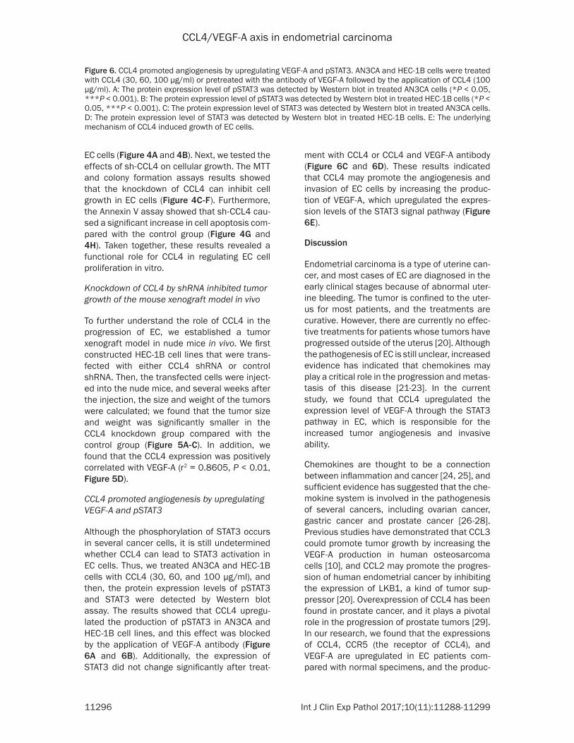
Although the phosphorylation of STAT3 occurs in several cancer cells, it is still undetermined whether CCL4 can lead to STAT3 activation in EC cells. Thus, we treated AN3CA and HEC-1B cells with CCL4 (30, 60, and 100 μg/ml), and then, the protein expression levels of pSTAT3 and STAT3 were detected by Western blot assay. The results showed that CCL4 upregu-lated the production of pSTAT3 in AN3CA and HEC-1B cell lines, and this effect was blocked by the application of VEGF-A antibody (Figure 6A and 6B). Additionally, the expression of STAT3 did not change significantly after treat-
ment with CCL4 or CCL4 and VEGF-A antibody (Figure 6C and 6D). These results indicated that CCL4 may promote the angiogenesis and invasion of EC cells by increasing the produc-tion of VEGF-A, which upregulated the expres-sion levels of the STAT3 signal pathway (Figure 6E).
Discussion
Endometrial carcinoma is a type of uterine can-cer, and most cases of EC are diagnosed in the early clinical stages because of abnormal uter-ine bleeding. The tumor is confined to the uter-us for most patients, and the treatments are curative. However, there are currently no effec-tive treatments for patients whose tumors have progressed outside of the uterus [20]. Although the pathogenesis of EC is still unclear, increased evidence has indicated that chemokines may play a critical role in the progression and metas-tasis of this disease [21-23]. In the current study, we found that CCL4 upregulated the expression level of VEGF-A through the STAT3 pathway in EC, which is responsible for the increased tumor angiogenesis and invasive ability.
Chemokines are thought to be a connection between inflammation and cancer [24, 25], and sufficient evidence has suggested that the che-mokine system is involved in the pathogenesis of several cancers, including ovarian cancer, gastric cancer and prostate cancer [26-28]. Previous studies have demonstrated that CCL3 could promote tumor growth by increasing the VEGF-A production in human osteosarcoma cells [10], and CCL2 may promote the progres-sion of human endometrial cancer by inhibiting the expression of LKB1, a kind of tumor sup-pressor [20]. Overexpression of CCL4 has been found in prostate cancer, and it plays a pivotal role in the progression of prostate tumors [29]. In our research, we found that the expressions of CCL4, CCR5 (the receptor of CCL4), and VEGF-A are upregulated in EC patients com-pared with normal specimens, and the produc-
Figure 6. CCL4 promoted angiogenesis by upregulating VEGF-A and pSTAT3. AN3CA and HEC-1B cells were treated with CCL4 (30, 60, 100 μg/ml) or pretreated with the antibody of VEGF-A followed by the application of CCL4 (100 μg/ml). A: The protein expression level of pSTAT3 was detected by Western blot in treated AN3CA cells (*P < 0.05, ***P < 0.001). B: The protein expression level of pSTAT3 was detected by Western blot in treated HEC-1B cells (*P < 0.05, ***P < 0.001). C: The protein expression level of STAT3 was detected by Western blot in treated AN3CA cells. D: The protein expression level of STAT3 was detected by Western blot in treated HEC-1B cells. E: The underlying mechanism of CCL4 induced growth of EC cells.
CCL4/VEGF-A axis in endometrial carcinoma
11297 Int J Clin Exp Pathol 2017;10(11):11288-11299
tion of CCL4, CCR5 and VEGF-A is associated with the clinical grade of the tumors in EC using an immunohistochemistry assay. We further confirmed the increase of CCL4 and VEGF-A using qRT-PCR. In addition, we demonstrated that the mRNA expression level of CCL4 is posi-tively correlated with VEGF-A in EC patients.
CC-chemokine receptor 5 (CCR5) is a G protein coupled receptor that functions as a chemo-kine receptor by binding to several chemokines, including CCL3, CCL4 and others [30]. Increas- ing evidence has supported that CCR5 is expressed on the surface of some tumor cells and is involved in angiogenesis via VEGF-A [10, 31]. In this research, we found that increa- sed CCL4 expression was positively correlated with the invasive ability of AN3CA and HEC-1B, and we also found that the antibody of CCR5 or VEGF-A could block this effect induced by CCL4. Moreover, ELISA and qRT-PCR results indicated that CCL4 promoted the expression level of VEGF-A in AN3CA and HEC-1B cells, and the upregulation of VEGF-A was also reversed by the antibodies of CCR5 or VEGF-A. These data suggested that CCL4 promoted the inva-sive ability and VEGF-A production by interact-ing with CCR5. VEGF-A plays a critical role in the growth and progression of tumor cells and is considered the most notable mediator of angio-genesis in various cancers [32, 33]. The tube formation assay demonstrated that CCL4 pro-moted angiogenesis by upregulating the expres-sion level of VEGF-A.
To further confirm the effects of CCL4 in EC cells, we knocked down the expression of CCL4 in HEC-1B cells by shRNA, and then we trans-ferred the cells into nude mice to establish a tumor xenograft model in vivo. The results showed that silenced CCL4 inhibited the growth of EC tumors in nude mice.
Signal transducers and activators of transcrip-tion (STAT3) is a member of the STAT family, which acts as transcription factors and are acti-vated by the phosphorylation of tyrosine [34]. Activated STAT3 has been found in several can-cers, including colorectal cancer, lung cancer and endometrial cancer [19, 35, 36]. In our results, we demonstrated that STAT3 was acti-vated after exposure to CCL4 (30, 60, 100 μg/ml) in AN3CA and HEC-1B cells, and the CCL4 (100 μg/ml) induced upregulation of pSTAT3 was reversed by the application of VEGF-A neu-
tralizing antibody. These results suggested that the phosphorylation of STAT3 may be involved in the CCL4-VEGF-A pathway, which is respon-sible for the progression of EC cells.
In conclusion, CCL4 was highly expressed in the EC cells, and it promoted the expression of VEGF-A by activating STAT3 via binding to CCR5. Additionally, VEGF-A promoted the invasive abil-ity and angiogenesis of the EC cells. These results support the possibility that CCL4 may be a novel therapeutic target in EC.
Acknowledgements
This study was supported by National Natural Science Foundation of China (No 81402165).
Disclosure of conflict of interest
None.
Address correspondence to: Ye Tian, Department of Radiotherapy & Oncology, The Second Affiliated Hos- pital of Soochow University, Suzhou 215004, China; Institute of Radiotherapy & Oncology, Soochow Uni- versity, Sanxiang Road No. 1055, Suzhou 215004, China. Tel: +86-0512-67783430; Fax: +86-0512-68284303; E-mail: [email protected]
References
[1] Jick H, Walker AM and Rothman KJ. The epi-demic of endometrial cancer: a commentary. Am J Public Health 1980; 70: 264-267.
[2] Trimble EL, Harlan LC, Clegg LX and Stevens JL. Pre-operative imaging, surgery and adjuvant therapy for women diagnosed with cancer of the corpus uteri in community practice in the United States. Gynecol Oncol 2005; 96: 741-748.
[3] Siegel R, Naishadham D and Jemal A. Cancer statistics, 2013. CA Cancer J Clin 2013; 63: 11-30.
[4] Cancer Genome Atlas Research N, Kandoth C, Schultz N, Cherniack AD, Akbani R, Liu Y, Shen H, Robertson AG, Pashtan I, Shen R, Benz CC, Yau C, Laird PW, Ding L, Zhang W, Mills GB, Kucherlapati R, Mardis ER and Levine DA. Inte-grated genomic characterization of endometri-al carcinoma. Nature 2013; 497: 67-73.
[5] Carlson MJ, Thiel KW and Leslie KK. Past, pres-ent, and future of hormonal therapy in recur-rent endometrial cancer. Int J Womens Health 2014; 6: 429-435.
[6] Gadducci A, Cosio S and Genazzani AR. Old and new perspectives in the pharmacological treatment of advanced or recurrent endome-

CCL4/VEGF-A axis in endometrial carcinoma
11298 Int J Clin Exp Pathol 2017;10(11):11288-11299
trial cancer: hormonal therapy, chemotherapy and molecularly targeted therapies. Crit Rev Oncol Hematol 2006; 58: 242-256.
[7] Gullino PM. Angiogenesis and oncogenesis. J Natl Cancer Inst 1978; 61: 639-643.
[8] Carmeliet P and Jain RK. Angiogenesis in can-cer and other diseases. Nature 2000; 407: 249-257.
[9] Matsumoto K and Ema M. Roles of VEGF-A sig-nalling in development, regeneration, and tu-mours. J Biochem 2014; 156: 1-10.
[10] Liao YY, Tsai HC, Chou PY, Wang SW, Chen HT, Lin YM, Chiang IP, Chang TM, Hsu SK, Chou MC, Tang CH and Fong YC. CCL3 promotes angiogenesis by dysregulation of miR-374b/VEGF-A axis in human osteosarcoma cells. On-cotarget 2016; 7: 4310-4325.
[11] Lira SA and Furtado GC. The biology of chemo-kines and their receptors. Immunol Res 2012; 54: 111-120.
[12] Zlotnik A and Yoshie O. Chemokines: a new classification system and their role in immuni-ty. Immunity 2000; 12: 121-127.
[13] Locati M, Deuschle U, Massardi ML, Martinez FO, Sironi M, Sozzani S, Bartfai T and Man-tovani A. Analysis of the gene expression pro-file activated by the CC chemokine ligand 5/RANTES and by lipopolysaccharide in human monocytes. J Immunol 2002; 168: 3557-3562.
[14] Erreni M, Bianchi P, Laghi L, Mirolo M, Fabbri M, Locati M, Mantovani A and Allavena P. Ex-pression of chemokines and chemokine recep-tors in human colon cancer. Methods Enzymol 2009; 460: 105-121.
[15] Milliken D, Scotton C, Raju S, Balkwill F and Wilson J. Analysis of chemokines and chemo-kine receptor expression in ovarian cancer as-cites. Clin Cancer Res 2002; 8: 1108-1114.
[16] Blum DL, Koyama T, M’Koma AE, Iturregui JM, Martinez-Ferrer M, Uwamariya C, Smith JA Jr, Clark PE and Bhowmick NA. Chemokine mark-ers predict biochemical recurrence of prostate cancer following prostatectomy. Clin Cancer Res 2008; 14: 7790-7797.
[17] Ma YR, Zhang S, Sun Y, Liu YY, Song Q and Hao YW. [MIP-1alpha promotes the migration abili-ty of Jurkat cell through human brain microvas-cular endothelial cell monolayer]. Zhongguo Shi Yan Xue Ye Xue Za Zhi 2014; 22: 35-39.
[18] Ding L, Li B, Zhao Y, Fu YF, Hu EL, Hu QG, Ni YH and Hou YY. Serum CCL2 and CCL3 as poten-tial biomarkers for the diagnosis of oral squa-mous cell carcinoma. Tumour Biol 2014; 35: 10539-10546.
[19] Zhu M, Che Q, Liao Y, Wang H, Wang J, Chen Z, Wang F, Dai C and Wan X. Oncostatin M acti-vates STAT3 to promote endometrial cancer
invasion and angiogenesis. Oncol Rep 2015; 34: 129-138.
[20] Pena CG, Nakada Y, Saatcioglu HD, Aloisio GM, Cuevas I, Zhang S, Miller DS, Lea JS, Wong KK, DeBerardinis RJ, Amelio AL, Brekken RA and Castrillon DH. LKB1 loss promotes endome- trial cancer progression via CCL2-dependent macrophage recruitment. J Clin Invest 2015; 125: 4063-4076.
[21] Wallace AE, Sales KJ, Catalano RD, Anderson RA, Williams AR, Wilson MR, Schwarze J, Wang H, Rossi AG and Jabbour HN. Prostaglandin F2alpha-F-prostanoid receptor signaling pro-motes neutrophil chemotaxis via chemokine (C-X-C motif) ligand 1 in endometrial adenocar-cinoma. Cancer Res 2009; 69: 5726-5733.
[22] Sakane R, Tsubamoto H, Sakata K, Inoue K, Ogino M, Shibahara H, Hao H and Hirota S. Ex-pression of chemokine ligand 18 in stage IA low-grade endometrial cancer. Anticancer Res 2014; 34: 5331-5336.
[23] Attar R, Agachan B, Kuran SB, Cacina C, Sozen S, Yurdum LM, Attar E and Isbir T. Association of CCL2 and CCR2 gene variants with endome-trial cancer in Turkish women. In Vivo 2010; 24: 243-248.
[24] Balkwill F and Mantovani A. Inflammation and cancer: back to Virchow? Lancet 2001; 357: 539-545.
[25] Coussens LM and Werb Z. Inflammation and cancer. Nature 2002; 420: 860-867.
[26] Son DS, Parl AK, Rice VM and Khabele D. Kera-tinocyte chemoattractant (KC)/human growth-regulated oncogene (GRO) chemokines and pro-inflammatory chemokine networks in mo- use and human ovarian epithelial cancer cells. Cancer Biol Ther 2007; 6: 1302-1312.
[27] Leung SY, Yuen ST, Chu KM, Mathy JA, Li R, Chan AS, Law S, Wong J, Chen X and So S. Ex-pression profiling identifies chemokine (C-C motif) ligand 18 as an independent prognostic indicator in gastric cancer. Gastroenterology 2004; 127: 457-469.
[28] Zhang J, Lu Y and Pienta KJ. Multiple roles of chemokine (C-C motif) ligand 2 in promoting prostate cancer growth. J Natl Cancer Inst 2010; 102: 522-528.
[29] Fang LY, Izumi K, Lai KP, Liang L, Li L, Miyamo-to H, Lin WJ and Chang C. Infiltrating macro-phages promote prostate tumorigenesis via modulating androgen receptor-mediated CC- L4-STAT3 signaling. Cancer Res 2013; 73: 5633-5646.
[30] Onuffer JJ and Horuk R. Chemokines, chemo-kine receptors and small-molecule antago-nists: recent developments. Trends Pharmacol Sci 2002; 23: 459-467.
[31] Barcelos LS, Coelho AM, Russo RC, Guabiraba R, Souza AL, Bruno-Lima G Jr, Proudfoot AE,

CCL4/VEGF-A axis in endometrial carcinoma
11299 Int J Clin Exp Pathol 2017;10(11):11288-11299
Andrade SP and Teixeira MM. Role of the che-mokines CCL3/MIP-1 alpha and CCL5/RAN-TES in sponge-induced inflammatory angio-genesis in mice. Microvasc Res 2009; 78: 148-154.
[32] Folkman J. Angiogenesis in cancer, vascular, rheumatoid and other disease. Nat Med 1995; 1: 27-31.
[33] Risau W. Mechanisms of angiogenesis. Nature 1997; 386: 671-674.
[34] Darnell JE Jr. STATs and gene regulation. Sci-ence 1997; 277: 1630-1635.
[35] Morikawa T, Baba Y, Yamauchi M, Kuchiba A, Nosho K, Shima K, Tanaka N, Huttenhower C, Frank DA, Fuchs CS and Ogino S. STAT3 expres-sion, molecular features, inflammation pat-terns, and prognosis in a database of 724 colorectal cancers. Clin Cancer Res 2011; 17: 1452-1462.
[36] Jiang R, Jin Z, Liu Z, Sun L, Wang L and Li K. Correlation of activated STAT3 expression with clinicopathologic features in lung adenocarci-noma and squamous cell carcinoma. Mol Di-agn Ther 2011; 15: 347-352.


![Original Article Upregulating miR-146a by physcion ... · Upregulating miR-146a by physcion reverses multidrug ... [20]. However, the role of physcion on hemato-logical malignancies](https://static.fdocuments.in/doc/165x107/5bc7678409d3f267298b9f31/original-article-upregulating-mir-146a-by-physcion-upregulating-mir-146a.jpg)

