Organic chemistry meets polymers, nanoscience ... · Organic chemistry meets polymers, nanoscience,...
9
1638 Organic chemistry meets polymers, nanoscience, therapeutics and diagnostics Vincent M. Rotello Review Open Access Address: Department of Chemistry, University of Massachusetts-Amherst, 710 North Pleasant Street, Amherst, Massachusetts 01003, USA Email: Vincent M. Rotello - [email protected] Keywords: organic synthesis, supramolecular, nanoparticle sensing Beilstein J. Org. Chem. 2016, 12, 1638–1646. doi:10.3762/bjoc.12.161 Received: 23 February 2016 Accepted: 18 July 2016 Published: 02 August 2016 This article is part of the Thematic Series "Supramolecular chemistry at the interface of biology, materials and medicine". Guest Editors: S. C. Zimmerman and E. V. Anslyn © 2016 Rotello; licensee Beilstein-Institut. License and terms: see end of document. Abstract The atom-by-atom control provided by synthetic organic chemistry presents a means of generating new functional nanomaterials with great precision. Bringing together these two very disparate skill sets is, however, quite uncommon. This autobiographical review provides some insight into how my program evolved, as well as giving some idea of where we are going. 1638 Review My roots – synthetic organic My interest in chemistry built on my incessant tinkering. When I was young, I was the sort that would take things apart to see how they worked. I also put them back together, occasionally with no extra parts… Eventually, the ability to create overcame the desire to dissect, and I started pursuing photography. My efforts in this domain progressed until I started considering art as a career. Alongside this artistic pathway, however, I de- veloped an interest in chemistry. What intrigued me at the start was connectivity – how atoms could be strung together. I explored both art and photography at Illinois Institute of Tech- nology, but science and scientists ended up deciding me. It started with Chem. 237, Organic Chemistry. This course was taught by Pete Johnson, who introduced me to retrosynthetic analysis, and in the process showed me how I could achieve the connectivity I had doodled when younger. This classroom work was followed up soon thereafter by practical training at the hands of Phil Garner. Phil was an old school 'guts and glory' synthetic type, and I learned from him everything I needed to about the practical side of synthetic organic. This work culmi- nated in the first paper I co-authored [1], where I learned painful lessons with the Fieser triangle at the twilight of the pen and ink era.
Transcript of Organic chemistry meets polymers, nanoscience ... · Organic chemistry meets polymers, nanoscience,...
1638
Organic chemistry meets polymers, nanoscience,therapeutics and diagnosticsVincent M. Rotello
Review Open Access
Address:Department of Chemistry, University of Massachusetts-Amherst, 710North Pleasant Street, Amherst, Massachusetts 01003, USA
Email:Vincent M. Rotello - [email protected]
Keywords:organic synthesis, supramolecular, nanoparticle sensing
Beilstein J. Org. Chem. 2016, 12, 1638–1646.doi:10.3762/bjoc.12.161
Received: 23 February 2016Accepted: 18 July 2016Published: 02 August 2016
This article is part of the Thematic Series "Supramolecular chemistry atthe interface of biology, materials and medicine".
Guest Editors: S. C. Zimmerman and E. V. Anslyn
© 2016 Rotello; licensee Beilstein-Institut.License and terms: see end of document.
AbstractThe atom-by-atom control provided by synthetic organic chemistry presents a means of generating new functional nanomaterials
with great precision. Bringing together these two very disparate skill sets is, however, quite uncommon. This autobiographical
review provides some insight into how my program evolved, as well as giving some idea of where we are going.
1638
ReviewMy roots – synthetic organicMy interest in chemistry built on my incessant tinkering. When
I was young, I was the sort that would take things apart to see
how they worked. I also put them back together, occasionally
with no extra parts… Eventually, the ability to create overcame
the desire to dissect, and I started pursuing photography. My
efforts in this domain progressed until I started considering art
as a career. Alongside this artistic pathway, however, I de-
veloped an interest in chemistry. What intrigued me at the start
was connectivity – how atoms could be strung together. I
explored both art and photography at Illinois Institute of Tech-
nology, but science and scientists ended up deciding me. It
started with Chem. 237, Organic Chemistry. This course was
taught by Pete Johnson, who introduced me to retrosynthetic
analysis, and in the process showed me how I could achieve the
connectivity I had doodled when younger. This classroom work
was followed up soon thereafter by practical training at the
hands of Phil Garner. Phil was an old school 'guts and glory'
synthetic type, and I learned from him everything I needed to
about the practical side of synthetic organic. This work culmi-
nated in the first paper I co-authored [1], where I learned
painful lessons with the Fieser triangle at the twilight of the pen
and ink era.
Beilstein J. Org. Chem. 2016, 12, 1638–1646.
1639
With my belly full of synthetic fire, I sought out a place to
pursue my trade. In the mid-80s Yale University was hotbed for
synthesis, and picking out an advisor was a difficult choice. I
ultimately chose Harry Wasserman, based on the freedom he
gave his group in choosing and attacking synthetic and physical
challenges. During this period I developed and finished a
variety of syntheses. My break came, however, when looking in
the back pages of the journal Heterocycles in the "New Natural
Products" section. I noticed a rather interesting new molecule
known as rapamycin that had the vicinal tricarbonyl motif in
vogue in the Wasserman group. When I suggested we go after
this molecule, Harry demurred, pointing out its complexity and
that "nobody would be interested". I kept my eye on the litera-
ture, however, and when Dave Williams published the "Central
America" fragment of the related macrolide FK-506, I urged
Harry to see if he could get some intermediate that I could use
to demonstrate our methodology for tricarbonyl synthesis. Dave
generously obliged by sending ~200 mg of an advanced inter-
mediate. With hands shaking, I cracked the vial and started on
the synthesis. Working with small-scale reactions (<1 mg), I
eventually worked out a high-yielding synthesis of the Williams
fragment [2]. This synthesis generated some buzz, catching the
eye of folks like Sam Danishefsky and facilitating my next
move.
Moving to LegolandSorting out what I wanted to do after grad school was a bit of a
challenge. In those days I knew I wanted to be an academic, but
what I wanted to do scientifically was an open question. I
started thinking about proposals for postdoctoral fellowships,
but the synthetic ideas I generated didn't fire me up like I
thought they should. I really enjoyed the power of organic syn-
thesis, but I wanted to do something with the molecules that I
laboriously fashioned. Once again my love of connectivity
kicked in, this time with supramolecular chemistry. I started
thinking of molecules instead of atoms as building blocks. I
looked around for professors with a like mind, and applied to
Julius Rebek at Pittsburgh. Between when I applied and when I
joined as an NSF postdoctoral fellow Julius had moved to MIT
for his brief stay in the Boston area before heading off to
Scripps.
While in the Rebek group I used my synthetic abilities while
gaining the insight into physical organic chemistry that has
informed the rest of my career. I started out working on self-
replicating systems [3], developing new systems that had novel
capabilities, including external regulation [4]. I also took on a
brutal project, focused on the synthesis of water-soluble analogs
of Kemp's triacid. This project was a massive effort, with a
huge number of reactions required to optimize the initial steps.
We did, however, obtain the desired receptors and observe
some interesting binding processes in water [5]. During this part
of my time in the group, Julius offered that if I stayed an addi-
tional year I could work on projects of my own. During this
time I discovered my inner mentor. Much to the annoyance of
my labmates, I built a veritable army of undergraduates,
pursuing supramolecular chemistry, along with a project in ful-
lerenes [6,7].
After finishing my postdoctoral work, I moved to the Univer-
sity of Massachusetts. I chose UMass based on its quality and
longstanding reputation for collaborative research. Upon arrival,
I collected a fired up group of graduate students and went to it. I
maintained my fullerenes project [8], and initiated a set of three
other supramolecular projects. Of these projects, our work on
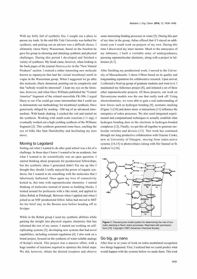
flavoenzyme models was the one that really took off. Using
electrochemistry, we were able to gain a real understanding of
how forces such as hydrogen bonding [9], aromatic stacking
(Figure 1) [10] and donor atom–π interactions [11] influence the
energetics of redox processes. We also used integrated experi-
mental and computational techniques to actually establish what
hydrogen bonding does to the electrons in hydrogen-bonded
complexes [12]. Finally, we put this all together to generate mo-
lecular switches and devices [13]. This work has continued
through our long productive collaboration with Graeme Cooke,
now at University of Glasgow, moving from redox-active
systems [14,15] to photovoltaics (along with Ifor Samuel at St.
Andrew’s) [16].
Figure 1: Flavoenzyme model system for determining the role of aro-matic stacking in flavin redox processes. Reprinted with permissionfrom [10]. Copyright (1997) American Chemical Society.
Go big, go nanoAfter four or so years of work on redox-modulated recognition
two things happened. First, I realized that we could predict what
would happen with the systems before we made them. This took
Beilstein J. Org. Chem. 2016, 12, 1638–1646.
1640
much of the fun out of the work. Simultaneously, I received
tenure, and tried to sort out what I wanted to do for my next
career phase. Our next move was into polymers, where we
started the conceptual journey we are still taking. The key ques-
tion we asked is "we know what happens when you have one
host–guest dyad, but what happens when you have 10, 50, 100
on a polymer?" On a straightforward level, we were able to
demonstrate that we could use non-covalent sidechain modifica-
tion between multivalent polymers and monovalent guests to
generate "plug and play polymers" (Figure 2) [17]. We also
showed that we could self-assemble a polymer around an elec-
troactive guest, effectively encapsulating and isolating it [18].
Figure 2: Recognition element-functionalized polymers for 'plug andplay' modification and self-assembly.
The research really became interesting when we started mixing
multivalent complementary polymers together. When we mixed
together diaminopyridine and uracil polymers together in
chloroform we generated a turbid solution. Under the micro-
scope we found that the turbidity surprisingly arose from vesic-
ular structures [19]. Through quite a bit of experimentation we
determined that the unprecedented self-assembly process was
driven by self-sorting of the polymer chains to provide vesicle
walls with denser recognition elements in the middle than at the
outside [20].
While we were working with polymers, we were just starting to
move into nanoparticles. As with the polymers, we started off
studying the interactions of recognition element-functionalized
nanoparticles with monovalent guests – in this case our old
friend flavin where we showed modulation of the flavin redox
potentials [21]. Taking this research one step further, we created
a nanoparticle with a mixed monolayer consisting of hydrogen
bonding and aromatic staking sidechains. When we incubated
this NP with flavin we observed an increase in binding over
time, i.e., we were able to template the particle to the guest [22].
We have since demonstrated this templation with peptides [23]
and are (still!) trying to definitively show templation to pro-
teins.
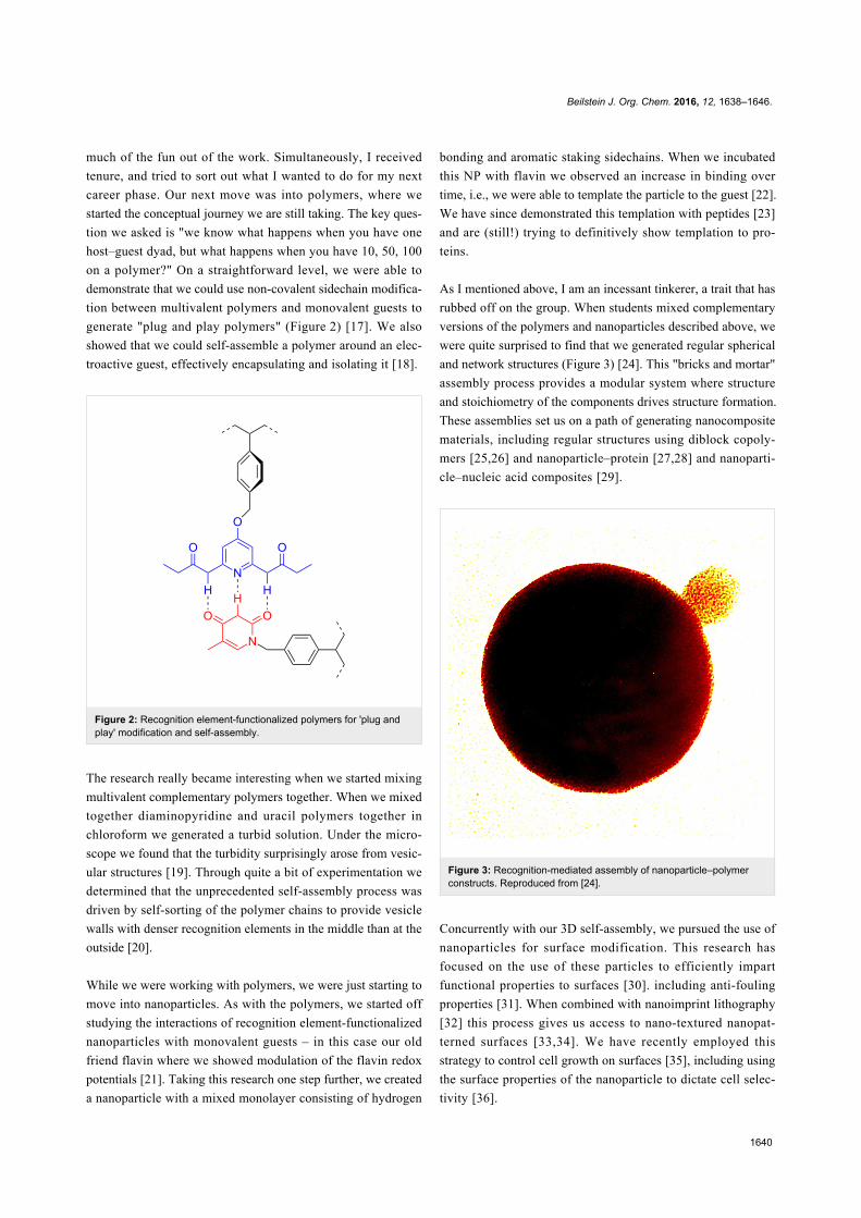
As I mentioned above, I am an incessant tinkerer, a trait that has
rubbed off on the group. When students mixed complementary
versions of the polymers and nanoparticles described above, we
were quite surprised to find that we generated regular spherical
and network structures (Figure 3) [24]. This "bricks and mortar"
assembly process provides a modular system where structure
and stoichiometry of the components drives structure formation.
These assemblies set us on a path of generating nanocomposite
materials, including regular structures using diblock copoly-
mers [25,26] and nanoparticle–protein [27,28] and nanoparti-
cle–nucleic acid composites [29].
Figure 3: Recognition-mediated assembly of nanoparticle–polymerconstructs. Reproduced from [24].
Concurrently with our 3D self-assembly, we pursued the use of
nanoparticles for surface modification. This research has
focused on the use of these particles to efficiently impart
functional properties to surfaces [30]. including anti-fouling
properties [31]. When combined with nanoimprint lithography
[32] this process gives us access to nano-textured nanopat-
terned surfaces [33,34]. We have recently employed this
strategy to control cell growth on surfaces [35], including using
the surface properties of the nanoparticle to dictate cell selec-
tivity [36].
Beilstein J. Org. Chem. 2016, 12, 1638–1646.
1641
Nanoparticles meet biologyOur move into nanocomposites coincided with our efforts to
interface materials and biology – the current core focus of the
lab. We started off looking at nucleic acids, where we showed
that cationic nanoparticles could bind to anionic DNA and
inhibit transcription [37]. We also developed a number of strate-
gies for delivery of small molecules, including glutathione-
mediated release of covalently attached thiols [38], as the effec-
tive release of drugs adsorbed into the cationic monolayers of
nanoparticles [39], and even photoactivatable drug [40] and
DNA release [41]. When we started looking at how particles
interact with proteins, however, very little was known about
how these materials would play together. We found out pretty
quickly that the answer was "not very well". Binding of the
nanoparticle induced protein denaturation, with loss of bioac-
tivity [42]. We hypothesized that this denaturation arose from
interaction of the protein with the hydrophobic elements of the
simple ligands we were using. This hypothesis led us to create
what we call the "tabula rasa" ligand [43], namely a ligand that
features a hydrophobic interior for self-assembly, and a short
tetra(ethylene glycol) layer to block interactions of the hydro-
phobic interiors with proteins [44]. These particles were indeed
"blank" (but not as blank as our later zwitterionic “corona-free’
particles [45]), and behaved like high molecular weight
poly(ethylene glycol) [46]. Once we appended simple anionic
and cationic recognition elements to the surface these system
bound proteins with high affinity [47] and some degree of selec-
tivity [48]. What was surprising is that not only did the
particle–protein binding process not denature the proteins, it
actually stabilizes them [46]! This result stills surprises
researchers who follow the dogma that proteins must denature
at interfaces. One of the other observations we made using
isothermal titration calorimetry is that nanoparticle–protein
interactions using the tabula rasa-based particles mimicked the
thermodynamics of protein–protein interactions quite well [49].
Once we were able to have proteins and nanoparticles work in
harmony [50] we started working on designing systems with
emergent behavior, i.e., where the particle–protein complex
behaves differently than either of the two components. A
prime example of this synergy is when we showed that nanopar-
ticle–enzyme complexes showed altered substrate selectivity
[51], with the particle dictating the enzyme kinetics by acting as
a "filter" for substrate and product [47]. Another area where we
demonstrated synergy is in the area of Pickering emulsions.
These emulsions are made by interfacial assembly of particles
at oil–water interfaces. We showed that nanoparticles and pro-
teins could be self-assembled at this interface [52], retaining
their activity. When the oil core was crosslinked, these systems
worked even better, enhancing enzyme activity [53], even under
extreme chemical and thermal conditions [54].
As we were learning the rules for nanoparticle-biological inter-
actions we started looking into applications for these self-
assembled materials. Our first real success came when we
demonstrated very efficient DNA transfection (i.e., gene
delivery) using gold nanoparticles [55]. In later work we im-
proved on our gene delivery vehicles [56], demonstrated the
delivery of siRNA [57] and enzymes [58] using nanoparticle
assemblies. All of these systems (as well as essentially all of the
other examples of nanomaterial delivery vehicles in the litera-
ture) occurred via endosomal uptake. The problem with this
route is that what goes into the endosome tends to stay in the
endosome, and eventually be degraded. Since most of the inter-
esting things in cells require access to the cytosol (including
materials destined to the nucleus), this entrapment is a major
limitation [59].
One of the beauties of supramolecular chemistry is its modu-
larity. We started looking into the use of the Pickering emul-
sions described above for delivery applications. There was a
challenge: nobody (including us) could generate emulsions with
diameters small enough (<200 nm) for use as in vivo delivery
vehicles [60]. Once again, supramolecular chemistry came to
the rescue. Anslyn showed that guanidinium groups bound
strongly to carboxylates [61]. This led to the surmise that this
interaction could be used to "pin" arginine-capped nanoparti-
cles to oil droplets comprised of fatty acids. This trick worked
remarkably well, providing ~150 nm nanocapsules. These
capsules were unstable in serum however. Reaching back to our
nanocomposite work, we were able to use bricks and mortar
assembly of anionic proteins (transferrin) and cationic nanopar-
ticles to create stable capsules [62]. These capsules delivered
hydrophobic drugs and dyes to cells very effectively. A
puzzling question arose however: dyes were delivered into cells
much faster than the particles on the outside of the capsule.
Clearly, endosomal uptake would result in identical rates of
uptake, leading us to surmise that uptake occurred through a
membrane fusion process.
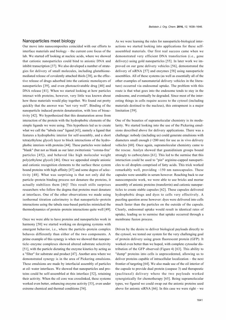
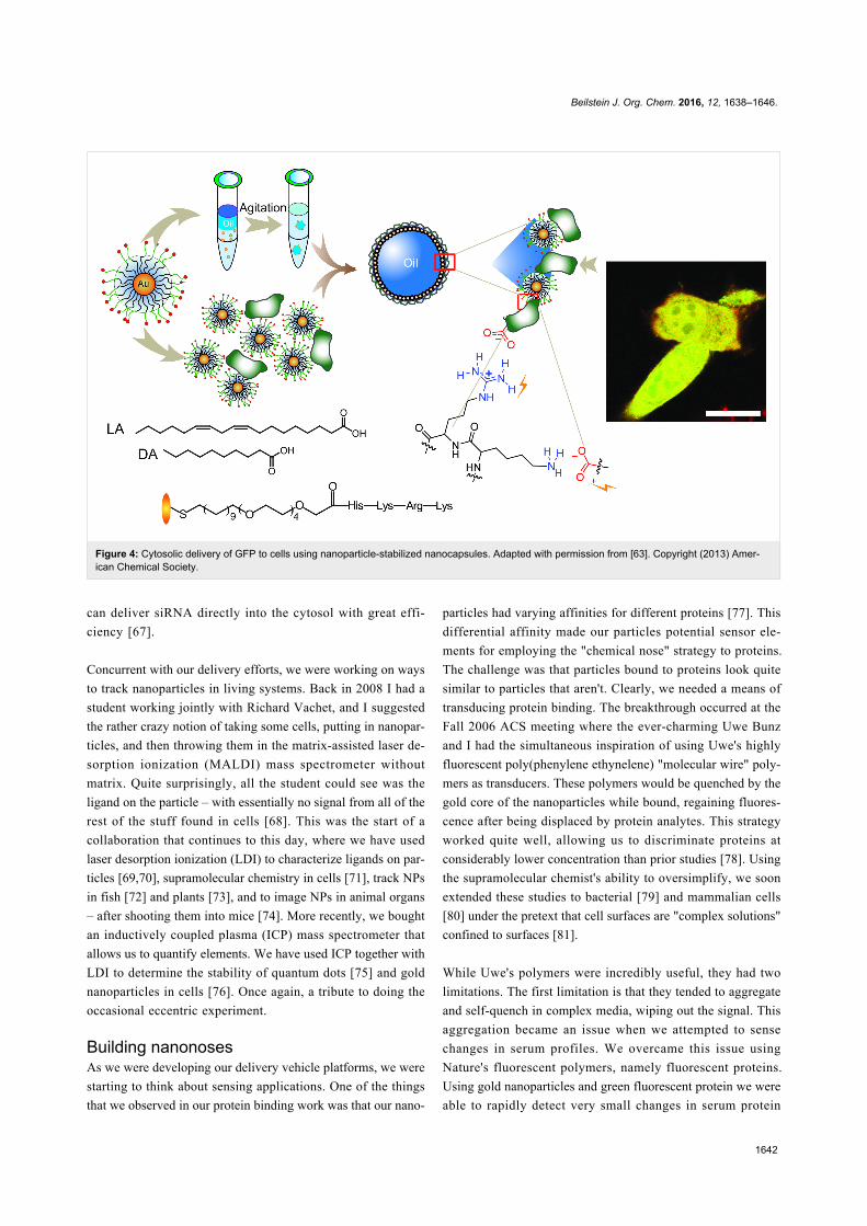
Driven by the desire to deliver biological payloads directly to
the cytosol, we tested our system for the very challenging goal
of protein delivery using green fluorescent protein (GFP). It
worked even better than we hoped, with complete cytosolar dis-
tribution of the GFP observed (Figure 4) [63]. This ability to
"dump" proteins into cells is unprecedented, allowing us to
deliver proteins capable of intracellular localization – the next
frontier of targeting [64]. We also made use of the oil interior of
the capsule to provide dual protein (caspase 3) and therapeutic
(paclitaxel) delivery where the two payloads worked
synergistically for chemotherapy [65]. Being supramolecular
types, we figured we could swap out the anionic proteins used
above for anionic siRNA [66]. In this case we were right – we
Beilstein J. Org. Chem. 2016, 12, 1638–1646.
1642
Figure 4: Cytosolic delivery of GFP to cells using nanoparticle-stabilized nanocapsules. Adapted with permission from [63]. Copyright (2013) Amer-ican Chemical Society.
can deliver siRNA directly into the cytosol with great effi-
ciency [67].
Concurrent with our delivery efforts, we were working on ways
to track nanoparticles in living systems. Back in 2008 I had a
student working jointly with Richard Vachet, and I suggested
the rather crazy notion of taking some cells, putting in nanopar-
ticles, and then throwing them in the matrix-assisted laser de-
sorption ionization (MALDI) mass spectrometer without
matrix. Quite surprisingly, all the student could see was the
ligand on the particle – with essentially no signal from all of the
rest of the stuff found in cells [68]. This was the start of a
collaboration that continues to this day, where we have used
laser desorption ionization (LDI) to characterize ligands on par-
ticles [69,70], supramolecular chemistry in cells [71], track NPs
in fish [72] and plants [73], and to image NPs in animal organs
– after shooting them into mice [74]. More recently, we bought
an inductively coupled plasma (ICP) mass spectrometer that
allows us to quantify elements. We have used ICP together with
LDI to determine the stability of quantum dots [75] and gold
nanoparticles in cells [76]. Once again, a tribute to doing the
occasional eccentric experiment.
Building nanonosesAs we were developing our delivery vehicle platforms, we were
starting to think about sensing applications. One of the things
that we observed in our protein binding work was that our nano-
particles had varying affinities for different proteins [77]. This
differential affinity made our particles potential sensor ele-
ments for employing the "chemical nose" strategy to proteins.
The challenge was that particles bound to proteins look quite
similar to particles that aren't. Clearly, we needed a means of
transducing protein binding. The breakthrough occurred at the
Fall 2006 ACS meeting where the ever-charming Uwe Bunz
and I had the simultaneous inspiration of using Uwe's highly
fluorescent poly(phenylene ethynelene) "molecular wire" poly-
mers as transducers. These polymers would be quenched by the
gold core of the nanoparticles while bound, regaining fluores-
cence after being displaced by protein analytes. This strategy
worked quite well, allowing us to discriminate proteins at
considerably lower concentration than prior studies [78]. Using
the supramolecular chemist's ability to oversimplify, we soon
extended these studies to bacterial [79] and mammalian cells
[80] under the pretext that cell surfaces are "complex solutions"
confined to surfaces [81].
While Uwe's polymers were incredibly useful, they had two
limitations. The first limitation is that they tended to aggregate
and self-quench in complex media, wiping out the signal. This
aggregation became an issue when we attempted to sense
changes in serum profiles. We overcame this issue using
Nature's fluorescent polymers, namely fluorescent proteins.
Using gold nanoparticles and green fluorescent protein we were
able to rapidly detect very small changes in serum protein
Beilstein J. Org. Chem. 2016, 12, 1638–1646.
1643
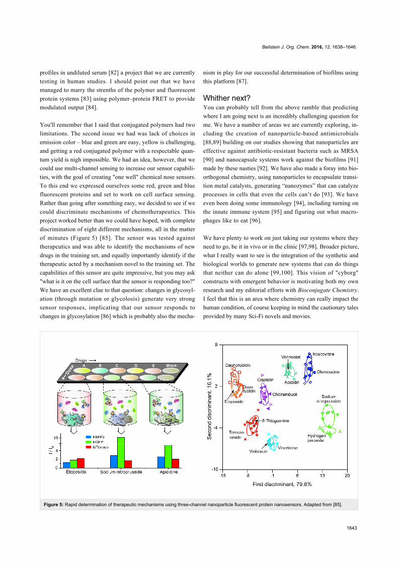
Figure 5: Rapid determination of therapeutic mechanisms using three-channel nanoparticle fluorescent protein nanosensors. Adapted from [85].
profiles in undiluted serum [82] a project that we are currently
testing in human studies. I should point out that we have
managed to marry the strenths of the polymer and fluorescent
protein systems [83] using polymer–protein FRET to provide
modulated output [84].
You'll remember that I said that conjugated polymers had two
limitations. The second issue we had was lack of choices in
emission color – blue and green are easy, yellow is challenging,
and getting a red conjugated polymer with a respectable quan-
tum yield is nigh impossible. We had an idea, however, that we
could use multi-channel sensing to increase our sensor capabili-
ties, with the goal of creating "one well" chemical nose sensors.
To this end we expressed ourselves some red, green and blue
fluorescent proteins and set to work on cell surface sensing.
Rather than going after something easy, we decided to see if we
could discriminate mechanisms of chemotherapeutics. This
project worked better than we could have hoped, with complete
discrimination of eight different mechanisms, all in the matter
of minutes (Figure 5) [85]. The sensor was tested against
therapeutics and was able to identify the mechanisms of new
drugs in the training set, and equally importantly identify if the
therapeutic acted by a mechanism novel to the training set. The
capabilities of this sensor are quite impressive, but you may ask
"what is it on the cell surface that the sensor is responding too?"
We have an excellent clue to that question: changes in glycosyl-
ation (through mutation or glycolosis) generate very strong
sensor responses, implicating that our sensor responds to
changes in glycosylation [86] which is probably also the mecha-
nism in play for our successful determination of biofilms using
this platform [87].
Whither next?You can probably tell from the above ramble that predicting
where I am going next is an incredibly challenging question for
me. We have a number of areas we are currently exploring, in-
cluding the creation of nanoparticle-based antimicrobials
[88,89] building on our studies showing that nanoparticles are
effective against antibiotic-resistant bacteria such as MRSA
[90] and nanocapsule systems work against the biofilms [91]
made by these nasties [92]. We have also made a foray into bio-
orthogonal chemistry, using nanoparticles to encapsulate transi-
tion metal catalysts, generating “nanozymes” that can catalyze
processes in cells that even the cells can’t do [93]. We have
even been doing some immunology [94], including turning on
the innate immune system [95] and figuring out what macro-
phages like to eat [96].
We have plenty to work on just taking our systems where they
need to go, be it in vivo or in the clinic [97,98]. Broader picture,
what I really want to see is the integration of the synthetic and
biological worlds to generate new systems that can do things
that neither can do alone [99,100]. This vision of "cyborg"
constructs with emergent behavior is motivating both my own
research and my editorial efforts with Bioconjugate Chemistry.
I feel that this is an area where chemistry can really impact the
human condition, of course keeping in mind the cautionary tales
provided by many Sci-Fi novels and movies.
Beilstein J. Org. Chem. 2016, 12, 1638–1646.
1644
Something about the authorLike anyone with a job, I live two lives. As you can probably
surmise, science moves me at work. As I travel, I also enjoy the
comradeship of scientists around the world, and can become
quite evangelical about the roles the scientific community can
serve to bridge gulfs between nations and cultures. To be
honest, I also enjoy seeing the world and its many wonders,
reveling in both the "big" sights and running at dawn in a new
town. And I have been accused of traveling on my stomach – I
do have a fondness for food .
At home, I am a family man (if not a Family Guy…). Coming
from the school that food equals love, my love of cooking is
likewise a major factor in my life. The skills I developed as a
synthetic chemist give me an understanding of ingredients and
techniques that allows me to cook cuisines from around the
world. Fortunately my wife and I are inveterate exercisers, and
my son's metabolism is still rapid enough to avoid (excessive)
weight gain. This exercise is typically done with our rather
neurotic Weimaraner Trudy, allowing us to get double duty
from our toil.
References1. Garner, P.; Park, J. M.; Rotello, V. Tetrahedron Lett. 1985, 26,
3299–3302. doi:10.1016/S0040-4039(00)98282-02. Wasserman, H. H.; Rotello, V. M.; Williams, D. R.; Benbow, J. W.
J. Org. Chem. 1989, 54, 2785–2786. doi:10.1021/jo00273a0043. Rotello, V.; Hong, J. I.; Rebek, J., Jr. J. Am. Chem. Soc. 1991, 113,
9422–9423. doi:10.1021/ja00024a0894. Hong, J.-I.; Feng, Q.; Rotello, V.; Rebek, J., Jr. Science 1992, 255,
848–850. doi:10.1126/science.255.5046.8485. Rotello, V. M.; Viani, E. A.; Deslongchamps, G.; Murray, B. A.;
Rebek, J., Jr. J. Am. Chem. Soc. 1993, 115, 797–798.doi:10.1021/ja00055a066
6. Rotello, V. M.; Howard, J. B.; Yadav, T.; Conn, M. M.; Viani, E.;Giovane, L. M.; Lafleur, A. L. Tetrahedron Lett. 1993, 34, 1561–1562.doi:10.1016/0040-4039(93)85006-I
7. Giovane, L. M.; Barco, J. W.; Yadav, T.; Lafleur, A. L.; Marr, J. A.;Howard, J. B.; Rotello, V. M. J. Phys. Chem. 1993, 97, 8560–8561.doi:10.1021/j100135a004
8. Guhr, K. I.; Greaves, M. D.; Rotello, V. M. J. Am. Chem. Soc. 1994,116, 5997–5998. doi:10.1021/ja00092a072
9. Breinlinger, E.; Niemz, A.; Rotello, V. M. J. Am. Chem. Soc. 1995,117, 5379–5380. doi:10.1021/ja00124a029
10. Breinlinger, E. C.; Rotello, V. M. J. Am. Chem. Soc. 1997, 119,1165–1166. doi:10.1021/ja9612110
11. Breinlinger, E. C.; Keenan, C. J.; Rotello, V. M. J. Am. Chem. Soc.1998, 120, 8606–8609. doi:10.1021/ja9809556
12. Rotello, V. M. J. Am. Chem. Soc. 1997, 119, 6833–6836.doi:10.1021/ja970801a
13. Deans, R.; Niemz, A.; Breinlinger, E. C.; Rotello, V. M.J. Am. Chem. Soc. 1997, 119, 10863–10864. doi:10.1021/ja9728740
14. Nandwana, V.; Samuel, I.; Cooke, G.; Rotello, V. M. Acc. Chem. Res.2013, 46, 1000–1009. doi:10.1021/ar300132r
15. Subramani, C.; Yesilbag, G.; Jordan, B. J.; Li, X.; Khorasani, A.;Cooke, G.; Sanyal, A.; Rotello, V. M. Chem. Commun. 2010, 46,2067–2069. doi:10.1039/b926746h
16. Ward, A. J.; Ruseckas, A.; Kareem, M. M.; Ebenhoch, B.;Serrano, L. A.; Al-Eid, M.; Fitzpatrick, B.; Rotello, V. M.; Cooke, G.;Samuel, I. D. W. Adv. Mater. 2015, 27, 2496–2500.doi:10.1002/adma.201405623
17. Ilhan, F.; Gray, M.; Rotello, V. M. Macromolecules 2001, 34,2597–2601. doi:10.1021/ma001700r
18. Galow, T. H.; Ilhan, F.; Cooke, G.; Rotello, V. M. J. Am. Chem. Soc.2000, 122, 3595–3598. doi:10.1021/ja993735g
19. Ilhan, F.; Galow, T. H.; Gray, M.; Clavier, G.; Rotello, V. M.J. Am. Chem. Soc. 2000, 122, 5895–5896. doi:10.1021/ja0011966
20. Uzun, O.; Xu, H.; Jeoung, E.; Thibault, R. J.; Rotello, V. M.Chem. – Eur. J. 2005, 11, 6916–6920. doi:10.1002/chem.200500809
21. Boal, A. K.; Rotello, V. M. J. Am. Chem. Soc. 1999, 121, 4914–4915.doi:10.1021/ja9905288
22. Boal, A. K.; Rotello, V. M. J. Am. Chem. Soc. 2000, 122, 734–735.doi:10.1021/ja993900s
23. Ghosh, P.; Verma, A.; Rotello, V. M. Chem. Commun. 2007,2796–2798. doi:10.1039/B705554D
24. Boal, A. K.; Ilhan, F.; DeRouchey, J. E.; Thurn-Albrecht, T.;Russell, T. P.; Rotello, V. M. Nature 2000, 404, 746–748.doi:10.1038/35008037
25. Frankamp, B. L.; Uzun, O.; Ilhan, F.; Boal, A. K.; Rotello, V. M.J. Am. Chem. Soc. 2002, 124, 892–893. doi:10.1021/ja0170605
26. Shenhar, R.; Jeoung, E.; Srivastava, S.; Norsten, T. B.; Rotello, V. M.Adv. Mater. 2005, 17, 2206–2210. doi:10.1002/adma.200500624
27. Srivastava, S.; Verma, A.; Frankamp, B. L.; Rotello, V. M. Adv. Mater.2005, 17, 617–621. doi:10.1002/adma.200400776
28. De, M.; Miranda, O. R.; Rana, S.; Rotello, V. M. Chem. Commun.2009, 2157–2159. doi:10.1039/b900552h
29. Srivastava, S.; Samanta, B.; Arumugam, P.; Han, G.; Rotello, V. M.J. Mater. Chem. 2007, 17, 52–55. doi:10.1039/B613887J
30. Jeoung, E.; Carroll, J. B.; Rotello, V. M. Chem. Commun. 2002,1510–1511. doi:10.1039/b201995g
31. Subramani, C.; Bajaj, A.; Miranda, O. R.; Rotello, V. M. Adv. Mater.2010, 22, 5420–5423. doi:10.1002/adma.201002851
32. Ofir, Y.; Moran, I. W.; Subramani, C.; Carter, K. R.; Rotello, V. M.Adv. Mater. 2010, 22, 3608–3614. doi:10.1002/adma.200904396
33. Subramani, C.; Ofir, Y.; Patra, D.; Jordan, B. J.; Moran, I. W.;Park, M.-H.; Carter, K. R.; Rotello, V. M. Adv. Funct. Mater. 2009, 19,2937–2942. doi:10.1002/adfm.200900805
34. Subramani, C.; Cengiz, N.; Saha, K.; Gevrek, T. N.; Yu, X.; Jeong, Y.;Bajaj, A.; Sanyal, A.; Rotello, V. M. Adv. Mater. 2011, 23, 3165–3169.doi:10.1002/adma.201101292
35. Subramani, C.; Saha, K.; Creran, B.; Bajaj, A.; Moyano, D. F.;Wang, H.; Rotello, V. M. Small 2012, 8, 1209–1213.doi:10.1002/smll.201102405
36. Tang, R.; Moyano, D. F.; Subramani, C.; Yan, B.; Jeoung, E.;Tonga, G. Y.; Duncan, B.; Yeh, Y.-C.; Jiang, Z.; Kim, C.; Rotello, V. M.Adv. Mater. 2014, 26, 3310–3314. doi:10.1002/adma.201306030
37. McIntosh, C. M.; Esposito, E. A.; Boal, A. K.; Simard, J. M.;Martin, C. T.; Rotello, V. M. J. Am. Chem. Soc. 2001, 123,7626–7629. doi:10.1021/ja015556g
38. Hong, R.; Han, G.; Fernández, J. M.; Kim, B.-j.; Forbes, N. S.;Rotello, V. M. J. Am. Chem. Soc. 2006, 128, 1078–1079.doi:10.1021/ja056726i

Beilstein J. Org. Chem. 2016, 12, 1638–1646.
1645
39. Kim, C. K.; Ghosh, P.; Pagliuca, C.; Zhu, Z.-J.; Menichetti, S.;Rotello, V. M. J. Am. Chem. Soc. 2009, 131, 1360–1361.doi:10.1021/ja808137c
40. Agasti, S.; Chompoosor, A.; You, C.-C.; Ghosh, P.; Kim, C. K.;Rotello, V. M. J. Am. Chem. Soc. 2009, 131, 5728–5729.doi:10.1021/ja900591t
41. Han, G.; You, C.-C.; Kim, B.-j.; Turingan, R. S.; Forbes, N. S.;Martin, C. T.; Rotello, V. M. Angew. Chem. 2006, 45, 3165–3169.doi:10.1002/anie.200600214
42. Fischer, N. O.; McIntosh, C. M.; Simard, J. M.; Rotello, V. M.Proc. Natl. Acad. Sci. U. S. A. 2002, 99, 5018–5023.doi:10.1073/pnas.082644099
43. Moyano, D. F.; Rotello, V. M. Langmuir 2011, 27, 10376–10385.doi:10.1021/la2004535
44. Hong, R.; Fischer, N. O.; Verma, A.; Goodman, C. M.; Emrick, T.;Rotello, V. M. J. Am. Chem. Soc. 2004, 126, 739–743.doi:10.1021/ja037470o
45. Moyano, D. F.; Saha, K.; Prakash, G.; Yan, B.; Kong, H.; Yazdani, M.;Rotello, V. M. ACS Nano 2014, 8, 6748–6755.doi:10.1021/nn5006478
46. Jordan, B. J.; Hong, R.; Gider, B.; Hill, J.; Emrick, T.; Rotello, V.Soft Matter 2006, 558–560. doi:10.1039/B603980D
47. You, C.-C.; Agasti, S. S.; De, M.; Knapp, M. J.; Rotello, V. M.J. Am. Chem. Soc. 2006, 128, 14612–14618. doi:10.1021/ja064433z
48. Chen, K.; Xu, Y.; Rana, S.; Miranda, O. R.; Dubin, P. L.;Rotello, V. M.; Sun, L. H.; Guo, X. H. Biomacromolecules 2011, 12,2552–2561. doi:10.1021/bm200374e
49. De, M.; You, C.-C.; Srivastava, S.; Rotello, V. M. J. Am. Chem. Soc.2007, 129, 10747–10753. doi:10.1021/ja071642q
50. Moyano, D. F.; Ray, M.; Rotello, V. M. MRS Bull. 2014, 39,1069–1073. doi:10.1557/mrs.2014.255
51. Hong, R.; Emrick, T.; Rotello, V. M. J. Am. Chem. Soc. 2004, 126,13572–13573. doi:10.1021/ja0461163
52. Samanta, B.; Yang, X.-C.; Ofir, Y.; Park, M.-H.; Patra, D.;Agasti, S. S.; Miranda, O. R.; Mo, Z.-H.; Rotello, V. M. Angew. Chem.2009, 48, 5341–5344. doi:10.1002/anie.200901590
53. Jeong, Y.; Duncan, B.; Park, M.-H.; Kim, C.; Rotello, V. M.Chem. Commun. 2011, 47, 12077–12079. doi:10.1039/c1cc14448k
54. Talbert, J. N.; Wang, L.-S.; Duncan, B.; Jeong, Y.; Andler, S. M.;Rotello, V. M.; Goddard, J. M. Biomacromolecules 2014, 15,3915–3922. doi:10.1021/bm500970b
55. Sandhu, K. K.; McIntosh, C. M.; Simard, J. M.; Smith, S. W.;Rotello, V. M. Bioconjugate Chem. 2002, 13, 3–6.doi:10.1021/bc015545c
56. Ghosh, P. S.; Kim, C.-K.; Han, G.; Forbes, N. S.; Rotello, V. M.ACS Nano 2008, 2, 2213–2218. doi:10.1021/nn800507t
57. Kim, S. T.; Chompoosor, A.; Yeh, Y.-C.; Agasti, S. S.; Solfiell, D. J.;Rotello, V. M. Small 2012, 8, 3253–3256. doi:10.1002/smll.201201141
58. Ghosh, P.; Yang, X.; Arvizo, R.; Zhu, Z.-J.; Agasti, S. S.; Mo, Z.;Rotello, V. M. J. Am. Chem. Soc. 2010, 132, 2642–2645.doi:10.1021/ja907887z
59. Fu, A.; Tang, R.; Hardie, J.; Farkas, M. E.; Rotello, V. M.Bioconjugate Chem. 2014, 25, 1602–1608. doi:10.1021/bc500320j
60. Patra, D.; Sanyal, A.; Rotello, V. M. Chem. – Asian J. 2010, 5,2442–2453. doi:10.1002/asia.201000301
61. Wiskur, S. L.; Lavigne, J. J.; Metzger, A.; Tobey, S. L.; Lynch, V.;Anslyn, E. V. Chem. – Eur. J. 2004, 10, 3792–3804.doi:10.1002/chem.200305737
62. Yang, X.-C.; Samanta, B.; Agasti, S. S.; Jeong, Y.; Zhu, Z.-J.;Rana, S.; Miranda, O. R.; Rotello, V. M. Angew. Chem., Int. Ed. 2011,50, 477–481. doi:10.1002/anie.201005662
63. Tang, R.; Kim, C. S.; Solfiell, D. J.; Rana, S.; Mout, R.;Velázquez-Delgado, E. M.; Chompoosor, A.; Jeong, Y.; Yan, B.;Zhu, Z.-J.; Kim, C.; Hardy, J. A.; Rotello, V. M. ACS Nano 2013, 7,6667–6673. doi:10.1021/nn402753y
64. Ray, M.; Tang, R.; Jiang, Z.; Rotello, V. M. Bioconjugate Chem. 2015,26, 1004–1007. doi:10.1021/acs.bioconjchem.5b00141
65. Kim, C. S.; Mout, R.; Zhao, Y.; Yeh, Y.-C.; Tang, R.; Jeong, Y.;Duncan, B.; Hardy, J. A.; Rotello, V. M. Bioconjugate Chem. 2015, 26,950–954. doi:10.1021/acs.bioconjchem.5b00146
66. Ding, Y.; Jiang, Z.; Saha, K.; Kim, C. S.; Kim, S. T.; Landis, R. F.;Rotello, V. M. Mol. Ther. 2014, 22, 1075–1083.doi:10.1038/mt.2014.30
67. Jiang, Y.; Tang, R.; Duncan, B.; Jiang, Z.; Yan, B.; Mout, R.;Rotello, V. M. Angew. Chem., Int. Ed. 2015, 54, 506–510.doi:10.1002/anie.201409161
68. Zhu, Z.-J.; Ghosh, P. S.; Miranda, O. R.; Vachet, R. W.; Rotello, V. M.J. Am. Chem. Soc. 2008, 130, 14139–14143. doi:10.1021/ja805392f
69. Yan, B.; Zhu, Z.-J.; Miranda, O. R.; Chompoosor, A.; Rotello, V. M.;Vachet, R. W. Anal. Bioanal. Chem. 2010, 396, 1025–1035.doi:10.1007/s00216-009-3250-6
70. Zhu, Z.-J.; Rotello, V. M.; Vachet, R. W. Analyst 2009, 134,2183–2188. doi:10.1039/b910428c
71. Yan, B.; Tonga, G. Y.; Hou, S.; Fedick, P. W.; Yeh, Y.-C.;Alfonso, F. S.; Mizuhara, T.; Vachet, R. W.; Rotello, V. M.Anal. Chem. 2014, 86, 6710–6714. doi:10.1021/ac501682y
72. Zhu, Z.-J.; Carboni, R.; Quercio, M. J., Jr..; Yan, B.; Miranda, O. R.;Anderton, D. L.; Arcaro, K. F.; Rotello, V. M.; Vachet, R. W. Small2010, 6, 2261–2265. doi:10.1002/smll.201000989
73. Zhu, Z.-J.; Wang, H.; Yan, B.; Zheng, H.; Jiang, Y.; Miranda, O. R.;Rotello, V. M.; Xing, B.; Vachet, R. W. Environ. Sci. Technol. 2012,46, 12391–12398. doi:10.1021/es301977w
74. Yan, B.; Kim, S. T.; Kim, C. S.; Saha, K.; Moyano, D. F.; Xing, Y.;Jiang, Y.; Roberts, A. L.; Alfonso, F. S.; Rotello, V. M.; Vachet, R. W.J. Am. Chem. Soc. 2013, 135, 12564–12567. doi:10.1021/ja406553f
75. Zhu, Z.-J.; Yeh, Y.-C.; Tang, R.; Yan, B.; Tamayo, J.; Vachet, R. W.;Rotello, V. M. Nat. Chem. 2011, 3, 963–968. doi:10.1038/nchem.1177
76. Zhu, Z.-J.; Tang, R.; Yeh, Y.-C.; Miranda, O. R.; Rotello, V. M.;Vachet, R. W. Anal. Chem. 2012, 84, 4321–4326.doi:10.1021/ac203408v
77. You, C.-C.; Agasti, S. S.; Rotello, V. M. Chem. – Eur. J. 2008, 14,143–150. doi:10.1002/chem.200701234
78. You, C.-C.; Miranda, O. R.; Gider, B.; Ghosh, P. S.; Kim, I.-B.;Erdogan, B.; Krovi, S. A.; Bunz, U. H. F.; Rotello, V. M.Nat. Nanotechnol. 2007, 2, 318–323. doi:10.1038/nnano.2007.99
79. Phillips, R. L.; Miranda, O. R.; You, C.-C.; Rotello, V. M.;Bunz, U. H. F. Angew. Chem. 2008, 47, 2590–2594.doi:10.1002/anie.200703369
80. Bajaj, A.; Miranda, O. R.; Kim, I.-K.; Phillips, R. L.; Jerry, D. J.;Bunz, U. H. F.; Rotello, V. M. Proc. Natl. Acad. Sci. U. S. A. 2009,106, 10912–10916. doi:10.1073/pnas.0900975106
81. Jiang, Z.; Le, N. D. B.; Gupta, A.; Rotello, V. M. Chem. Soc. Rev.2015, 44, 4264–4274. doi:10.1039/C4CS00387J
82. De, M.; Rana, S.; Akpinar, H.; Miranda, O. R.; Arvizo, R. R.;Bunz, U. H. F.; Rotello, V. M. Nat. Chem. 2009, 1, 461–465.doi:10.1038/nchem.334
83. Moyano, D. F.; Rana, S.; Bunz, U. H. F.; Rotello, V. M.Faraday Discuss. 2011, 152, 33–42. doi:10.1039/c1fd00024a
Beilstein J. Org. Chem. 2016, 12, 1638–1646.
1646
84. Rana, S.; Elci, S. G.; Mout, R.; Singla, A. K.; Yazdani, M.; Bender, M.;Bajaj, A.; Saha, K.; Bunz, U. H. F.; Jirik, F. R.; Rotello, V. M.J. Am. Chem. Soc. 2016, 138, 4522–4529. doi:10.1021/jacs.6b00067
85. Rana, S.; Le, N. D. B.; Mout, R.; Saha, K.; Tonga, G. Y.;Bain, R. E. S.; Miranda, O. R.; Rotello, C. M.; Rotello, V. M.Nat. Nanotechnol. 2015, 10, 65–69. doi:10.1038/nnano.2014.285
86. Rana, S.; Le, N. D. B.; Mout, R.; Duncan, B.; Elci, S. G.; Saha, K.;Rotello, V. M. ACS Cent. Sci. 2015, 1, 191–197.doi:10.1021/acscentsci.5b00126
87. Li, X.; Kong, H.; Mout, R.; Saha, K.; Moyano, D. F.; Robinson, S. M.;Rana, S.; Zhang, X.; Riley, M. A.; Rotello, V. M. ACS Nano 2014, 8,12014–12019. doi:10.1021/nn505753s
88. Li, X.; Rotello, V. M. Nanomedicine 2011, 6, 1295–1296.doi:10.2217/nnm.11.129
89. Wang, L.-S.; Gupta, A.; Rotello, V. M. ACS Infect. Dis. 2016, 2, 3–4.doi:10.1021/acsinfecdis.5b00116
90. Li, X.; Robinson, S. M.; Gupta, A.; Saha, K.; Jiang, Z.; Moyano, D. F.;Sahar, A.; Riley, M. A.; Rotello, V. M. ACS Nano 2014, 8,10682–10686. doi:10.1021/nn5042625
91. Li, X.; Yeh, Y.-C.; Giri, K.; Mout, R.; Landis, R. F.; Prakash, Y. S.;Rotello, V. M. Chem. Commun. 2015, 51, 282–285.doi:10.1039/C4CC07737G
92. Duncan, B.; Li, X.; Landis, R. F.; Kim, S. T.; Gupta, A.; Wang, L.-S.;Ramanathan, R.; Tang, R.; Boerth, J. A.; Rotello, V. M. ACS Nano2015, 9, 7775–7782. doi:10.1021/acsnano.5b01696
93. Tonga, G. Y.; Jeong, Y.; Duncan, B.; Mizuhara, T.; Mout, R.; Das, R.;Kim, S. T.; Yeh, Y.-C.; Yan, B.; Hou, S.; Rotello, V. M. Nat. Chem.2015, 7, 597–603. doi:10.1038/nchem.2284
94. Saha, K.; Rahimi, M.; Yazdani, M.; Kim, S. T.; Moyano, D. F.; Hou, S.;Das, R.; Mout, R.; Rezaee, F.; Mahmoudi, M.; Rotello, V. M.ACS Nano 2016, 10, 4421–4430. doi:10.1021/acsnano.6b00053
95. Moyano, D. F.; Goldsmith, M.; Solfiell, D. J.; Landesman-Milo, D.;Miranda, O. R.; Peer, D.; Rotello, V. M. J. Am. Chem. Soc. 2012, 134,3965–3967. doi:10.1021/ja2108905
96. Moyano, D. F.; Liu, Y.; Peer, D.; Rotello, V. M. Small 2016, 12, 76–82.doi:10.1002/smll.201502273
97. Tonga, G. Y.; Moyano, D. F.; Kim, C. S.; Rotello, V. M.Curr. Opin. Colloid Interface Sci. 2014, 19, 49–55.doi:10.1016/j.cocis.2014.03.004
98. Kim, C. S.; Tonga, G. Y.; Solfiell, D.; Rotello, V. M.Adv. Drug Delivery Rev. 2013, 65, 93–99.doi:10.1016/j.addr.2012.08.011
99. Tonga, G. Y.; Saha, K.; Rotello, V. M. Adv. Mater. 2014, 26, 359–370.doi:10.1002/adma.201303001
100.Mizuhara, T.; Moyano, D. F.; Rotello, V. M. Nano Today 2016, 11,31–40. doi:10.1016/j.nantod.2015.11.002
License and TermsThis is an Open Access article under the terms of the
Creative Commons Attribution License
(http://creativecommons.org/licenses/by/2.0), which
permits unrestricted use, distribution, and reproduction in
any medium, provided the original work is properly cited.
The license is subject to the Beilstein Journal of Organic
Chemistry terms and conditions:
(http://www.beilstein-journals.org/bjoc)
The definitive version of this article is the electronic one
which can be found at:
doi:10.3762/bjoc.12.161