
Organ-on-a-chip engineering: Toward bridging the gap ...
24
Biomicrofluidics 14, 041501 (2020); https://doi.org/10.1063/5.0011583 14, 041501 © 2020 Author(s). Organ-on-a-chip engineering: Toward bridging the gap between lab and industry Cite as: Biomicrofluidics 14, 041501 (2020); https://doi.org/10.1063/5.0011583 Submitted: 22 April 2020 . Accepted: 22 June 2020 . Published Online: 14 July 2020 Qasem Ramadan , and Mohammed Zourob COLLECTIONS This paper was selected as Featured ARTICLES YOU MAY BE INTERESTED IN Overcoming hurdles to organ-on-a-chip technology Scilight 2020, 291106 (2020); https://doi.org/10.1063/10.0001619 Atomic force microscopy techniques highlighted for solar fuels research Scilight 2020, 291110 (2020); https://doi.org/10.1063/10.0001560 Machine learning helps researchers gain crucial understanding into quantum foundations Scilight 2020, 281107 (2020); https://doi.org/10.1063/10.0001610
Transcript of Organ-on-a-chip engineering: Toward bridging the gap ...

Biomicrofluidics 14, 041501 (2020); https://doi.org/10.1063/5.0011583 14, 041501
© 2020 Author(s).
Organ-on-a-chip engineering: Towardbridging the gap between lab and industry Cite as: Biomicrofluidics 14, 041501 (2020); https://doi.org/10.1063/5.0011583Submitted: 22 April 2020 . Accepted: 22 June 2020 . Published Online: 14 July 2020
Qasem Ramadan , and Mohammed Zourob
COLLECTIONS
This paper was selected as Featured
ARTICLES YOU MAY BE INTERESTED IN
Overcoming hurdles to organ-on-a-chip technologyScilight 2020, 291106 (2020); https://doi.org/10.1063/10.0001619
Atomic force microscopy techniques highlighted for solar fuels researchScilight 2020, 291110 (2020); https://doi.org/10.1063/10.0001560
Machine learning helps researchers gain crucial understanding into quantum foundationsScilight 2020, 281107 (2020); https://doi.org/10.1063/10.0001610

Organ-on-a-chip engineering: Toward bridging thegap between lab and industry
Cite as: Biomicrofluidics 14, 041501 (2020); doi: 10.1063/5.0011583
View Online Export Citation CrossMarkSubmitted: 22 April 2020 · Accepted: 22 June 2020 ·Published Online: 14 July 2020
Qasem Ramadana) and Mohammed Zourob
AFFILIATIONS
Alfaisal University, Al Zahrawi Street, Riyadh 11533, Kingdom of Saudi Arabia
a)Author to whom correspondence should be addressed: [email protected]
ABSTRACT
Organ-on-a-chip (OOC) is a very ambitious emerging technology with a high potential to revolutionize many medical and industrialsectors, particularly in preclinical-to-clinical translation in the pharmaceutical arena. In vivo, the function of the organ(s) is orchestrated bya complex cellular structure and physiochemical factors within the extracellular matrix and secreted by various types of cells. The trend inin vitro modeling is to simplify the complex anatomy of the human organ(s) to the minimal essential cellular structure “micro-anatomy”instead of recapitulating the full cellular milieu that enables studying the absorption, metabolism, as well as the mechanistic investigation ofdrug compounds in a “systemic manner.” However, in order to reflect the human physiology in vitro and hence to be able to bridge the gapbetween the in vivo and in vitro data, simplification should not compromise the physiological relevance. Engineering principles have longbeen applied to solve medical challenges, and at this stage of organ-on-a-chip technology development, the work of biomedical engineers,focusing on device engineering, is more important than ever to accelerate the technology transfer from the academic lab bench to specializedproduct development institutions and to the increasingly demanding market. In this paper, instead of presenting a narrative review of theliterature, we systemically present a synthesis of the best available organ-on-a-chip technology from what is found, what has been achieved,and what yet needs to be done. We emphasized mainly on the requirements of a “good in vitro model that meets the industrial need” interms of the structure (micro-anatomy), functions (micro-physiology), and characteristics of the device that hosts the biological model.Finally, we discuss the biological model–device integration supported by an example and the major challenges that delay the OOC technol-ogy transfer to the industry and recommended possible options to realize a functional organ-on-a-chip system.
Published under license by AIP Publishing. https://doi.org/10.1063/5.0011583
I. INTRODUCTION
Pre-clinical drug screening, along with the Toxicity Testingfor the 21st Century (TT21C), aims to transform toxicity testingfrom a system reliant on high-dose animal studies to one basedprimarily on human-relevant in vitro models. The phylogeneticdistance between laboratory animals and humans,1–4 the discrep-ancy between current in vitro systems and the human body,5 andthe limitations of in silico modeling6 have generated the need fornew solutions to the ever-increasing demand for safety screeningof new substances. The inherent complexity of interconnectedtissues in animal models makes it difficult to elucidate and trackthe physiological events that characterize the interaction betweenan animal’s organs and exogenic factors. Therefore, translationalmedical research should be focused more on complex humanfactors and conditions, rather than relying on animal models. Onthe other hand, while the simplicity of the traditional in vitro
models makes them robust and suitable for high throughput research,7
unfortunately, only little biological relevance is provided to thecomplex biological tissues of the human body.
Organ-on-a-chip (OOC) is an emerging trans-disciplinarytechnology as a result of the recent advances in microtechnology(particularly microfluidics), cell biology, physiology, and tissueengineering and is driven by the need for low cost and reliableanimal-alternative in vitro models for drug screening in mostlengthy and costly product developments (Fig. 1). The target of theOOC technology is to develop effective and translatable integratedmicrophysiological models for investigating the physiologicalevents that characterize the interaction between organs, immunesystem, and exogenic (e.g., pharmaceutics and nutraceutics) stimuliin health and disease states. This would be achieved by recapitulat-ing the key structure and functions of a specific human tissue or anetwork of functional organs in vitro.
Biomicrofluidics REVIEW scitation.org/journal/bmf
Biomicrofluidics 14, 041501 (2020); doi: 10.1063/5.0011583 14, 041501-1
Published under license by AIP Publishing.

Cell patterning, with defined spatial positioning and stable cellgrowth, in microfluidic chips have been widely used for fundamen-tal cell biology8,9 and various applications such as tissue engineer-ing,10,11 neuron networks,12 cell-based biosensors,13,14 and drugscreening.15 However, the concept of long-term culture of heteroge-neous cells in perfusion microfluidic chips, i.e., OOC, was triggeredby the pioneering work of Schuler’s group, termed as micro-cellculture analog (μCCA), in 2004,16 and Ingber’s group17,18 in 2010,which aim to accelerate drug discovery processes and ultimatelyreplacing animal testing as a more accurate and affordable in vitroplatform for drug development and personalized medicine.
OOC systems would represent powerful tools for providingphysiologically relevant in vitro disease models that faithfully repro-duce the key physiological aspects of the complex human.However, achieving this goal is still beyond the capability of thecurrently available microfluidic technology used in research labs.Despite the tremendous effort by various research groups all overthe world on building in vitro microphysiological models or OOCsof various tissues or organs, for example, liver,19–26 lung,18,27–33
gut,18,34–39 kidney,40–44 skin,45–52 bone,53–56 adipose,57–59 heart,60,61
brain/blood–brain barrier,62–66 vasculature,67–71 and diseases suchas cancer,72–75 diabetes,59 infection,76,77 and thrombosis,78 the tech-nology gap between the lab models and industry/clinic adoptablemodels is still dramatically wide. The vast majority of the currentOOC devices rely on simple microfluidic chips that consist ofeither planner or double-layered channels with a porous mem-brane. Such devices were implemented in academia labs or bystart-up companies that lack the capability to invest in technologi-cal development of OOC engineering systems (hardware).Therefore, despite the large number of OOC studies publishedduring the last decade, only scattered biological data that mainlydemonstrate the co-culture of two or three cell types as a model ofa specific human organ and characterization of cell–cell interactionand simple functional assays are available. Several excellent reviewson the OOC technology were recently published,79–83 which sur-veyed the landscape of OOC technology and presented the recent
development in OOC. Bhatia and Ingber79 discussed the technicalchallenges that must be overcome to develop organ-on-a-chipmodels into acceptable predictive models of human physiology andsketched the possible directions in future related research. Skardalet al.81 described the progress that has been made to generatecomplex multi-organoid body-on-a-chip platforms and their appli-cations and discussed the impact of this technology on drug andtoxicology screening, disease modeling, and personalized medicine.Mencattini et al.82 emphasized the role of time-lapse microscopyand machine learning approaches on the advances of OOC tech-nology, while van den Berg et al.83 discussed the potential use ofOOC in personalized and precision medicine.
A careful look at the current OOC research landscape revealsthat the state of the art of this technology is still dominated byproof-of-concept studies that aim to reproduce a specific tissue ororgan-like structure in vitro “X-on-a-chip.” “X” here is an organ ortissue. Most of these studies share a similar chip structure andmethodology by co-culturing relevant cells together in close prox-imity by varying the cell types in different studies. With only a fewexceptions, there is still a severe lack in deep and focused accumu-lated research on a single organ. To develop multi-organ-basedmodels, it is necessary to gain a deep understanding of the cell–cellinteraction and overall tissue structure and function of a singleorgan before integrating with another functional organ.
A successful OOC in vitro model relies on device engineering,cell biology, and biomarker discovery (Fig. 1). The lack of develop-ment in these areas would slow the advances toward the realizationof reliable in vitro models. In this paper, we focus on the engineer-ing development aspect by surveying the literature landscape andhighlighting the state-of-art OOC engineering in both academiaand industry. We emphasized the requirements of a “good in vitromodel that meets the industrial need” in terms of the structure(micro-anatomy), functions (micro-physiology), and characteristicsof the device that host the biological model. We will discuss thecurrent manufacturing technologies used in OOC and, finally, willsketch the possible pathways that may push the boundaries of thistechnology toward transferring the technology from the lab to fab-rication and hopefully to market.
II. FROM LAB-ON-A-CHIP TO ORGAN-ON-A-CHIP
A. What is organ-on-a-chip?
OOC is an emerging trans-disciplinary technology that over-laps with tissue engineering and lab-on-a-chip technologies, ben-efited from recent advances in microtechnology (particularlymicrofluidics), cell biology, physiology, and tissue engineeringand driven by the need of low cost and reliable animal-alternativein vitro models for drug screening. An OOC device can bedefined as a microfluidic-based perfusion device that hosts a(co-)culture of cells in vitro and aims to recapitulate specificstructure(s), function(s), and key aspects of human metabolismof a certain tissue or an organ in normal and pathological physi-ology. Thanks to the well-defined and precise features producedby microfabrication, OOC would enable key aspects of livingorgans, including physiologically relevant tissue microarchitecture,spatiotemporal cell–cell interaction, and extracellular microenviron-ments. Currently, OOC devices are fabricated by soft-lithographic
FIG. 1. The advancement of organ-on-a-chip technology relies on the develop-ment of three main components: the cell source, chip technology, and biomarkerdiscovery. The drawings of the biological items, cells, drug, and circulation, areavailable online from https://smart.servier.com, licensed under a CreativeCommons Attribution 3.0 Unported License.
Biomicrofluidics REVIEW scitation.org/journal/bmf
Biomicrofluidics 14, 041501 (2020); doi: 10.1063/5.0011583 14, 041501-2
Published under license by AIP Publishing.

processes, with polydimethylsiloxane (PDMS) and glass represent-ing the common fabrication materials owing to their optical proper-ties, which enables live-cell imaging.
In vivo, it is challenging to isolate the interactions between twoorgans because they are embedded within the complex whole-bodyenvironment; signals released by each organ are diluted into thebloodstream and delivered to many other tissues. OOC would enabletracking the cell–cell/tissue–tissue signals in isolation in a simplestructure or within a complex structure with precise spatiotemporalcontrol, therefore, providing a better understanding of the contribu-tion of a specific cell type within the tissue milieu or organ.
B. OOC’s microfluidics: From the flat “lab-on-a-chip” tothe 3D “organ-on-a-chip”
The fabrication of micro-electro-mechanical systems (MEMS)has benefited from the well-established semiconductor microfabri-cation technology, namely, lithography and etching, employing asilicon substrate, the material used to fabricate integrated circuits(ICs), for creating planar miniaturized features with unprecedentedhigh precision and high throughput capabilities. MEMS and micro-fabrication technologies have pushed the boundaries of miniaturi-zation beyond electrical and optical systems to explore newapplications such as microfluidics. The prominent microfluidicstechnology enables handling of minute amounts of fluids as low asa few picoliters in a network of microchannels and manipulation ofvarious biochemical reactions at very small volumes. In the early1990s, Manz et al. proposed that MEMS/microfluidic technologiescould be applied to biological analysis, and by using tiny planarchannels, many laboratory processes including sample preparation,separation, and detection can be carried out using very smallvolumes.84 This emerging “flat-microfluidic” technology, which wasenabled by the surface and bulk micromachining techniques, hasshown a spectacular growth over the last three decades and sparkedthe development of a wide spectrum of on-chip assays for molecu-lar and cellular analyses ranging from in vitro diagnostic, personal-ized medicine, and infectious disease, among others. However,advances in cell-based applied research (e.g., cell culture) in minia-turized device were much slower with limited diffusion in biomedi-cal practice compared to other biological applications such as DNAmicroarray technology.
Since its introduction by Harrison,85 the process of growingeukaryotic cells in vitro remained basically unchanged for almost acentury. This is partially due to the dynamic nature of the biologicalprocesses in cell growth that require continuous monitoring of thecell environment. With a plethora of recent publications, it has beenshown that the utilization of microfabrication and microfluidics incell biology practice enables high spatial resolution of cell positioningand patterning and opens a new avenue to increase the resolution ofanalysis and sampling throughput that push the boundaries of cellbiology toward more advances in “cellomics” science.
C. From monoculture to co-culture andorgan-on-a-chip
Cell co-culture is a cell cultivation setup, in which two or moredifferent type of cells are grown with some degree of contactbetween them86 to synthesize a physiologically relevant multicellular
system. The motivations for creating such systems include studyingthe interaction of heterogeneous population of cells and creatinghuman-based biomimetic tissue models for pre-clinical drug screen-ing. It becomes evident that the cellular phenotype is produced bycomplex interactions between genotypes and strongly affected bythe extracellular environment. For example, the crosstalk betweencancer cells and accessory cells fuels and shapes tumor develop-ment,87 and it was demonstrated that that inflammatory immunecells are essential players of cancer-related inflammation.88 Theorgan structure is characterized by the intricate composition ofvarious specialized cell types arranged in precise geometries thatlead to a complex interaction between cells and the microenviron-ment to enable the organ function that is achieved by a well-definedtissue interface and geometrical orientation. Mimicking the in vivocellular heterostructure within the microfluidic system requiresdesigning complex microfluidic systems. Cell positioning and cell–cell separation distance in the co-culture system needs to be care-fully chosen to ensure relevance to the ultimate application and toensure relevant substance exchange between the two cell types. Suchcell–cell separation can be achieved in microfluidic devices usingcompartmentalization. Using microfabrication technology allowscreating a unique structure with the desirable scaling and precisecontrol of chemical and physical and flow conditions with cellularscale spatial resolution in three-dimensional space. For example, theinter-compartment fluid exchange or diffusion can be achieved byplacing a semi-porous barrier between the compartments withporosity that is custom designed to enable the desired permeabilityand diffusion and thickness that allows cell–cell communicationand some degree of contact.
III. OOC ENGINEERING: REQUIREMENTS OF THEMICROFLUIDIC SYSTEMS
OOC functions are different from other lab-on-chip (LOC)systems because they host a viable cell co-culture in an artificialmilieu for a relatively long period of time and recreates in vivoconditions in a micro-scale bioreactor. The optimum function ofthe OOC system is to provide the tools and environment thatenable studying of the cellular assembly in vitro that mimics theircounterpart in vivo and capturing the spatiotemporal cellularbehavior when exposed to exogenic stimuli and substances.Furthermore, miniaturization would enable the integration ofprocess control, sensors, imaging systems, and other analyticalcomponents. Therefore, the design of an OOC system needs totake into account all these functional parameters; however, thesemay vary depending on the in vitro model of interest, e.g., liver,skin, heart, etc. The following are the general requirements ofOOC system engineering that would facilitate achieving themajor functions and enabling translation of the in vivo milieuinto a physiologically relevant in vitro system.
A. Steady, continuous supply of cell nutrients andwaste removal in a stable perfusion circuit
The cellular system in the OOC recapitulates the interaction ofcells/tissue with blood and circulating substances. To enable this, areliable fluidic circuit needs to be designed that ensures stable fluid
Biomicrofluidics REVIEW scitation.org/journal/bmf
Biomicrofluidics 14, 041501 (2020); doi: 10.1063/5.0011583 14, 041501-3
Published under license by AIP Publishing.

flow at the designated flow rate with possible flow modulation aswell as media dilution. Due to their small volume, microfluidicchannels are prone to bubble generation, which severely impactsthe experimental conditions; therefore, the fluidic circuit mustinclude the bubble removing/prevention mechanism. Over thecourse of the culture, cell population density varies (in general,cases increase), and in the case of co-culture, different cell popu-lations may have different proliferation profiles that requirenutrient supply adjustment accordingly. Perfusion is a major dif-ferentiating factor of OOC, which is accomplished by retainingcells within a channel/chamber, while exchanging the nutrient-carrying medium to sustain cell growth and viability. OOCsystems maintain cells over much longer periods by continuouslyfeeding the cells with fresh media and removing cell waste whilekeeping the cells live in culture. In general, the following are themajor advantages of perfusion (Fig. 2):
(i) By continuous nutrient supply and waste removal, the nutri-ent levels are maintained for optimal growing conditionswhile the cell waste product is removed to avoid cell toxicity;
(ii) During the culture process, cells secrete the protein of inter-est into the flowing media, which can be sampled and ana-lyzed either in situ or off the chip.
(iii) Preventing the exposure of the drug/stimuli of interest toexcessive waste that may cause deviation of the planned con-ditions (e.g., drift in the pH value).
(iv) By optimizing the cell-to-liquid ratio within the OOC micro-fluidic chip, less culture media will be used thereby reducingthe overall cost of culture.
(v) Providing physiologically relevant shear stress on the cellmembrane. In vivo endothelial cells, in particular, developand differentiate under flow-induced shear stress. Therefore,to mimic the action of such shear stress in vitro, endothelialcells should be cultured under steady flow with a calculatedflow rate for prolonged time to induce the physiologicalmechanical force.
(vi) Inducing liquid exchange through the compartmentalizedfluidic system and enabling time-dependent sampling fordownstream analysis.
Figure 2 schematically depicts a simple OOC chip with majorfunctional components. Cell–cell interaction in a heterogeneouscellular culture system can be achieved by employing a semi-porousmembrane that separates two vertically stacked fluidic channelswhere two different cell types can be grown. The two cell types arephysically separated but fluidically connected due to the diffusionthrough the porous membrane. For instance, epithelial cells (e.g.,intestinal,36 epidermal,46 and lung alveolar17) can be cultured onthe upper surface of the porous membrane (apical chamber), andother parenchymal cells are grown in the lower (basolateral)chamber. Besides maintaining in vivo-like biochemical conditionsand continuous nutrients and waste removal, microfluidic technol-ogy also enables mimicking the mechanical forces on cells andtissue such as shear stresses and stretching. For example, using twoside hollow channels adjacent to the elastic porous membrane(Fig. 2) and cyclic vacuum suction induced cyclic mechanicalstretching of the adherent cell layers, which recapitulates thebreathing action of lung.17
B. Device fabrication materials
The main problem that still hinders the progress of OOCdevice development is the material that is used for fabrication andthe lack of standards that govern possible mass production of chipssimilar to the well-established semiconductor industry. Currently,the vast majority of LOC and OOC devices are fabricated by PDMSusing the soft lithography technique. PDMS is a great rapid proto-typing material that enabled the research community to use it in aplethora of applications and produce high impact results due to theease of fabrication and rapid molding process, thanks to its elastic-ity, gas permeability, biocompatibility, and good optical clarity.However, PDMS absorbs small biomolecules and other organiccompounds and drugs, which limit its applications as an OOC
FIG. 2. Schematic drawing of a simple OOC device withmajor functions: fluid perfusion, nutrient supply, wasteremoval, mechanical force application, and downstreamsampling.
Biomicrofluidics REVIEW scitation.org/journal/bmf
Biomicrofluidics 14, 041501 (2020); doi: 10.1063/5.0011583 14, 041501-4
Published under license by AIP Publishing.

material. In addition, despite the ease of rapid fabrication in thelab, PDMS may not be suitable to be mass produced due to thelack of industrial standards. Recently, a number of other materialswere used for the fabrication of microfluidic/LOC/OOC devicesthat aim to overcome the shortcoming of PDMS such as polysty-rene (PS)89 and polymethylmethacrylate (PMMA),90,91 which isreviewed elsewhere.92,93 Table I shows a list of recently used materi-als in LOC applications and their advantages and disadvantages.The materials are compared in terms of the key OOC-relevantproperties, namely, biocompatibility, transparency, elasticity, andmanufacturability.
C. Tissue architecture: Cell–cell and tissue–tissueinterface
Cellular organization reflects the tissue function as cells/tissueand organs communicate by secreting various soluble factors andextracellular vesicles that mediate peripheral crosstalk with the cir-culatory system.106 The connection of different organ modules invitro can greatly affect their functionality and effectiveness.Inter-organ communication is mainly studied through a systemiccommon medium that interconnects different tissue/organmodules to mimic the circulating blood that can transport nutri-ents, soluble factors, cell metabolites, and drugs and mediatesorgan crosstalk. Developing a common cell culture medium for themaintenance of phenotypes and functions of all organs in the OOCsystem is a challenging task that demands innovative solutions.Due to the central role of liver in metabolism, several multi-organsystems, with a liver module, employed liver culture media as thecommon medium to ensure hepatic functions in the co-culturesystem.107,108
OOC is a sophisticated form of cell culture architecture thatensures precise cellular positioning and in vivo-like cell polarizationby providing a template on which cells can reproduce a complexassembly and mimic the actual tissue organization. To construct anin vitro organotypic cellular structure with fluidic/bio/chemicalexchange, microfluidic systems are fabricated to enable organizingof various types of cells with the appropriate tissue architecture.Three-dimensional (3D) cell culture is utilized to create multicellu-lar structures or spheroids. However, the tissue architectures invivo, in many cases, do not contain spheroidal tissue. Therefore,cell culture methods need to force the formation of cell assemblyand their extracellular milieu to enable the relevant physiologicalstructure before using the in vitro model for analysis. For instance,polarity is an inevitable architectural feature of organs, which iscreated by the asymmetrical distribution of proteins in the cellmembrane and determined by the formation of cell–cell tight junc-tions (TJs) that separate the basolateral and apical membrane.109 Itis the major characteristic of the epithelial, endothelial, and liverstructure and function, and it can be produced in a prober extracel-lular matrix (ECM) or cell culture process.110
Microfluidic technology has enabled the integration of variouscell types and/or organs in a single fluidic circuit that allow organ–organ crosstalk while preserving individual organ functionality andmimics the in vivo role of vascular perfusion. A straightforwardapproach is to use common cell culture medium to support all thecell types/organs within the integrated system. However, thisapproach is limited to tissues that are already mature and pheno-typically stable.111 In microfluidic systems, multi-tissue structurescould be hosted within a compartmentalized system that is sepa-rated by an endothelial barrier to mimic the tissue–blood interfac-ing in the body such that tissue-specific media could be maintained
TABLE I. List of common materials used in the fabrication of LOC and OOC devices.
Materials Biocompatibility
Opticalproperty
(transparency)
Mechanicalproperty(elasticity)
Chemicalresistance Manufacturability
Cost(Ref. 16) Reference
PDMS Good Good Goodelasticity
Poor Lab-based softlithography
No scale up production
High 94–96
Polymethylmethacrylate(PMMA)
Good Good Rigid Good Microinjectionmolding,
hot embossing, casting,reactive ion etching,mechanical milling
(CNC), lasermicromachining
Low 90, 91, 97,and 98
Polystyrene (PS) Good Good Rigid Poor Injection molding,hot embossing
Low 89 and 99
Polyimide (PI) Good Poor Poor Good Photosensitive,lithography
High 100–102
Polycarbonate (PC) Good Poor Rigid Good Injection molding,hot embossing
Low 99 and 103
Cyclic olefin copolymer(COC)
Good Poor Rigid Good Injection molding,hot embossing
Moderate 104–105
Biomicrofluidics REVIEW scitation.org/journal/bmf
Biomicrofluidics 14, 041501 (2020); doi: 10.1063/5.0011583 14, 041501-5
Published under license by AIP Publishing.
in each compartment to support and mature each tissue in anoptimal manner, while enabling the crosstalk among the tissueunits via vascular flow.106 The design of the compartmentalizedfluidic system for OOC, which ensures a relevant in vitro system,involves several critical parameters such as the relative size of indi-vidual compartment (i.e., the size of the hosted organ), the order inwhich the organs are connected, the tissue orientation, and theperfusion rate within each compartment. Since the cell metabo-lism varies from organ to organ and during cell maturity, a care-fully designed OOC system could provide a deeper understandingof cell metabolism and organ–organ interaction. The design ofOOC is mainly driven by the physiological parameters that needto be recapitulated in vitro. This could be achieved by a minimalfunctional structure of an individual organ or integrated organs
by selecting the key cell models [from either cell lines or theinduced pluripotent stem cell (iPSC) source], biochemical stimuli(e.g., drugs and toxins), and physical stimuli (e.g., hydrodynamic,mechanical, and electrical).
To achieve a physiologically relevant organotypic structureand functional coupling that enable cell nutrients supply, chemical,and paracrine signaling, OOCs are integrated in various connectionstrategies such as
(i) Convection-based fluidic transfer (manual pipetting or throughtubing): This simple connection method does not require amicro-fabricated channel to connect the fluidic chambers butprovides flexibility for integration of several individual organsand enables organ–organ via secreted factors.111–118 However,
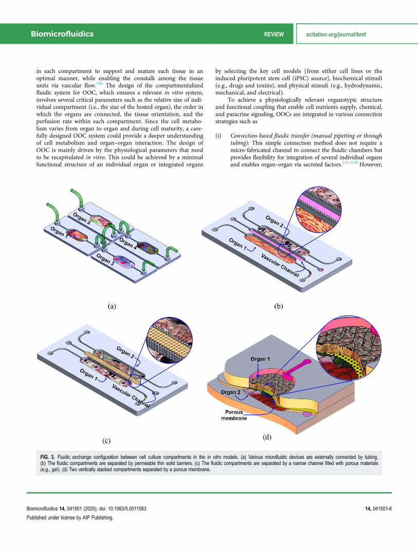
FIG. 3. Fluidic exchange configuration between cell culture compartments in the in vitro models. (a) Various microfluidic devices are externally connected by tubing.(b) The fluidic compartments are separated by permeable thin solid barriers. (c) The fluidic compartments are separated by a narrow channel filled with porous materials(e.g., gel). (d) Two vertically stacked compartments separated by a porous membrane.
Biomicrofluidics REVIEW scitation.org/journal/bmf
Biomicrofluidics 14, 041501 (2020); doi: 10.1063/5.0011583 14, 041501-6
Published under license by AIP Publishing.

it fails to recapitulate the physiological flow between organsand is limited to the use of organs that can be supported bythe same culture media [Fig. 3(a)].
(ii) Connection through porous barriers/gel with planar orienta-tion: In this configuration, cells are assembled/cultured in atwo-dimensional (2D) organization within a planar compart-mentalized microfluidic structure. The multi-compartmentsin such structures are physically separated by semi-porousvertical barriers [Fig. 3(b)]. So, cells of different types are cul-tured in close proximity to each other such that they arephysically isolated but fluidically and chemically con-nected.119,120 The semi-porous barriers are characterized byan array of small pores that allow only liquid but not cells tobe exchanged through the adjacent compartments. The sizeof these pores could be adjusted to specific requirement (e.g.,type/size of the cells) to control the mass transfer betweenthe different compartments. The porous channels can befilled with porous materials (e.g., hydrogel) to enable biomi-metic vascular structures between different organs [Fig. 3(c)].In sequence, this would enable chemical/biological interac-tions (paracrine signaling) between the cells in different neigh-boring compartments. The flow profile, rate, and direction ofthe inter-compartment flow can be controlled using eitherinternal or external pumps or valves. Taking advantage of theporous barriers structure, multi-cell type cultures can bearranged in the desired order where the interaction betweenthe cells/tissue within the various compartments is defined bythe direction of the perfusion flow. The perfusion micro-poreswith the combination of micro-flow in the adjacent compart-ment can be utilized to generate chemical gradients whendesired;121 hence, the composition and concentration ofchemicals within the side compartment can be selectivelyadjusted. The horizontal order of organs is a common struc-ture due to the ease of fabrication and suitability to study theinteraction of specific organ/cells with the immune system.
Table II lists examples of OOC modes that are demonstratedin planar order.
(iii) Connection through porous barriers with vertical orientation:In this configuration, two-layer, or more, fluidic compart-ments are vertically stacked and interfaced through a porousmembrane, which is sandwiched between them such that twoor more cell types can be co-cultured in close proximity toeach other in two vertical orientations. In this structure, aporous membrane with optimized thickness and pore size canbe used to physically separate the two cellular structures. Inaddition, the porous membrane also can be used as a sub-strate for culturing two types of cell on both sides. When epi-thelial/endothelial/epidermal cells are cultured on the uppersurface of the membrane and the corresponding parenchymaltissue on the lower side, this structure would mimic the struc-ture and function of important parts of the human body,which are biological barriers such as the small intestine, lungparenchyma, skin, and blood vessels that generally control theinteraction of the body with drugs, food, and the environ-ment. The lower compartment can host a model of a specificorgan such as the liver, adipose, muscle, bone, etc. This struc-ture can be used to recapitulate the transport (absorption anddistribution) of bioactive materials or drug through the epi-thelial and measuring the bioavailability of these substancesin the target organs and its metabolic profile. Furthermore,the lower compartment/channel can be used to host circulat-ing immune cells to model the activation of the immunesystem after the transport of a foreign substance through theepithelia. Table III lists examples of OOC modes that aredemonstrated in a vertical orientation.
D. Allometric scaling
Very little research has been carried out on the rational designof cell–cell or tissue–tissue interaction in in vitro models that
TABLE II. List of OOC examples that are demonstrated in a planar organization/order.
Organ model(s) Used cells Key studied parameterPhysiological parameters/
readout Reference
Liver, bone marrow,uterine cancer
HepG2/C3A, MEG-01,MES-SA, MESSA/DX-5
Drug mixtures for potential efficacyin treating multidrug resistant
cancers
Inhibiting MES-SA/DX-5cell proliferation
122
Intestine, liver, cancer,and connective tissue
HCT-116, HepaRG, Caco-2,TIG-121 fibroblasts
Pneumatic pressure driven mediumcirculation platform with amicroplate-sized format
Evaluation of the effects ofprodrugs on multiple organ
models
116
Breast cancer, bone,muscle, andmicrovascular network
hBM-MSCs, HUVECs,osteoblast-differentiated cells
(OBs), C2C12
Bone- and muscle-mimickingmicroenvironments through a
microvascular networkconcentrically wrapped with mural
cells
Cancer cell extravasation;extravasation rates and
microvasculaturepermeabilities
123
Adipose-immune cells Human primary adipocytesand U937
Simultaneous multiplexedmeasurements of pro-inflammatory
cytokinesThe immune-metabolic correlation
Pro-inflammatory cytokinesand glucose uptake
120
Biomicrofluidics REVIEW scitation.org/journal/bmf
Biomicrofluidics 14, 041501 (2020); doi: 10.1063/5.0011583 14, 041501-7
Published under license by AIP Publishing.

faithfully reflect that in vivo. A good OOC design needs to be takeninto account for the relationship between cells, cell metabolism,and exchange in the human body using allometric rules in order toestablish appropriate cell ratios in the system in a rationalmanner.127 Current design approaches are based on relative sizesand not functions.128 Two different allometric scaling models (i.e.,the cell number scaling model and the metabolic scaling model)need to be considered127 to enable the predictive potential. Forexample, Ucciferri et al.127 proposed connecting hepatocytes withendothelial cells before adding other cells or to construct an invitro model of biotransformation and distribution in liver.Therefore, the hepatocytes in the model are scaled with reference tothe basal metabolism, whereas the endothelium is scaled using thesurface area of the human vascular system. As cells are usuallyplated in monolayers, the allometric design process begins by con-sidering the metabolism of a two-dimensional culture of humanhepatocytes in a single module. By integrating in vivo and in vitrodata allometric scaling would enable more accurate prediction ofhuman pharmacokinetics/pharmacodynamics.
E. Stable performance over extended periods of time
The OOC system is a miniaturized bioreactor that hosts thehybrid cellular structure for extended periods of time for studyingthe time-dependent cellular behavior that depends on the type ofcells/tissue and the downstream analysis. To achieve reliablein vitro results, it is important to maintain stable cell viability andcontrol the phenotypic stability in their artificial milieu during therelatively long-life span of the OOC model. The life cycle ofthe OOC system can be divided into three stages: (i) creating themodel structure, (ii) model’s functional characterization, and (iii)model validation and testing (Fig. 4). These processes and function-alities should drive the engineering design of the OOC systemwhen transferring the prototype to product development. Such amethodology is not common in the bioengineering product withinthe LOC domain, but, despite the lack of standards, the industrial
design applied to mechanical and electrical products can beadopted in the OOC development.129 In general, regardless of thetype of the in vitro model under study, the longevity of use ofthe model is crucial to enable reliable and consistent results andat the same time to allow repeatable and consistent experiments onthe same set of cells/tissues. The following summarizes the requiredprocess control during the stages of the OOC model:
1. Model structure (micro-anatomy)
During the stage of building the structure of the in vitromodel, it is important to control the environment and monitor thecell growth and phenotyping. Depending on the in vitro modelunder study, the controlled components in the system need to bealigned to the intended structure and functional parameters. Ingeneral, the following parameters are important in most of theOOC in vitro models:
(1) The microfluidic dynamic properties: The dynamic of the perfu-sion flow is a crucial parameter in the dynamic cell culture.Therefore, it is important to take the fluid dynamic within theOOC system into consideration during the design of the chip.These include the geometry and dimensions of the channels/chambers and the time-dependent pressure gradient, whichneed to be optimized based on the required cell–liquid/nutri-ents interaction and the human physiological data. In addition,the flow/pump mode needs to be initially defined to configurethe type of flow such as steady or pulsatile flow. In the secondcase, the driving frequency should be set within a physiologi-cally relevant range (i.e., in the range of then human heartbeatof 60–180 beats per minute or a frequency of 1–3 Hz). Thefluid flow within the microfluidic channel imposes mechanicalshear stresses and results in strain on cells and tissues, whichare integral parts of the cellular microenvironment that modu-lates the proliferation, differentiation, phenotype, and migra-tion. These actions in sequence affect the function ofmulticellular systems in organ-level health and disease.130
TABLE III. List of OOC examples that are demonstrated in the vertical organization/order.
Organ model(s) Used cells Key studied parameterPhysiological
parameters/readout Reference
Gut-immune cells Caco-2 and U937 Caco-2 as a protection layer TEER andinflammatorycytokines
37
Skin-immune cells HaCaT and U937 HaCaT as a protection layer TEER andinflammatorycytokines
46
Gut-liver andskin-liver
Human primary cryopreserved hepatocytes andKupffer cells, Caco2 and HT29 cells
Integration barrier tissue withparenchymal forpharmacokinetics
Permeability 107 and 124
Blood–brain barrier Human microvascular endothelial (BMVECs)from the iPS cell line IMR90-4 cells and
primary human astrocytes
Differentiation under hypoxicconditions
Permeability anddrug transport
125
Lung airway Primary airway epithelial hAECs and humanlung microvascular endothelial cells
Lung inflammatory Cytokine secretion 126
Biomicrofluidics REVIEW scitation.org/journal/bmf
Biomicrofluidics 14, 041501 (2020); doi: 10.1063/5.0011583 14, 041501-8
Published under license by AIP Publishing.

FIG. 4. General presentation of the integrated OOC tools, device, process, and the required functions through the development of the in vitro model stages (modelbuilding, functional characterization, and testing).
Biomicrofluidics REVIEW scitation.org/journal/bmf
Biomicrofluidics 14, 041501 (2020); doi: 10.1063/5.0011583 14, 041501-9
Published under license by AIP Publishing.

(2) The physicochemical parameters (temperature, pH, oxygen,CO2): The optimum micro-bioreactor could supply cells withdynamic profiles of nutrients, oxygen, and growth factors and,at the same time, control the physical and chemical environ-ments of the cell culture with in-line monitoring modality.Maintaining ideal culture temperatures is vital for optimal cellgrowth, where mammalian cells thrive at around 37 °C. ThepH value is also critical to cellular function. The cell culturepH may deviate from the optimum ranges due to the build-upof acidic metabolites by cultures that have grown too dense orgrown too long in that medium and the low oxygen levels andcontamination by fast-growing bacteria or fungi. The vastmajority of cell culture media contain carbonate-based buffersthat work with elevated CO2 levels in the incubator to stabilizethe cell culture pH131 as they are also present in vivo. In arecent study, Zhang et al.111 demonstrated an integrated pHand oxygen sensor within a compact micro-bioreactor. The pHsensor detects the changes in the light absorption of phenolred in the culture medium to translate into a voltage change,while the oxygen-quenchable luminescent dye [Ru(dpp)3]2+ Cl2 – tris(4,7-diphenyl-1,10-phenanthroline)ruthenium(II)chloride was used for the oxygen sensor. A comprehensivereview of various instrumented in vitro microphysiologicalsystems has been presented by Soucy et al.132 Recently,Kirkstall133 developed the Quasi Vivo® System, which consistsof interconnected cell culture chambers and a peristaltic pumpto create a continuous flow of media over cells. The QuasiVivo® system is available with three different culture chambers(QV500, QV600, and QV900), which enable monitoring ofvariables during the cell culture experiment. The system alsofits with the standard cell culture inserts (Transwell).
(3) Mass transfer at the tissue–fluid interface: To mimic in vivocross-organ interactions, reliable fluidic connections betweenorgan modules (fluidic chambers) are essential. The architec-ture of the fluidic networks in multi-organ systems can have alarge impact on tissue/organ crosstalk. The perfusion circuit inrecirculating microfluidic systems allows mimicking blood cir-culation and facilitates communications among organs. One ofthe key challenges in OOC systems, particularly when involv-ing 3D cell culture or a multi-organ model, is to maintainacceptable cell viability during the course of cell culture.Therefore, nutrients, oxygen, growth factors, and other regula-tory molecules have to be efficiently transported from the bulkof culture media and through the multi-tissue structure (inter-nal mass transfer).134 The organization of the cellular systemand the order or orientation of the tissue in the multi-organsystem have a significant impact on the inter-tissue mass trans-fer, which is generally dependent on the combination of diffu-sion and convection mechanisms, the porosity and size of themicrofluidic system, and the diffusion rate through the bioma-terial.134 Taking into account the mass transfer parameters isan essential consideration to achieve long-term viability of themodel. For example, the fluidic barrier between various celltype chambers needs to be designed and numerically simulatedprior to designing the compartmentalized microfluidic system.The most important parameters are the porosity, the thickness,and the material (e.g., hydrogel) of the structure. Improving
the microfluidic design will result in efficient mass transfer. Inparticular, it is critical to maintain a balance between oxygendelivery to cells and their oxygen consumption. Oxygen trans-fer is also a key parameter in the cell culture setup that requiresmonitoring due to the poor solubility of oxygen in the culturemedium135 and taking into consideration the diffusion distanceof oxygen in tissue of 100–200 μm.136
2. Functional characterization (micro-physiology)
In the downstream stages, to gain confidence in the structuredin vitro model, it is necessary to evaluate it by demonstrating the rele-vant in vivo physiology and assess the analogy between the in vitromodel and its in vivo counterpart. The type of functional componentsdepends on the organ(s) under study. The following are some ofmajor characteristic parameters that are commonly studied:
a. Differentiation. One of the key challenges in the OOC tech-nology is to obtain functional, human, organ-specific parenchymalcells that can produce specific human physiological characteristics.Various human-based cell types are currently commercially avail-able, which assisted to push the boundaries of this technology. Inaddition, induced pluripotent stem cells (iPSCs) have also beenused to create specialized organ-specific tissues. For example, thestructure of the in vitro model representing the small intestine forpharmacokinetics screening/drug absorption should include themajor cell types: enterocytes, Paneth cells, and goblet cells with arelevant in vivo ratio. However, since it is difficult to include all thenecessary features in a single chip, the complexity of the modelvaries depending on the specific desired function. Monitoring ofcell differentiation within the OOC system is essential to ensure themodel relevancy during the course of culture. For example, a rapidloss of “liver” phenotype during culture was observed, which makesthe culture system not amenable to repeat-dose studies.137 In fact,OOC technology could be also used to enhance stem cell differenti-ation in vitro and to study patient-specific developmental responsesfor personalized medicine.138
b. Nutrient and metabolite profile in cell culture (e.g., glucoseand lactate). Assessing the metabolic activity of a cell culture pro-vides a detailed understanding of the physiological state of cellculture. In general, in large-scale bioreactors, glucose and lactateconcentrations are usually measured using colorimetric assays andbulky electrochemical detection systems, which require manual andtedious sample preparation that is not suitable for monitoring overextended periods of time. Prill et al.139 demonstrated an automateddetection of drug-induced changes in cellular viability by continu-ous monitoring of glucose consumption and lactate secretion of ahepatic tumor cell line. Using microfluidically addressed electro-chemical sensors, the system probes the mitochondrial apoptoticpathway by exposing the cells to the complex I inhibitor rotenone,which reduces OXPHOS that in turn upregulates other catabolicmechanisms, like anaerobic glycolysis. Bavli et al.140 reported real-time monitoring of mitochondrial respiration using two-frequencyphase modulation of tissue-embedded phosphorescent microprobesin a liver-on-chip device. The computer-controlled microfluidicswitchboard allowed contiguous electrochemical measurements of
Biomicrofluidics REVIEW scitation.org/journal/bmf
Biomicrofluidics 14, 041501 (2020); doi: 10.1063/5.0011583 14, 041501-10
Published under license by AIP Publishing.

glucose and lactate, providing real-time analysis of minute shiftsfrom oxidative phosphorylation to anaerobic glycolysis, an earlyindication of mitochondrial stress.
c. Permeability of epithelial tissue barriers. A key characteristicof epithelial tissue structures, such as gastrointestinal (GI) tract epi-thelium, blood vessel endothelium, and skin epidermis is the tightarrangement of cells that strongly express intercellular tight junc-tions (TJs), which play an important role in modulating the trans-port of nutrients/drugs from the apical side to the basolateral bloodstream. This barrier is not static but can be modulated by specificstimuli. For example, the increased permeability of intestinal epi-thelium has been linked to the pathogenesis of inflammatory boweldisease, ulcerative colitis, Crohn’s disease, and food allergies. Onthe other hand, a transient increase in paracellular transport couldimprove the bioavailability of desirable bioactive compounds,which normally are poorly absorbed.141
One of the common methods of monitoring epithelial integ-rity is based on the measurement of transepithelial electrical resis-tance (TEER) of barrier forming cells grown on porous membranesusing two sets of electrodes that are connected to a volt-ohmmeter.This non-invasive method can be applied to living cells withoutmarkers and allows them to be monitored during growth and dif-ferentiation. TEER reflects the resistance to the passage of ionsthrough the physiological epithelial barrier and is recognized asone of the most accurate and sensitive measures of epithelial integ-rity and permeability. TEER can also be used to measure the effectof different stimuli on the barrier permeability and any biologicalresponse of the underlying cells within the lower chamber. Thesequantitative data provide an effective tool to predict the influenceof tissue exposure to chemical, biological, electrical, thermal, andmechanical stimuli related to organ inflammation (e.g., skin andgut), toxicity, and immune modulation. The electrical behavior of acell/cell layer is frequency dependent. The cell membrane is apoorly conducting lipid bilayer, and at low frequencies, the cellbehaves as an insulator. As the frequency increases, the electricfield lines penetrate the cell so that the cytoplasmic resistance ofthe cell can be probed.142 Therefore, frequency-dependent imped-ance measurements would provide a wealth of information notonly about the cell layer integrity but also about the molecularpathway of transported bio/chemical substances through the epithe-lial monolayer because impedance measurements allow for the sep-aration of paracellular resistance (governed by TJ properties) fromtranscellular resistance (determined by conductive structures resid-ing in the cell membranes).
d. Response to mechanical stimulation. Among the earlierOOC models is the lung-on-a-chip model,17 a biomimetic micro-system that recapitulates the microarchitecture and dynamic micro-environment of the alveolar–capillary unit of the human lung. Thealveolar epithelial cells were exposed to air to induce differentiationand the formation of the alveolar–epithelium barrier tissue. Torecapitulate the breathing action of lung, cyclic vacuum suction wasapplied to two side hollow chambers adjacent to the cell culturechannels to induce cyclic mechanical stretching of the adherent celllayers. The mechanical strain also showed to enhance the uptake ofthe epithelial layer of nanoparticles and stimulates their transport
into the underlying microvascular channel. Besides the lung, anumber of tissues in the human body are exposed to mechanicalstrain, such as the heart, bone, skin, gut, and urothelium.143 Recentresearch on the role of mechanical forces in wound healing andrepair opens the possibility of targeting mechano-transductionpathways to reduce scar formation.144 Epidermal keratinocytes anddermal fibroblasts can sense the mechanical force, which, togetherwith soluble and immobilized signal cues, are responsible for theobserved wound healing response.143 The ability to reproduce invivo-like strain on tissue culture in vitro would enable the investiga-tion of the mechanical–biochemical signal interaction and targetspecific pathways associated with mechanical strain.145
e. Migration. Motility is an essential feature of live cells duringall the stages of cell life. Monitoring cell migration is important forunderstanding a variety of physiological and pathological processesin the tissue in vitro models such as organogenesis, cancer metasta-sis, and inflammation. Data obtained from these tests may allowfor an understanding of how a particular cell type migrates orresponds to a chemical stimulation and migrates toward it. Themost common cell migration assays are wound healing and thetranswell migration and invasion assays.146,147 Most of microfluidic-based cell migration studies have focused on single cell types ratherthan heterogeneous cellular structure, which dramatically simplifiesthe in vivo conditions. More sophisticated tools may be required tostudy the tissue-level cell migration for clinical applications. As anexample of OOC-based cell migration study is the cancer metastasis.During metastasis, cancer cells migrate from their initial location todistant organs that lead to the formation of new tumor, whichinvolves cell–cell interaction and chemotaxis.148,149 Zervantonakiset al.72 developed a microfluidic-based assay to recreate the tumor–vascular interface to test the effect of biochemical factors from theinteracting cells on carcinoma cell intravasation. The study showedthat signaling with macrophages via secretion of TNFα results inendothelial barrier impairment and the intravasation rates increaseas validated with live imaging. It was also found that endothelialbarrier impairment is associated with a higher number and fasterdynamics of tumor cell–endothelial cell interactions. In anotherstudy, Lei et al.150 developed a compartmentalized microfluidic chipto examine the interaction between neurons and cancer cells. It wasobserved that the nerve bundles to guide a directional migration ofcancer cells. The on-chip model allows the screening of compoundsthat inhibit cancer cell migration along neurites in vitro.
f. Immune competence: Response to toxic or pathogenicsubstances. The communication between parenchymal cells andimmune cells is a key characteristic in any biologically active invitro model, which takes place via either soluble factors or directcell–cell interaction at the crossroad of drug/nutrient metabolismand immune modulation. Inflammation is an immunologicalmechanism that assists in the removal of infectious and otherforeign substances, which consists of a cascade of immunological,physiological, and behavioral processes, and is orchestrated bysoluble immune signaling molecules, called cytokines.151,152 Thisalso leads to a variety of cellular responses in tissues including ele-vated permeability in microvessels, attachment of circulating cellsto the vessels in the vicinity of the injury site, migration of several
Biomicrofluidics REVIEW scitation.org/journal/bmf
Biomicrofluidics 14, 041501 (2020); doi: 10.1063/5.0011583 14, 041501-11
Published under license by AIP Publishing.

cell types, cell apoptosis, and growth of new tissue and bloodvessels.152,153 The response of various epithelial in vitro models toallergen exposure was studied using OOC systems such as the smallintestine,36–37 skin,46,154 and lung/airway.17,155
3. Testing, validation, and screening
The use of OOC devices for screening depends on successfuldevice validation to ensure that the biological functions reproducedin vitro are representative of the native tissues in vivo, which iscrucial to enable adopting this technology in industrial applica-tions. A direct comparison against the current industry standardsas well as between different technologies might be necessary for thetranslational OOC technology and to increase the confidence in theoutputs of OOC systems.156 However, to date, validating studies ofOOC systems are very limited and the vast majority of OOCstudies focus on the demonstration of the micro-anatomy of spe-cific tissue or organ and characterization of simple functions. Ewartet al.157 describe the main steps and key considerations that need tobe taken in order to enable OOC technology adoption into theindustrial laboratory. Written by scientists from 13 pharmaceuticalcompanies and partners at the National Institutes of Health (NIH),this article captures a consensus view on the progression strategy tofacilitate and accelerate the adoption of this valuable technology.
In general, there are three primary uses of OOC systems thatprovide initial value for industry:157
(1) Mechanistic investigation: The OOC system will enable rapidassessment of new drug mechanisms in pharmacology or toxic-ity. The technical standards for use in mechanistic investigationwould be defined by the biology or pathobiology of the site ofaction to be investigated.157
(2) Preclinical safety screening: The key functionality of in vitromodels is to provide in vitro datasets that can accuratelypredict the adverse effects in vivo. Such compounds with highrisk profile to be deprioritized, while those with a lower riskprofile are brought forward.158
(3) Drug absorption, distribution, metabolism, and excretion(ADME): ADME assays are critical in gaining insights into themetabolism and potential drug interactions. Robust in vivo pre-dictions can be achieved with well-characterized in vitromodels with the combination of computational methods(physiology-based pharmacokinetics/toxicokinetics).
IV. BIOLOGICAL INTEGRATION (CELL/TISSUEHETEROGENEITY)
The function of the organ(s) is orchestrated by complex cellu-lar structures and physiochemical factors within the ECM andsecreted by various types of cells. Each organ in the human bodycontains a diverse type of cells that are necessary to perform itsfunction.159–164 Knowing the cellular structure (microanatomy) ofthe target organ is essential to determine the accurate biologicalmodel of the OOC system. Various OOC systems with multiple celltypes have been or are being constructed, but none have yet tocapture the full diversity of cell types within an organ that is highlycomplex, therefore, posing a tremendous challenge to mimic. It is
unrealistic to expect an OOC system that recapitulates the full cel-lular structure of a healthy/diseased organ in the near future.Nonetheless, it is still important to study cellular heterogeneity oftarget tissue/organs and identify the best subset of cells thatprovide a sufficient physiological representation that models thestructure of that organ. By creating the minimal structure and ana-lyzing its function, the response of the organ(s) model can beextrapolated, which enables capturing the specific functions ofinterest. For example, integration of the GI tract, liver, and adiposerepresent organ-level heterogeneity of the “the gut–liver–adiposeaxis” as illustrated in Fig. 5. The GI tract is a complex environmentwhere human epithelial cells, commensal bacteria, and opportunis-tic pathogens constantly interact with each other and are faced witha continuous flow of nutrients and metabolites. The microarchitec-ture of the GI tract barrier (epithelium) includes a single layered orpseudostratified columnar epithelium enterocytes, Paneth cells,mucus-secreting goblet cells, hormone-secreting enteroendocrinecells, and antigen-sampling microfold (M) cells165 (Fig. 5). On theother hand, the microarchitecture of the liver forms a 3D structurethat consists of epithelial and mesenchymal cells arranged in repeti-tive microscopic units. The major cell types that constitute theminimal structure of the liver are hepatocytes, hepatic stellate cells,fibroblasts, and Kupffer cells, which also include vessels and biliaryduct parenchymal distributions.166 An engineered system caninclude the GI tract module in close vicinity to a liver module toemulate the drugs/nutrients metabolization by the liver immedi-ately after GI tract absorption before reaching the systemic circula-tion. Furthermore, to study the obesity-associated metabolicpathophysiology, adipose tissue can be integrated into the system,which further increases the complexity of the model. This complexinterconnected model would enable studying the absorption,metabolism, and mechanistic investigation of drug compounds in asystemic manner (Fig. 5). A recent review by Mertz et al.167
addresses the advances in characterization of cell heterogeneity andcell plasticity and how it impacts tissue and organ function.
V. ENGINEERING SYSTEM INTEGRATION
Some of the obvious advantages of OOC compared to conven-tional multi-well plates are the dynamic interactions betweenseveral organs that enable reproducing specific functions in vitroand the ability to perform time resolved measurements at differentcheck points. This would require in situ, real-time, non-destructivedetection assays, rather than end point assays. OOCs are highlyintegrated and complex systems by nature as they reflect humanphysiology. To construct a sufficiently physiologically relevant invitro model, the minimal number of cell subsets of cells must beincluded in the co-culture system. Integration of additional celltype/tissue/organ into the system adds another level of unavoidablecomplexity. In consequence, the complexity of the biological modelwould require engineering a complex device. Different cell typesmay require different culture medium, nutrients, and growthfactors and may not be compatible to co-culture before differentia-tion/maturation. In addition, cell organization/order and interfac-ing vary in different tissue and organs as discussed earlier. Suchcomplexity puts the OOC technology at risk of delay of marketpenetration and acceptance by the targeted end users. Furthermore,
Biomicrofluidics REVIEW scitation.org/journal/bmf
Biomicrofluidics 14, 041501 (2020); doi: 10.1063/5.0011583 14, 041501-12
Published under license by AIP Publishing.

new medical technologies require competent health professionalswho are well-trained to adopt such advanced technology. In conse-quence, this would pose a high assay cost that is prone to a highrate of use-related errors, which raises an important question “Canthis complex technology be commercialized?”
OOC technology is progressing through two parallel develop-ment tasks: cell models (biological model) and device engineering;however, the recent significant development in cell biology (e.g.,iPSCs) does not accompany the same speed of OOC device engi-neering. To accelerate this translational technology, reducing thegap between the academic research and industry and, therefore,
enabling smoother adaptation by the end user, many challengesmust yet be overcome to meet the requirements listed above torealize market-driven products. The ability to perform a morpho-logical non-invasive in-line assessment is advantageous for assess-ing the structural integrity and drug- or disease-induced structuralchanges. Sensors and imaging modalities integration and enablingreal-time sampling of the cellular products and multi-channelmonitoring would also be attractive features.
Engineering OOC devices may be advantageous to the well-established semiconductor industry, as it is initially the case of thefabrication of the single device, such as integrated circuits (ICs),
FIG. 5. An example of biological model integration: inter-connected GI tract, liver, and adipose represent organ-level heterogeneity of the “the gut–liver–adipose axis.”
FIG. 6. Depicted integrated OOC system with disposableorgan chips (OOCs) and multi-function operating system(OS).
Biomicrofluidics REVIEW scitation.org/journal/bmf
Biomicrofluidics 14, 041501 (2020); doi: 10.1063/5.0011583 14, 041501-13
Published under license by AIP Publishing.

printed circuit boards (PCBs), and field-programmable gate arrays(FPGAs). OOC chips are single-use devices that need not be expen-sive to be adopted by the end user; therefore, one approach is tominimize the complexity of the fluidic chip that hosts the organo-typic culture while maximizing the functionality of the operatingunit. Figure 6 depicts an integrated OOC system with disposableorgan chips (OCs) and a multi-function operating system (OS).
VI. OOC IN THE INDUSTRY
OOC technology is still in its beginning stage and has consid-erable interest among researchers in academia with increasingnumber of publications that cover various aspects of physiology.However, as we emphasized earlier, tremendous effort is needed totackle the technological challenges to enable transferring this tech-nology from the laboratory to the industry. A rough estimation ofthe highest technology readiness level (TRL), which describes thematurity of a technology, among current industry players can bepositioned at TRL4, which refers to technology validation in thelaboratory environment,168 while the TRL for technology that istested in the application environment is TRL9. OOC technologyhas gained a lot of momentum toward commercialization fuelledby support from governmental funders as well as the industry, inparticular, the pharmaceutical industry. Two major initiativesactively working in this domain are listed in Table IV.
In 2012, the National Institutes of Health (NIH), led by theNational Center for Advancing Translational Sciences (NCATS),launched the Tissue Chip program. The program is a joint collabo-ration with the U.S. Food and Drug Administration (FDA) and theDefense Advanced Research Projects Agency (DARPA), whichawarded 12 projects that supported the development OOC systemsthat represent human organs and 7 projects that explored the useof stem and progenitor cells to differentiate into multiple cell typesthat represent the cellular architecture within organ systems. In2014, NIH funded the next phase of the tissue chip program toimprove ways of predicting drug safety and effectiveness. The nextphase aimed to link individual organs on chips to develop a humanmulti-organ model system to replicate the complex human responseto drug exposure. Ultimately, linking a major human organ modelin one system to form a human-body-on-a-chip model intends toaccelerate the translation of these basic discoveries into the clinicand industry. In 2016, NCATS with a partnership with theCenter for the Advancement of Science in Space (CASIS),launched the Tissue Chips in Space initiative, which aims to helprefine the Tissue Chip platforms for in-flight experiments at theInternational Space Station U.S. National Laboratory (ISSNational Lab), so that scientists can better understand diseasesand translate their findings to affect human health on Earth. In2017, NCATS announced 13 additional awards to develop 3DTissue Chip research platforms that model diseases and test drugefficacy before clinical trials. In 2018, NCATS announced theTissue Chip Consortium awardees to develop tissue chip modelsfor pain, opioid addiction, and type 2 diabetes. In order toprovide a way for independent testing and validation of platformsdeveloped by Tissue Chip for drug screening, NCATS awardedthree testing centers to assess the chips’ robustness and provideinputs for further improvement of the devices.
Over the past few years, several companies and start-ups havelunched OOC-based devices or tools for tissue/organ modeling(Table V). Among these products, MIMETAS OrganoPlate® hasshown many uses in the literature. The MIMETAS OrganoPlate®features 96 (with two-lane) or 40 (with three-lane) independentculture cells, each supporting one in-gel culture and one/two perfu-sion channels [Fig. 7(a)]. The system employs the Phaseguides™technology as meniscus pinning barriers that enable precise,barrier-free definition of culture matrices and cells in three dimen-sions.169 The pump-less media flow is achieved by using a gravity-driven leveling technique. TissUse GmbH (Berlin, Germany)introduced a variety of HUMIMIC Chips, such as HUMIMICChip2, HUMIMIC Chip3, HUMIMIC Chip4, and HUMIMICChip XX/XY, for different in vitro modeling purposes [Fig. 7(b)].The organ models are connected by microfluidic channels, whichare covered with human dermal microvascular endothelial cells.178
A peristaltic on-chip pump enables an in vivo-like nutrient andoxygen supply to the cells and generates pulsatile shear stress in anadjustable range. In 2010, the lung-on-a-chip system was developedby Ingber’s group17 at the Wyss Institute, which mimics the func-tion of a lung alveolus, including the breathing mechanism.Emulate Inc. (Boston, USA) is currently commercializing a varietyof organ chips based on this platform [Fig. 7(c)]. The basic chipcomprises two microfluidic chambers separated by a semi-porousmembrane with two side vacuum channels. Applying a cyclicvacuum through the side channels causes the cell layer on themembrane to stretch, mimicking natural stretching during inhala-tion of the lung. Another interesting OOC chip is provided byAIMBiotech, which comprises a gel region with adjoining mediachannels separated from the gel channel by trapezoidal posts[Fig. 7(d)]. The chip allows 3D cell culture in the gel channel andimmune cell (e.g., T cell) circulation in the adjacent channels.
VII. INDUSTRY TRANSFER HURDLES
The ultimate goal of OOC technology is to emulate humanorgans and eventually the whole body in health and disease statesand to enable mimicking the interaction of drugs with body. Themajority of OOC studies focus mainly on the biological characteri-zation of the cellular components with limited effort toward deviceengineering. The main hurdle delaying the maturation of OOCdevice engineering is the fact that every OOC study uses differentsets of cells to investigate different biological/medical effects. Mostof these studies share a similar simple device structure that relies onvarious peripheral tools to provide the necessary cell culture envi-ronment or capture cell signaling. To develop a reliable single-organOOC system, a key cellular structure needs to be present. Also, aprecise cellular manipulation within the OOC system and a detailedunderstanding of the human body’s complex response to xenobiot-ics are necessary. This requires engineering a robust device thatenables a long-term cell manipulation and signal capturing withoutcompromising the cellular system viability. Increasing the biologicalcomplexity, i.e., employing more cell types or adding additionalorgan(s) to the model, would require more complex fluidic net-works, hence increasing the complexity of the OOC device.
The drug industry is well established, which involves complex,costly, and regulated process in a highly competitive environment.187
Biomicrofluidics REVIEW scitation.org/journal/bmf
Biomicrofluidics 14, 041501 (2020); doi: 10.1063/5.0011583 14, 041501-14
Published under license by AIP Publishing.

Therefore, adopting a new technology within this environment mayinvolve risking the drug production. On the other hand, OOC is arelatively young technology with most of its related research imple-mented as exploratory studies outside the mainstream pharmaceuti-cal development and is not mature enough to be adopted as a partof ongoing drug development programs. Despite the tremendouseffort focused on OOC device prototyping among many start-upsaround the world, penetrating of the OOC technology into the
pharmaceutical industry is still facing many scientific (as describedabove), manufacturing, and industrial challenges. OOC chips needto be reliable, affordable, scalable, and mass manufacturable.Current OOC devices require skilled scientific personnel to operateand rely on various peripheral equipment during the cell cultureprocess, such as incubators, pumps, automated dispensers, sensors,and actuators, as well as model characterization and readout, suchas optical imaging devices. It becomes crucial to enable the OOC
TABLE IV. The two major initiatives actively working toward the advancement of OOC technology.
Initiative (where)Funding agencies and
organizers Academia involved Industry partners Activities
Tissue Chip(USA) (https://ncats.nih.gov/tissuechip)
National Institute ofHealth (NIH), NationalCenter for advancingTranslational Science
(NCATS), Food and DrugAdministration (FDA),Defense Advanced
Research Projects Agency(DARPA)
Columbia University,Duke University,
Harvard University,Massachusetts Institute
of Technology,University of
Wisconsin–Madison,Northwestern University,University of California(Berkeley), University ofPittsburgh, University ofWashington (Seattle),Vanderbilt University,Washington University(St. Louis), CornellUniversity, Duke
University, University ofCalifornia (Irvine),Johns Hopkins
University, University ofFlorida, Stanford
University
GlaxoSmithKline (GSK),Pfizer, Inc. AstraZeneca,Children’s Hospital of
Philadelphia, Emulate, Inc.,Boston
1. Cell sourcing: Exploring thepotential of stem andprogenitor cells to
differentiate into multiplecell types that represent thecellular architecture withinorgan systems. These couldact as a source of cells topopulate tissue chips.
2. Organ models: Heart–liver–vascular systems, muscletissue, cardiopulmonarysystem, cancer metastasis,neural toxicity, female
reproductive tract, cardiactissue, liver sinusoid,
muscle-circulatory system,neurovascular system, lung,cartilage–bone, epithelialmucosa with sensory
Neurons and microbiome3. Tissue Chips in Space:
Experimenting human cellsand tissue in space
environment (e.g., effect ofmicrogravity)
ORCHID(Europe) (https://h2020-orchid.eu/)
European Union’sHorizon 2020 researchand innovation program
The Institute for humanOrgan and DiseaseModel technologies
(hDMT), theNetherlands
University of Twente,Eindhoven University,University of Delft,Leiden University,
Erasmus Medical Center,the Hubrecht Institute,Fraunhofer, IMEC, andUniversidad Zaragoza
Genmab and Galapagos,Leiden University medicalcenter, TNO, AmsterdamUMC, Hubrecht OrganoidTechnology (HUB), and
Philips
1. Organ-on-Chip technologyplatforms: stem cells, deviceplatform, standardized/artificial extracellularmatrix platform, high
throughput data analysisplatform
2. Organ models: Brain, cancer,eye, gut–liver, muscle,
blood vessel, cardiovascular,brain and gastrointestinaldisease interactions, and
human (intestinal)microbiome
Biomicrofluidics REVIEW scitation.org/journal/bmf
Biomicrofluidics 14, 041501 (2020); doi: 10.1063/5.0011583 14, 041501-15
Published under license by AIP Publishing.

TABLE V. Companies and start-ups actively working in the area of OOC.
Company(founding year),website Technology Product(s)
Structure andcharacteristics
Applications(examples) Reference
Hμrel Corp. (2006)www.hurelcorp.com
• Mainly based onmulti-well plates forcell co-culture.Microfluidic co-culturedevices also studiedbut not listed asproducts
8 species and 10 models,e.g., HμRELhuman™HμRELhumanPool™
HμRELrat™HμRELratWH™HμRELprimate™HμRELminipig™HμRELdog™
HμRELmouse™HμRELrabbit™HμRELcat™
• Multi-well cultureplates Human/animal co-culturemodels
• Multi-readout,repeat-dosehepatotoxicityscreening
Studying the cellularresponse, metaboliccompetency, andpharmacokinetic
interactions amongmultiple tissues and
organsViral chronicity inliver disease models
169 and170
Mimetas (2013),www.mimetas.com
• Phaseguides™ enableprecise, barrier-freedefinition of culturematrices and cells in3D, supporting cell–cell interactions
OrganoPlate®PhaseGuide™
OrganoPlate® 2-lane,OrganoPlate® 3-lane,OrganoPlate® Graft,OrganoPlate® Caco-2,
OrganoTEER®,OrganoFlow® L
• 96-well basedmicrofluidic 3D cellculture plate
• Pump-lessperfusionmicrofluidic liquidhandling platform
Pancreatic cancer,blood vessel, kidneyproximal tube, CNStoxicology, blood–brain barrier, gut,angiogenesis, liver,
breast cancer
171–177
TissUse (2010)www.tissuse.com
• Multi-Organ-Chipplatform
• On-chip micro-pump
HUMIMIC ChipHUMIMIC StarterHUMIMIC AutoLabHUMIMIC AutoPlant
Organ compartmentsof varying sizeOn-chip pump
Combination ofmulti-organs, e.g.,
intestine, liver, kidney,and neuronal tissue
Human skin
178–180
Emulate (2013)www.emulatebio.com
Human EmulationSystem for multi-organ
cultureChip-lab equipment
interfacing
Organ-ChipsPod™ Portable ModuleZoë™ Culture ModuleOrb™ Hub Module
Bio-kit for Kidney, liverand intestine
Stretchable plasticchip with porous
membraneAllow organ chips tobe transported andplaced on standardmicroscopes for
imagingUp to 12 Organ Chip
combinations
lung, liver, intestine,and kidney
181–184
AIM Biotech (2012)www.aimbiotech.com
• Microfluidic device for3D cell culture
• Hydrogel injectable• Gas permeable• Multicellular co-culture
Microscope slide formatchips
Compatible with allpolymerizable gels
Enables the control ofinterstitial flow acrossthe 3D gel region
Rapid media exchangethrough vacuum
aspiration
Immunotherapy,neurobiology, andvascular biology
185–186
4DBio (2014) www.4designbiosciences.com
• VMO platform:vascularized chambernetwork
Vascularized 96-wellmicrofluidic plate
Channels are linedwith endothelial cellsand act as arteriole
and venule, connectedby a network of livingcapillaries within a
physiologicextracellular matrix
Tumor, metastasis, andvascular pathology
Biomicrofluidics REVIEW scitation.org/journal/bmf
Biomicrofluidics 14, 041501 (2020); doi: 10.1063/5.0011583 14, 041501-16
Published under license by AIP Publishing.

devices beyond the research laboratory bench by either developingrobust instrumentations particularly for OOCs or enabling theinterface of OOC devices with the classical analytical tools.
Manufacturing of OOC chips for industrial use is a majorhurdle that the OOC technology faces as the adopted fabricationmethodologies are originated and extended from academic labs.OOC fabrication requires a multidisciplinary approach of engi-neers, fabrication expertise, material scientists, and biologists. Inthis respect, it is important to apply mature manufacturing exper-tise to accelerate device engineering and prototyping processes.Industry aligned funds, with strong collaboration with the pharmaindustry, need to be directed toward solving the major challenges
facing the industry transfer including device engineering, fabrica-tion material, cell–material interface, biological complexity, andsensors integration, among others, and enabling chips prototyp-ing beyond the academic labs. Figure 8 depicts a possible roadmap of OOC production with some of the associated challenges.One possible approach is to develop a modular universal OOCplatform that enables hosting a variety of organ-on-a-chip orbody-on-a-chip models. Early communication with stakeholders(researcher, regulators, suppliers, and end users) would helpidentify the industry needs in early stages of development.Depending on the nature of the assay, OOC systems (products)can be designed to provide a single or multi-organ in vitro
TABLE V. (Continued.)
Company(founding year),website Technology Product(s)
Structure andcharacteristics
Applications(examples) Reference
AxoSim (2014)www.axosim.com
• Nerve-on-a-Chip® NERVESIM™BrainSim™
iPSC derivedspheroids in a 3D
culture environment
Neurotoxicityneurodegenerative
diseases
CNBio (2009)www.cn-bio.com
• 3D cell culture• Liver-on-a-chip• Barrier models
PhysioMimix:Multi-organ interactions,ADME, and toxicity
Benchtop fluidiccontrol
Up to six MPS platescan be run for
multiple independentexperiments
Toxicology, drugmetabolism, anddisease modeling
AlveoliX AG(2015), www.alveolix.com
Lung-on-chip model AXLung-on-Chip System Ultrathin membranebreathing motion
Toxicity
Hesperos Inc.(2015) www.hesperosinc.com
Multi-organ fluidic chip Organ models: heart–liver, heart–liver–skeletal
muscle–neuron,neuromuscular junction,
heart–liver–cancer
Serum-free cellmedium
Gravity flow system
Customizedhuman-on-a-chip
systems
Kirkstall (2006)www.kirkstall.com
Interconnected cellculture flow system
QV500QV600QV900
Produced frommedical-grade silicone
Compatible withcommercially availabletranswells and inserts
Liver, brain, cardio,respiratory, kidney,
and gut
Nortisbio (2015)https://www.nortisbio.com
• Perfusion systemplatform
• Perfusion through bothtissue lumen
ParVivo™ perfusionsystem
Plug and play Chipswith pre-established
tissues
Toxicity and efficacytesting
Synvivohttps://www.synvivobio.com/
• 3D tissue/organ-on-chip models
SynBBB (blood–brainbarrier model)
SynTumor (variouscancer models)
SynRAM (inflammationmodel for rolling
adhesion and migrationassays)
SynTox (Toxicology)
Air liquid interface(ALI)
In vivo-like airwaystructureReal-time
visualization
Toxicity assaysBiomarker analysis
Therapeutic screening
Biomicrofluidics REVIEW scitation.org/journal/bmf
Biomicrofluidics 14, 041501 (2020); doi: 10.1063/5.0011583 14, 041501-17
Published under license by AIP Publishing.

model. Ultimately, body-on-a-chip can be realized by integratingall the key organs in one system.
Although there are only a few young companies developingOOC devices such as those listed in Table V, they are, in fact,showing great progress in providing exploratory platforms forfeasibility studies with exciting innovations at increasing speed.Additionally, many major pharmaceutical and consumer care
companies are involved, in collaboration with the companiesmentioned above, in feasibility studies and have started to buildtheir own OOC R&D investigational units. For example, EmulateInc. has started a collaboration program with Johnson &Johnson company (NJ, USA)188 for drug candidate screening.TissUse GmbH is also collaborating with Bayer AG (Leverkusen,Germany)189 and AstrZeneca190 to explore the potential use of
FIG. 7. (a) The microfluidic microtiter plate OrganoPlate from MIMETAS with three-line 40 integrated microfluidic chips. A gel channel (blue) holds the extracellular matrix(ECM) in place through the PhaseGuide’s pressure barrier function. Cells can be introduced in the middle lane (gel channel) to create a 3D cell culture while medium flowthrough the adjacent channels. Reproduced with permission from Kramer et al., J. Mol. Sci. 20, 4647 (2019), Copyright 2019 MDPI.171 (b) The multi-organ chip platform(Chip3) from TissUse GmbH. The chip comprises five assembled layers: glass slide, PDMS layer with microfluidic channels, adapter layer, cell culture insert layer, and res-ervoir layer. Reproduced with permission from Schimek et al., Sci. Rep. 10, 7865 (2020). Copyright 2020 Nature Research. (c) The organ chip from Emulate. The chip wasused to construct an in vitro model of the human lung alveolus. Epithelial cells are cultivated on top of a semi-porous membrane, and human pulmonary microvascularendothelial cells are cultivated on the bottom of the membrane. Applying a cyclic vacuum through the side chambers causes the cell layer to stretch, mimicking naturalstretching during inhalation. Reproduced with permission from Kasendra et al., Sci. Rep. 8, 2871 (2018). Copyright 2018 Nature Research.182–184 (d) AIM Bioterch cellculture chip comprises a gel region with adjoining media channels separated from the gel channel by trapezoidal posts. Reproduced with permission from Zervantonakiset al., Proc. Natl. Acad. Sci. U.S.A. 109(34), 13515–13520 (2012). Copyright 2012 the National Academy of Sciences of the United States of America.186
Biomicrofluidics REVIEW scitation.org/journal/bmf
Biomicrofluidics 14, 041501 (2020); doi: 10.1063/5.0011583 14, 041501-18
Published under license by AIP Publishing.
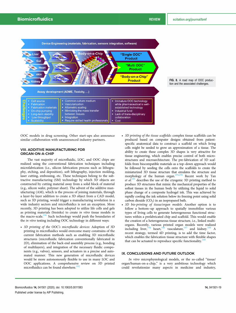
OOC models in drug screening. Other start-ups also announcesimilar collaboration with unannounced industry partners.
VIII. ADDITIVE MANUFACTURING FORORGAN-ON-A-CHIP
The vast majority of microfluidic, LOC, and OOC chips arerealized using the conventional fabrication techniques includingmicrofabrication (i.e., silicon fabrication process such as lithogra-phy, etching, and deposition), soft lithography, injection molding,laser cutting, embossing, etc. These techniques belong to the sub-tractive manufacturing (SM) technology by which 3D objects areconstructed by cutting material away from a solid block of material(e.g., silicon wafer, polymer sheet). The advent of the additive man-ufacturing (AM), which is the process of joining materials, througha layer-by-layer addition to create a 3D object from a CAD model,such as 3D printing, would trigger a manufacturing revolution in awide industry sectors and microfluidics is not an exception. Morerecently, 3D printing has been adopted to utilize life cells and gelsas printing materials (bioinks) to create in vitro tissue models inthe macro-scale.191 Such technology would push the boundaries ofthe in vitro testing including OOC technology in different ways:
• 3D printing of the OOC’s microfluidic devices: Adoption of 3Dprinting in microfluidics would overcome many constrains of thecurrent fabrication methods such as enabling 3D microfluidicstructures (microfluidic fabrication conventionally fabricated in2D), elimination of the back-end assembly process (e.g., bondingof multilayers), and integration of the necessary fluidic compo-nents (e.g., valves), sensors, and actuators in a precise and auto-mated manner. This new generation of microfluidic deviceswould be more autonomously flexible to use in many LOC andOOC applications. A comprehensive review on 3D printedmicrofluidics can be found elsewhere.192
• 3D printing of the tissue scaffolds: complex tissue scaffolds can beproduced based on computer designs obtained from patient-specific anatomical data to construct a scaffold on which livingcells might be seeded to grow an approximation of a tissue. Theability to create these complex 3D shapes is very attractive fortissue engineering, which enables precise control of both micro-structures and microarchitecture. The pre-fabrication of 3D scaf-folds from biocompatible materials as a top–down approach wouldbe followed by seeding the cells onto the scaffolds to create theminiaturized 3D tissue structure that emulates the structure andmorphology of the human organ.193,194 Recent work by Tanet al.195 describes the use of the cryogenic 3D printing method toproduce 3D structures that mimic the mechanical properties of thesoftest tissues in the human body by utilizing the liquid to solidphase change of a composite hydrogel ink. This was achieved byrapidly cooling the ink solution below its freezing point using solidcarbon dioxide (CO2) in an isopropanol bath.
• 3D bio-printing of tissue/organ models: Another option is tofollow a bottom–up approach to spatially immobilize varioustypes of living cells to generate heterogeneous functional struc-tures within a prefabricated chip and scaffold. This would enablethe creation of a heterogeneous tissue structure, i.e., linked multi-organs. Recently, various printed organ models were realizedincluding liver,194 heart,196 vasculature,197 and kidney.198 Arecent strategy, termed 4D printing, is to add the time factor,which enables the fabrication tissue structure with flexible shapesthat can be actuated to reproduce specific functionality.199
IX. CONCLUSIONS AND FUTURE OUTLOOK
In vitro microphysiological models, or the so-called “tissue/organ/human-on-a-chip,” is a very ambitious technology whichcould revolutionize many aspects in medicine and industry,
FIG. 8. A road map of OOC produc-tion and the associated challenges.
Biomicrofluidics REVIEW scitation.org/journal/bmf
Biomicrofluidics 14, 041501 (2020); doi: 10.1063/5.0011583 14, 041501-19
Published under license by AIP Publishing.

particularly for preclinical-to-clinical translation in the pharmaceu-tical arena. However, many technical challenges, as well as stand-ardization and regulatory endorsement, are still ahead and need tobe overcome before adopting this technology as an alternativeto animal models and transferring them from the laboratory toindustry. To realize physiologically relevant in vitro models, acomplex functional tissue structure “micro-anatomy” needs to beestablished, and this would require devising tools for cell andreagent manipulating, monitoring, sensing, and readout capturing.Many significant works were recently demonstrated for inter-connected multi-organ systems for drug screening, which show ahigh momentum toward the realization of the “human-on-a-chip”concept. However, individual organ models need to be establishedto increase their reliability and maximize the key physiologicalrobustness. Research in iPSCs is actively mobilized for transferringto many applications including OOC technology, and these cellsform an attractive option to be adopted for organ and disease mod-eling as well as personalized medicine. Trans-disciplinary researchinvolving stem cell biologists, engineers, physiologists, and pharma-ceutical industry partners is necessary to enable crossing the boun-dary between the lab and industry. Furthermore, the last few yearswitnessed the emergence of several partnerships between pharma-ceutical corporations, start-ups, and academia that demonstrate theimportance of this technology that carries a tremendous potentialin mitigating the current business needs in the drug discovery andpromise commercial success.
OOC is an engineering driven technology and is benefitedfrom well-established multi-engineering disciplines includingmicrofabrication, microfluidics, microscopy, sensors, etc. Recently,the convergence of 3D (bio) printing with microfluidics, LOC, andOOCs adds an extra momentum toward the enabling the technol-ogy and its commercialization. OOCs would represent a paradigmshift in physiology research as they would enable continuous moni-toring of living cells and rapid detection of mode of action, allow-ing an unprecedented advance in our understanding of living cellsand how they respond to stimuli and providing a wealth of infor-mation at low cost and in a short time. But OOC technology goesfar beyond the pharmaceutical industry and holds great promise todeepen our understanding of the complex interactions between thebody and exogenic stimuli and provides a more accurate assessmentof the unknown effects of drugs, food, chemicals, pathogens, andenvironmental toxins.
DATA AVAILABILITY
The data that support the findings of this study are availablewithin the article.
REFERENCES1S. H. Curry, Ann. N. Y. Acad. Sci. 993, 69–74 (2003).2A. Knight, J. Bailey, and J. Balcombe, Altern. Lab. Anim. 34, 19–27 (2006).3N. Shanks, R. Greek, and J. Greek, Philos. Ethics Humanit. Med. 4(2), (pub-lished online 2009).4J. Seok, H. S. Warren, A. G. Cuenca, M. N. Mindrinos, H. V. Baker, W. Xu,D. R. Richards, G. P. McDonald-Smith, H. Gao, L. Hennessy, C. C. Finnerty,C. M. López, S. Honari, E. E. Moore, J. P. Minei, J. Cuschieri, P. E. Bankey,J. L. Johnson, J. Sperry, A. B. Nathens, T. R. Billiar, M. A. West, M. G. Jeschke,
M. B. Klein, R. L. Gamelli, N. S. Gibran, B. H. Brownstein, C. Miller-Graziano,S. E. Calvano, P. H. Mason, J. P. Cobb, L. G. Rahme, S. F. Lowry, R. V. Maier,L. L. Moldawer, D. N. Herndon, R. W. Davis, W. Xiao, and R. G. Tompkins,Proc. Natl. Acad. Sci. U.S.A. 110, 3507–3512 (2013).5C. Gentile, Curr. Stem Cell. Res. Ther. 11, 652–665 (2016).6L. Geris, Y. Guyot, J. Schrooten, and I. Papantoniou, Interface Focus 6,20150105 (2016).7A. Astashkina, B. Mann, and D. W. Grainger, Pharmacol. Ther. 134, 82–106(2102).8N. Xia, C. K. Thodeti, T. P. Hunt, Q. B. Xu, M. Ho, G. M. Whitesides,R. Westervelt, and D. E. Ingber, FASEB J. 22, 1649–1659 (2008).9R. A. Desai, L. Gao, S. Raghavan, W. F. Liu, and C. S. Chen, J. Cell Sci. 122,905–911 (2009).10C. T. Ho, R. Z. Lin, R. J. Chen, C. K. Chin, S. E. Gong, H. Y. Chang,H. L. Peng, L. Hsu, T. R. Yew, S. F. Chang, and C. F. Liu, Lab Chip 13,3578–3587 (2013).11A. Khademhosseini, R. Langer, J. Borenstein, and J. P. Vacanti, Proc. Natl.Acad. Sci. U.S.A. 103, 2480–2487 (2006).12Y. Takayama, N. Kotake, T. Haga, T. Suzuki, and K. Mabuchi, J. Biosci.Bioeng. 114, 92–95 (2012).13W. F. Hynes, N. J. Doty, T. I. Zarembinski, M. P. Schwartz, M. W. Toepke,W. L. Murphy, S. K. Atzet, R. Clark, J. A. Melendez, and N. C. Cady, Biosensors4, 28–44 (2014).14H. Jiang, D. Jiang, P. Zhu, F. Pi, J. Ji, C. Sun, J. Sun, and X. Sun, Biosens.Bioelectron. 3, 126–133 (2016).15Z. B. Liu, Y. Zhang, J. J. Yu, A. F. T. Mak, Y. Li, and M. Yang, Sens. ActuatorsB Chem. 143, 776–783 (2010).16A. Sin, K. C. Chin, M. F. Jamil, Y. Kostov, G. Rao, and M. L. Shuler,Biotechnol. Prog. 20(1), 338–345 (2004).17D. Huh, B. D. Matthews, A. Mammoto, M. Montoya-Zavala, H. Y. Hsin, andD. E. Ingber, Science 328(5986), 1662–1668 (2010).18H. J. Kim, D. Huh, G. Hamilton, and D. E. Ingber, Lab Chip 12, 2165–2174(2012).19A. Carraro, W. M. Hsu, K. M. Kulig, W. S. Cheung, M. L. Miller,E. J. Weinberg, E. F. Swart, M. Kaazempur-Mofrad, J. T. Borenstein,J. P. Vacanti, and C. Neville, Biomed. Microdevices 10, 795–805 (2008).20P. J. Lee, P. J. Hung, and L. P. Lee, Biotechnol. Bioeng. 97, 1340–1346 (2007).21B. J. Kane, M. J. Zinner, M. L. Yarmush, and M. Toner, Anal. Chem. 78,4291–4298 (2006).22P. Chao, T. Maguire, E. Novik, K. C. Cheng, and M. L. Yarmush, Biochem.Pharmacol. 78, 625–632 (2009).23A. Legendre, R. Baudoin, G. Alberto, P. Paullier, M. Naudot, T. Bricks,J. Brocheton, S. Jacques, J. Cotton, and E. Leclerc, J. Pharm. Sci. 102, 3264–3276(2013).24E. Novik, T. J. Maguire, P. Chao, K. C. Cheng, and M. L. Yarmush, Biochem.Pharmacol. 79, 1036–1044 (2010).25B. Delalat, C. Cozzi, S. R. Ghaemi, G. Polito, F. H. Kriel, T. D. Michl,F. J. Harding, C. Priest, G. Barillaro, and N. H. Voelcker, Adv. Funct. Mater. 28,1801825 (2018).26J. B. Lee, J. S. Park, Y. M. Shin, D. H. Lee, J. K. Yoon, D. H. Kim, U. H. Ko,Y. T. Kim, S. H. Bae, and H. J. Sung, Adv. Funct. Mat. 29, 1900075 (2019).27D. Huh, H. Fujioka, Y. C. Tung, N. Futai, R. Paine, J. B. Grotberg, andS. Takayama, Proc. Natl. Acad. Sci. U.S.A. 104, 18886–18891 (2007).28D. Huh, D. C. Leslie, B. D. Matthews, J. P. Fraser, S. Jurek, G. A. Hamilton,K. S. Thorneloe, M. A. McAlexander, and D. E. Ingber, Sci. Transl. Med. 4,159ra147 (2012).29C. S. Fritsche, O. Simsch, E. J. Weinberg, B. Orrick, C. Stamm,M. R. Kaazempur-Mofrad, J. T. Borenstein, R. Hetzer, and J. P. Vacanti, Int.J. Artif. Organs 32, 701–710 (2009).30H. Tavana, P. Zamankhan, P. J. Christensen, J. B. Grotberg, and S. Takayama,Biomed. Microdevices 13, 731–742 (2011).31M. Felder, A. O. Stucki, J. D. Stucki, T. Geiser, and O. T. Guenat, Integr. Biol.6, 1132–1140 (2014).
Biomicrofluidics REVIEW scitation.org/journal/bmf
Biomicrofluidics 14, 041501 (2020); doi: 10.1063/5.0011583 14, 041501-20
Published under license by AIP Publishing.

32M. Felder, B. Trueeb, A. O. Stucki, S. Borcard, J. D. Stucki, B. Schnyder,T. Geiser, and O. T. Guenat, Front. Bioeng. Biotechnol. 22, 3 (2019).33A. O. Stucki, J. D. Stucki, S. R. R. Hall, M. Felder, Y. Mermoud, R. A. Schmid,T. Geiserce, and O. T. Guenat, Lab Chip. 15, 1302–1310 (2015).34W. Shin and H. J. Kim, Proc. Natl. Acad. Sci. U.S.A. 115, E10539–E10547(2018).35H. J. Kim and D. E. Ingber, Integr. Biol. 5, 1130–1140 (2013).36Q. Ramadan, H. Jafarpoorchekab, C. Huang, P. Silacci, S. Carrara, G. Koklu,J. Ghaye, J. Ramsden, C. Ruffert, G. Vergeres, and M. A. M. Gijs, Lab Chip 13,196 (2013).37Q. Ramadan and L. Jing, Biomed. Microdevices 18(1), 11 (2016).38S. B. N. Gourikutty, S. Y. Chiam, and Q. Ramadan, J. Micromech. Microeng.27(12) 124004 (2017).39W. Shin, A. Wu, M. W. Massidda, C. Foster, N. Thomas, D. W. Lee, H. Koh,Y. W. Ju, J. H. Kim, and H. J. Kim, Front. Bioeng. Biotechnol. 7, 13 (2019).40K. J. Jang and K. Y. Suh, Lab Chip 10, 36–42 (2010).41R. Baudoin, L. Griscom, M. Monge, C. Legallais, and E. Leclerc, Biotechnol.Prog. 23, 1245–1253 (2007).42K. J. Jang, A. P. Mehr, G. A. Hamilton, L. A. McPartlin, S. Chung, K. Y. Suh,and D. E. Ingber, Integr. Biol. 5, 1119–1129 (2013).43A. I. Astashkina, B. K. Mann, G. D. Prestwich, and D. W. Grainger,Biomaterials 33, 4700–4711 (2012).44T. M. Desrochers, E. Palma, and D. L. Kaplan, Adv. Drug Deliv. Rev. 69–70,67–80 (2014).45B. Ataç, I. Wagner, R. Horland, R. Lauster, U. Marx, A. G. Tonevitsky,R. P. Azarc, and G. Lindner, Lab Chip 13, 3555–3561 (2013).46Q. Ramadan and C. W. Ting, Lab Chip 16, 1899 (2016).47M. Alberti, Y. Dancik, G. Sriram, B. Wu, Y. L. Teo, Z. Feng,M. Bigliardi, R. G. Wu, Z. P. Wang, and P. L. Bigliardi, Lab Chip 17,1625–1634 (2017).48S. Lee, S. P. Jin, Y. K. Kim, G. Y. Sung, J. H. Chung, and J. H. Sung, Biomed.Microdevices 19, 22–35 (2017).49H. J. Song, H. Y. Lim, W. Chun, K. C. Choi, J. H. Sung, and G. Y. Sung, J. Ind.Eng. Chem. 56, 375–381 (2017).50H. J. Song, H. Y. Lim, W. Chun, K. C. Choi, T. Y. Lee, J. H. Sun, andG. Y. Sun, J. Ind. Eng. Chem. 60, 355–359 (2018).51G. Sriram, M. Alberti, Y. Dancik, B. Wu, R. Wu, Z. Feng, S. Ramasamy,P. L. Bigliardi, M. Bigliardi, and Z. P. Wang, Mater. Today 21(4), 326–340(2018).52H. E. Abaci, Z. Guo, A. Coffman, B. Gillette, W. H. Lee, S. K. Sia, andA. M. Christiano, Adv. Health Mater. 5, 1800–1807 (2016).53S. H. Park, W. Y. Sim, B. H. Min, S. S. Yang, A. Khademhosseini, andD. L. Kaplan, PLoS ONE 7, e46689 (2012).54Y. Zhang, Z. Gazit, G. Pelled, D. Gazit, and G. Vunjak-Novakovic, Integr. Biol.3, 39–47 (2011).55S. Hao, L. Ha, G. Cheng, Y. Wan, Y. Xia, D. M. Sosnoski, A. M. Mastro, andS. Y. Zheng, Small 14(12), 1702787 (2018).56A. Marturano-Kruik, M. M. Nava, K. Yeager, A. Chramiec, L. Hao,S. Robinson, E. Guo, M. T. Raimondi, and G. Vunjak-Novakovic, Proc. Natl.Acad. Sci. U.S.A. 115(6), 1256–1261 (2018).57Y. Liu, P. Kongsuphol, S. B. N. Gourikutty, and Q. Ramadan, Biomed.Microdevices 19(3), 18 (2017).58P. Loskill, T. Sezhian, K. M. Tharp, F. T. Lee-Montiel, S. Jeeawoody,W. M. Reese, P. H. Zushin, A. Stahl, and K. E. Healy, Lab Chip 7(9), 1645–1654(2017).59P. Kongsuphol, S. Gupta, Y. X. Liu, S. B. N. Gourikutt, S. K. Biswas, andQ. Ramadan, Sci. Rep. 9, 4887 (2019).60A. Agarwal, J. A. Goss, A. Cho, M. L. McCain, and K. K. Parker, Lab Chip 3,3599–3608 (2013).61F. Qian, C. Huang, Y. D. Lin, A. N. Ivanovskaya, T. J. O’Hara, R. H. Booth,C. J. Creek, H. A. Enright, D. A. Soscia, A. M. Belle, R. Liao, F. C. Lightstone,K. S. Kulp, and E. K. Wheeler, Lab Chip 17, 1732–1739 (2017).62R. Booth and H. Kim, Lab Chip 12, 1784–1792 (2012).
63O. Kilic, D. Pamies, E. Lavell, P. Schiapparelli, Y. Feng, T. Hartung,A. Quinones-Hinojosa, H. Guerrero-Cazares, and A. Levchenko, Lab Chip 16,4152–4162 (2016).64L. M. Griep, F. Wolbers, B. de Wagenaar, P. M. Braak, B. B. Weksler,I. A. Romero, P. O. Couraud, I. Vermes, A. D. van der Meer, and A. van denBerg, Biomed. Microdevices 15, 145–150 (2013).65N. R. Wevers, D. G. Kasi, T. Gray, K. J. Wilschut, B. Smith, R. van Vught,F. Shimizu, Y. Sano, T. Kanda, G. Marsh, S. J. Trietsch, P. Vulto, H. L. Lanz, andB. Obermeier, Fluids Barriers CNS 15, 23 (2018).66S. Jeong, S. Kim, J. Buonocore, J. Park, C. J. Welsh, J. Li, and A. Han, IEEETrans. Biomed. Eng. 65(2), 431–439 (2018).67Y. Zheng, J. Chen, M. Craven, N. W. Choi, S. Totorica, A. Diaz-Santana,P. Kermani, B. Hempstead, C. Fischbach-Teschl, J. A. López, and A. D. Stroock,Proc. Natl. Acad. Sci. U.S.A. 109, 9342–9347 (2012).68K. Schimek, M. Busek, S. Brincker, B. Groth, S. Hoffmann, R. Lauster,G. Lindner, A. Lorenz, U. Menzel, F. Sonntag, H. Walles, U. Marx, andR. Horland, Lab Chip 13, 3588–3598 (2013).69J. S. Jeon, S. Bersini, M. Gilardi, G. Dubini, J. L. Charest, M. Moretti, andR. D. Kamm, Proc. Natl. Acad. Sci. U.S.A. 112, E818–E818 (2015).70N. K. R. Pandian, R. G. Mannino, W. A. Lam, and A. Jain, Curr. Opin.Biomed. Eng. 5, 29–34 (2018).71R. G. Mannino, N. K. Pandian, A. Jain, and W. A. Lam, Curr. Opin. Biomed.Eng. 5, 13–20 (2017).72I. K. Zervantonakis, S. K. Hughes-Alford, J. L. Charest, J. S. Condeelis,F. B. Gertler, and R. D. Kamm, Proc. Natl. Acad. Sci. U.S.A. 109, 13515–13520(2012).73V. S. Shirure, Y. Bi, M. B. Curtis, A. Lezia, M. M. Goedegebuure,S. P. Goedegebuure, R. Aft, R. C. Fieldsbd, and S. C. George, Lab Chip 18,3687–3702 (2018).74H. F. Tsai, A. Trubelja, A. Q. Shen, and G. Bao, J. R. Soc. Interface 14(131),20170137 (2017).75M. R. Carvalho, D. Barata, L. M. Teixeira, S. Giselbrecht, R. L. Reis, J. M. Oliveira,R. Truckenmüller, and P. Habibovic, Sci. Adv. 5(5), eaaw1317 (2019).76Y. Zhu, J. W. Warrick, K. Haubert, D. J. Beebe, and J. Yin, Biomed.Microdevices 11(3), 565–570 (2009).77A. M. Ortega-Prieto, J. K. Skelton, S. N. Wai, E. Large, M. Lussignol,G. Vizcay-Barrena, D. Hughes, R. A. Fleck, M. Thursz, M. T. Catanese, andM. Dorner, Nat. Commun. 9, 682 (2018).78Y. S. Zhang, F. Davoudi, P. Walch, A. Manbachi, X. Luo, V. Dell’Erba,A. K. Miri, H. Albadawi, A. Arneri, X. Li, X. Wang, M. R. Dokmeci,A. Khademhosseini, and R. Oklua, Lab Chip 16(21), 4097–4105 (2016).79N. S. Bhatia and D. E. Ingber, Nat. Biotechnol. 32(8), 760–772 (2014).80A. R. Perestrelo, A. C. P. Águas, A. Rainer, and G. Forte, Sensors 15,31142–31170 (2015).81A. Skardal, T. Shup, and A. Atala, Drug Discovery Today 21(9), 1399–1411(2016).82A. Mencattini, F. Mattei, G. Schiavoni, A. Gerardino, L. Businaro, C. DiNatale, and E. Martinelli, Front. Pharmacol. 10, 100 (2019).83A. van den Berg, C. L. Mummery, R. Passier, and A. D. van der Meer, LabChip 19, 198–205 (2019).84A. Manz, N. Graber, and H. M. Widmer, Sens. Actuators B Chem. 1(1–6),244–248 (1990).85R. G. Harrison, M. J. Greenman, F. B. Mall, and C. M. Jackson, Anat. Rec. 1,116–128 (2005).86L. Goers, P. Freemont, and K. M. Polizzi, J. R. Soc. Interface 11, 20140065(2014).87H. Gonzalez, C. Hagerling, and Z. Werb, Genes Dev. 32, 1267–1284 (2018).88D. Hanahan and R. A. Weinberg, Cell 144, 646–674 (2011).89H. Li, Y. Fan, R. Kodzius, R. Kodzius, and I. G. Foulds, Microsyst. Technol.18, 373–379 (2012).90C. Matellan and A. E. R. Hernández, Sci. Rep. 8, 6971 (2018).91A. Liga, J. A. S. Morton, and M. Kersaudy-Kerhoas, Microfluid. Nanofluid. 20,164 (2016).
Biomicrofluidics REVIEW scitation.org/journal/bmf
Biomicrofluidics 14, 041501 (2020); doi: 10.1063/5.0011583 14, 041501-21
Published under license by AIP Publishing.

92C. W. Tsao, Micromachines 7, 225 (2016).93B. K. Gale, A. R. Jafek, C. J. Lambert, B. L. Goenner, H. Moghimifam,U. C. Nze, and S. K. Kamarapu, Inventions 3, 60 (2018).94Y. Xia and G. M. Whitesides, Angew. Chem. Int. Ed. Engl. 37, 550–575(1998).95M. A. Eddings, M. A. Johnson, and B. K. Gale, J. Micromech. Microeng. 18,067001 (2008).96M. A. Unger, H. P. Chou, T. Thorsen, A. Scherer, and S. R. Quake, Science288, 113–116 (2000).97D. J. Guckenberger, T. E. de Groot, A. D. M. Wan, D. J. Beebe, andE. W. K. Young, Lab Chip 15, 2364–2378 (2015).98O. Rahmanian and D. L. DeVoe, Lab Chip 13, 1102–1108 (2013).99J. Y. Jiang, J. Zhan, W. Yue, M. Yang, C. Yi, and C. W. Li, RSC Adv. 5, 36036(2015).100P. C. Constantin, M. Aflori, R. F. Damian, and R. D. Rusu, Materials 12,3166 (2019).101B. Hopp, T. Smausz, B. Papdi, Z. Bor, A. Szabó, L. Kolozsvári, C. Fotakis, andA. Nógrádi, Appl. Phys. A 93, 45–49 (2008).102K. H. Moon, B. Chae, K. S. Kim, S. W. Lee, and Y. M. Jung, Polymers 11, 489(2019).103Y. Liu, D. Ganser, A. Schneider, R. Liu, P. Grodzinski, and N. Kroutchinina,Anal. Chem. 73(17), 4196–4201 (2001).104P. S. Nunes, P. D. Ohlsson, O. Ordeig, and J. P. Kutter, Microfluid.Nanofluid. 9, 145–161 (2010).105N. Keller, T. M. Nargang, M. Runck, F. Kotz, A. Striegel, K. Sachsenheimer,D. Klemm, K. Länge, M. Worgull, C. Richter, D. Helmer, and B. E. Rapp, LabChip 16, 1561–1564 (2016).106K. Ronaldson-Bouchard and G. Vunjak-Novakovic, Cell Stem Cell 22, 310(2018).107I. Maschmeyer, A. K. Lorenz, K. Schimek, T. Hasenberg, A. P. Ramme,J. Hübner, M. Lindner, C. Drewell, S. Bauer, A. Thomas, N. S. Sambo,F. Sonntag, R. Lauster, and U. Marx, Lab Chip 15(12), 2688–2699 (2015).108E. M. Materne, A. P. Ramme, A. P. Terrasso, M. Serra, P. M. Alves, C. Brito,D. A. Sakharov, A. G. Tonevitsky, R. Lauster, and U. Marx, J. Biotechnol. 205,36–46 (2015).109A. Wodarz and I. Nathke, Nat. Cell Biol. 9, 1016–1024 (2007).110S. A. Lelièvre, T. Kwok, and S. Chittiboyina, Toxicol. In Vitro 45(Pt 3),287–295 (2017).111Y. S. Zhang, J. Alemana, S. R. Shina, T. Kilica, D. Kima, S. A. M. Shaegh,S. Massa, R. Riahi, S. Chaea, N. Hu, H. Avci, W. Zhang, A. Silvestria,A. S. Nezhad, A. Manbohi, F. De Ferrari, A. Polini, G. Calzone, N. Shaikh,P. Alerasool, E. Budina, J. Kang, N. Bhise, J. Ribasa, A. Pourmand, A. Skardald,T. Shupe, C. E. Bishop, M. R. Dokmeci, A. Atala, and A. Khademhosseini, Proc.Natl. Acad. Sci. U.S.A. 14(12), E2293–E2302 (2017).112G. J. Mahler, M. B. Esch, R. P. Glahn, and M. L. Shuler, Biotechnol. Bioeng.104(1), 193–205 (2009).113F. Sonntag, N. Schilling, K. Mader, M. Gruchow, U. Klotzbach, G. Lindner,R. Horland, I. Wagner, R. Lauster, S. Howitz, S. Hoffmann, and U. Marx,J. Biotechnol. 148, 70–75 (2010).114Z. T. Yu, K. Kamei, H. Takahashi, C. J. Shu, X. Wang, G. W. He,R. Silverman, C. G. Radu, O. N. Witte, K. M. Lee, and H. R. Tseng, Biomed.Microdevices 11, 547–555 (2009).115L. Vernetti, A. Gough, N. Baetz, S. Blutt, J. R. Broughman, J. A. Brown,J. Foulke-Abel, N. Hasan, J. In, E. Kelly, O. Kovbasnjuk, J. Repper,N. Senutovitch, J. Stabb, C. Yeung, N. C. Zachos, M. Donowitz, M. Estes,J. Himmelfarb, G. Truskey, J. P. Wikswo, and D. L. Taylor, Sci. Rep. 7, 42296(2017).116T. Satoh, S. Sugiura, K. Shin, R. Onuki-Nagasaki, S. Ishida, K. Kikuchi,M. Kakiki, and T. Kanamori, Lab Chip 18, 115–125 (2018).117V. van Duinen, A. van den Heuvel, S. J. Trietsch, H. L. Lanz, J. M. van Gils,A. J. van Zonneveld, P. Vulto, and T. Hankemeier, Sci. Rep. 7, 18071 (2017).118C. Oleaga, C. Bernabini, A. S. T. Smith, B. Srinivasan, M. Jackson,W. McLam, V. Platt, R. Bridges, Y. Cai, N. Santhanam, B. Berry, S. Najjar,
N. Akanda, X. Guo, C. Martin, G. Ekman, M. B. Esch, J. Langer, G. Ouedraogo,J. Cotovio, L. Breton, M. L. Shuler, and J. J. Hickman, Sci. Rep. 6, 20030 (2016).119C. Y. Chan, V. N. Gora Michael, E. DeRosa, T. J. Huang, and P. K. Yuen,Biomicrofluidics 8, 046505 (2014).120Y. X. Liu, P. Kongsuphol, S. Y. Chiam, Q. X. Zhang, S. B. N. Gourikutty,S. Saha, S. K. Biswas, and Q. Ramadan, Lab Chip 18, 3550–3560 (2018).121S. Parittotokkaporn, A. Dravid, M. Bansal, Z. Aqrawe, D. Svirskis, V. Suresh,and S. J. O’Carroll, Biomed. Microdevices 21, 77 (2019).122D. A. Tatosian and M. L. Shuler, Bioengineering 103(1), 187–198 (2009).123J. S. Jeona, S. Bersinib, M. Gilardic, G. D. J. L. Charestf, M. Morettic, andR. D. Kamm, Proc. Natl. Acad. Sci. U.S.A. 12(1), 214–219 (2015).124I. Maschmeyer, T. Hasenberg, A. Jaenicke, M. Lindner, A. K. Lorenz, J. Zech,L. A. Garbe, F. Sonntag, P. Hayden, S. Ayehunie, R. Lauster, U. Marx, andE. M. Materne, Eur. J. Pharm. Biopharm. 95(Pt. A), 77–87 (2015).125T. E. Park, N. Mustafaoglu, A. Herland, R. Hasselkus, R. Mannix,E. A. FitzGerald, R. Prantil-Baun, A. Watters, O. Henry, M. Benz, H. Sanchez,H. J. McCrea, L. C. Goumnerova, H. W. Song, S. P. Palecek, E. Shusta, andD. E. Ingber, Nat. Commun. 10, 2621 (2019).126K. H. Benam, R. Villenave, C. Lucchesi, A. Varone, C. Hubeau, H. H. Lee,S. E. Alves, M. Salmon, T. C. Ferrante, J. C. Weaver, A. Bahinski,G. A. Hamilton, and D. E. Ingber, Nat. Methods 13, 151–157 (2016).127N. Ucciferri, L. T. Sbrana, and A. Ahluwalia, Front. Bioeng. Biotechnol. 2, 74(2014).128C. Maass, C. L. Stokes, L. G. Griffith, and M. Cirit, Integr. Biol. 9(4),290–302 (2017).129C. F. Mandenius, Bioengineering 5, 56 (2018).130K. Kaarj and J. Y. Yoon, Micromachines 10, 700 (2019).131S. C. Baicu and M. J. Taylor, Cryobiology 45(1), 33–48 (2002).132J. R. Soucy, A. J. Bindas, A. N. Koppes, and R. A. Koppes, iScience 21,521–548 (2019).133See https://www.kirkstall.com/why-quasi-vivo for more info on the QuasiVivo® System.134N. Salehi-Nik, G. Amoabediny, B. Pouran, H. Tabesh, M. Ali Shokrgozar,N. Haghighipour, N. Khatibi, F. Anisi, K. Mottaghy, and B. Zandieh-Doulabi,Biomed. Res. Int. 2013, 762132.135I. Martin, D. Wendt, and M. Heberer, Trends Biotechnol. 22(2), 80–86 (2004).136G. F. Muschler, C. Nakamoto, and L. G. Griffith, J. Bone Jt. Surg. A 86(7),1541–1558 (2004).137J. J. Sutherland, R. A. Jolly, K. M. Goldstein, and J. L. Stevens, PLoS Comput.Biol. 12, e1004847 (2016).138D. E. Ingber, Development 145, 156125 (2018).139S. Prill, M. S. Jaeger, and C. Duschl, Biomicrofluidics 8(3), 034102 (2014).140D. Bavli, S. Prill, E. Ezra, G. Levy, M. Cohen, M. Vinken, J. Vanfleteren,M. Jaeger, and Y. Nahmias, Proc. Natl. Acad. Sci. U.S.A. 113(16), E2231–E2240(2016).141A. Kosińska and W. Andlauer, Food Chem. 135(3), 999–1005 (2012).142T. Sun, E. J. Swindle, J. E. Collins, J. A. Holloway, D. E. Davies, andH. Morgan, Lab Chip 10, 1611–1617 (2010).143O. T. Guenat and F. Berthiaume, Biomicrofluidics 12, 042207 (2018).144L. A. Barnes, C. D. Marshall, T. Leavitt, M. S. Hu, A. L. Moore,J. G. Gonzalez, M. T. Longaker, and G. C. Gurtner, Adv. Wound Care 7(2),47–56 (2018).145R. Krishnan, J. Park, C. Y. Seow, P. V. Lee, and A. G. Stewart, TrendsPharmacol. Sci. 37(2), 87–100 (2016).146R. D. Castellone, N. R. Leffler, L. Dong, and L. V. Yang, Cancer Lett. 312(2),197–208 (2011).147A. Albini, Pathol. Oncol. Res. 4(3), 230–241 (1998).148D. X. Nguyen, P. D. Bos, and J. Massagué, Nat. Rev. Cancer 9, 274 (2009).149F. Calvo and E. Sahai, Curr. Opin. Cell Biol. 23, 621–629 (2011).150Y. Lei, J. Li, N. Wang, X. Yang, Y. Hamada, Q. Li, W. Zheng, and X. Jiang,Integr. Biol. 8, 359–367 (2016).151N. T. Ashley, Z. M. Weil, and R. J. Nelson, Annu. Rev. Ecol. Evol. Syst. 43,385–406 (2012).
Biomicrofluidics REVIEW scitation.org/journal/bmf
Biomicrofluidics 14, 041501 (2020); doi: 10.1063/5.0011583 14, 041501-22
Published under license by AIP Publishing.

152Q. Ramadan and M. A. M. Gijs, Lab Chip 15, 614 (2015).153G. W. Schmid-Schonbein, Annu. Rev. Biomed. Eng. 8, 93–151 (2006).154A. Pupovac, B. Senturk, C. Griffoni, K. Maniura-Weber, M. Rottmar, andS. L. McArthur, Adv. Healthcare Mater. 7(12), e1701405 (2018).155M. B. Esch, J. H. Sung, J. Yang, C. Yu, J. Yu, J. C. March, and M. L. Shuler,Biomed. Microdevices 14(5), 895–906 (2012).156B. Zhang, A. Korolj, B. Fook, L. Lai, and M. Radisic, Nat. Rev. Mater. 3, 257(2018).157L. Ewart, K. Fabre, A. Chakilam, Y. Dragan, D. B. D. J. Eswaraka, J. Gan,P. Guzzie-Peck, M. Otieno, C. G. Jeong, D. A. Keller, S. M. de Morais,J. A. Phillips, W. Proctor, R. Sura, T. Van Vleet, D. Watson, Y. Will, D. Tagle,and B. Berridge, Exp. Biol. Med. 242, 1579–1585 (2017).158J. M. McKim, Comb. Chem. High Throughput Screening 3(2), 188–206 (2010).159L. G. Griffith and M. A. Swartz, Nat. Rev. Mol. Cell Biol. 7, 211–224 (2006).160K. M. Yamada and E. Cukierman, Cell 130(4), 601–610 (2007).161J. H. Sung, M. B. Esch, J. M. Prot, C. J. Long, A. Smith et al., Lab Chip 13,1201–1212 (2013).162C. P. Huang, J. Lu, H. Seon, A. P. Lee, L. A. Flanagan et al., Lab Chip 9,1740–1748 (2009).163I. K. Zervantonakis, C. R. Kothapalli, S. Chung, R. Sudo, and R. D. Kamm,Biomicrofluidics 5, 13406 (2011).164L. Ronnov-Jessen, O. W. Petersen, and M. J. Bissell, Physiol. Rev. 76, 69–125(1996).165T. Sato, R. G. Vries, H. J. Snippert, M. Van De Wetering, N. Barker,D. E. Stange, J. H. Van Es, A. Abo, P. Kujala, P. J. Peters, and H. Clevers, Nature459, 262–265 (2009).166L. J. A. Didio, E. Gaudio, and S. Correr, in Biopathology of the Liver, editedby P. M. Motta (Springer, Dordrecht, 1988).167D. R. Mertz, T. Ahmed, and S. Takayama, Lab Chip 18(16), 2378–2395 (2018).168J. Straub, Aerosp. Sci. Technol. 46, 312–320 (2015).169T. J. Maguire, E. Novik, P. Chao, J. Barminko, Y. Nahmias, M. L. Yarmush,and K.-C. Cheng, Curr. Drug Metab. 10(10), 1192–1199 (2009).170T. Oh, J. H. Sung, D. A. Tatosian, M. L. Shuler, D. Kim, Cytometry Part A71A, 857–865 (2007).171B. Kramer, L. de Haan, M. Vermeer, T. Olivier, T. Hankemeier, P. Vulto,J. Joore, and H. L. Lanz, Int. J. Mol. Sci. 20, 4647 (2019).172L. Liu, Y. Koo, T. Russell, E. Gay, Y. Li, and Y. Yun, PLoS One 15(3),e0230335 (2020).173A. Petrosyan, P. Cravedi, V. Villani, A. Angeletti, J. Manrique, A. Renieri,R. E. De Filippo, L. Perin, and S. D. Sacco, Nat. Commun. 10, 3656 (2019).174C. Beaurivage, E. Naumovska, Y. X. Chang, E. D. Elstak, A. Nicolas,H. Wouters, G. van Moolenbroek, H. L. Lanz, S. J. Trietsch, J. Joore, P. Vulto,R. A. J. Janssen, K. S. Erdmann, J. Stallen, and D. Kurek, Int. J. Mol. Sci. 20,5661 (2019).175V. van Duinen, D. Zhu, C. Ramakers, A. J. van Zonneveld, P. Vulto, andT. Hankemeier, Angiogenesis 22, 157–165 (2019).176V. van Duinen, W. Stam, V. Borgdorff, A. Reijerkerk, V. Orlova, P. Vulto,T. Hankemeier, and A. Jan van Zonneveld, J. Vis. Exp. 153, e59678 (2019).177L. Liu, Y. Koo, T. Russell, E. Gay, Y. Li, and Y. Yun, PLoS One 15(3),e0230335 (2020).178K. Schimek, S. Frentzel, K. Luettich, D. Bovard, I. Rütschle, L. Boden,F. Rambo, H. Erfurth, E. M. Dehne, A. Winter, U. Marx, and J. Hoeng, Sci. Rep.10, 7865 (2020).
179T. Hasenberg, S. Mühleder, A. Dotzler, S. Bauer, K. Labuda, W. Holnthoner,H. Redl, R. Lauster, and U. Marx, J. Biotechnol. 216, 1–10 (2015).180I. Vahav, L. J. van den Broek, M. Thon, H. N. Monsuur, S. W. Spiekstra,B. Atac, R. J. Scheper, R. Lauster, G. Lindner, and U. Marx, J. Tissue Eng.Regener. Med. 14, 761–773 (2020).181M. Kasendra, A. Tovaglieri, A. Sontheimer-Phelps, S. Jalili-Firoozinezhad,A. Bein, A. Chalkiadaki, W. Scholl, C. Zhang, H. Rickner, C. A. Richmond,H. Li, D. T. Breault, and D. E. Ingber, Sci. Rep. 8, 2871 (2018).182D. Sheyn, D. Cohn-Yakubovich, S. Ben-David, S. De Mel, V. Chan,C. Hinojosa, N. Wen, G. A. Hamilton, D. Gazit, and Z. Gazit, Microfluid.Nanofluid. 23, 99 (2019).183M. Kasendra, R. Luc, J. Yin, D. V. Manatakis, G. Kulkarni, C. Lucchesi,J. Sliz, A. Apostolou, L. Sunuwar, J. Obrigewitch, K. J. Jang, G. A. Hamilton,M. Donowitz, and K. Karalis, eLife 9, e50135 (2020).184S. Peel, A. M. Corrigan, B. Ehrhardt, K. J. Jang, P. Caetano-Pinto,M. Boeckeler, J. E. Rubins, K. Kodella, D. B. Petropolis, J. Ronxhi, G. Kulkarni,A. J. Foster, D. Williams, G. A. Hamiltonb, and L. Ewart, Lab Chip 19, 410–421(2019).185J. H. Park, M. J. Kim, W. J. Kim, K.-D. Kwon, K.-T. H. B. T. Choi, S.-Y. Lee,and H. K. Shin, Cancer Lett. 478, 71–81 (2020).186M. Campisi, Y. Shin, T. Osaki, C. Hajal, V. Chiono, and R. D. Kamm,Biomaterials 180, 117–129 (2018).187U. Marx, T. Akabane, T. B. Andersson, B. Elizabeth, M. Beilmann, S. Beken,S. Brendler-Schwaab, M. Cirit et al., Altern. Anim. Exp. (published online2020).188See https://www.emulatebio.com/press/strategic-collaboration for the pressrelease on the collaboration between Emulate Inc. and Janssen Biotech, Inc.189See https://www.tissuse.com/en/news/press-releases/ for the press release onthe collaboration between TissUse GmbH and Bayer AG.190S. Bauer, C. W. Huldt, K. P. Kanebratt, I. Durieux, D. Gunne, S. Andersson,L. Ewart, W. G. Haynes, I. Maschmeyer, A. Winter, C. Ämmälä, U. Marx, andT. B. Andersson, Sci. Rep. 7, 14620 (2017).191W. Peng, D. Unutmaz, and I. T. Ozbolat, Trends Biotechnol. 34(9), 722–732(2016).192G. Weisgrab, A. Ovsianikov, and P. F. Costa, Adv. Mater. Technol. 4(10),1900275 (2019).193T. J. Hinton, Q. Jallerat, R. N. Palchesko, J. H. Park, M. S. Grodzicki,H. J. Shue, M. H. Ramadan, A. R. Hudson, and A. W. Feinberg, Sci. Adv. 1,e1500758 (2015).194J. W. Lee, Y. J. Choi, W. J. Yong, F. Pati, J. H. Shim, K. S. Kang, I. H. Kang,J. Park, and D. W. Cho, Biofabrication 8, 015007 (2016).195Z. C. Tan, C. Parisi, L. Di Silvio, D. Dini, and A. E. Forte, Sci. Rep. 7, 16293(2017).196L. P. B. Guerzoni, Y. Tsukamoto, D. B. Gehlen, D. Rommel, T. Haraszti,M. Akashi, and L. De Laporte, Biomacromolecules 20, 3746–3754 (2019).197B. Grigoryan, S. J. Paulsen, D. C. Corbett, D. W. Sazer, C. L. Fortin,A. J. Zaita, P. T. Greenfield, N. J. Calafat, J. P. Gounley, A. H. Ta, F. Johansson,A. Randles, J. E. Rosenkrantz, J. D. Louis-Rosenberg, P. A. Galie, K. R. Stevens,and J. S. Mille, Science 364, 458–464 (2019).198N. Y. C. Lin, K. A. Homan, S. S. Robinson, D. B. Kolesky, N. Duarte,A. Moisan, and J. A. Lewis, Proc. Natl. Acad. Sci. U.S.A. 116, 5399–5404 (2019).199B. Gao, Q. Yang, X. Zhao, G. Jin, Y. Ma, and F. Xu, Trends Biotechnol. 34,746–756 (2016).
Biomicrofluidics REVIEW scitation.org/journal/bmf
Biomicrofluidics 14, 041501 (2020); doi: 10.1063/5.0011583 14, 041501-23
Published under license by AIP Publishing.


![Microfluidic Organ‐on‐a‐Chip Technology for ...web.me.iastate.edu/hashemi/Caplin_Hashemi_Organ_Chip_Montaza… · [ 2 ] Organ-on-a-chip technology provides a practical solu-tion](https://static.fdocuments.in/doc/165x107/5f83c00f8cebff40bb7a548c/microfluidic-organaonaaachip-technology-for-webme-2-organ-on-a-chip.jpg)