Orexin neurons are indispensable for stress-induced thermogenesis in mice
13
J Physiol 588.21 (2010) pp 4117–4129 4117 Orexin neurons are indispensable for stress-induced thermogenesis in mice Wei Zhang 1 , Jinko Sunanaga 2,3 , Yoshiko Takahashi 2,3 , Taketsugu Mori 2 , Takeshi Sakurai 4 , Yuichi Kanmura 3 and Tomoyuki Kuwaki 1,2 1 Department of Molecular and Integrative Physiology, Chiba University Graduate School of Medicine, Chiba 260-8670, Japan Departments of 2 Physiology and 3 Anesthesiology & Critical Care Medicine, Kagoshima University Graduate School of Medical & Dental Sciences, Kagoshima 890-8544, Japan 4 Department of Molecular Neuroscience and Integrative Physiology, Graduate School of Medical Science, Kanazawa University, Ishikawa 920-8640, Japan Orexin neurons contribute to cardiovascular, respiratory and analgesic components of the fight-or-flight response against stressors. Here, we examined whether the same is true for stress-induced hyperthermia. We used prepro-orexin knockout mice (ORX-KO) and orexin neuron-ablated mice (ORX-AB) in which the latter lack not only orexin, but also other putative neurotransmitter/modulators contained in the orexin neurons. In response to repetitive insertion of a temperature probe into their rectum (handling stress), ORX-KO mice showed a normal temperature change as compared to that of wild-type littermates (WT) while ORX-AB showed an attenuated response. Stress-induced expression of uncoupling protein-1, a key molecule in non-shivering thermogenesis in the brown adipose tissue (BAT), was also blunted in ORX-AB but not in ORX-KO. When the BAT was directly activated by a β3 adrenergic agonist, there was no difference in the resultant BAT temperature among the groups, indicating that BAT per se was normal in ORX-AB. In WT and ORX-KO, handling stress activated orexin neurons (as revealed by increased expression of c-Fos) and the resultant hyperthermia was largely blunted by pre-treatment with a β3 antagonist. This observation further supports the notion that attenuated stress-induced hyperthermia in ORX-AB mice was caused by a loss of orexin neurons and abnormal BAT regulation. This study pointed out, for the first time, the possible importance of co-existent neurotransmitter/modulators in the orexin neurons for stress-induced hyperthermia and the importance of integrity of the orexin neurons for full expression of multiple facets of the fight-or-flight response. (Received 19 June 2010; accepted after revision 28 August 2010; first published online 31 August 2010) Corresponding author T. Kuwaki: Department of Physiology, Kagoshima University Graduate School of Medical and Dental Sciences, Sakuragaoka 8-35-1, Kagoshima 890-8544, Japan. Email: [email protected] Abbreviations BAT, brown adipose tissue; DMH, dorsomedial hypothalamus; ORX-AB, orexin neuron-ablated mice; ORX-KO, orexin knockout mice; PBS, phosphate-buffered saline; REM, rapid eye movement; UCP, uncoupling protein; WT AB , wild-type littermate of the orexin neuron-ablated mice; WT KO , wild-type littermate of the orexin knockout mice. Introduction Cold exposure and immune challenge inhibit GABAergic neurons in the preoptic area (Lazarus et al. 2007; Nakamura & Morrison, 2008) resulting in disinhibition of multiple thermogenic systems located in the dorso- medial hypothalamus (DMH) and medullary raphe pallidus nucleus (Cano et al. 2003; Nakamura et al. 2004; W. Zhang and J. Sunanaga contributed equally to this work. Hodges et al. 2008). These thermogenic nuclei activate sympathetic preganglionic neurons in the spinal cord, resulting in cutaneous vasoconstriction and activation of brown adipose tissue (BAT) that ultimately raises body temperature (Ootsuka & McAllen, 2006; Yoshida et al. 2009). Although we know less about the thermo- regulatory pathways underlying stress-induced hyper- thermia, involvement of the DMH–BAT system has been proposed (DiMicco & Zaretsky, 2007). We previously demonstrated that cardiorespiratory excitation during the fight-or-flight response (defense C 2010 The Authors. Journal compilation C 2010 The Physiological Society DOI: 10.1113/jphysiol.2010.195099
Transcript of Orexin neurons are indispensable for stress-induced thermogenesis in mice

J Physiol 588.21 (2010) pp 4117–4129 4117
Orexin neurons are indispensable for stress-inducedthermogenesis in mice
Wei Zhang1, Jinko Sunanaga2,3, Yoshiko Takahashi2,3, Taketsugu Mori2, Takeshi Sakurai4, Yuichi Kanmura3
and Tomoyuki Kuwaki1,2
1Department of Molecular and Integrative Physiology, Chiba University Graduate School of Medicine, Chiba 260-8670, JapanDepartments of 2Physiology and 3Anesthesiology & Critical Care Medicine, Kagoshima University Graduate School of Medical & Dental Sciences,Kagoshima 890-8544, Japan4Department of Molecular Neuroscience and Integrative Physiology, Graduate School of Medical Science, Kanazawa University,Ishikawa 920-8640, Japan
Orexin neurons contribute to cardiovascular, respiratory and analgesic components of thefight-or-flight response against stressors. Here, we examined whether the same is true forstress-induced hyperthermia. We used prepro-orexin knockout mice (ORX-KO) and orexinneuron-ablated mice (ORX-AB) in which the latter lack not only orexin, but also otherputative neurotransmitter/modulators contained in the orexin neurons. In response to repetitiveinsertion of a temperature probe into their rectum (handling stress), ORX-KO mice showed anormal temperature change as compared to that of wild-type littermates (WT) while ORX-ABshowed an attenuated response. Stress-induced expression of uncoupling protein-1, a keymolecule in non-shivering thermogenesis in the brown adipose tissue (BAT), was also blunted inORX-AB but not in ORX-KO. When the BAT was directly activated by a β3 adrenergic agonist,there was no difference in the resultant BAT temperature among the groups, indicating thatBAT per se was normal in ORX-AB. In WT and ORX-KO, handling stress activated orexinneurons (as revealed by increased expression of c-Fos) and the resultant hyperthermia waslargely blunted by pre-treatment with a β3 antagonist. This observation further supports thenotion that attenuated stress-induced hyperthermia in ORX-AB mice was caused by a lossof orexin neurons and abnormal BAT regulation. This study pointed out, for the first time,the possible importance of co-existent neurotransmitter/modulators in the orexin neurons forstress-induced hyperthermia and the importance of integrity of the orexin neurons for fullexpression of multiple facets of the fight-or-flight response.
(Received 19 June 2010; accepted after revision 28 August 2010; first published online 31 August 2010)Corresponding author T. Kuwaki: Department of Physiology, Kagoshima University Graduate School of Medical andDental Sciences, Sakuragaoka 8-35-1, Kagoshima 890-8544, Japan. Email: [email protected]
Abbreviations BAT, brown adipose tissue; DMH, dorsomedial hypothalamus; ORX-AB, orexin neuron-ablated mice;ORX-KO, orexin knockout mice; PBS, phosphate-buffered saline; REM, rapid eye movement; UCP, uncoupling protein;WTAB, wild-type littermate of the orexin neuron-ablated mice; WTKO, wild-type littermate of the orexin knockout mice.
Introduction
Cold exposure and immune challenge inhibit GABAergicneurons in the preoptic area (Lazarus et al. 2007;Nakamura & Morrison, 2008) resulting in disinhibitionof multiple thermogenic systems located in the dorso-medial hypothalamus (DMH) and medullary raphepallidus nucleus (Cano et al. 2003; Nakamura et al. 2004;
W. Zhang and J. Sunanaga contributed equally to this work.
Hodges et al. 2008). These thermogenic nuclei activatesympathetic preganglionic neurons in the spinal cord,resulting in cutaneous vasoconstriction and activationof brown adipose tissue (BAT) that ultimately raisesbody temperature (Ootsuka & McAllen, 2006; Yoshidaet al. 2009). Although we know less about the thermo-regulatory pathways underlying stress-induced hyper-thermia, involvement of the DMH–BAT system has beenproposed (DiMicco & Zaretsky, 2007).
We previously demonstrated that cardiorespiratoryexcitation during the fight-or-flight response (defense
C© 2010 The Authors. Journal compilation C© 2010 The Physiological Society DOI: 10.1113/jphysiol.2010.195099
4118 W. Zhang and others J Physiol 588.21
response) was attenuated in orexin-deficient mice (Kayabaet al. 2003; Zhang et al. 2006a). However, it is currentlyunclear whether the same is true for stress-induced hyper-thermia. We expected an affirmative result from tworeasons. First, orexin neurons locate not only in theperifornical area (so-called defense area) and the lateralhypothalamic area but also in the DMH (Elias et al. 1998;Peyron et al. 1998; Nambu et al. 1999). Second, at leastsome orexin neurons project to the medullary raphe, oneof the thermogenic nuclei described above (Peyron et al.1998; Nambu et al. 1999).
A thermoregulatory role for orexin has previouslybeen proposed. Intracerebroventricular administration oforexin increased body temperature (Monda et al. 2001;Zheng et al. 2005). Cold exposure increased orexin mRNAin the hypothalamus (Ida et al. 2000). Transneuronalretrograde transport of pseudorabies virus from the BATidentified the caudal raphe neurons with orexinergicinnervation (Berthoud et al. 2005) and orexin-containingneurons in the hypothalamus (Oldfield et al. 2002).Orexin knockout (ORX-KO) mice showed elevated bodytemperature during sleep (Mochizuki et al. 2006) andorexin neuron-ablated (ORX-AB) mice had an attenuatedbody temperature fluctuation (Zhang et al. 2007).
Orexin neurons contain not only orexin but also otherputative neurotransmitter/modulator candidates such asglutamate (Abrahamson et al. 2001; Rosin et al. 2003;Torrealba et al. 2003), dynorphin (Chou et al. 2001),galanin (Hakansson et al. 1999) and nitric oxide (Chenget al. 2003). We can evaluate possible roles for these sub-stances by comparing the phenotypes of ORX-KO andORX-AB mice because the latter lack these substancesin addition to orexin (Chou et al. 2001). However, thephenotypes tested thus far were very similar, at least inregard to feeding behaviour (Hara et al. 2005) and cardio-respiratory changes during the fight-or-flight response(Zhang et al. 2006b; Kuwaki et al. 2008). Regulationof rapid-eye-movement (REM) sleep appears to be anexception, since ORX-AB mice experienced more andlonger REM sleep than ORX-KO mice (Kantor et al.2009). Dynorphin and glutamate may act synergisticallywith orexin to promote wakefulness (Arrigoni et al.2010). Nevertheless, the precise role(s) of the substancesco-localized with orexin are largely unknown.
The aim of the present study was to examine whetherorexin and co-localized substances in orexin neuronscontribute to stress-induced hyperthermia. We hypo-thesized that both ORX-KO and ORX-AB mice wouldshow a similar attenuation of stress-induced hyperthermiabecause this was the case in studies of the cardiorespiratorycomponent of the fight-or-flight response (Zhang et al.2006b; Kuwaki et al. 2008). In addition, we examinedthe relative importance of heat production in BAT andprevention of heat loss by vasoconstriction in a stress-induced hyperthermia protocol. We found, contrary
to our expectations, that ORX-AB but not ORX-KOmice have a blunted stress-induced hyperthermia. Theresults indicate the possible importance of co-existentneurotransmitter/modulators in the orexin neurons forstress-induced hyperthermia and the importance ofintegrity of the orexin neurons for full expression ofmultiple facets of the fight-or-flight response.
Methods
Ethical approval
All experimental procedures were performed inaccordance with the guiding principles for the care and useof animals in the field of physiological sciences publishedby the Physiological Society of Japan (2003) and wereapproved by the Institutional Animal Use Committees atChiba University and Kagoshima University.
Animals
Animals used in this study were 5- to 8-month-oldmale ORX-KO mice, ORX-AB mice and correspondingwild-type littermates (WTKO and WTAB). The ORX-KOmice have a nuclear translocation signal plus the LacZ(also known as β-galactosidase) gene inserted into theprepro-orexin gene and do not produce orexin-A and -B(Chemelli et al. 1999). ORX-KO mice were maintainedas heterozygotes and crossed to obtain null mutants andWTKO mice. In ORX-AB mice, almost all (>98%) oforexin neurons are ablated by 4 months of age throughexpression of the neurotoxin ataxin-3, under the controlof the human orexin promoter (Hara et al. 2001). Thus, bycomparison to the phenotype of ORX-KO mice, ORX-ABmice allow us to test the roles of other neurotransmitters(e.g. glutamate and dynorphin) produced in the orexinneurons (Chou et al. 2001; Rosin et al. 2003). ORX-ABwere also maintained as heterozygotes and crossed withC57BL/6 (Clea, Japan) to obtain ORX-AB and WTAB mice.To maximize genetic homogeneity, we backcrossed theORX-KO and ORX-AB mice with C57BL/6 for more thanten generations. The genotype of ORX-KO and ORX-ABmice was identified by PCR of DNA extracted from thetail, as has been reported (Terada et al. 2008; Zhang et al.2009). All mice were housed in a room maintained at22–24◦C, with lights on at 07.00 h and off at 19.00 h. Micehad food and tap water available ad libitum. The animalswere isolated 2–7 days preceding the experiment and singlyhoused in small cages. All the animal experiments wereperformed in a quiet air-conditioned (22–24◦C) roomfrom 13.00–19.00 h to avoid possible influence of circadianrhythm on body temperature.
C© 2010 The Authors. Journal compilation C© 2010 The Physiological Society

J Physiol 588.21 Orexin neurons in stress-induced hyperthermia 4119
Handling stress and measurement of rectaltemperature
The temperature measurement itself was used as a stressoraccording to the method described elsewhere (van derHeyden et al. 1997; Bouwknecht et al. 2007). Animal cageswere brought to the experimental room around 12.00 hand left there for 1–2 h as an acclimation period. Eachmouse was picked up on the wire ceiling of its homecage and gently immobilized by hand. A thermistor probe(RET-3, Physitemp, Clifon, NJ, USA), dipped into mineraloil before inserting, was placed into the rectum. After astable rectal temperature was measured for 10 s, the animalwas released into its home cage. The whole procedure(picking up of the animal, insertion of probe, waiting forstabilization and release of the animal) was completedwithin 30 s. The same procedure was repeated at 10 minintervals for 2 h. We call these procedures ‘handling stress’in this paper.
The relative importance of heat production in theBAT and the prevention of heat loss by vasoconstrictionwas also examined in the handling stress procedure.For this purpose, a β3 receptor selective antagonist,SR59230A (Tocris; dissolved in saline, 5 mg kg−1), or an α1adrenergic antagonist, prazosin (Sigma; dissolved in 3.3%polyethylene glycol, 1 mg kg−1), was intraperitoneallyadministered 60 min prior to the handling stress.β3 andα1receptors are the predominant subtypes at the sympatheticnerve terminals in the BAT and blood vessels, respectively.Only one drug or a vehicle was tested in a naıve animal.Doses of the drugs were chosen according to publishedreports (Lecci et al. 1990; Manara et al. 1996).
Telemetric measurement of body temperature
To assess the suitability of the repetitive probe insertionmethod to examine stress-induced hyperthermia, wecompared the abdominal temperature measured byan indwelling telemetric device (TA10ETA-F10, DataSciences International, MN, USA) with rectal temperaturemeasured by probe insertion described above. Forimplantation of the telemetric transducer, mice wereanaesthetized with 2–3% isoflurane and given anantibiotic (cephalosporin, 50 mg kg−1, S.C.). After arecovery period of 1 week, the same handling stressprocedure was performed as described above together withthe continuous telemetric measurement of abdominaltemperature. Telemetric data were averaged for the 2 minjust before the temperature probe was inserted into therectum.
Quantitative real-time PCR
Expression of mitochondrial uncoupling protein(UCP)-1, a major contributor to non-shivering thermo-
genesis in the BAT (Cannon & Nedergaard, 2004), wasdetermined using quantitative real-time PCR. For thispurpose, interscapular BAT was quickly dissected on icefrom decapitated mice just after the 4th measurementof rectal temperature when the temperature reacheda maximal plateau in the WTAB mice (see Results).BAT tissues were weighed and stored in RNAlatersolution (Qiagen, Tokyo, Japan) at –80◦C before use.Total RNA was extracted using the RNeasy kit (Qiagen)according to the manufacturer’s protocol and cDNAwas reverse-transcribed using random primers. Possiblecontamination of genomic DNA was prevented byDNase-I treatment before cDNA synthesis and the qualityof the obtained cDNA was checked by PCR using a primerset for β-actin DNA (5′-ATCCGTAAAGACCTCTATGC-3′
and 5′-ACGCAGCTCAGTAACAGTC-3′) spanning theintron between exon 5 and exon 6. We confirmed that thePCR product was only from cDNA (286 bp) but not fromgenomic DNA (411 bp).
Quantitative real-time PCR was performed (Light-Cycler, Roche Diagnostics, Mannheim, Germany) usingTaqMan primers to amplify a fragment of mouse UCP-1(Mm00494069 m1, Applied Biosystems, Carlsbad, CA,USA) and mouse β-actin (4352341E, Applied Biosystems).Each sample was determined in triplicate. Results ofUCP-1 were normalized to β-actin.
BAT function test
To examine whether the BAT tissue per se was functionalor not, the temperature response to a β3 receptor selectiveagonist, CL316243 (Stronberg & Pietri-Rouxel, 1996),was tested in chloralose- (75 mg kg−1, I.P.) and urethane-(750 mg kg−1, I.P.) anaesthetized mice warmed with aheating pad (set at 33◦C). The adequacy of anaesthesiawas confirmed by absence of the withdrawal reflex to apinching stimulus. A thermo probe (IT-23, Physitemp)was implanted in the BAT and the skin sutured. After30 min of control recording, saline (10 μl g−1, I.P.) andCL316243 (Tocris, dissolved in saline, 1 mg kg−1) weresequentially administered in this order with an interval of2 h. BAT temperature was recorded for another 2 h untilthe temperature reached maximum.
Immunohistochemistry
Activation of orexin neurons by handling stress wasexamined by double immunohistochemical staining fororexin and c-Fos, a marker of neural activation (Hughes& Dragunow, 1995), using a similar method to thosepreviously published (Watanabe et al. 2005; Sunanagaet al. 2009; Zhang et al. 2009). In brief, after thecompletion of 2 h of rectal temperature measurement,the mice were deeply anaesthetized by an I.P. injection
C© 2010 The Authors. Journal compilation C© 2010 The Physiological Society

4120 W. Zhang and others J Physiol 588.21
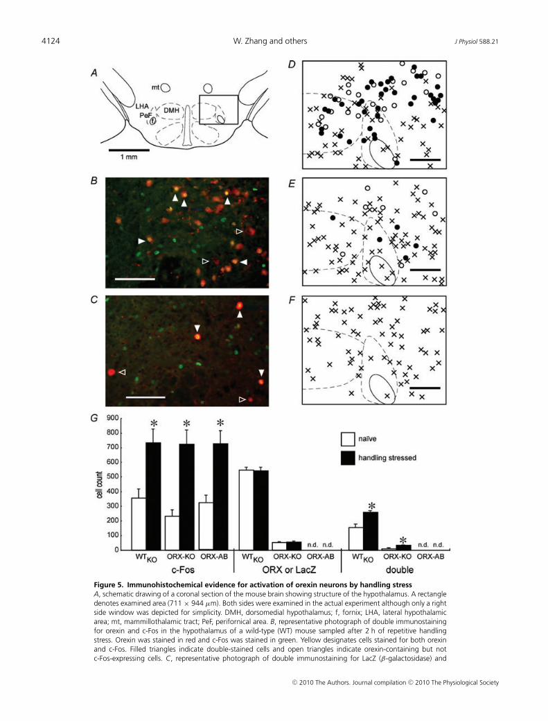
of urethane (1.6 g kg−1) and transcardially perfused with0.01 M phosphate-buffered saline (PBS) followed by afixative solution containing 4% paraformaldehyde in PBS.Brains were excised and post-fixed in the same fixativesolution for 48 h at 4◦C. Control brains were obtained fromnaıve animals. After cryoprotection with 30% sucrose,serial transverse frozen sections (40 μm) were cut fromthe brain tissue that included the hypothalamus. Everyfourth section was collected and sequentially incubatedwith PBS containing 0.3% Triton-X 100 and 1% normalgoat serum for 30 min, rabbit anti-c-Fos antiserum(1/1000, Oncogene Research Products, San Diego, CA,USA) for 1 h, biotinylated goat anti-rabbit IgG anti-body (1/200, Vector Laboratories, Burlingham, CA, USA)for 90 min and rabbit anti-orexin antiserum (1/1000,Peptide Institute, Minohshi, Osaka, Japan) for 1 h at theroom temperature. The anti-orexin antiserum recognizesmouse/human orexin A and B but not glucagon-likepeptide-1. Finally, tissue was incubated with Alexa Fluor488 streptavidin conjugate (1/200, Molecular Probes,Eugene, OR, USA) and Alexa Fluor 568-labelled goatanti-rabbit IgG antibody (1/200, Molecular Probes) for90 min in a dark box. The sections were then mountedon a glass slide and examined with a fluorescencemicroscope (Biorevo BZ-8000, Keyence, Osaka, Japan).Images were recorded at ×100 with a 48-bit digital camera(711 × 944 μm window). The photographic frame wasset so that the fornix would be bottom and midline ofthe window (Fig. 5A). To confirm the specificity of theantibodies, incubations without primary or secondaryantibody were conducted as a negative control in eachexperiment and no signal was observed.
For ORX-KO mice that do not express orexin, we tookadvantage of the animal model which has a nuclear trans-location signal plus the LacZ gene instead of the orexingene (Chemelli et al. 1999). Brain sections were preparedby the same method described above and incubated with amouse monoclonal anti-LacZ antibody (1/1000, Promega,Madison, WI, USA) instead of anti-orexin antiserum. Inaddition, Alexa Fluor 568-labelled goat anti-mouse IgGantibody (1/200, Molecular Probes) was used instead ofAlexa Fluor 568-labelled goat anti-rabbit IgG antibody.
The number of single-labelled (c-Fos, orexin or LacZ)and double-labelled (orexin plus c-Fos or LacZ plus c-Fos)cells was determined in a blinded manner to the treatment(stressed or naıve).
Statistics
The effect of repeated handling stress on rectaltemperature and that of β3 agonist on BAT temperaturewas assessed by ANOVA with genotype as the mainfactor and time as a repeated measure. When appropriate,within-subjects effect over time and between-subjectseffects at each time point was determined by Dunnett’s
post hoc test. Effect of the β3- or α1-antagonist onhandling stress-induced hyperthermia was assessed byone-way ANOVA with drugs (blocker or vehicle) as mainfactor and time as a repeated measure. When appropriate,a within-subjects effect over time was determined byDunnett’s test and between-subjects effects at each timepoint was determined by unpaired 2-tailed t test. UCP-1measurements and immunohistochemistry results wereanalysed by one-way ANOVA. Data are presented asmean ± S.E.M. Differences were considered significant atP < 0.05.
Results
Handling stress-induced hyperthermia can beaccurately determined by repetitive probe insertion
Temperature measurement itself was used as a stressorin the following study. Therefore, it may be questionablewhether the first measurement can be thought as thebaseline value without stimulation or whether it isalready distorted by the handling process. To examine thisquestion, six WTAB mice were used to compare abdominaltemperature measured by an indwelling telemetric devicewith rectal temperature measured by probe insertion(Fig. 1). There was no difference between the rectaltemperature obtained by the first probe insertion and theabdominal temperature obtained by the telemeter at thecorresponding time point (36.3 ± 0.1◦C in either method)and no deflection was observed (�T = 0.0 ± 0.1◦C,range = −0.5 to +0.3). In addition, there was nosignificant difference in the measured values between thetwo methods at any time point.
Blunted stress-induced hyperthermia in ORX-AB micebut not in ORX-KO mice
We have previously shown that the stress-induced cardio-respiratory responses were significantly attenuated inORX-KO and ORX-AB mice (Kayaba et al. 2003; Zhanget al. 2006a). Here, we examined whether the same wastrue for stress-induced hyperthermia, a different facet ofthe fight-or-flight response.
There was no difference in initial rectal temperatureamong the genotypes. It was 36.3 ± 0.1◦C in WTKO
mice (n = 22), 36.2 ± 0.1◦C in ORX-KO mice (n = 37),36.1 ± 0.1◦C in WTAB mice (n = 21) and 36.1 ± 0.1◦Cin ORX-AB mice (n = 32). In WTKO and WTAB mice,rectal temperature increased by >1.0◦C at the thirdmeasurement (20 min after the initial measurement)(Fig. 2). Repetitive handling stress every 10 min did notresult in a further increase and the rectal temperatureremained higher than the baseline throughout theremainder of the experimental period, although amoderate decline was observed in the very late phase(100–120 min).
C© 2010 The Authors. Journal compilation C© 2010 The Physiological Society
J Physiol 588.21 Orexin neurons in stress-induced hyperthermia 4121
Figure 1. Comparison between body temperaturemeasurement by rectal temperature probeinsertion and abdominal telemetry in wild-typemiceTo confirm that the temperature measurement by arectal probe accurately reflects the body temperaturejust before probe insertion but subsequently triggershyperthermia, rectal temperature was repetitivelymeasured at 10 min intervals while abdominaltemperature was continuously measured usingradio-telemetry. Data are mean ± S.E.M. of 6 wild-typemice. Arrows indicate the timing of the insertion oftemperature probe into the animal’s rectum.
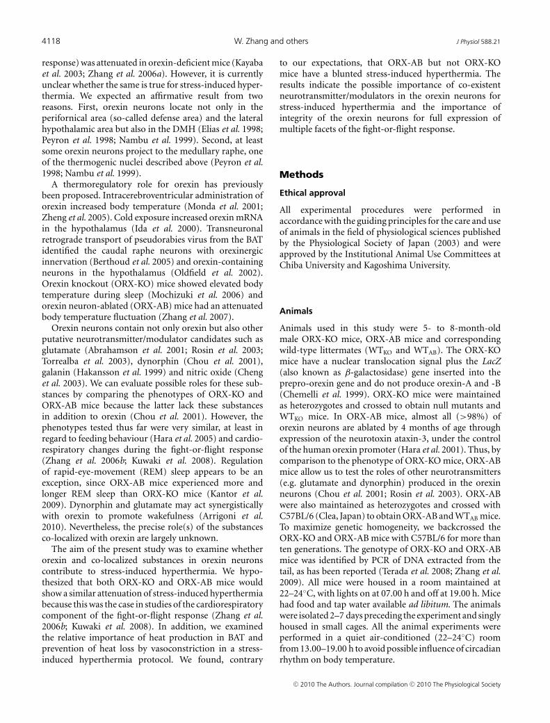
In contrast to our expectation, a little slower but largelysimilar temperature change was observed in ORX-KOmice (Fig. 2). Meanwhile, responses in ORX-AB mice werevery slow (peaking at 70 min) and small (0.4 ± 0.1◦C at thepeak). Overall, rectal temperature in ORX-AB mice wassignificantly lower than WTAB mice from 10 to 120 minafter the initial application of the handling stress.
Blunted stress-induced expression of UCP-1in ORX-AB mice but not in ORX-KO mice
To examine the possible contribution of UCP-1 expressionto the stress-induced hyperthermia, we determined UCP-1mRNA levels using the quantitative RT-PCR technique.
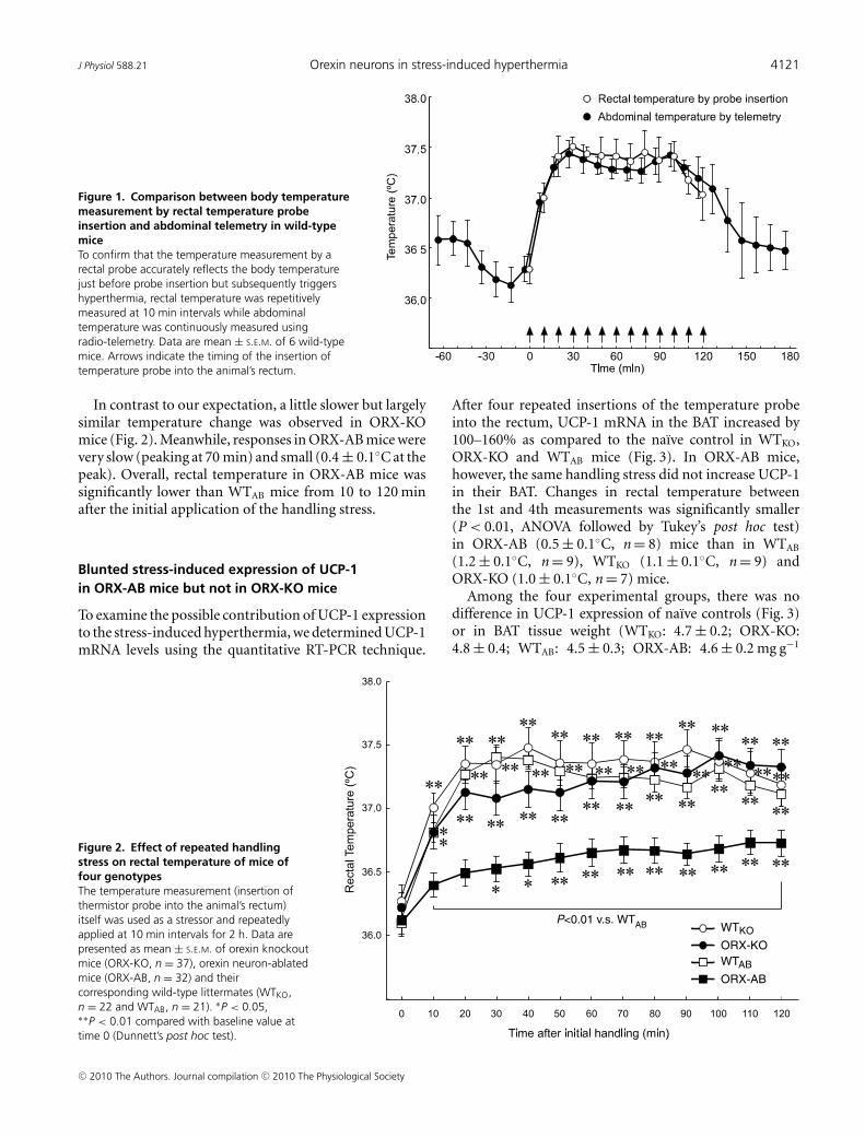
After four repeated insertions of the temperature probeinto the rectum, UCP-1 mRNA in the BAT increased by100–160% as compared to the naıve control in WTKO,ORX-KO and WTAB mice (Fig. 3). In ORX-AB mice,however, the same handling stress did not increase UCP-1in their BAT. Changes in rectal temperature betweenthe 1st and 4th measurements was significantly smaller(P < 0.01, ANOVA followed by Tukey’s post hoc test)in ORX-AB (0.5 ± 0.1◦C, n = 8) mice than in WTAB
(1.2 ± 0.1◦C, n = 9), WTKO (1.1 ± 0.1◦C, n = 9) andORX-KO (1.0 ± 0.1◦C, n = 7) mice.
Among the four experimental groups, there was nodifference in UCP-1 expression of naıve controls (Fig. 3)or in BAT tissue weight (WTKO: 4.7 ± 0.2; ORX-KO:4.8 ± 0.4; WTAB: 4.5 ± 0.3; ORX-AB: 4.6 ± 0.2 mg g−1
P<0.01 v.s. WTAB WTKO
WTAB
ORX-KO
ORX-AB
Figure 2. Effect of repeated handlingstress on rectal temperature of mice offour genotypesThe temperature measurement (insertion ofthermistor probe into the animal’s rectum)itself was used as a stressor and repeatedlyapplied at 10 min intervals for 2 h. Data arepresented as mean ± S.E.M. of orexin knockoutmice (ORX-KO, n = 37), orexin neuron-ablatedmice (ORX-AB, n = 32) and theircorresponding wild-type littermates (WTKO,n = 22 and WTAB, n = 21). ∗P < 0.05,∗∗P < 0.01 compared with baseline value attime 0 (Dunnett’s post hoc test).
C© 2010 The Authors. Journal compilation C© 2010 The Physiological Society
4122 W. Zhang and others J Physiol 588.21
body weight). These results indicated that the lack ofstress-induced increase of UCP-1 in ORX-AB mice wasnot due to abnormality in the BAT in itself but to anabnormality in the central nervous system.
Preserved BAT function in ORX-AB mice
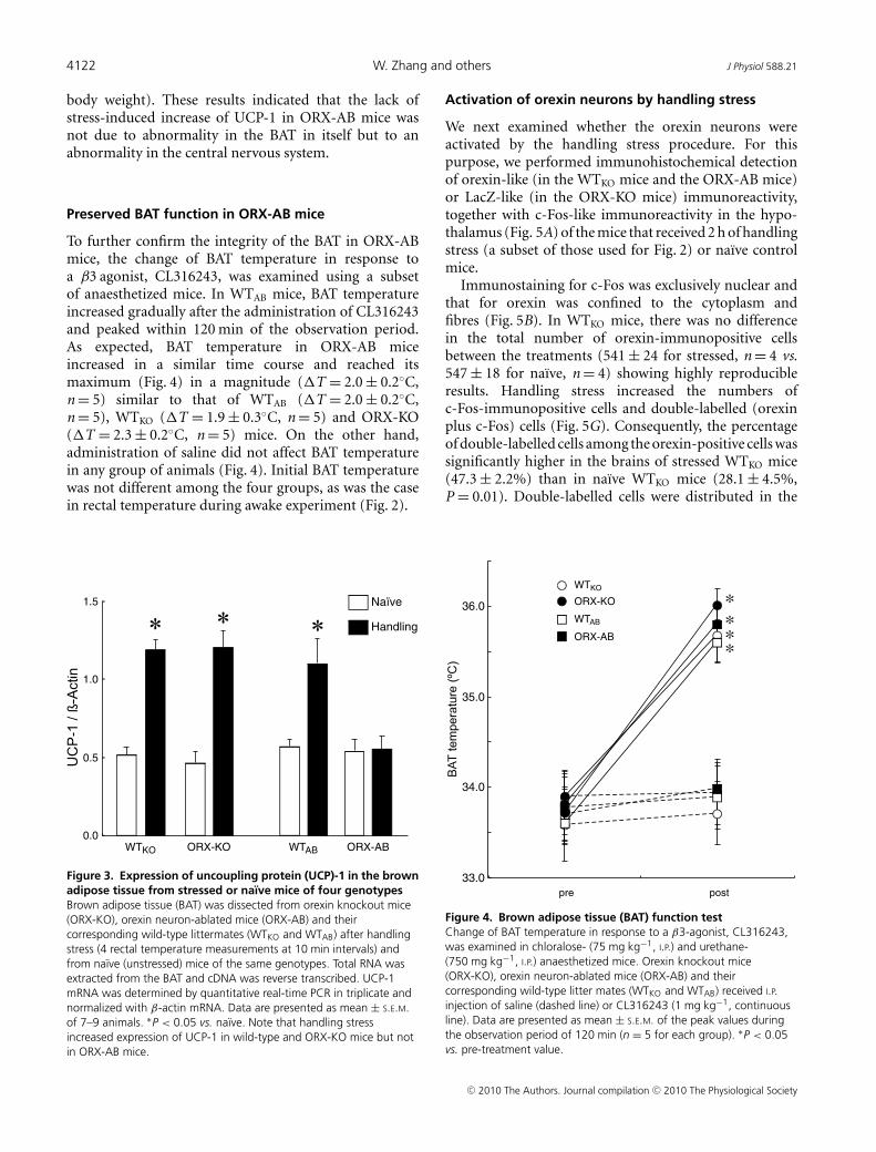
To further confirm the integrity of the BAT in ORX-ABmice, the change of BAT temperature in response toa β3 agonist, CL316243, was examined using a subsetof anaesthetized mice. In WTAB mice, BAT temperatureincreased gradually after the administration of CL316243and peaked within 120 min of the observation period.As expected, BAT temperature in ORX-AB miceincreased in a similar time course and reached itsmaximum (Fig. 4) in a magnitude (�T = 2.0 ± 0.2◦C,n = 5) similar to that of WTAB (�T = 2.0 ± 0.2◦C,n = 5), WTKO (�T = 1.9 ± 0.3◦C, n = 5) and ORX-KO(�T = 2.3 ± 0.2◦C, n = 5) mice. On the other hand,administration of saline did not affect BAT temperaturein any group of animals (Fig. 4). Initial BAT temperaturewas not different among the four groups, as was the casein rectal temperature during awake experiment (Fig. 2).
0.0
0.5
1.0
1.5
WTABORX-KOWTKO ORX-AB
Naïve
Handling
Figure 3. Expression of uncoupling protein (UCP)-1 in the brownadipose tissue from stressed or naıve mice of four genotypesBrown adipose tissue (BAT) was dissected from orexin knockout mice(ORX-KO), orexin neuron-ablated mice (ORX-AB) and theircorresponding wild-type littermates (WTKO and WTAB) after handlingstress (4 rectal temperature measurements at 10 min intervals) andfrom naıve (unstressed) mice of the same genotypes. Total RNA wasextracted from the BAT and cDNA was reverse transcribed. UCP-1mRNA was determined by quantitative real-time PCR in triplicate andnormalized with β-actin mRNA. Data are presented as mean ± S.E.M.of 7–9 animals. ∗P < 0.05 vs. naıve. Note that handling stressincreased expression of UCP-1 in wild-type and ORX-KO mice but notin ORX-AB mice.
Activation of orexin neurons by handling stress
We next examined whether the orexin neurons wereactivated by the handling stress procedure. For thispurpose, we performed immunohistochemical detectionof orexin-like (in the WTKO mice and the ORX-AB mice)or LacZ-like (in the ORX-KO mice) immunoreactivity,together with c-Fos-like immunoreactivity in the hypo-thalamus (Fig. 5A) of the mice that received 2 h of handlingstress (a subset of those used for Fig. 2) or naıve controlmice.
Immunostaining for c-Fos was exclusively nuclear andthat for orexin was confined to the cytoplasm andfibres (Fig. 5B). In WTKO mice, there was no differencein the total number of orexin-immunopositive cellsbetween the treatments (541 ± 24 for stressed, n = 4 vs.547 ± 18 for naıve, n = 4) showing highly reproducibleresults. Handling stress increased the numbers ofc-Fos-immunopositive cells and double-labelled (orexinplus c-Fos) cells (Fig. 5G). Consequently, the percentageof double-labelled cells among the orexin-positive cells wassignificantly higher in the brains of stressed WTKO mice(47.3 ± 2.2%) than in naıve WTKO mice (28.1 ± 4.5%,P = 0.01). Double-labelled cells were distributed in the
33.0
34.0
35.0
36.0
pre post
ORX-KO
ORX-AB
WTKO
WTAB
*
***
Figure 4. Brown adipose tissue (BAT) function testChange of BAT temperature in response to a β3-agonist, CL316243,was examined in chloralose- (75 mg kg−1, I.P.) and urethane-(750 mg kg−1, I.P.) anaesthetized mice. Orexin knockout mice(ORX-KO), orexin neuron-ablated mice (ORX-AB) and theircorresponding wild-type litter mates (WTKO and WTAB) received I.P.injection of saline (dashed line) or CL316243 (1 mg kg−1, continuousline). Data are presented as mean ± S.E.M. of the peak values duringthe observation period of 120 min (n = 5 for each group). ∗P < 0.05vs. pre-treatment value.
C© 2010 The Authors. Journal compilation C© 2010 The Physiological Society

J Physiol 588.21 Orexin neurons in stress-induced hyperthermia 4123
DMH, perifornical area and lateral hypothalamic area(Fig. 5D).
In the ORX-KO mice, immunostaining for both LacZand c-Fos was exclusively nuclear (Fig. 5C). There was nodifference in the total numbers of LacZ-immunopositivecells between the treatments (56 ± 6 for stressed, n = 3vs. 54 ± 4 for naıve, n = 4) showing reproducible results.Handling stress significantly increased the numbers ofc-Fos-immunopositive cells and double-labelled (LacZplus c-Fos) cells (Fig. 5G). Consequently, the percentageof double-labelled cells in the LacZ-positive populationwas significantly higher in the brains of stressedORX-KO mice (61.6 ± 4.4%) than in naıve ORX-KO mice(18.0 ± 8.2%, P = 0.01). As was the case in the WTKO mice,double-labelled cells were distributed in the DMH, peri-fornical area and lateral hypothalamic area (Fig. 5E).
No orexin-like immunoreactivity was observed in theORX-AB mice, as expected (not shown). Of note, therewas no difference in the distribution (Fig. 5D–F) or thenumbers (Fig. 5G) of c-Fos-positive cells among stressedWTKO, stressed ORX-KO and stressed ORX-AB, indicatingthat handling stress activated the cells in the examined areain ORX-KO and ORX-AB similarly to those in WTKO.
Heat production in BAT as the main cause of handlingstress-induced hyperthermia
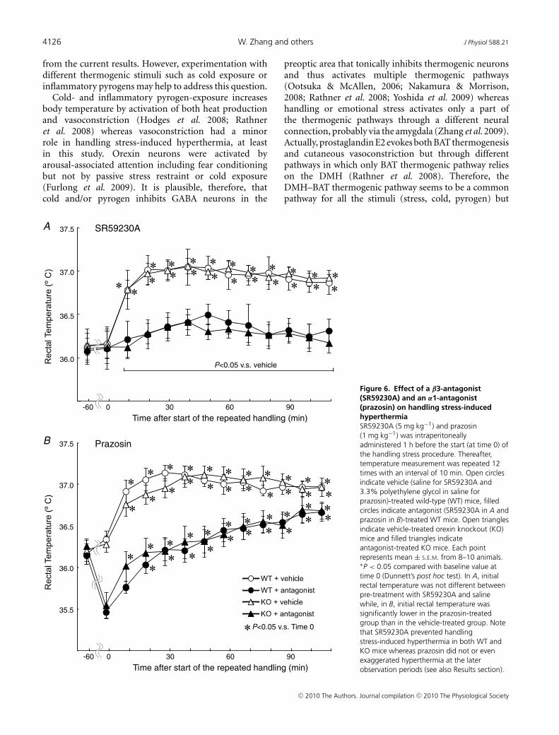
Body temperature is regulated by the balance betweenheat production and heat loss. To examine therelative importance of these processes in handlingstress-induced hyperthermia, a β3 receptor selectiveantagonist, SR59230A, or an α1 adrenergic antagonist,prazosin, was intraperitoneally administered 60 min priorto the start of the handling stress procedure.
SR59230A did not affect basal rectal temperaturein either WTKO mice or ORX-KO mice (Fig. 6A).However, SR59230A significantly attenuated the hyper-thermia that was induced by repetitive handlingstress in both WTKO (�T from time 0 to thepeak = 0.9 ± 0.1◦C in vehicle-treated, n = 10 vs.0.4 ± 0.1◦C in SR59230A-treated, n = 10, P < 0.01)and ORX-KO mice (�T from time 0 to thepeak = 0.9 ± 0.1◦C in vehicle-treated, n = 10 vs.0.3 ± 0.1◦C in SR59230A-treated, n = 10, P < 0.01).
Prazosin reduced rectal temperature in the restingcondition in both WTKO mice and ORX-KO mice(Fig. 6B). Even though prazosin was effectivewhen the handling stress was started, it did notinhibit or even exaggerate the subsequent handlingstress-induced hyperthermia in both WTKO (�T fromtime 0 to the peak = 0.8 ± 0.1◦C in vehicle-treated,n = 8 vs. 1.2 ± 0.1◦C in prazosin-treated, n = 9,P < 0.01) and ORX-KO mice (�T from time 0 to
the peak = 0.8 ± 0.1◦C in vehicle-treated, n = 8 vs.1.2 ± 0.1◦C in prazosin-treated, n = 10, P < 0.01).
These data, together with the result of UCP-1 expressionin Fig. 3, indicated that heat production in the BAT,rather than prevention of heat loss by cutaneous vaso-constriction, was the important determinant of thehandling stress-induced hyperthermia, at least under theseexperimental conditions. On the other hand, regulation ofskin blood flow seemed more important than BAT thermo-genesis for basal body temperature regulation.
Discussion
In our previous studies using ORX-KO and ORX-AB mice,we demonstrated that endogenous orexin contributes tocardiovascular, respiratory and analgesic components ofthe fight-or-flight response (Kayaba et al. 2003; Watanabeet al. 2005; Zhang et al. 2006a,b, 2009). We hypo-thesized that the same would be true for stress-inducedhyperthermia. Contrary to our expectations, ORX-KOmice showed normal temperature change in responseto handling stress while ORX-AB mice showed theexpected attenuation of stress-induced hyperthermia(Fig. 2). UCP-1, a key molecule in non-shivering thermo-genesis, did not increase in the BAT of ORX-AB mice inresponse to the handling stress (Fig. 3). BAT of ORX-ABmice seemed to be functional since its temperatureincreased in response to a β3 agonist (Fig. 4). In addition,ORX-AB mice seemed to be able to sense stressful stimulibecause c-Fos expression in their hypothalamus wascomparable to those in WT and ORX-KO mice (Fig. 5).Therefore, it seems plausible that ORX-AB mice havenormal BAT but its activation is attenuated during thehandling stress, probably due to the absence of orexinneurons. In both WT and ORX-KO mice, handling stressactivated orexin neurons (Fig. 5) and the subsequenthyperthermia largely depended on thermogenesis in theBAT but not on prevention of heat loss by vasoconstriction(Fig. 6). Together, these observations support the notionthat attenuated stress-induced hyperthermia in ORX-ABwas caused by a loss of orexin neurons and abnormalBAT regulation. This study pointed out, for the firsttime, the possible importance of the co-existent neuro-transmitter/modulator candidates in the orexin neuronsfor stress-induced hyperthermia and the importance ofintegrity of the orexin neurons for full expression ofmultiple facets of the fight-or-flight response.
Methodological considerations
In this study, the temperature measurement itself was usedas a stressor. Therefore, it may be questionable whetherthe result in the first measurement can be thought asthe baseline value without stimulation or whether it is
C© 2010 The Authors. Journal compilation C© 2010 The Physiological Society
4124 W. Zhang and others J Physiol 588.21
Figure 5. Immunohistochemical evidence for activation of orexin neurons by handling stressA, schematic drawing of a coronal section of the mouse brain showing structure of the hypothalamus. A rectangledenotes examined area (711 × 944 μm). Both sides were examined in the actual experiment although only a rightside window was depicted for simplicity. DMH, dorsomedial hypothalamus; f, fornix; LHA, lateral hypothalamicarea; mt, mammillothalamic tract; PeF, perifornical area. B, representative photograph of double immunostainingfor orexin and c-Fos in the hypothalamus of a wild-type (WT) mouse sampled after 2 h of repetitive handlingstress. Orexin was stained in red and c-Fos was stained in green. Yellow designates cells stained for both orexinand c-Fos. Filled triangles indicate double-stained cells and open triangles indicate orexin-containing but notc-Fos-expressing cells. C, representative photograph of double immunostaining for LacZ (β-galactosidase) and
C© 2010 The Authors. Journal compilation C© 2010 The Physiological Society

J Physiol 588.21 Orexin neurons in stress-induced hyperthermia 4125
already distorted by the handling process. To examine thisquestion, we compared core temperature measured byan indwelling telemetric device with rectal temperaturemeasured by probe insertion. We confirmed that thiswas a needless concern (Fig. 1), at least in the presentexperimental procedure (temperature measurement byinsertion of a probe into the rectum completed within30 s), presumably because of the buffering effect of bodymass to a rapid change in BAT temperature.
In ORX-KO mice that do not express orexin, weidentified ‘orexin neurons’ by immunostaining for LacZwhich is expressed in this neuronal population (Chemelliet al. 1999). Although some neurons did express LacZ,the number of LacZ-immunopositive neurons (∼50 permouse) was far fewer than that of orexin-immunopositiveneurons in the WT mice (∼500 per mouse). A similarlow penetration of LacZ expression was observed inone ORX-KO heterozygote animal (56/547 = 10%). Thereason for incomplete penetration of LacZ expressionis unknown (Sakurai et al. 1999), although similarphenomena have been observed in other transgenic mice(McGowan et al. 1989; Mercer et al. 1991). LacZ immuno-staining in ORX-KO mice is, therefore, not a completesubstitute for orexin immunostaining in WT mice and theresults were somewhat biased. Nevertheless, we think wecan conclude from the present results that at least some ofthe ‘orexin neurons’ in ORX-KO mice were activated byhandling stress because the total number of LacZ-positivecells was not different between naıve and stressed ORX-KOmice. In addition, activation of the hypothalamic area byhandling stress seemed not different between WT andORX-KO mice because the distribution and the totalnumber of c-Fos-positive cells were comparable betweenthe two.
State-dependent role of orexin neuronsin thermoregulation
Basal body temperature was not different among threegenotypes. Therefore, orexin and orexin neurons do not
c-Fos in the hypothalamus of an orexin knockout (ORX-KO) mouse sampled after 2 h of repetitive handling stress.LacZ gene was introduced into the ORX-KO mice instead of the normal orexin gene. LacZ was stained in redand c-Fos was stained in green. Yellow designates cells stained for both LacZ and c-Fos. Filled triangles indicatedouble-stained cells and open triangles indicate LacZ-containing but not c-Fos expressing cells. Bar, 100 μm in Band C. D, typical distribution of double-stained cells (filled circle), orexin-positive but not c-Fos positive cells (opencircle), and c-Fos-positive but not orexin-positive cells (×) in the examination window of a WT mouse. E, typicaldistribution of double-stained cells (filled circle), LacZ-positive but not c-Fos-positive cells (open circle), and c-Fospositive but not LacZ-positive cells (×) in the examination window of a ORX-KO mouse. F, typical distribution ofc-Fos-positive cells (×) in the examination window of a orexin neuron-ablated (ORX-AB) mouse. Note that thereis no apparent difference in the distribution of c-Fos-positive cells among D, E and F. Bar, 200 μm in D–F. G,numbers of c-Fos-, orexin (ORX)- and LacZ-immunopositive cells and double-stained cells (c-Fos and ORX in WTor in ORX-AB, and c-Fos and LacZ in ORX-KO) in the hypothalamus. Every 4th section in an animal (6 sectionsper mouse) was examined. Data are presented as mean ± S.E.M. of 4 mice in a group except for handling-stressedORX-KO mice (n = 3). ∗P < 0.05 vs. naıve. No orexin-immunopositive cell was detected (n.d.) in ORX-AB mice.
seem to participate in body temperature regulation whenthe animal is at rest. This notion is in line with thereports showing normal body temperature in ORX-KO(Mochizuki et al. 2006) and ORX-AB mice (Zhang et al.2007) when the animal is awake without any specificstimulus.
On the other hand, this study showed that abnormalitiesin the thermoregulatory system became apparent whenhandling stress was applied to the ORX-AB mice. Thisobservation was in line with our previous reports showingnormal basal ventilation and an attenuated respiratoryaugmentation in response to the stimulation to theamygdala (Zhang et al. 2009) or the hypothalamus (Zhanget al. 2006a) that mimics the condition under stress.Abnormality in respiratory regulation was also apparentin ORX-KO and ORX-AB mice after repetitive inter-mittent hypoxia that mimics sleep apnoea (Kuwaki, 2008;Terada et al. 2008; Toyama et al. 2009). Taken together,orexin neurons plausibly regulate body temperature ina state-dependent and feedforward manner to fit with abodily demand associated with behavioural and metabolicchanges.
Location of the orexin neuronsin the thermoregulatory circuit
There seems to be two possibilities for location of theorexin neurons in handling stress-induced thermogenicpathway. One possibility is that some of the orexin neuronsper se function as DMH-raphe thermogenic neurons(Cano et al. 2003; DiMicco & Zaretsky, 2007). Anatomicalconnection data support this notion (Oldfield et al. 2002;Berthoud et al. 2005). Some orexin neurons in DMHwere in fact activated by handling stress in this study(Fig. 5D and E). Another possibility is that some orexinneurons receive stress information (Zhang et al. 2009) and,in turn, activate the DMH-raphe thermogenic neuronsthrough their projection to the DMH (Peyron et al. 1998).We cannot discriminate between the two possibilities
C© 2010 The Authors. Journal compilation C© 2010 The Physiological Society
4126 W. Zhang and others J Physiol 588.21
from the current results. However, experimentation withdifferent thermogenic stimuli such as cold exposure orinflammatory pyrogens may help to address this question.
Cold- and inflammatory pyrogen-exposure increasesbody temperature by activation of both heat productionand vasoconstriction (Hodges et al. 2008; Rathneret al. 2008) whereas vasoconstriction had a minorrole in handling stress-induced hyperthermia, at leastin this study. Orexin neurons were activated byarousal-associated attention including fear conditioningbut not by passive stress restraint or cold exposure(Furlong et al. 2009). It is plausible, therefore, thatcold and/or pyrogen inhibits GABA neurons in the
preoptic area that tonically inhibits thermogenic neuronsand thus activates multiple thermogenic pathways(Ootsuka & McAllen, 2006; Nakamura & Morrison,2008; Rathner et al. 2008; Yoshida et al. 2009) whereashandling or emotional stress activates only a part ofthe thermogenic pathways through a different neuralconnection, probably via the amygdala (Zhang et al. 2009).Actually, prostaglandin E2 evokes both BAT thermogenesisand cutaneous vasoconstriction but through differentpathways in which only BAT thermogenic pathway relieson the DMH (Rathner et al. 2008). Therefore, theDMH–BAT thermogenic pathway seems to be a commonpathway for all the stimuli (stress, cold, pyrogen) but
Time after start of the repeated handling (min)
36.0
36.5
37.0
37.5
-60 0 60 9030
35.5
36.0
36.5
37.0
37.5
Time after start of the repeated handling (min)
-60 0 60 9030
P<0.05 v.s. Time 0
P<0.05 v.s. vehicle
SR59230A
PrazosinB
A
Figure 6. Effect of a β3-antagonist(SR59230A) and an α1-antagonist(prazosin) on handling stress-inducedhyperthermiaSR59230A (5 mg kg−1) and prazosin(1 mg kg−1) was intraperitoneallyadministered 1 h before the start (at time 0) ofthe handling stress procedure. Thereafter,temperature measurement was repeated 12times with an interval of 10 min. Open circlesindicate vehicle (saline for SR59230A and3.3% polyethylene glycol in saline forprazosin)-treated wild-type (WT) mice, filledcircles indicate antagonist (SR59230A in A andprazosin in B)-treated WT mice. Open trianglesindicate vehicle-treated orexin knockout (KO)mice and filled triangles indicateantagonist-treated KO mice. Each pointrepresents mean ± S.E.M. from 8–10 animals.∗P < 0.05 compared with baseline value attime 0 (Dunnett’s post hoc test). In A, initialrectal temperature was not different betweenpre-treatment with SR59230A and salinewhile, in B, initial rectal temperature wassignificantly lower in the prazosin-treatedgroup than in the vehicle-treated group. Notethat SR59230A prevented handlingstress-induced hyperthermia in both WT andKO mice whereas prazosin did not or evenexaggerated hyperthermia at the laterobservation periods (see also Results section).
C© 2010 The Authors. Journal compilation C© 2010 The Physiological Society

J Physiol 588.21 Orexin neurons in stress-induced hyperthermia 4127
the cutaneous vasoconstriction pathway seems to bypassDMH and orexin neurons.
Possible neurotransmitter/modulators in the orexinneurons for handling stress-induced hyperthermia
We have no data at present to determine whichneurotransmitter/modulator in the orexin neurons isimportant for stress-induced hyperthermia. Dynorphinseems to be a hypothermic neuropeptide because intra-cerebroventricular (I.C.V.) administration decreased bodytemperature (Handler et al. 1994) and the content ofdynorphin in the brain was high during hibernation whenthe body temperature is low (Cui et al. 1996). Galaninalso seems to be hypothermic because I.C.V. administrationinhibited endotoxin-induced fever (Lyudyno et al. 2001)and promoted sleep (Steiger, 2007). Glutamate seemsto be a possible candidate because microinjection ofglutamate receptor agonists into the raphe pallidusactivated sympathetic nerve activity to the BAT and micro-injection of glutamate receptor antagonists into the raphepallidus inhibited an activation of BAT sympathetic nerveevoked by stimulation to the DMH (Cao & Morrison,2006). However, it should be clarified whether theglutamate-synthesizing DMH thermoregulatory neuronsare orexin neurons or not. Future experiments will benecessary to distinguish among these possibilities.
References
Abrahamson EE, Leak RK & Moore RY (2001). Thesuprachiasmatic nucleus projects to posterior hypothalamicarousal systems. Neuroreport 12, 435–440.
Arrigoni E, Mochizuki T & Scammell TE (2010). Activation ofthe basal forebrain by the orexin/hypocretin neurons. ActaPhysiol (Oxf) 198, 223–235.
Berthoud H-R, Patterson LM, Sutton GM, Morrison C &Zheng H (2005). Orexin inputs to caudal raphe neuronsinvolved in thermal, cardiovascular, and gastrointestinalregulation. Histochem Cell Biol 123, 147–156.
Bouwknecht JA, Olivier B & Paylor RE (2007). Thestress-induced hyperthermia paradigm as a physiologicalanimal model for anxiety: a review of pharmacological andgenetic studies in the mouse. Neurosci Biobehav Rev 31,41–59.
Cannon B & Nedergaard J (2004). Brown adipose tissue:function and physiological significance. Physiol Rev 84,277–359.
Cano G, Passerin AM, Schiltz JC, Card JP, Morrison SF & SvedAF (2003). Anatomical substrates for the central control ofsympathetic outflow to interscapular adipose tissue duringcold exposure. J Comp Neurol 460, 303–326.
Cao W-H & Morrison SF (2006). Glutamate receptors in theraphe pallidus mediate brown adipose tissue thermogenesisevoked by activation of dorsomedial hypothalamic neurons.Neuropharmacol 51, 426–437.
Chemelli RM, Willie JT, Sinton CM, Elmquist JK, Scammell T,Lee C et al. (1999). Narcolepsy in orexin knockout mice:molecular genetics of sleep regulation. Cell 98,437–451.
Cheng SB, Kuchiiwa S, Gao HZ, Kuchiiwa T & Nakagawa S(2003). Morphological study of orexin neurons in thehypothalamus of the Long-Evans rat, with special referenceto co-expression of orexin and NADPH-diaphorase or nitricoxide synthase activities. Neurosci Res 46, 53–62.
Chou TC, Lee CE, Lu J, Elmquist JK, Hara J, Willie JT et al.(2001). Orexin (hypocretin) neurons contain dynorphin. JNeurosci 21, RC168.
Cui Y, Lee TF & Wang LCH (1996). In vivo microdialysis studyon changes in septal dynorphin and β-endorphin activitiesin active and hibernating Columbian ground squirrels. BrainRes 710, 271–274.
DiMicco JA & Zaretsky DV (2007). The dorsomedialhypothalamus: a new player in thermoregulation. Am JPhysiol Regul Integr Comp Physiol 292, R47–R63.
Elias CF, Saper CB, Maratos-Flier E, Tritos NA, Lee C, Kelly Jet al. (1998). Chemically defined projections linking themediobasal hypothalamus and the lateral hypothalamic area.J Comp Neurol 402, 442–459.
Furlong TM, Vianna DML, Liu L & Carrive P (2009).Hypocretin/orexin contributes to the expression of some butnot all forms of stress and arousal. Eur J Neurosci 30,1603–1614.
Hakansson M, de Lecea L, Sutcliffe JG, Yanagisawa M & MeisterB (1999). Leptin receptor- and STAT3-immunoreactivities inhypocretin/orexin neurones of the lateral hypothalamus. JNeuroendocrinol 11, 653–663.
Handler CM, Piliero TC, Geller EB & Adler MW (1994). Effectof ambient temperature on the ability of mu-, kappa- anddelta-selective opioid agonists to modulate thermoregulatorymechanisms in the rat. J Pharmacol Exp Ther 268,847–855.
Hara J, Beuckmann CT, Nambu T, Willie JT, Chemelli RM,Sinton CM et al. (2001). Genetic ablation of orexin neuronsin mice results in narcolepsy, hypophagia, and obesity.Neuron 30, 345–354.
Hara J, Yanagisawa M & Sakurai T (2005). Difference in obesityphenotype between orexin-knockout mice and orexinneuron-deficient mice with same genetic background andenvironmental conditions. Neurosci Lett 380, 239–242.
Hodges MR, Tattersall GJ, Harris MB, McEvoy SD, RichersonDN, Deneris ES et al. (2008). Defects in breathing andthermoregulation in mice with near-complete absence ofcentral serotonin neurons. J Neurosci 28, 2495–2505.
Hughes P & Dragunow M (1995). Induction ofimmediate-early genes and the control ofneurotransmitter-regulated gene expression within thenervous system. Pharmacol Rev 47, 133–178.
Ida T, Nakahara K, Murakami T, Hanada R, Nakazato M &Murakami N (2000). Possible involvement of orexin in thestress reaction in rats. Biochem Biophys Res Commun 270,318–323.
Kantor S, Mochizuki T, Janisiewicz AM, Clark E, Nishino S &Scammell TE (2009). Orexin neurons are necessary for thecircadian control of REM sleep. Sleep 32, 1127–1134.
C© 2010 The Authors. Journal compilation C© 2010 The Physiological Society

4128 W. Zhang and others J Physiol 588.21
Kayaba Y, Nakamura A, Kasuya Y, Ohuchi T, Yanagisawa M,Komuro I, Fukuda Y & Kuwaki T (2003). Attenuated defenseresponse and low basal blood pressure in orexin knockoutmice. Am J Physiol Regul Integr Comp Physiol 285,R581–R593.
Kuwaki T (2008). Orexinergic modulation of breathing acrossvigilance states. Respir Physiol Neurobiol 164, 204–212.
Kuwaki T, Zhang W, Nakamura A & Deng BS (2008).Emotional and state-dependent modification ofcardiorespiratory function: role of orexinergic neurons.Autonom Neurosci 142, 11–16.
Lazarus M, Yoshida K, Coppari R, Bass CE, Mochizuki T,Lowell BB & Saper CB (2007). EP3 prostaglandin receptorsin the median preoptic nucleus are critical for feverresponses. Nat Neurosci 10, 1131–1133.
Lecci A, Borsini F, Volterra G & Meli A (1990).Pharmacological validation of a novel animal model ofanticipatory anxiety in mice. Psychopharmacol 101, 255–261.
Lyudyno VI, Krasnova IN, Smirnova MP & Klimenko VM(2001). Antipyretic effect of neuropeptide galanin inendotoxin-induced fever. Bull Exp Biol Med 131, 60–63.
McGowan R, Campbell R, Peterson A & Sapienza C (1989).Cellular mosaicism in the methylation and expression ofhemizygous loci in the mouse. Genes Develop 3, 1669–1676.
Manara L, Badone D, Baroni M, Boccardi G, Cecchi R, Croci Tet al. (1996). Functional identification of rat atypicalß-adrenoceptors by the first ß3-selective antagonists,aryloxypropanolaminotetralins. Brit J Pharmacol 117,435–442.
Mercer EH, Hoyle GW, Kapur RP, Brinster RL & Palmiter RD(1991). The dopamine ß-hydroxylase gene promoter directsexpression of E. coli lacZ to sympathetic and other neuronsin adult transgenic mice. Neuron 7, 703–716.
Mochizuki T, Klerman EB, Sakurai T & Scammell TE (2006).Elevated body temperature during sleep in orexin knockoutmice. Am J Physiol Regul Integr Comp Physiol 291,R533–R540.
Monda M, Viggiano A, Mondola P & Luca VD (2001).Inhibition of prostaglandin synthesis reduces hyperthermicreactions induced by hypocretin-1/orexin A. Brain Res 909,68–74.
Nakamura K, Matsumura K, Hubschle T, Nakamura Y, HiokiH, Fujiyama F et al. (2004). Identification of sympatheticpremotor neurons in medullary raphe regions mediatingfever and other thermoregulatory functions. J Neurosci 24,5370–5380.
Nakamura K & Morrison SF (2008). A thermoregulatorypathway that controls body temperature. Nat Neurosci 11,62–71.
Nambu T, Sakurai T, Mizukami K, Hosoya Y, Yanagisawa M &Goto K (1999). Distribution of orexin neurons in the adultrat brain. Brain Res 827, 243–260.
Oldfield BJ, Giles ME, Watson A, Anderson C, Colvill LM &Mckinley MJ (2002). The neurochemical characterisation ofhypothalamic pathways projecting polysynaptically to brownadipose tissue in the rat. Neuroscience 110, 515–526.
Ootsuka Y & McAllen RM (2006). Comparison between tworat sympathetic pathways activated in cold defense. Am JPhysiol Regul Integr Comp Physiol 291, R589–R595.
Peyron C, Tighe DK, Van Den Pol AN, de Lecea L, Heller HC,Sutcliffe JG & Kilduff TS (1998). Neurons containinghypocretin (orexin) project to multiple neuronal systems. JNeurosci 18, 9996–10015.
Rathner J, Madden C & Morrison S (2008). Central pathwayfor spontaneous and prostaglandin E2-evoked cutaneousvasoconstriction. Am J Physiol Regul Integr Comp Physiol295, R343–R354.
Rosin DL, Weston MC, Sevigny CP, Stornetta RL & GuyenetPG (2003). Hypothalamic orexin (hypocretin) neuronsexpress vesicular glutamate transporters VGLUT1 orVGLUT2. J Comp Neurol 465, 593–603.
Sakurai T, Moriguchi T, Furuya K, Kajiwara N, Nakamura T,Yanagisawai M & Goto K (1999). Structure and function ofhuman prepro-orexin gene. J Biol Chem 274, 17771–17776.
Steiger A (2007). Neurochemical regulation of sleep. J Psych Res41, 537–552.
Stronberg AD & Pietri-Rouxel F (1996). Function andregulation of the ß3-adrenoceptor. Trends Pharmacol Sci 17,373.
Sunanaga J, Deng BS, Zhang W, Kanmura Y & Kuwaki T(2009). CO2 activates orexin-containing neurons in mice.Respir Physiol Neurobiol 166, 184–186.
Terada J, Nakamura A, Zhang W, Yanagisawa M, Kuriyama T,Fukuda Y & Kuwaki T (2008). Ventilatory long-termfacilitation in mice can be observed both during sleep andwake periods and depends on orexin. J Appl Physiol 104,499–507.
Torrealba F, Yanagisawa M & Saper CB (2003). Colocalizationof orexin A and glutamate immunoreactivity in axonterminals in the tuberomammillary nucleus in rats. Neurosci119, 1033–1044.
Toyama S, Sakurai T, Tatsumi K & Kuwaki T (2009).Attenuated phrenic long-term facilitation in orexinneuron-ablated mice. Respir Physiol Neurobiol 168, 295–302.
Van Der Heyden JAM, Zethof TJJ & Olivier B (1997).Stress-induced hyperthermia in singly housed mice. PhysiolBehav 62, 463–470.
Watanabe S, Kuwaki T, Yanagisawa M, Fukuda Y & ShimoyamaM (2005). Persistent pain and stress activate pain-inhibitoryorexin pathways. Neuroreport 16, 5–8.
Yoshida K, Li X, Cano G, Lazarus M & Saper CB (2009).Parallel preoptic pathways for thermoregulation. J Neurosci29, 11954–11964.
Zhang S, Zeitzer JM, Sakurai T, Nishino S & Mignot E (2007).Sleep/wake fragmentation disrupts metabolism in a mousemodel of narcolepsy. J Physiol 581, 649–663.
Zhang W, Sakurai T, Fukuda Y & Kuwaki T (2006a). Orexinneuron-mediated skeletal muscle vasodilation and shift ofbaroreflex during defense response in mice. Am J PhysiolRegul Integr Comp Physiol 290, R1654–R1663.
Zhang W, Shimoyama M, Fukuda Y & Kuwaki T (2006b).Multiple components of the defense response depend onorexin: Evidence from orexin knockout mice and orexinneuron-ablated mice. Autonom Neurosci 126–127, 139–145.
Zhang W, Zhang N, Sakurai T & Kuwaki T (2009). Orexinneurons in the hypothalamus mediate cardiorespiratoryresponses induced by disinhibition of the amygdala and bednucleus of the stria terminalis. Brain Res 1262, 25–37.
C© 2010 The Authors. Journal compilation C© 2010 The Physiological Society
J Physiol 588.21 Orexin neurons in stress-induced hyperthermia 4129
Zheng H, Patterson LM & Berthoud H-R (2005). Orexin-Aprojections to the caudal medulla and orexin-induced c-Fosexpression, food intake, and autonomic function. J CompNeurol 485, 127–142.
Author contributions
Conception and design: T.K. Data collection: W.Z., J.S., Y.T. andT.M. Analysis and interpretation: W.Z., J.S., T.S., Y.K. and T.K.Article drafting and revisions: W.Z., J.S. and T.K. All authorsapproved the final version of the manuscript. Experiments weredone at Chiba University and Kagoshima University.
Acknowledgements
We thank Dr Thomas S. Kilduff for valuable discussion andhelp in editing the manuscript. We thank Ms Orie Iwaya forher technical assistance. We also thank Dr Mieko Kurosawa, DrTakashi Miki and Dr Yoshitoshi Kasuya for their technical advicein identification of brown adipose tissue and PCR procedure.Part of the work was supported by the Grants-in Aid for ScientificResearch from the Ministry of Education, Science, Culture andSports, Japan.
Author’s present address
W. Zhang: Division of Cardiovascular Disease, Department ofMedicine, University of Alabama at Birmingham, Birmingham,AL 35294-0007, USA.
C© 2010 The Authors. Journal compilation C© 2010 The Physiological Society