Orbitofrontal and limbic signatures of empathic concern ...
13
Behavioural neurology Orbitofrontal and limbic signatures of empathic concern and intentional harm in the behavioral variant frontotemporal dementia Sandra Baez a,b,c , Juan P. Morales b , Andrea Slachevsky h,i,j,k , Teresa Torralva a,b , Cristian Matus f,g , Facundo Manes a,b,c,e and Agustin Ibanez a,b,c,d,e,* a Institute of Cognitive Neurology (INECO) & Institute of Neuroscience, Favaloro University, Buenos Aires, Argentina b UDP-INECO Foundation Core on Neuroscience (UIFCoN), Faculty of Psychology, Diego Portales University, Santiago, Chile c National Scientific and Technical Research Council (CONICET), Argentina d Universidad Aut onoma del Caribe, Barranquilla, Colombia e ACR Centre of Excellence in Cognition and its Disorders, Macquarie University, Sydney, NSW, Australia f Fundaci on M edica San Cristobal, Santiago, Chile g Hospital de Carabineros de Chile, Santiago, Chile h Departamento de Fisiopatologı´a, ICBM y Departamento de Ciencias Neurol ogicas Oriente, Facultad de Medicina, Universidad de Chile, Santiago, Chile i Unidad de Neurologı´a Cognitiva y Demencias, Departamento de Neurologı´a Oriente, Facultad de Medicina, Universidad de Chile y Servicio de Neurologı´a, Hospital del Salvador, Santiago, Chile j Centro de Investigaci on Avanzada en Educaci on, Universidad de Chile, Santiago, Chile k Servicio de Neurologı´a, Clı´nica Alemana, Santiago, Chile article info Article history: Received 19 May 2015 Revised 28 September 2015 Accepted 12 November 2015 Reviewed 11 September 2015 Action editor Brad Dickerson Published online 24 November 2015 Keywords: bvFTD Intentional harm Empathy Intentionality comprehension abstract Perceiving and evaluating intentional harms in an interpersonal context engages both cognitive and emotional domains. This process involves inference of intentions, moral judgment, and, crucially, empathy towards others' suffering. This latter skill is notably impaired in behavioral variant frontotemporal dementia (bvFTD). However, the relation- ship between regional brain atrophy in bvFTD and deficits in the above-mentioned abilities is not well understood. The present study investigated how gray matter (GM) atrophy in bvFTD patients correlates with the perception and evaluation of harmful actions (attri- bution of intentionality, evaluation of harmful behavior, empathic concern, and moral judgment). First, we compared the behavioral performance of 26 bvFTD patients and 23 healthy controls on an experimental task (ET) indexing intentionality, empathy, and moral cognition during evaluation of harmful actions. Second, we compared GM volume in pa- tients and controls using voxel-based morphometry (VBM). Third, we examined brain re- gions where atrophy might be associated with specific impairments in the patient group. * Corresponding author. Laboratory of Experimental Psychology & Neuroscience (LPEN), Institute of Cognitive Neurology (INECO) & CONICET, Pacheco de Melo 1860, Buenos Aires 1126, Argentina. E-mail addresses: [email protected] (S. Baez), [email protected] (J.P. Morales), [email protected] (A. Slachevsky), ttor- [email protected] (T. Torralva), [email protected] (C. Matus), [email protected] (F. Manes), [email protected] (A. Ibanez). Available online at www.sciencedirect.com ScienceDirect Journal homepage: www.elsevier.com/locate/cortex cortex 75 (2016) 20 e32 http://dx.doi.org/10.1016/j.cortex.2015.11.007 0010-9452/© 2015 Elsevier Ltd. All rights reserved.
Transcript of Orbitofrontal and limbic signatures of empathic concern ...

www.sciencedirect.com
c o r t e x 7 5 ( 2 0 1 6 ) 2 0e3 2
Available online at
ScienceDirect
Journal homepage: www.elsevier.com/locate/cortex
Behavioural neurology
Orbitofrontal and limbic signatures of empathicconcern and intentional harm in the behavioralvariant frontotemporal dementia
Sandra Baez a,b,c, Juan P. Morales b, Andrea Slachevsky h,i,j,k,Teresa Torralva a,b, Cristian Matus f,g, Facundo Manes a,b,c,e andAgustin Ibanez a,b,c,d,e,*
a Institute of Cognitive Neurology (INECO) & Institute of Neuroscience, Favaloro University, Buenos Aires, Argentinab UDP-INECO Foundation Core on Neuroscience (UIFCoN), Faculty of Psychology, Diego Portales University,
Santiago, Chilec National Scientific and Technical Research Council (CONICET), Argentinad Universidad Aut�onoma del Caribe, Barranquilla, Colombiae ACR Centre of Excellence in Cognition and its Disorders, Macquarie University, Sydney, NSW, Australiaf Fundaci�on M�edica San Cristobal, Santiago, Chileg Hospital de Carabineros de Chile, Santiago, Chileh Departamento de Fisiopatologı́a, ICBM y Departamento de Ciencias Neurol�ogicas Oriente, Facultad de Medicina,
Universidad de Chile, Santiago, Chilei Unidad de Neurologı́a Cognitiva y Demencias, Departamento de Neurologı́a Oriente, Facultad de Medicina,
Universidad de Chile y Servicio de Neurologı́a, Hospital del Salvador, Santiago, Chilej Centro de Investigaci�on Avanzada en Educaci�on, Universidad de Chile, Santiago, Chilek Servicio de Neurologı́a, Clı́nica Alemana, Santiago, Chile
a r t i c l e i n f o
Article history:
Received 19 May 2015
Revised 28 September 2015
Accepted 12 November 2015
Reviewed 11 September 2015
Action editor Brad Dickerson
Published online 24 November 2015
Keywords:
bvFTD
Intentional harm
Empathy
Intentionality comprehension
* Corresponding author. Laboratory of ExpeCONICET, Pacheco de Melo 1860, Buenos Air
E-mail addresses: [email protected] ([email protected] (T. Torralva), cmatusy@gmhttp://dx.doi.org/10.1016/j.cortex.2015.11.0070010-9452/© 2015 Elsevier Ltd. All rights rese
a b s t r a c t
Perceiving and evaluating intentional harms in an interpersonal context engages both
cognitive and emotional domains. This process involves inference of intentions, moral
judgment, and, crucially, empathy towards others' suffering. This latter skill is notably
impaired in behavioral variant frontotemporal dementia (bvFTD). However, the relation-
ship between regional brain atrophy in bvFTD and deficits in the above-mentioned abilities
is not well understood. The present study investigated how gray matter (GM) atrophy in
bvFTD patients correlates with the perception and evaluation of harmful actions (attri-
bution of intentionality, evaluation of harmful behavior, empathic concern, and moral
judgment). First, we compared the behavioral performance of 26 bvFTD patients and 23
healthy controls on an experimental task (ET) indexing intentionality, empathy, and moral
cognition during evaluation of harmful actions. Second, we compared GM volume in pa-
tients and controls using voxel-based morphometry (VBM). Third, we examined brain re-
gions where atrophy might be associated with specific impairments in the patient group.
rimental Psychology & Neuroscience (LPEN), Institute of Cognitive Neurology (INECO) &es 1126, Argentina.Baez), [email protected] (J.P. Morales), [email protected] (A. Slachevsky), ttor-ail.com (C. Matus), [email protected] (F. Manes), [email protected] (A. Ibanez).
rved.

c o r t e x 7 5 ( 2 0 1 6 ) 2 0e3 2 21
Moral judgment
Gray matter atrophy
Finally, we explored whether the patients' deficits in intentionality comprehension and
empathic concern could be partially explained by regional GM atrophy or impairments in
other relevant factors, such as executive functions (EFs). In bvFTD patients, atrophy of
limbic structures (amygdala and anterior paracingulate cortex e APC) was related to im-
pairments in intentionality comprehension, while atrophy of the orbitofrontal cortex (OFC)
was associated with empathic concern deficits. Intentionality comprehension impairments
were predicted by EFs and orbitofrontal atrophy predicted deficits in empathic concern.
Thus, although the perception and evaluation of harmful actions are variously compro-
mised in bvFTD, deficits in empathic concern may be central to this syndrome as they are
associated with one of the earliest atrophied region. More generally, our results shed light
on social cognition deficits in bvFTD and may have important clinical implications.
© 2015 Elsevier Ltd. All rights reserved.
1. Introduction
Suppose you are at a birthday party and a man suddenly ap-
pears holding a large knife. This person walks to the table
where the cake is placed. How would you react? Would you
feel threatened? Probably you would not startle because you
would quickly understand that he does not intend to hurt
anybody; he is simply going to cut the birthday cake. Now
imagine that this man deliberately attacks someone with the
knife. You would then automatically identify his behavior as
harmful, feel empathic concern for the victim, and assess the
action as morally wrong. The ability to detect intentional
harms is early processed by frontotemporal networks (Hesse
et al., 2015), and involves several skills, such as intention-
ality detection, empathy, and moral judgment (Decety &
Cacioppo, 2012; Decety, Michalska, & Kinzler, 2012; Escobar
et al., 2014). These abilities are essential for human survival
and successful social interaction.
Our estimation of an action's harmfulness depends on
whether we perceive it as intentional or accidental (Decety,
Michalska, & Akitsuki, 2008; Decety et al., 2012). In addition,
empathic concern is higher when a person inflicts pain on
another one intentionally rather than accidentally (Decety
et al., 2012). By the same token, the estimation of how
severely punished an actor should be in the above situations
depends on the assessment of his/her intentionality (Treadway
et al., 2014). Thus, detection of the intentionality plays a crucial
role in how harmful actions are perceived, and it also affects
moral judgments and empathic responses (Decety et al., 2008).
Observing intentional harms elicits empathic reactions
(Bernhardt & Singer, 2012; Decety et al., 2012) critical for suc-
cessful social functioning (Rankin, Kramer, & Miller, 2005).
Loss of empathy is a cardinal symptom of the behavioral
variant of frontotemporal dementia (bvFTD), which consti-
tutes a clue for early diagnosis (Piguet, Hornberger, Mioshi, &
Hodges, 2011; Rascovsky et al., 2011). Patients with bvFTD
lack concern for other's feelings (Rankin et al., 2006) and are
described by relatives as selfish and self-centered (Hsieh,
Irish, Daveson, Hodges, & Piguet, 2013). Empathy changes
may thus be presumed to underlie this population's diffi-
culties in interpersonal judgment, emotions, behavior, and
social functioning (Lough et al., 2006; Piguet et al., 2011;
Rascovsky et al., 2011).
At the neuroanatomical level, empathy processes engage a
broad network including the insula, the anterior cingulate
cortex (ACC), the supplementary motor area (SMA), the
amygdala, the orbitofrontal cortex (OFC), and the tempor-
oparietal junction (Bernhardt & Singer, 2012; Fan, Duncan, de
Greck, & Northoff, 2011; Singer & Lamm, 2009). Notably,
several studies on bvFTD have reported a reduction of gray
matter (GM) in most of these areas (Rosen et al., 2002; Seeley
et al., 2008). However, at present, the relationship between
atrophy in such regions and the empathy deficits observed in
bvFTD patients is not well understood.
Only three studies have investigated the neural basis of
empathic impairments in bvFTD. Two of them (Eslinger,
Moore, Anderson, & Grossman, 2011; Rankin et al., 2006) re-
ported associations between caregivers' ratings on a self-
report questionnaire and reduced GM volumes in ventrome-
dial frontal regions, the right SMA, the right subcallosal gyrus,
the bilateral temporal poles, the right fusiform gyrus, and the
right amygdala. The third study (Cerami et al., 2014) compared
two components of empathy (intention attribution and
emotion attribution), and correlated them with reduced GM
density within the mentalizing network. Specifically, emotion
attribution performance in the patients correlated with GM
reduction in the right amygdala, left insula, and posterior-
superior temporal sulcus eextending into the temporopar-
ietal junction. However, neither these nor any other studies
have yet investigated how the atrophy pattern of bvFTD cor-
relates with empathic responses (and their related evaluation
of intentionality and moral judgment) elicited by the percep-
tion of harm to others.
Over the last decade, several studies in healthy and clinical
populations have employed representations of harmful ac-
tions to others (e.g., infliction of pain) as proxies to investigate
different aspects of empathy, intention attribution, andmoral
judgment. The evidence thus demonstrates that the percep-
tion of harmful actions robustly induces empathic and moral
responses (Decety & Cacioppo, 2012; Decety et al., 2012;
Escobar et al., 2014; Treadway et al., 2014). At the neural
level, prefrontal and limbic regions impaired in bvFTD (Rosen
et al., 2002; Seeley et al., 2008) have been particularly associ-
ated with the perception and evaluation of harmful actions
(Decety et al., 2008, 2012; Hesse et al., 2015). Specifically,
perceiving an individual who intentionally hurts another
person triggers an early amygdala boost (Hesse et al., 2015),
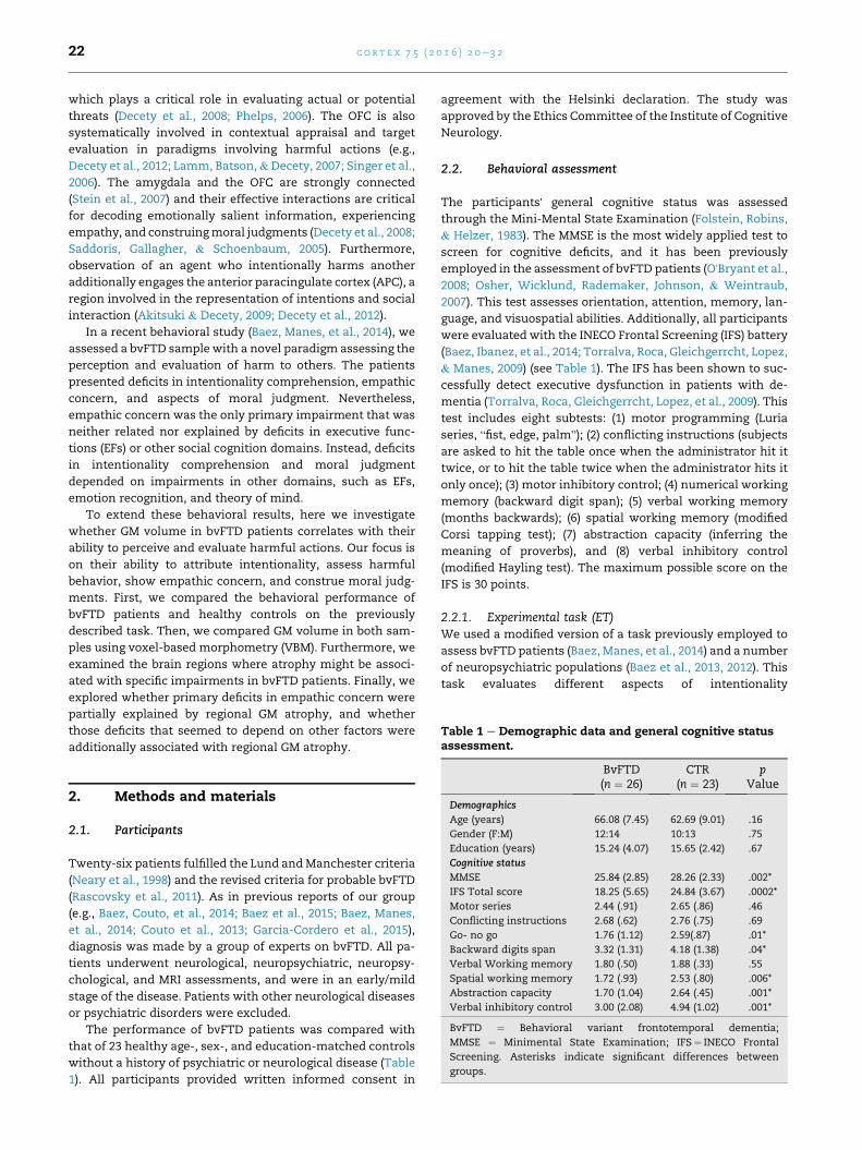
Table 1 e Demographic data and general cognitive statusassessment.
c o r t e x 7 5 ( 2 0 1 6 ) 2 0e3 222
which plays a critical role in evaluating actual or potential
threats (Decety et al., 2008; Phelps, 2006). The OFC is also
systematically involved in contextual appraisal and target
evaluation in paradigms involving harmful actions (e.g.,
Decety et al., 2012; Lamm, Batson,&Decety, 2007; Singer et al.,
2006). The amygdala and the OFC are strongly connected
(Stein et al., 2007) and their effective interactions are critical
for decoding emotionally salient information, experiencing
empathy, and construingmoral judgments (Decety et al., 2008;
Saddoris, Gallagher, & Schoenbaum, 2005). Furthermore,
observation of an agent who intentionally harms another
additionally engages the anterior paracingulate cortex (APC), a
region involved in the representation of intentions and social
interaction (Akitsuki & Decety, 2009; Decety et al., 2012).
In a recent behavioral study (Baez, Manes, et al., 2014), we
assessed a bvFTD sample with a novel paradigm assessing the
perception and evaluation of harm to others. The patients
presented deficits in intentionality comprehension, empathic
concern, and aspects of moral judgment. Nevertheless,
empathic concern was the only primary impairment that was
neither related nor explained by deficits in executive func-
tions (EFs) or other social cognition domains. Instead, deficits
in intentionality comprehension and moral judgment
depended on impairments in other domains, such as EFs,
emotion recognition, and theory of mind.
To extend these behavioral results, here we investigate
whether GM volume in bvFTD patients correlates with their
ability to perceive and evaluate harmful actions. Our focus is
on their ability to attribute intentionality, assess harmful
behavior, show empathic concern, and construe moral judg-
ments. First, we compared the behavioral performance of
bvFTD patients and healthy controls on the previously
described task. Then, we compared GM volume in both sam-
ples using voxel-based morphometry (VBM). Furthermore, we
examined the brain regions where atrophy might be associ-
ated with specific impairments in bvFTD patients. Finally, we
explored whether primary deficits in empathic concern were
partially explained by regional GM atrophy, and whether
those deficits that seemed to depend on other factors were
additionally associated with regional GM atrophy.
BvFTD(n ¼ 26)
CTR(n ¼ 23)
pValue
Demographics
Age (years) 66.08 (7.45) 62.69 (9.01) .16
Gender (F:M) 12:14 10:13 .75
Education (years) 15.24 (4.07) 15.65 (2.42) .67
Cognitive status
MMSE 25.84 (2.85) 28.26 (2.33) .002*
IFS Total score 18.25 (5.65) 24.84 (3.67) .0002*
Motor series 2.44 (.91) 2.65 (.86) .46
Conflicting instructions 2.68 (.62) 2.76 (.75) .69
Go- no go 1.76 (1.12) 2.59(.87) .01*
Backward digits span 3.32 (1.31) 4.18 (1.38) .04*
Verbal Working memory 1.80 (.50) 1.88 (.33) .55
Spatial working memory 1.72 (.93) 2.53 (.80) .006*
Abstraction capacity 1.70 (1.04) 2.64 (.45) .001*
Verbal inhibitory control 3.00 (2.08) 4.94 (1.02) .001*
BvFTD ¼ Behavioral variant frontotemporal dementia;
MMSE ¼ Minimental State Examination; IFS¼ INECO Frontal
Screening. Asterisks indicate significant differences between
groups.
2. Methods and materials
2.1. Participants
Twenty-six patients fulfilled the Lund andManchester criteria
(Neary et al., 1998) and the revised criteria for probable bvFTD
(Rascovsky et al., 2011). As in previous reports of our group
(e.g., Baez, Couto, et al., 2014; Baez et al., 2015; Baez, Manes,
et al., 2014; Couto et al., 2013; Garcia-Cordero et al., 2015),
diagnosis was made by a group of experts on bvFTD. All pa-
tients underwent neurological, neuropsychiatric, neuropsy-
chological, and MRI assessments, and were in an early/mild
stage of the disease. Patients with other neurological diseases
or psychiatric disorders were excluded.
The performance of bvFTD patients was compared with
that of 23 healthy age-, sex-, and education-matched controls
without a history of psychiatric or neurological disease (Table
1). All participants provided written informed consent in
agreement with the Helsinki declaration. The study was
approved by the Ethics Committee of the Institute of Cognitive
Neurology.
2.2. Behavioral assessment
The participants' general cognitive status was assessed
through the Mini-Mental State Examination (Folstein, Robins,
& Helzer, 1983). The MMSE is the most widely applied test to
screen for cognitive deficits, and it has been previously
employed in the assessment of bvFTD patients (O'Bryant et al.,2008; Osher, Wicklund, Rademaker, Johnson, & Weintraub,
2007). This test assesses orientation, attention, memory, lan-
guage, and visuospatial abilities. Additionally, all participants
were evaluated with the INECO Frontal Screening (IFS) battery
(Baez, Ibanez, et al., 2014; Torralva, Roca, Gleichgerrcht, Lopez,
& Manes, 2009) (see Table 1). The IFS has been shown to suc-
cessfully detect executive dysfunction in patients with de-
mentia (Torralva, Roca, Gleichgerrcht, Lopez, et al., 2009). This
test includes eight subtests: (1) motor programming (Luria
series, “fist, edge, palm”); (2) conflicting instructions (subjects
are asked to hit the table once when the administrator hit it
twice, or to hit the table twice when the administrator hits it
only once); (3) motor inhibitory control; (4) numerical working
memory (backward digit span); (5) verbal working memory
(months backwards); (6) spatial working memory (modified
Corsi tapping test); (7) abstraction capacity (inferring the
meaning of proverbs), and (8) verbal inhibitory control
(modified Hayling test). The maximum possible score on the
IFS is 30 points.
2.2.1. Experimental task (ET)We used a modified version of a task previously employed to
assess bvFTD patients (Baez, Manes, et al., 2014) and a number
of neuropsychiatric populations (Baez et al., 2013, 2012). This
task evaluates different aspects of intentionality

c o r t e x 7 5 ( 2 0 1 6 ) 2 0e3 2 23
comprehension, empathy, andmoral judgment in the context
of intentional and accidental harms. The ET consists of 25
animated scenarios (11 intentional, 11 accidental, 3 neutral)
involving two individuals. Each scenario consists of 3 digital
color pictures presented in a successive manner to imply
motion. The durations of the first, second, and third pictures
in each animation were 500, 200, and 1000 msec, respectively.
The three following types of situations were depicted: (1)
intentional harm, in which one person deliberately inflicts
pain on another (e.g., one person purposely steps on someone
else's toe); (2) accidental harm,where accidentally inflicts pain
on another; and (3) control or neutral situations involving no
harm (e.g., one hands a flower to another).
Importantly, since the protagonists' faces were not visible,
participants were blind to their facial emotional reactions.
However, body expressions and postures provided sufficient
information about the victim's emotional reaction and the
agent's intention. In this abbreviated version, participants
were asked to respond four different questions that effectively
reveal empathy impairments in bvFTD (Baez, Manes, et al.,
2014) and other neuropsychiatric conditions (Baez et al.,
2013, 2012). These questions evaluated: (a) intentionality
comprehension (was the action done on purpose?), (b) evalu-
ation of the perpetrator's harmful behavior (how bad was the
intention?), (c) empathic concern (how sad do you feel for the
victim?), and (d) punishment (how much penalty does this
action deserve?). The question about intentionality was
answered selecting “Yes” or “No”. The other questions were
answered using a computer-based visual analog scale ranging
from �9 to 9 ethese numbers were not visible to participants.
The meaning of the scale extremes depends on the question.
For example, in the question “how sad do you feel for the hurt
person?”, one extreme of the bar reads “I feel very sad” and the
other extreme reads “I don't feel sad at all”. We measured
accuracy for the intentionality question and ratings and raw
reaction times (RTs) for the other questions. RTs measure-
ments indicated the lapse between presentation of the ques-
tion and the participant's response. Before testing, to ensure
correct understanding of the instructions, we administered a
shorter training version of the task involving similar
situations.
2.3. MRI scanning
Participants were scanned in a 1.5 T Phillips Intera scanner
equipped with a standard head coil. A T1-weighted spin echo
sequence was used to generate 120 contiguous axial slices
(TR ¼ 2300 msec; TE ¼ 13 msec; flip angle ¼ 68�;FOV ¼ 256� 256 mm; matrix size ¼ 256 � 240; in-plane
resolution ¼ 1 � 1 mm; slice thickness ¼ 1 mm).
2.4. Data analysis
2.4.1. Behavioral dataWe compared demographic and neuropsychological data be-
tween samples using ANOVA tests; categorical variables were
analyzed through X2 tests. The ratings and RTs for each
question were analyzed independently using a 2 (group:
bvFTD vs controls) � 3 (condition: intentional, accidental,
neutral) factorial ANOVA. When a significant interaction
between group and condition was found, we examined
between-group differences in ratings or RTs using the Tukey'sHSD post-hoc test. Intra-group comparisons were also con-
ducted via repeated measures ANOVA. Differences among
conditions (intentional harm, accidental harm, and neutral
situations) were examined with Tukey's HSD post-hoc tests.
Given that working memory has been particularly associated
with empathy (Ze, Thoma, & Suchan, 2014) and mentalizing
skills (Gordon & Olson, 1998), we re-analyzed intentionality
comprehension and empathic concern data covarying for
backward digit span and spatial working memory scores. Ef-
fect sizes were calculated through partial eta (h2) tests. The
statistical significance level was set at p < .05.
2.4.2. VBM analysisImages were preprocessed using the DARTEL Toolbox, in
accordance with previously described procedures (Ashburner
& Friston, 2000). Then, modulated 12-mm full-width half-
maximum kernel-smoothed (Good et al., 2001) images were
normalized to the MNI space and analyzed through general
linear models for 2nd level analyses on SPM-8 software. To
explore regional GM reduction in the bvFTD group relative to
controls, we performed a two-sample comparison, including
total intracranial volume as a confounding covariate (p < .05,
FWE corrected at cluster level, extent threshold ¼ 100 voxels).
2.4.3. Relationship between atrophic brain regions andspecific impairments in bvFTD patientsIn the bvFTD group, we performed seven multiple regression
analyses in SPM-8 to identify atrophied brain regions that
were associatedwith impaired performance on the ET (one for
each measure showing significant differences between pa-
tients and controls: (1) intentionality comprehension of acci-
dental harms, harmful behavior ratings for (2) neutral and (3)
intentional harms, empathic concern for (4) neutral and (5)
intentional harms, and punishment ratings for (6) neutral and
(7) intentional harms). In order to explore the relationship
between regional GM reduction and the deficits observed in
bvFTD patients, these analyses were restricted to areas of
significant GMatrophy in patients relative to controls. Age and
total intracranial volume were included as covariates of no
interest (p < .05 uncorrected, extend threshold ¼ 50 voxels).
2.4.4. Other factors related to intentionality and empathicconcern impairments in bvFTD patientsFinally, using SPSS 22.0, we conducted multiple regression
analyses to explore whether impairments in the two ET
measures associated with reduced GM volumes (as evidenced
by the previous regression analyses) were partially explained
by (a) GM volume in areas related with ET performance, or (b)
other relevant factors, such as EFs or sex. We estimated two
different models in which the measures significantly associ-
ated with GM reductions in the bvFTD group were separately
considered as dependent variables. The first model included
intentionality comprehension of accidental harms as the
dependent variable. In the second one, the dependent variable
consisted in empathic concern ratings for intentional harms.
The predictors in the first and the second models were GM
volume values from the clusters significantly associated with
(i) intentionality comprehension of accidental harms and (ii)

c o r t e x 7 5 ( 2 0 1 6 ) 2 0e3 224
empathic concern for intentional harms, respectively. In
addition, the variables of group, sex, and total IFS score were
included as predictors in both models. Sex was included as a
predictor since several studies (e.g., Baron-Cohen &
Wheelwright, 2004; Preis & Kroener-Herwig, 2012; Toussaint
&Webb, 2005) have reported higher empathy levels in women
than men. The IFS was selected as a predictor because it in-
cludes several EFs subtests, which robustly detect executive
dysfunction in bvFTD (Torralva, Roca, Gleichgerrcht, Lopez,
et al., 2009). Both patients and controls were included in
these regression analyses. The statistical significance level
was set at p < .05.
3. Results
3.1. Behavioral data
Demographic and neuropsychological data is shown in Table
1. Behavioral results are summarized in Fig. 1.
Regarding intentionality comprehension, we observed a
significant interaction between group and condition [F (2,
94) ¼ 5.88, p< .01, h2 ¼ .18]. A post-hoc analysis (Tukey's HSD,
MS¼ 640.70, df ¼ 140.96) revealed lower comprehension of the
intentionality of accidental harms in patients than in controls
(p< .01). Group differences in intentionality comprehension
for accidental harm remained significant after adjusting for
working memory [F(1, 44) ¼ 4.41, p< .05, h2 ¼ .11]. Backward
digit span (p ¼ .30) or spatial working memory (p ¼ .13) did not
show a significant effect on intentionality comprehension. In
patients, intra-group comparisons via repeated-measures
ANOVA showed significant differences in the intentionality
comprehension among the three conditions [F(2, 48) ¼ 12.21,
p < .001, h2 ¼ .33]. A post-hoc comparison (Tukey HSD,
MS ¼ 821.60, df ¼ 48) revealed that intentionality
Fig. 1 e Behavioral results during the evaluation of harmful actio
* (A) Intentionality comprehension accuracy; (B) Harmful behav
ratings. NS ¼ neutral situations, IH ¼ intentional harms, AH ¼
comprehension of intentional (p < .001) and neutral situations
(p < .001) was higher than the comprehension of accidental
harms. In controls, there were no significant differences
among the three conditions [F(2, 44) ¼ .99, p ¼ .37, h2 ¼ .04].
Furthermore, a significant interaction between group and
condition was observed in ratings of harmful behavior [F (2,
94) ¼ 11.27, p< .01, h2 ¼ .19]. A post-hoc analysis (Tukey HSD,
MS ¼ 12.01, df ¼ 117.50) showed that patients had higher rat-
ings than controls for neutral (p< .01) and accidental (p< .05)
situations. Intra-group comparisons showed significant dif-
ferences in harmful behavior ratings among the three condi-
tions in both patients [F(2, 48) ¼ 45.83, p < .001, h2 ¼ .67] and
controls [F(2, 44) ¼ 126.92, p < .001, h2 ¼ .85]. Post-hoc com-
parisons [patients:(Tukey's HSD, MS ¼ 7.58, df ¼ 48), controls:
(Tukey's HSD,MS¼ 8.02, df¼ 44)] revealed that, in both groups,
intention-to-harm ratings were higher for intentional harm
than for neutral (p < .001) and accidental (p < .001) situations.
Furthermore, in both groups harmful behavior ratings for
accidental harm were higher than for neutral situations
(p < .001).
We also found a significant interaction between group and
condition in empathic concern ratings [F (2, 94)¼ 21.04, p< .01,
h2 ¼ .18]. A post-hoc analysis (Tukey HSD, MS ¼ 12.29,
df ¼ 107.74) revealed that patients rated intentional harms
lower (p< .05) and neutral situations higher (p< .01) than
controls. Between-group differences in ratings of empathic
concern for intentional harm remained significant after
adjusting for working memory [F(1,44) ¼ 17.82, p < .001,
h2 ¼ .33]. Backward digit span (p ¼ .29) or spatial working
memory (p ¼ .79) had no significant effect on empathic
concern. Intra-group comparisons showed significant differ-
ences in empathic concern ratings among the three condi-
tions in both patients [F(2,48) ¼ 32.54, p < .001, h2 ¼ .57] and
controls [F(2, 44) ¼ 146.62, p < .001, h2 ¼ .86]. Post-hoc com-
parisons [patients: (Tukey's HSD, MS ¼ 8.15, df ¼ 48), controls:
ns. Significant differences between groups are indicated by
ior ratings; (C) Empathic concern ratings; (D) Punishment
accidental harms.

c o r t e x 7 5 ( 2 0 1 6 ) 2 0e3 2 25
(Tukey's HSD,MS ¼ 6.53, df ¼ 44)] revealed that in both groups
empathic concern ratings for intentional and accidental harm
were higher than for neutral situations (p < .001). In controls,
empathic concern ratings for intentional harm were higher
than for accidental harm (p < .001). This difference was not
observed in bvFTD patients.
There was also a significant interaction between group and
condition [F (2, 94) ¼ 12.50, p< .01, h2 ¼ .21] in punishment
ratings. A post-hoc analysis (Tukey's HSD, MS ¼ 14.61,
df ¼ 103.94) showed that patients rated neutral (p< .01) and
accidental (p< .01) situations higher than controls. Intra-
group comparisons revealed significant differences in pun-
ishment ratings among the three conditions in both patients
[F(2, 48) ¼ 42.86, p < .001, h2 ¼ .64] and controls [F(2,
44)¼ 159.02, p< .001, h2¼ .87]. Post-hoc comparisons [patients:
(Tukey's HSD, MS ¼ 10.65, df ¼ 48), controls: (Tukey's HSD,
MS ¼ 6.28, df ¼ 44)] revealed that in both groups punishment
ratings were higher for intentional harm than for neutral
(p < .001) and accidental (p < .001) situations. Furthermore, in
both groups punishment ratings for accidental harm were
higher than for neutral situations (p < .001).
No RT differences were observed between groups. Means
and standard deviations are shown in supplementary mate-
rial (Table S1).
3.2. VBM results
3.2.1. BvFTD brain atrophyRelative to controls, bvFTD patients exhibited an atrophy
pattern similar to those reported in previous studies (Kipps,
Nestor, Acosta-Cabronero, Arnold, & Hodges, 2009; Rosen
et al., 2002; Seeley, Crawford, Zhou, Miller, & Greicius, 2009;
Whitwell et al., 2009) (see Fig. 2 and Table 2). One cluster
located in the frontal lobes included the bilateral OFC and the
right ventromedial prefrontal cortex, and extended to the
right insula and the right ACC. Moreover, two clusters of
Fig. 2 e Regions of significant GM volume loss in the bvFTD grou
significant GM reduction were found in the left and right
temporal lobes, respectively. The first cluster included the left
inferior temporal gyrus and extended to the fusiform gyrus.
The second cluster comprised voxels in the right amygdala,
the hippocampus, and the parahippocampal gyrus.
3.2.2. Atrophied brain regions related to specific impairmentsin bvFTD patientsIn the bvFTD group, we performed seven multiple regression
analyses to identify atrophied brain regions that were asso-
ciated with impaired performance on the ET (one for each
measure showing significant differences between patients
and controls). In bvFTD patients, lower accuracy in inten-
tionality comprehension was positively associated with lower
GM volumes in two clusters, involving limbic structures, such
as the right amygdala (extending to the hippocampus), and
the APC (Fig. 3A). Furthermore, lower empathic concern was
positively associated with decreased GM volumes in the left
OFC (gyrus rectus) (Fig. 3A). No significant associations were
found between ratings of harmful behavior, empathic concern
or punishment for neutral or accidental situations and atro-
phied brain regions. In summary, in bvFTD patients lower
intentionality comprehension for accidental harms was
associated with greater atrophy in the right amygdala and the
APC. Lower empathic concern was associated with greater
atrophy in the left OFC.
3.2.3. Are impairments in intentionality and empathicconcern partially explained by GM volume in atrophied brainareas?We conducted two additional multiple regression analyses.
First, we explored whether GM volumes in the regions associ-
ated with ET impairments were enough to explain impair-
ments in intentionality comprehension for accidental harm
and empathic concern for intentional harm (the only two
measures associated with atrophied areas). Second, we
p compared with the control group (p < .05, FWE-corrected).

Table 2 e Regions of significant atrophy in bvFTD patients compared with controls.
Region Cluster k x y z Peak t Peak z
Rigth amygdala 3045 28 4 �16 6.88 5.67
Right hippocampus 34 �7 �24 6.24 5.28
Right parahippocampal gyrus 28 1 �30 5.87 5.04
Right gyrus rectus 3177 10 19 �22 6.81 5.63
Right middle frontal gyrus, orbital part 13 46 �3 5.12 4.53
Right anterior cingulate cortex 4 43 12 4.78 4.28
Left inferior temporal gyrus 290 �45 �16 �27 5.42 4.74
Left middle temporal lobe �36 �3 �18 4.76 4.27
p < .05, FWE corrected at cluster level.
c o r t e x 7 5 ( 2 0 1 6 ) 2 0e3 226
assessed whether other relevant factors, such as EFs or sex,
were also associated with these impairments. A first model
including intentionality for accidental harms as the dependent
variable [F (5, 43) ¼ 6.01, p< .01] showed that executive func-
tioning (beta ¼ .35) and group (beta ¼ .31) predicted compre-
hension of intentionality behind accidental harms, explaining
41% of the variance. Thus, higher accuracy in intentionality
comprehension was associated with higher executive func-
tioning and with control group membership. We carried out a
second model with empathic concern for intentional harms as
the dependent variable (Fig. 3B). This model [F (4, 43) ¼ 10.26,
p< .01] evidenced that GM volume in the left OFC (beta ¼ .31)
and group (beta ¼ .61) was significantly associated with
empathic concern ratings, explaining 69% of the variance. This
indicates that higher empathic concern was associated to
larger GM volume in the OFC and with control group mem-
bership. Standardized coefficients are shown in Table 3.
4. Discussion
This is the first study investigating the relationship between
regional GM reduction in bvFTD patients and different aspects
involved in the evaluation of intentional harm. Furthermore,
Fig. 3 e Atrophied brain regions related to behavioral impairme
associated with intentionality comprehension of accidental har
Significant associations between GM volume in the left OFC an
we explored whether intentionality comprehension and
empathic concern deficits in bvFTD patients were partially
explained by regional GM atrophy or by executive dysfunction.
Results showed that difficulties to assess intentionality in
accidental harms were associated with lower GM volumes in
limbic structures (amygdala and APC). Moreover, deficits in
empathic concernwere associatedwith atrophy of the left OFC.
Impairments in harmful behavior and punishment ratings for
neutral and accidental situations were not associated with GM
volume in atrophied brain regions. Additionally, deficits in
empathic concern were partially explained by atrophy of the
OFC but not by EFs. Conversely, impairments in intentionality
comprehension were predicted by EFs but not by specific
regional atrophy. These results provide further evidence of a
primary deficit in empathic concern in bvFTD associatedwith a
key early region of atrophy. The identification of primary
empathy impairments and their relationship with the atrophy
pattern of bvFTD patientsmay be useful to establish behavioral
patterns and to predict disease progression.
4.1. Behavioral results
We replicated the findings of a recent behavioral study in
bvFTD patients (Baez, Manes, et al., 2014). Regarding
nts in bvFTD patients. (A) Regions of reduced GM volume
ms and empathic concern for intentional harms; (B)
d ratings of empathic concern for intentional harms.

Table 3 e Standardized coefficients of the multipleregression models.
Predictors Model I DV:Intentionality
Model II DV:Empathic concern
b p b p
Group .31 .04 .61 .001
Gender .19 .17 .04 .76
IFS total score .35 .02 �.22 .13
Right amygdala GMV .08 .66
Right paracingulate
cortex GMV
�.04 .80
Left gyrus rectus GMV .31 .03
DV ¼ dependent variable; IFS¼ INECO frontal screening;
GMV ¼ gray matter volume.
c o r t e x 7 5 ( 2 0 1 6 ) 2 0e3 2 27
intentionality comprehension, patients showed difficulties in
distinguishing accidental from neutral and intentional harms.
This is consistent with previous demonstrations that bvFTD
patients cannot normally infer the intentionality behind
others' actions (Cerami et al., 2014; Gregory et al., 2002; Poletti,
Enrici, & Adenzato, 2012; Torralva, Roca, Gleichgerrcht,
Bekinschtein, & Manes, 2009) or understand ambiguous
emotional scenes (Fernandez-Duque, Hodges, Baird, & Black,
2010). Contextual cues help to bias the intrinsic meaning of
ambiguous targets (Amoruso et al., 2014; Bar, 2004), particu-
larly in the interpretation of harmful actions (Melloni, Lopez,
& Ibanez, 2013) and other social cognition skills (Ibanez,
Kotz, Barrett, Moll, & Ruz, 2014; Ibanez & Manes, 2012). In
line with a previous study in healthy subjects (Decety et al.,
2012), the results of intra-group analyses showed that
comprehension of intentionality was higher for intentional
than accidental harm. This suggests that situations of the
latter kind are less clear and explicit, thus increasing the level
of ambiguity and the demands in the attribution of
intentionality.
In addition, patients rated the harmful behavior for neutral
and accidental situations higher than controls. Intentionality
detection is a decisive step in determining whether an action
is malicious (Decety et al., 2012). Inability to infer the in-
tentions of others' actions may affect harmful behavior rat-
ings. Moreover, patients with bvFTD tend to overattribute bad
intentions to the agent (Gregory et al., 2002; Kipps & Hodges,
2006), even if the action was unintentional.
Compared to controls, bvFTD patients showed higher
empathic concern ratings for neutral situations. However, in
neutral situations nobody is being hurt, which suggests that
the patients misinterpreted these scenarios and provided
unexpected empathic concern ratings. In addition, we found
that patients showed lower empathic concern ratings for
intentional harm. In agreement with these results, intra-
group comparisons showed that, unlike bvFTD patients, con-
trols provided higher empathic concern ratings for intentional
than accidental harm. Supporting our results, previous
studies (Eslinger et al., 2011; Lough et al., 2006; Rankin et al.,
2006) have reported diminished levels of empathic concern
in bvFTD as rated by relatives or caregivers.
Regarding aspects related tomoral judgment, patients gave
higher punishment ratings to neutral and accidental
situations than controls. However, neutral situations did not
represent a wrong action and accidental harms may go un-
punished, regardless of their magnitude (Treadway et al.,
2014; Young & Saxe, 2009). Thus, these findings again sug-
gest deficits in inferring the intentionality of the action and in
attributing bad intentions even when this was not the pur-
pose. Moral reasoning relies on both affective and cognitive
processes to integrate intentions and action consequences
(Decety et al., 2012). In agreement with previous reports (Baez,
Couto, et al., 2014; Lough et al., 2006; Mendez, 2006; Mendez,
Anderson, & Shapira, 2005), our results suggest that such as-
pects of moral reasoning are impaired in bvFTD.
4.2. Relationship between GM volume and intentionalitycomprehension and empathy impairments in bvFTDpatients
Consistent with previous reports (Rosen et al., 2002; Seeley
et al., 2009), our results showed that bvFTD patients exhibi-
ted the classically reported atrophy pattern involving the
frontal (ventromedial prefrontal, orbitofrontal, and cingulate
cortices), insular, and temporal (parahippocampal, fusiform
gyri, amygdala and hippocampus) lobes.
Multiple regression analysis revealed that impairments in
comprehension of intentionality behind accidental harmwere
associated with lower GM volumes in limbic structures, such
as the amygdala and the APC, although this ability is also
associated to EFs (see below). The amygdala plays a critical
role in detecting intentional harm in social contexts (Hesse
et al., 2015) and in the emotional assessment of morally
salient scenarios (Shenhav & Greene, 2014). In line with these
results, multiple regression analysis revealed that impair-
ments in comprehension of intentionality behind accidental
harm were associated with lower GM volumes in limbic
structures, such as the amygdala and the APC. Moreover, the
amygdala is the only structure which discriminates between
intentional and accidental harm in an early time window
(<200 msec) and predicts their classification as intentional or
unintentional (Hesse et al., 2015). Furthermore, our results are
coherent with those of a previous study in bvFTD (Eslinger
et al., 2011) showing that changes in cognitive empathy (as
rated by caregivers) are related to atrophy in the right dorso-
lateral prefrontal cortex, the left caudate, and the right
amygdala. In addition, the cognitive aspects of empathy have
been associated to mentalizing (Zaki & Ochsner, 2012), a
fundamental ability to empathize with others by considering
theirmental states. This ability is compromised subsequent to
amygdala damage (Fine, Lumsden,& Blair, 2001; Stone, Baron-
Cohen, Calder, Keane, & Young, 2003). The APC is also a key
region for mentalizing (Gallagher & Frith, 2003; Walter, Abler,
Ciaramidaro, & Erk, 2005; Walter et al., 2004). Specifically, the
APC is implicated in the representation of intentions of an
agent involved in social interaction, regardless of whether this
interaction is observed or imagined (Walter et al., 2005, 2004).
Thus, our results align with previous evidence implicating the
amygdala and the APC in the perception of harm and in the
inference of intentionality of others' actions.Also, in bvFTD patients, impairments in empathic concern
for intentional harms were associated with decreased GM
volumes in the OFC (gyrus rectus). According with present
c o r t e x 7 5 ( 2 0 1 6 ) 2 0e3 228
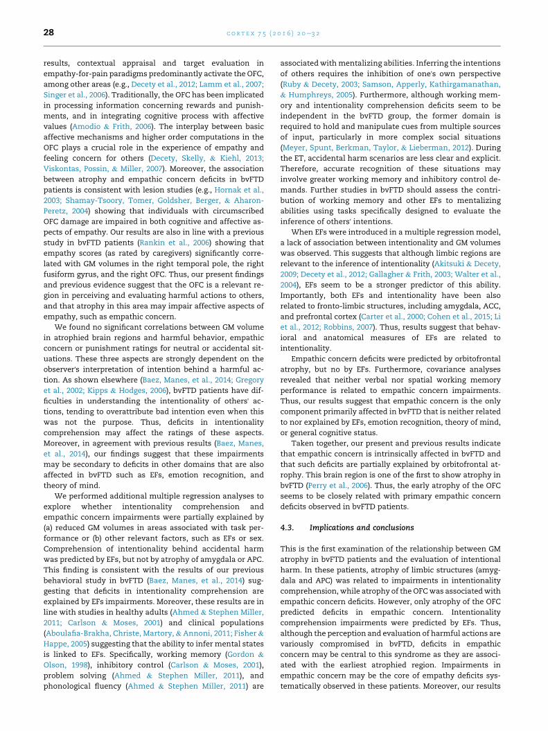
results, contextual appraisal and target evaluation in
empathy-for-pain paradigms predominantly activate the OFC,
among other areas (e.g., Decety et al., 2012; Lamm et al., 2007;
Singer et al., 2006). Traditionally, the OFC has been implicated
in processing information concerning rewards and punish-
ments, and in integrating cognitive process with affective
values (Amodio & Frith, 2006). The interplay between basic
affective mechanisms and higher order computations in the
OFC plays a crucial role in the experience of empathy and
feeling concern for others (Decety, Skelly, & Kiehl, 2013;
Viskontas, Possin, & Miller, 2007). Moreover, the association
between atrophy and empathic concern deficits in bvFTD
patients is consistent with lesion studies (e.g., Hornak et al.,
2003; Shamay-Tsoory, Tomer, Goldsher, Berger, & Aharon-
Peretz, 2004) showing that individuals with circumscribed
OFC damage are impaired in both cognitive and affective as-
pects of empathy. Our results are also in line with a previous
study in bvFTD patients (Rankin et al., 2006) showing that
empathy scores (as rated by caregivers) significantly corre-
lated with GM volumes in the right temporal pole, the right
fusiform gyrus, and the right OFC. Thus, our present findings
and previous evidence suggest that the OFC is a relevant re-
gion in perceiving and evaluating harmful actions to others,
and that atrophy in this area may impair affective aspects of
empathy, such as empathic concern.
We found no significant correlations between GM volume
in atrophied brain regions and harmful behavior, empathic
concern or punishment ratings for neutral or accidental sit-
uations. These three aspects are strongly dependent on the
observer's interpretation of intention behind a harmful ac-
tion. As shown elsewhere (Baez, Manes, et al., 2014; Gregory
et al., 2002; Kipps & Hodges, 2006), bvFTD patients have dif-
ficulties in understanding the intentionality of others' ac-tions, tending to overattribute bad intention even when this
was not the purpose. Thus, deficits in intentionality
comprehension may affect the ratings of these aspects.
Moreover, in agreement with previous results (Baez, Manes,
et al., 2014), our findings suggest that these impairments
may be secondary to deficits in other domains that are also
affected in bvFTD such as EFs, emotion recognition, and
theory of mind.
We performed additional multiple regression analyses to
explore whether intentionality comprehension and
empathic concern impairments were partially explained by
(a) reduced GM volumes in areas associated with task per-
formance or (b) other relevant factors, such as EFs or sex.
Comprehension of intentionality behind accidental harm
was predicted by EFs, but not by atrophy of amygdala or APC.
This finding is consistent with the results of our previous
behavioral study in bvFTD (Baez, Manes, et al., 2014) sug-
gesting that deficits in intentionality comprehension are
explained by EFs impairments. Moreover, these results are in
line with studies in healthy adults (Ahmed & Stephen Miller,
2011; Carlson & Moses, 2001) and clinical populations
(Aboulafia-Brakha, Christe, Martory, &Annoni, 2011; Fisher&
Happe, 2005) suggesting that the ability to infer mental states
is linked to EFs. Specifically, working memory (Gordon &
Olson, 1998), inhibitory control (Carlson & Moses, 2001),
problem solving (Ahmed & Stephen Miller, 2011), and
phonological fluency (Ahmed & Stephen Miller, 2011) are
associatedwithmentalizing abilities. Inferring the intentions
of others requires the inhibition of one's own perspective
(Ruby & Decety, 2003; Samson, Apperly, Kathirgamanathan,
& Humphreys, 2005). Furthermore, although working mem-
ory and intentionality comprehension deficits seem to be
independent in the bvFTD group, the former domain is
required to hold and manipulate cues from multiple sources
of input, particularly in more complex social situations
(Meyer, Spunt, Berkman, Taylor, & Lieberman, 2012). During
the ET, accidental harm scenarios are less clear and explicit.
Therefore, accurate recognition of these situations may
involve greater working memory and inhibitory control de-
mands. Further studies in bvFTD should assess the contri-
bution of working memory and other EFs to mentalizing
abilities using tasks specifically designed to evaluate the
inference of others' intentions.When EFs were introduced in a multiple regression model,
a lack of association between intentionality and GM volumes
was observed. This suggests that although limbic regions are
relevant to the inference of intentionality (Akitsuki & Decety,
2009; Decety et al., 2012; Gallagher & Frith, 2003; Walter et al.,
2004), EFs seem to be a stronger predictor of this ability.
Importantly, both EFs and intentionality have been also
related to fronto-limbic structures, including amygdala, ACC,
and prefrontal cortex (Carter et al., 2000; Cohen et al., 2015; Li
et al., 2012; Robbins, 2007). Thus, results suggest that behav-
ioral and anatomical measures of EFs are related to
intentionality.
Empathic concern deficits were predicted by orbitofrontal
atrophy, but no by EFs. Furthermore, covariance analyses
revealed that neither verbal nor spatial working memory
performance is related to empathic concern impairments.
Thus, our results suggest that empathic concern is the only
component primarily affected in bvFTD that is neither related
to nor explained by EFs, emotion recognition, theory of mind,
or general cognitive status.
Taken together, our present and previous results indicate
that empathic concern is intrinsically affected in bvFTD and
that such deficits are partially explained by orbitofrontal at-
rophy. This brain region is one of the first to show atrophy in
bvFTD (Perry et al., 2006). Thus, the early atrophy of the OFC
seems to be closely related with primary empathic concern
deficits observed in bvFTD patients.
4.3. Implications and conclusions
This is the first examination of the relationship between GM
atrophy in bvFTD patients and the evaluation of intentional
harm. In these patients, atrophy of limbic structures (amyg-
dala and APC) was related to impairments in intentionality
comprehension, while atrophy of the OFCwas associatedwith
empathic concern deficits. However, only atrophy of the OFC
predicted deficits in empathic concern. Intentionality
comprehension impairments were predicted by EFs. Thus,
although the perception and evaluation of harmful actions are
variously compromised in bvFTD, deficits in empathic
concern may be central to this syndrome as they are associ-
ated with the earliest atrophied region. Impairments in
empathic concern may be the core of empathy deficits sys-
tematically observed in these patients. Moreover, our results

c o r t e x 7 5 ( 2 0 1 6 ) 2 0e3 2 29
suggest that adequate executive functioning and preserved
GM volume in the amygdala and the APC are relevant to
comprehend the intentionality, while GM integrity in the OFC
is crucial for feeling empathic concern for others.
Some limitations of this study should be mentioned.
Although we used a somewhat liberal significance level for
some of the VBM multiple regressions, we controlled for age
and total intracranial volume and ruled them out as con-
founds. Moreover, given our moderate sample size, more
subtle associations may have been missed due to low statis-
tical power. While our sample size is large enough for the
multiple regression analyses performed (Green, 1991; Stevens,
2002; Tabachnick & Fidell, 1989), further studies should assess
the structural correlates of intentionality comprehension and
empathic concern in larger groups.
One of the strengths of the current study is its reliance on a
more ecological design that circumvents some limitations of
self-report questionnaires. The task employed here detected
deficits in intentionality comprehension, empathy, and moral
judgment. These results emphasize the value of tasks
involving real-life social scenarios (Burgess, Alderman, Volle,
Benoit, & Gilbert, 2009; Ibanez & Manes, 2012; Torralva, Roca,
Gleichgerrcht, Bekinschtein, et al., 2009), as evidenced by
their greater sensitivity in the clinical assessment of neuro-
psychiatric populations. From a clinical perspective, given
that adequate empathic functioning is an important element
of higher social functioning (Rankin et al., 2005), primary
empathy impairments should be considered in the assess-
ment and early treatment of bvFTD.
In conclusion, our study highlights the importance of
limbic structures in comprehending intentionality and, more
particularly, of the OFC in the early empathy changes
observed in bvFTD. Longitudinal studies are needed to test
whether atrophy of this brain region may predict disease
progression based on empathic concern levels. Moreover,
future studies should explore the relationship between the
atrophy pattern and different aspects of the evaluation of
harmful actions in other variants of frontotemporal dementia.
A more subtle understanding of these complex social cogni-
tive deficits in bvFTD will improve assessment in clinical
settings. Furthermore, insights into relevant factors contrib-
uting to social impairments in bvFTD patients may shed light
on potential strategies for early diagnosis and for the devel-
opment of cognitive stimulation programs.
Acknowledgments
This study was supported by grants from Comisi�on Nacional
de Investigaci�on Cientı́fica y Tecnol�ogica/FONDECYT Regular
(1130920 and 1140114), PICT 2012-0412, and PICT 2012-1309,
CONICET, and the INECO Foundation. The authors declare no
competing financial interests.
Supplementary data
Supplementary data related to this article can be found at
doi:10.1016/j.cortex.2015.11.007.
r e f e r e n c e s
Aboulafia-Brakha, T., Christe, B., Martory, M. D., & Annoni, J. M.(2011). Theory of mind tasks and executive functions: asystematic review of group studies in neurology. Journal ofNeuropsychology, 5, 39e55. http://dx.doi.org/10.1348/174866410X533660.
Ahmed, F. S., & Stephen Miller, L. (2011). Executive functionmechanisms of theory of mind. Journal of Autism andDevelopmental Disorders, 41, 667e678. http://dx.doi.org/10.1007/s10803-010-1087-7.
Akitsuki, Y., & Decety, J. (2009). Social context and perceivedagency affects empathy for pain: an event-related fMRIinvestigation. NeuroImage, 47, 722e734. http://dx.doi.org/10.1016/j.neuroimage.2009.04.091.
Amodio, D. M., & Frith, C. D. (2006). Meeting of minds: the medialfrontal cortex and social cognition. Nature reviews.Neuroscience, 7, 268e277. http://dx.doi.org/10.1038/nrn1884.
Amoruso, L., Sedeno, L., Huepe, D., Tomio, A., Kamienkowski, J.,Hurtado, E., et al. (2014). Time toTango: expertise and contextualanticipation during action observation.NeuroImage, 98, 366e385.http://dx.doi.org/10.1016/j.neuroimage.2014.05.005.
Ashburner, J., & Friston, K. J. (2000). Voxel-basedmorphometryethe methods. NeuroImage, 11, 805e821. http://dx.doi.org/10.1006/nimg.2000.0582.
Baez, S., Couto, B., Torralva, T., Sposato, L. A., Huepe, D.,Montanes, P., et al. (2014a). Comparing moral judgments ofpatients with frontotemporal dementia and frontal stroke.JAMA Neurology, 71, 1172e1176. http://dx.doi.org/10.1001/jamanogyeurol.2014.347.
Baez, S., Herrera, E., Villarin, L., Theil, D., Gonzalez-Gadea, M. L.,Gomez, P., et al. (2013). Contextual social cognitionimpairments in schizophrenia and bipolar disorder. PLoS One,8, e57664. http://dx.doi.org/10.1371/journal.pone.0057664.
Baez, S., Ibanez, A., Gleichgerrcht, E., Perez, A., Roca, M.,Manes, F., et al. (2014). The utility of IFS (INECO FrontalScreening) for the detection of executive dysfunction inadults with bipolar disorder and ADHD. Psychiatry Research,216, 269e276. http://dx.doi.org/10.1016/j.psychres.2014.01.020.
Baez, S., Kanske, P., Matallana, D., Montanes, P., Reyes, P.,Slachevsky, A., et al. (2015). Integration of intention andoutcome for moral judgment in frontotemporal dementia:brain structural signatures. Neurodegenerative Diseases. http://dx.doi.org/10.1159/000441918.
Baez, S., Manes, F., Huepe, D., Torralva, T., Fiorentino, N.,Richter, F., et al. (2014). Primary empathy deficits infrontotemporal dementia. Frontiers in Aging Neuroscience, 6,262. http://dx.doi.org/10.3389/fnagi.2014.00262.
Baez, S., Rattazzi, A., Gonzalez-Gadea, M. L., Torralva, T.,Vigliecca, N. S., Decety, J., et al. (2012). Integrating intentionand context: assessing social cognition in adults withAsperger syndrome. Frontiers in Human Neuroscience, 6, 302.http://dx.doi.org/10.3389/fnhum.2012.00302.
Bar, M. (2004). Visual objects in context. Nature ReviewsNeuroscience, 5, 617e629. http://dx.doi.org/10.1038/nrn1476.
Baron-Cohen, S., &Wheelwright, S. (2004). The empathy quotient:an investigation of adults with Asperger syndrome or highfunctioning autism, and normal sex differences. Journal ofAutism and Developmental Disorders, 34, 163e175.
Bernhardt, B. C., & Singer, T. (2012). The neural basis of empathy.Annual Review of Neuroscience, 35, 1e23. http://dx.doi.org/10.1146/annurev-neuro-062111-150536.
Burgess, P. W., Alderman, N., Volle, E., Benoit, R. G., & Gilbert, S. J.(2009). Mesulam's frontal lobe mystery re-examined.Restorative Neurology and Neuroscience, 27, 493e506. http://dx.doi.org/10.3233/RNN-2009-0511.

c o r t e x 7 5 ( 2 0 1 6 ) 2 0e3 230
Carlson, S. M., & Moses, L. J. (2001). Individual differences ininhibitory control and children's theory of mind. ChildDevelopment, 72, 1032e1053.
Carter, C. S., Macdonald, A. M., Botvinick, M., Ross, L. L.,Stenger, V. A., Noll, D., et al. (2000). Parsing executiveprocesses: strategic vs. evaluative functions of the anteriorcingulate cortex. Proceedings of the National Academy of Sciencesof the United States of America, 97, 1944e1948.
Cerami, C., Dodich, A., Canessa, N., Crespi, C., Marcone, A.,Cortese, F., et al. (2014). Neural correlates of empathicimpairment in the behavioral variant of frontotemporaldementia. Alzheimer's & Dementia: The Journal of the Alzheimer'sAssociation, 10, 827e834. http://dx.doi.org/10.1016/j.jalz.2014.01.005.
Cohen, N., Margulies, D. S., Ashkenazi, S., Schaefer, A.,Taubert, M., Henik, A., et al. (2015). Using executive controltraining to suppress amygdala reactivity to aversiveinformation. NeuroImage. http://dx.doi.org/10.1016/j.neuroimage.2015.10.069.
Couto, B., Manes, F., Montanes, P., Matallana, D., Reyes, P.,Velasquez, M., et al. (2013). Structural neuroimaging of socialcognition in progressive non-fluent aphasia and behavioralvariant of frontotemporal dementia. Frontiers in HumanNeuroscience, 7, 467. http://dx.doi.org/10.3389/fnhum.2013.00467.
Decety, J., & Cacioppo, S. (2012). The speed of morality: a high-density electrical neuroimaging study. Journal ofNeurophysiology, 108, 3068e3072. http://dx.doi.org/10.1152/jn.00473.2012.
Decety, J., Michalska, K. J., & Akitsuki, Y. (2008). Who caused thepain? an fMRI investigation of empathy and intentionality inchildren. Neuropsychologia, 46, 2607e2614. http://dx.doi.org/10.1016/j.neuropsychologia.2008.05.026.
Decety, J., Michalska, K. J., & Kinzler, K. D. (2012). The contributionof emotion and cognition to moral sensitivity: aneurodevelopmental study. Cerebral Cortex, 22, 209e220.http://dx.doi.org/10.1093/cercor/bhr111.
Decety, J., Skelly, L. R., & Kiehl, K. A. (2013). Brain response toempathy-eliciting scenarios involving pain in incarceratedindividuals with psychopathy. JAMA Psychiatry, 70, 638e645.http://dx.doi.org/10.1001/jamapsychiatry.2013.27.
Escobar, M. J., Huepe, D., Decety, J., Sedeno, L., Messow, M. K.,Baez, S., et al. (2014). Brain signatures of moral sensitivity inadolescents with early social deprivation. Scientific Reports, 4,5354. http://dx.doi.org/10.1038/srep05354.
Eslinger, P. J., Moore, P., Anderson, C., & Grossman, M. (2011).Social cognition, executive functioning, and neuroimagingcorrelates of empathic deficits in frontotemporal dementia.Journal of Neuropsychiatry and the Clinical Neurosciences, 23,74e82. http://dx.doi.org/10.1176/appi.neuropsych.23.1.74.
Fan, Y., Duncan, N. W., de Greck, M., & Northoff, G. (2011). Is therea core neural network in empathy? an fMRI based quantitativemeta-analysis. Neuroscience and Biobehavioral Reviews, 35,903e911. http://dx.doi.org/10.1016/j.neubiorev.2010.10.009.
Fernandez-Duque, D., Hodges, S. D., Baird, J. A., & Black, S. E.(2010). Empathy in frontotemporal dementia and Alzheimer'sdisease. Journal of Clinical and Experimental Neuropsychology, 32,289e298. http://dx.doi.org/10.1080/13803390903002191.
Fine, C., Lumsden, J., & Blair, R. J. (2001). Dissociation between'theory of mind' and executive functions in a patient withearly left amygdala damage. Brain: A Journal of Neurology, 124,287e298.
Fisher, N., & Happe, F. (2005). A training study of theory of mindand executive function in children with autistic spectrumdisorders. Journal of Autism and Developmental Disorders, 35,757e771. http://dx.doi.org/10.1007/s10803-005-0022-9.
Folstein, M. F., Robins, L. N., & Helzer, J. E. (1983). The mini-mentalstate examination. Archives of General Psychiatry, 40, 812.
Gallagher, H. L., & Frith, C. D. (2003). Functional imaging of ‘theoryof mind’. Trends in Cognitive Sciences, 7, 77e83.
Garcia-Cordero, I., Sedeno, L., Fraiman, D., Craiem, D., de laFuente, L. A., Salamone, P., et al. (2015). Stroke andNeurodegeneration induce different connectivity aberrationsin the insula. Stroke, 46, 2673e2677.
Good, C. D., Johnsrude, I. S., Ashburner, J., Henson, R. N.,Friston, K. J., & Frackowiak, R. S. (2001). A voxel-basedmorphometric study of ageing in 465 normal adult humanbrains. NeuroImage, 14, 21e36. http://dx.doi.org/10.1006/nimg.2001.0786.
Gordon, A. C., & Olson, D. R. (1998). The relation betweenacquisition of a theory of mind and the capacity to hold inmind. Journal of Experimental Child Psychology, 68, 70e83. http://dx.doi.org/10.1006/jecp.1997.2423.
Green, S. (1991). How many subjects does it take to do aregression analysis. Multivariate Behavioral Research, 26,499e510.
Gregory, C., Lough, S., Stone, V., Erzinclioglu, S., Martin, L., Baron-Cohen, S., et al. (2002). Theory of mind in patients with frontalvariant frontotemporal dementia and Alzheimer's disease:theoretical and practical implications. Brain, 125, 752e764.
Hesse, E., Mikulan, E., Decety, J., Sigman, M., Garcia, M., Silva, W.,et al. (2015). Early detection of intentional harm in the humanamygdala. Brain: A Journal of Neurology. http://dx.doi.org/10.1093/brain/awv336.
Hornak, J., Bramham, J., Rolls, E. T., Morris, R. G., O'Doherty, J.,Bullock, P. R., et al. (2003). Changes in emotion aftercircumscribed surgical lesions of the orbitofrontal andcingulate cortices. Brain: A Journal of Neurology, 126, 1691e1712.http://dx.doi.org/10.1093/brain/awg168.
Hsieh, S., Irish, M., Daveson, N., Hodges, J. R., & Piguet, O. (2013).When one loses empathy: its effect on carers of patients withdementia. Journal of Geriatric Psychiatry and Neurology, 26,174e184. http://dx.doi.org/10.1177/0891988713495448.
Ibanez, A., Kotz, S. A., Barrett, L., Moll, J., & Ruz, M. (2014). Situatedaffective and social neuroscience. Frontiers in HumanNeuroscience, 8, 547. http://dx.doi.org/10.3389/fnhum.2014.00547.
Ibanez, A., & Manes, F. (2012). Contextual social cognition and thebehavioral variant of frontotemporal dementia. Neurology, 78,1354e1362. http://dx.doi.org/10.1212/WNL.0b013e3182518375.
Kipps, C. M., & Hodges, J. R. (2006). Theory of mind infrontotemporal dementia. Social Neuroscience, 1, 235e244.http://dx.doi.org/10.1080/17470910600989847.
Kipps, C. M., Nestor, P. J., Acosta-Cabronero, J., Arnold, R., &Hodges, J. R. (2009). Understanding social dysfunction in thebehavioural variant of frontotemporal dementia: the role ofemotion and sarcasm processing. Brain: A Journal of Neurology,132, 592e603. http://dx.doi.org/10.1093/brain/awn314.
Lamm, C., Batson, C. D., & Decety, J. (2007). The neural substrateof human empathy: effects of perspective-taking andcognitive appraisal. Journal of Cognitive Neuroscience, 19, 42e58.http://dx.doi.org/10.1162/jocn.2007.19.1.42.
Li, C. T., Hsieh, J. C., Wang, S. J., Yang, B. H., Bai, Y. M., Lin, W. C.,et al. (2012). Differential relations between fronto-limbicmetabolism and executive function in patients with remittedbipolar I and bipolar II disorder. Bipolar Disorders, 14, 831e842.http://dx.doi.org/10.1111/bdi.12017.
Lough, S., Kipps, C. M., Treise, C., Watson, P., Blair, J. R., &Hodges, J. R. (2006). Social reasoning, emotion and empathy infrontotemporal dementia. Neuropsychologia, 44, 950e958.
Melloni, M., Lopez, V., & Ibanez, A. (2013). Empathy andcontextual social cognition. Cognitive, Affective & BehavioralNeuroscience. http://dx.doi.org/10.3758/s13415-013-0205-3.
Mendez, M. F. (2006). What frontotemporal dementia revealsabout the neurobiological basis of morality.Medical Hypotheses,67, 411e418. http://dx.doi.org/10.1016/j.mehy.2006.01.048.

c o r t e x 7 5 ( 2 0 1 6 ) 2 0e3 2 31
Mendez, M. F., Anderson, E., & Shapira, J. S. (2005). Aninvestigation of moral judgement in frontotemporaldementia. Cognitive and Behavioral Neurology, 18, 193e197.
Meyer, M. L., Spunt, R. P., Berkman, E. T., Taylor, S. E., &Lieberman, M. D. (2012). Evidence for social working memoryfrom a parametric functional MRI study. Proceedings of theNational Academy of Sciences of the United States of America, 109,1883e1888. http://dx.doi.org/10.1073/pnas.1121077109.
Neary, D., Snowden, J. S., Gustafson, L., Passant, U., Stuss, D.,Black, S., et al. (1998). Frontotemporal lobar degeneration: aconsensus on clinical diagnostic criteria. Neurology, 51,1546e1554.
O'Bryant, S. E., Humphreys, J. D., Smith, G. E., Ivnik, R. J., Graff-Radford, N. R., Petersen, R. C., et al. (2008). Detecting dementiawith the mini-mental state examination in highly educatedindividuals. Archives of Neurology, 65, 963e967. http://dx.doi.org/10.1001/archneur.65.7.963.
Osher, J. E., Wicklund, A. H., Rademaker, A., Johnson, N., &Weintraub, S. (2007). The mini-mental state examination inbehavioral variant frontotemporal dementia and primaryprogressive aphasia. American Journal of Alzheimer's Disease andOther Dementias, 22, 468e473. http://dx.doi.org/10.1177/1533317507307173.
Perry, R. J., Graham, A., Williams, G., Rosen, H., Erzinclioglu, S.,Weiner, M., et al. (2006). Patterns of frontal lobe atrophy infrontotemporal dementia: a volumetric MRI study. Dementiaand Geriatric Cognitive Disorders, 22, 278e287. http://dx.doi.org/10.1159/000095128.
Phelps, E. A. (2006). Emotion and cognition: insights from studiesof the human amygdala. Annual Review of Psychology, 57,27e53. http://dx.doi.org/10.1146/annurev.psych.56.091103.070234.
Piguet, O., Hornberger, M., Mioshi, E., & Hodges, J. R. (2011).Behavioural-variant frontotemporal dementia: diagnosis,clinical staging, and management. Lancet Neurology, 10,162e172. http://dx.doi.org/10.1016/S1474-4422(10)70299-4.
Poletti, M., Enrici, I., & Adenzato, M. (2012). Cognitive andaffective Theory of Mind in neurodegenerative diseases:neuropsychological, neuroanatomical and neurochemicallevels. Neuroscience and Biobehavioral Reviews, 36, 2147e2164.http://dx.doi.org/10.1016/j.neubiorev.2012.07.004.
Preis, M. A., & Kroener-Herwig, B. (2012). Empathy for pain: theeffects of prior experience and sex. European Journal of Pain,16, 1311e1319. http://dx.doi.org/10.1002/j.1532-2149.2012.00119.x.
Rankin, K. P., Gorno-Tempini, M. L., Allison, S. C., Stanley, C. M.,Glenn, S., Weiner, M. W., et al. (2006). Structural anatomy ofempathy in neurodegenerative disease. Brain, 129, 2945e2956.http://dx.doi.org/10.1093/brain/awl254.
Rankin, K. P., Kramer, J. H., & Miller, B. L. (2005). Patterns ofcognitive and emotional empathy in frontotemporal lobardegeneration. Cognitive and Behavioral Neurology, 18, 28e36.
Rascovsky, K., Hodges, J. R., Knopman, D., Mendez, M. F.,Kramer, J. H., Neuhaus, J., et al. (2011). Sensitivity of reviseddiagnostic criteria for the behavioural variant offrontotemporal dementia. Brain: A Journal of Neurology, 134,2456e2477. http://dx.doi.org/10.1093/brain/awr179.
Robbins, T. W. (2007). Shifting and stopping: fronto-striatalsubstrates, neurochemical modulation and clinicalimplications. Philosophical Transactions of the Royal Society ofLondon Series B Biological Sciences, 362, 917e932. http://dx.doi.org/10.1098/rstb.2007.2097.
Rosen, H. J., Gorno-Tempini, M. L., Goldman, W. P., Perry, R. J.,Schuff, N., Weiner, M., et al. (2002). Patterns of brain atrophyin frontotemporal dementia and semantic dementia.Neurology, 58, 198e208.
Ruby, P., & Decety, J. (2003). What you believe versus what youthink they believe: a neuroimaging study of conceptual
perspective-taking. The European Journal of Neuroscience, 17,2475e2480.
Saddoris, M. P., Gallagher, M., & Schoenbaum, G. (2005). Rapidassociative encoding in basolateral amygdala depends onconnections with orbitofrontal cortex. Neuron, 46, 321e331.http://dx.doi.org/10.1016/j.neuron.2005.02.018.
Samson, D., Apperly, I. A., Kathirgamanathan, U., &Humphreys, G. W. (2005). Seeing it my way: a case of aselective deficit in inhibiting self-perspective. Brain, 128,1102e1111. http://dx.doi.org/10.1093/brain/awh464.
Seeley, W. W., Crawford, R., Rascovsky, K., Kramer, J. H.,Weiner, M., Miller, B. L., et al. (2008). Frontal paralimbicnetwork atrophy in very mild behavioral variantfrontotemporal dementia. Archives of Neurology, 65, 249e255.http://dx.doi.org/10.1001/archneurol.2007.38.
Seeley, W. W., Crawford, R. K., Zhou, J., Miller, B. L., &Greicius, M. D. (2009). Neurodegenerative diseases targetlarge-scale human brain networks. Neuron, 62, 42e52. http://dx.doi.org/10.1016/j.neuron.2009.03.024.
Shamay-Tsoory, S. G., Tomer, R., Goldsher, D., Berger, B. D., &Aharon-Peretz, J. (2004). Impairment in cognitive and affectiveempathy in patients with brain lesions: anatomical andcognitive correlates. Journal of Clinical and ExperimentalNeuropsychology, 26, 1113e1127. http://dx.doi.org/10.1080/13803390490515531.
Shenhav, A., & Greene, J. D. (2014). Integrative moral judgment:dissociating the roles of the amygdala and ventromedialprefrontal cortex. Journal of Neuroscience, 34, 4741e4749.
Singer, T., & Lamm, C. (2009). The social neuroscience ofempathy. Annals of the New York Academy of Sciences, 1156,81e96. http://dx.doi.org/10.1111/j.1749-6632.2009.04418.x.
Singer, T., Seymour, B., O'Doherty, J. P., Stephan, K. E., Dolan, R. J.,& Frith, C. D. (2006). Empathic neural responses are modulatedby the perceived fairness of others. Nature, 439, 466e469.http://dx.doi.org/10.1038/nature04271.
Stein, J. L., Wiedholz, L. M., Bassett, D. S., Weinberger, D. R.,Zink, C. F., Mattay, V. S., et al. (2007). A validated network ofeffective amygdala connectivity. NeuroImage, 36, 736e745.http://dx.doi.org/10.1016/j.neuroimage.2007.03.022.
Stevens, J. P. (2002). Applied multivariate statistics for the socialsciences. Mahwah, NJ: Erlbaum.
Stone, V. E., Baron-Cohen, S., Calder, A., Keane, J., & Young, A.(2003). Acquired theory of mind impairments in individualswith bilateral amygdala lesions. Neuropsychologia, 41, 209e220.
Tabachnick, B. G., & Fidell, L. S. (1989). Using multivariate statistics.Cambridge, MA: Harper & Row.
Torralva, T., Roca, M., Gleichgerrcht, E., Bekinschtein, T., &Manes, F. (2009a). A neuropsychological battery to detectspecific executive and social cognitive impairments in earlyfrontotemporal dementia. Brain, 132, 1299e1309.
Torralva, T., Roca, M., Gleichgerrcht, E., Lopez, P., & Manes, F.(2009b). INECO Frontal Screening (IFS): a brief, sensitive, andspecific tool to assess executive functions in dementia. Journalof the International Neuropsychological Society: JINS, 15, 777e786.http://dx.doi.org/10.1017/S1355617709990415.
Toussaint, L., & Webb, J. R. (2005). Gender differences in therelationship between empathy and forgiveness. The Journal ofSocial Psychology, 145, 673e685. http://dx.doi.org/10.3200/SOCP.145.6.673-686.
Treadway, M. T., Buckholtz, J. W., Martin, J. W., Jan, K.,Asplund, C. L., Ginther, M. R., et al. (2014). Corticolimbic gatingof emotion-driven punishment. Nature Neuroscience, 17,1270e1275. http://dx.doi.org/10.1038/nn.3781.
Viskontas, I. V., Possin, K. L., & Miller, B. L. (2007). Symptoms offrontotemporal dementia provide insights into orbitofrontalcortex function and social behavior. Annals of the New YorkAcademy of Sciences, 1121, 528e545. http://dx.doi.org/10.1196/annals.1401.025.

c o r t e x 7 5 ( 2 0 1 6 ) 2 0e3 232
Walter, H., Abler, B., Ciaramidaro, A., & Erk, S. (2005). Motivatingforces of human actions. Neuroimaging reward and socialinteraction. Brain Research Bulletin, 67, 368e381. http://dx.doi.org/10.1016/j.brainresbull.2005.06.016.
Walter, H., Adenzato, M., Ciaramidaro, A., Enrici, I., Pia, L., &Bara, B. G. (2004). Understanding intentions in socialinteraction: the role of the anterior paracingulate cortex.Journal of Cognitive Neuroscience, 16, 1854e1863. http://dx.doi.org/10.1162/0898929042947838.
Whitwell, J. L., Przybelski, S. A., Weigand, S. D., Ivnik, R. J.,Vemuri, P., Gunter, J. L., et al. (2009). Distinct anatomicalsubtypes of the behavioural variant of frontotemporaldementia: a cluster analysis study. Brain: A Journal of
Neurology, 132, 2932e2946. http://dx.doi.org/10.1093/brain/awp232.
Young, L., & Saxe, R. (2009). Innocent intentions: a correlationbetween forgiveness for accidental harm and neural activity.Neuropsychologia, 47, 2065e2072. http://dx.doi.org/10.1016/j.neuropsychologia.2009.03.020.
Zaki, J., & Ochsner, K. N. (2012). The neuroscience of empathy:progress, pitfalls and promise. Nature Neuroscience, 15,675e680. http://dx.doi.org/10.1038/nn.3085.
Ze, O., Thoma, P., & Suchan, B. (2014). Cognitive and affectiveempathy in younger and older individuals. Aging & MentalHealth, 18, 929e935. http://dx.doi.org/10.1080/13607863.2014.899973.