Oral Pathology lecture2 - Columbia · PDF file“liver colored”, alveolar bone loss...
16
Pediatric Oral Pathology Kavita Kohli, DDS Associate Professor of Clinical Dentistry K. Kohli, DDS Topics • Newborn lesions • Infections • Ulcerative and vesiculobullous lesions • Pigmented, vascular and red lesions • Exophytic lesions • Gingival Enlargements K. Kohli, DDS Lesions in Newborns • D/D – Keratin Cysts – Congenital Epulis – Natal/Neonatal Teeth
Transcript of Oral Pathology lecture2 - Columbia · PDF file“liver colored”, alveolar bone loss...
1
Pediatric Oral PathologyKavita Kohli, DDS
Associate Professor of Clinical Dentistry
K. Kohli, DDS
Topics• Newborn lesions• Infections• Ulcerative and vesiculobullous lesions• Pigmented, vascular and red lesions• Exophytic lesions• Gingival Enlargements
K. Kohli, DDS
Lesions in Newborns• D/D
– Keratin Cysts – Congenital Epulis– Natal/Neonatal Teeth
2
K. Kohli, DDS
Keratin Cysts of the Newborn
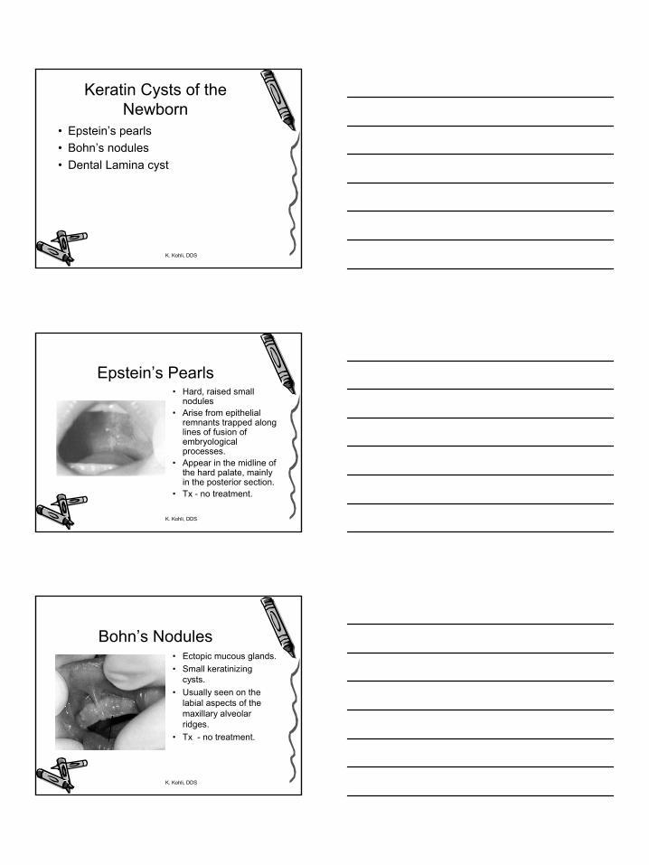
• Epstein’s pearls• Bohn’s nodules• Dental Lamina cyst
K. Kohli, DDS
Epstein’s Pearls• Hard, raised small
nodules• Arise from epithelial
remnants trapped along lines of fusion of embryological processes.
• Appear in the midline of the hard palate, mainly in the posterior section.
• Tx - no treatment.
K. Kohli, DDS
Bohn’s Nodules• Ectopic mucous glands. • Small keratinizing
cysts. • Usually seen on the
labial aspects of the maxillary alveolar ridges.
• Tx - no treatment.

3
K. Kohli, DDS
Dental Lamina Cyst• Usually seen on the crest of the
alveolus• Remnants of the dental lamina.• Tx - no treatment.
K. Kohli, DDS
Congenital Epulis of the Newborn
• Relatively rare, seen in neonates(at birth), of unknown origin, with proliferation of mesenchymal cells.
• Equal distribution between mx and md.
• Females > males.• Usually firm,
pedunculated,pink, smooth, solitary.
• Tx - often regress with time, but may need to be excised, recurrence is uncommon.
K. Kohli, DDS
Natal/Neonatal Teeth• Natal - seen present at birth.• Neonatal - seen within 30
days of birth.• In almost all cases it is the
early eruption of a primary incisor.
• Usually only 5/6th of the crown is formed and the mobility arises from no root development.
• Tx - nursing issues, firms up as root develops, may be extracted if aspiration a possibility.
4
K. Kohli, DDS
Oral Infections• D/D -
– Bacterial– Viral– Fungal
K. Kohli, DDS
Bacterial Infections• Odontogenic• Scarlet fever• Tuberculosis• Atypical mycobacterial infection• Actinomycosis• Syphillis• Impetigo
• Osteomyelitis
K. Kohli, DDS
Odontogenic Infections• Acute - sick child, raised temp., red swollen
face. • Chronic - sinus tract present, mobile and/or
discolored tooth, halitosis.• Tx -
– remove the cause and local drainage and debridement,– May admit if spikes in temp. seen, facial space involvement
suspected or seen &/or dehydrated. – Antibiotics - only if systemic involvement seen, or if child is
immunocompromised. Pen family first drug of choice.

5
K. Kohli, DDS
Osteomyelitis• Some times an odontogenic infection can
lead to osteomyelitis in the mandible. • Radiographically - moth eaten appearance. • Tx - curettage to remove bony sequestra,
antibiotics (after culture and sensitivity test) for at least 6 weeks.
K. Kohli, DDS
Viral Infections• Primary herpetic gingivostomatitis• Herpes labialis• Herpangina• Hand, foot and mouth disease• Infectious mononucleosis• Varicella
K. Kohli, DDS
Primary Herpetic Gingivostomatitis
• Most common cause of severe oral ulcerations in children over the age of 6 mos (peaks at 14 mos).
• Caused by Herpes Simplex Type 1. • Incubation period of 3-5 days with a prodromal 48 hour h/o
irritability, lymphadenopathy, pyrexia and malaise. • Stomatitis seen, with gingival tissues become red and
edematous. • Vesicles seen any where on oral mucosa and rapidly break
down to form very painful ulcers. Solitary ulcers (<3mm) seen and some times larger ulcers with irregular margins are seen when there is coalescence of individual lesions.
• Self limiting and ulcers heal spontaneously without scarring within 10-14 days.
6
K. Kohli, DDS
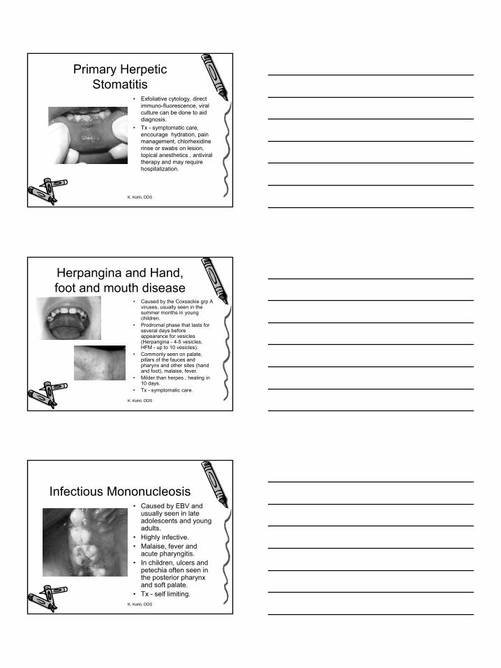
Primary Herpetic Stomatitis
• Exfoliative cytology, direct immuno-fluorescence, viral culture can be done to aid diagnosis.
• Tx - symptomatic care, encourage hydration, pain management, chlorhexidine rinse or swabs on lesion, topical anesthetics , antiviral therapy and may require hospitalization.
K. Kohli, DDS
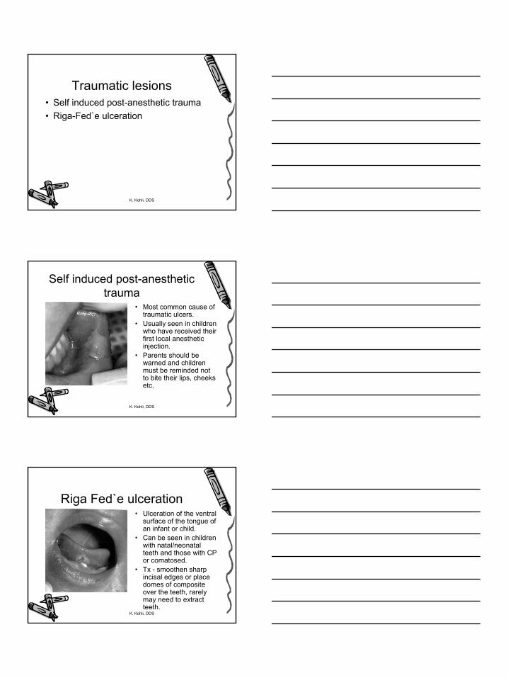
Herpangina and Hand, foot and mouth disease
• Caused by the Coxsackie grp A viruses, usually seen in the summer months in young children.
• Prodromal phase that lasts for several days before appearance for vesicles (Herpangina - 4-5 vesicles, HFM - up to 10 vesicles).
• Commonly seen on palate, pillars of the fauces and pharynx and other sites (hand and foot), malaise, fever.
• Milder than herpes , healing in 10 days.
• Tx - symptomatic care.
K. Kohli, DDS
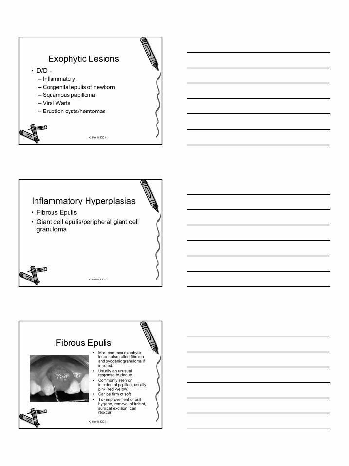
Infectious Mononucleosis• Caused by EBV and
usually seen in late adolescents and young adults.
• Highly infective. • Malaise, fever and
acute pharyngitis. • In children, ulcers and
petechia often seen in the posterior pharynx and soft palate.
• Tx - self limiting.

7
K. Kohli, DDS
Varicella• Highly contagious virus. • Seen as chicken pox in
children and as shingles in adults.
• Prodromal phase of malaise and fever for 24 hours, followed by crops of pruriticvesicles.
• 50% of children have oral lesions.
• Tx - self limiting, resolves in 7-10 days, supportive and palliative.
K. Kohli, DDS
Fungal Infections• Candidiasis
– Common oral organism, but usually does not cause infection unless host is immunocompromised.
– Acute pseudomembranous - in infants seen as Thrush. White scrapable plaques that reveal an erythematous base. In older children, seen in immunocompromised ones who are under active treatment - like CT, RT, broad spectrum ab.’s and steroids.
– Median rhomboid glossitis - seen on dorsal surface of the tongue (usually anterior to the vallate papillae). Can be a response to broad spectrum ab.’s.
• Tx - antifungal for 4 weeks (Nystatin, Ampho B, Fluconazole or Ketoconazole.
K. Kohli, DDS
Ulcerative and Vesiculobullous Lesions
• D/D -– Traumatic– Infective (already discussed)– Others
8
K. Kohli, DDS
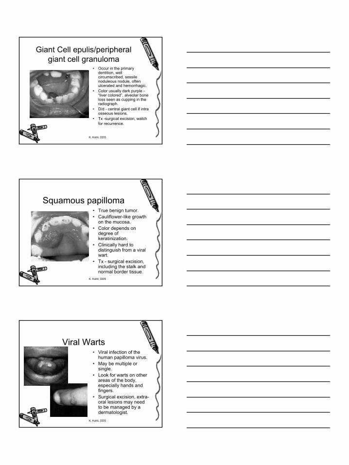
Traumatic lesions• Self induced post-anesthetic trauma• Riga-Fed`e ulceration
K. Kohli, DDS
Self induced post-anesthetic trauma
• Most common cause of traumatic ulcers.
• Usually seen in children who have received their first local anesthetic injection.
• Parents should be warned and children must be reminded not to bite their lips, cheeks etc.
K. Kohli, DDS
Riga Fed`e ulceration• Ulceration of the ventral
surface of the tongue of an infant or child.
• Can be seen in children with natal/neonatal teeth and those with CP or comatosed.
• Tx - smoothen sharp incisal edges or place domes of composite over the teeth, rarely may need to extract teeth.

9
K. Kohli, DDS
Others• Recurrent aphthous ulceration• Erythema multiforme• Stevens-Johnson syndrome• Behcets syndrome• Epidermolysis bullosa• Lupus erythematosus• Neutropenic ulceration
K. Kohli, DDS
Recurrent aphthous ulceration
• Estimated to affect up to 20% of the population. • Seems to have a genetic predisposition, cause
unknown• 3 types -
• Minor aphthae - majority of cases, crops of shallow ulcers up to 5mm, non-keratinized mucosa, typical yellow pseudomebranous slough with an erythematous border.
• Major aphthae - involves the kertinized mucosa, larger ulcers, last longer.
• Herpetiform ulceration
K. Kohli, DDS
Recurrent aphthous ulceration
• Tx - symptomatic care w/ mouth rinses (chlorhexidine, tetracycline,benzydamine hydrochloride, benadryl, xylocaine), heals within 10-14 days without scarring for minor, but with scarring in major.

10
K. Kohli, DDS
Pigmented, Vascular and Red lesions
• D/D -– Vascular– Pigmented– Others
K. Kohli, DDS
Vascular Lesions• Hemangioma• Other vascular malformations• Hematoma• Petechiae and purpura• Hereditary haemorrhagic telangiectasia• Sturge-Weber syndrome• Maffuci’s syndrome
K. Kohli, DDS
Hemangioma• Endothelial
hamartomas,• Typically present at
birth, may grow with the infant, but then regress with time and may even completely disappear
• Tx - none required other than observation, may be a cosmetic concern.

11
K. Kohli, DDS
Petechiae and Purpura• Petechiae - small pinpoint submucosal or
subcutaneous hemorrhages.• Purpura or ecchymoses present as larger
collections of blood. • Usually seen in patients with severe bleeding
disorders or coagulopathies, leukemia etc. • Initially bright red in color, change to a bluish-
brown hue with time as the extravasated blood is metabolized.
K. Kohli, DDS
Pigmented lesions• Melanotic neuroectodermal tumor of
infancy• Peutz-Jeghers Syndrome• Addision’s disease
K. Kohli, DDS
Other Red lesions• Giant cell epulis/peripheral giant cell granuloma• Eruption cyst• Langerhans cell histiocytosis• Geographic tongue• Fissured tongue• Median rhomboid glossitis - already
discussed• Heavy metal toxicity

12
K. Kohli, DDS
Eruption Cyst or hematoma
• Follicular enlargement appearing just before the eruption of tooth.
• Blue-black in color (may contain blood).
• Tx - none unless infected, reassure the child and parent, follicle will rupture, but may need to surgically opened if infected.
K. Kohli, DDS
Geographic tongue• Also known as glossitis
migrans, benign migratory glossitis, erythema migrans or wandering rash of the tongue.
• Areas of depapillation and erythema with a heaped up keratinized margin on the lateral and dorsal surface of the tongue - map like area that changes.
• Tx - none, may prescribe chlorhexidine mouthwash and/or topical steroids when child in pain
K. Kohli, DDS
Fissured Tongue• Also known as plicated
tongue, scrotal tongue, fissured tongue or lingua secta.
• Usually see fissures that run perpendicular to the lateral borders
• Commonly seen in children w/ Downs.
• About 20% will also have geographic tongue or associated c/ Melkersson-Rosenthal Syndrome.
• Tx - none
13
K. Kohli, DDS
Exophytic Lesions• D/D -
– Inflammatory– Congenital epulis of newborn– Squamous papilloma– Viral Warts– Eruption cysts/hemtomas
K. Kohli, DDS
Inflammatory Hyperplasias• Fibrous Epulis• Giant cell epulis/peripheral giant cell
granuloma
K. Kohli, DDS
Fibrous Epulis• Most common exophytic
lesion, also called fibroma and pyogenic granuloma if infected.
• Usually an unusual response to plaque.
• Commonly seen on interdental papillae, usually pink (red -yellow).
• Can be firm or soft• Tx - improvement of oral
hygiene, removal of irritant, surgical excision, can reoccur.
14
K. Kohli, DDS
Giant Cell epulis/peripheral giant cell granuloma
• Occur in the primary dentition, well circumscribed, sessile noduleous nodule, often ulcerated and hemorrhagic.
• Color usually dark purple -“liver colored”, alveolar bone loss seen as cupping in the radiograph.
• D/d - central giant cell if intra osseous lesions.
• Tx -surgical excision, watch for recurrence.
K. Kohli, DDS
Squamous papilloma• True benign tumor.• Cauliflower-like growth
on the mucosa. • Color depends on
degree of keratinization.
• Clinically hard to distinguish from a viral wart.
• Tx - surgical excision, including the stalk and normal border tissue.
K. Kohli, DDS
Viral Warts• Viral infection of the
human papilloma virus. • May be multiple or
single. • Look for warts on other
areas of the body, especially hands and fingers.
• Surgical excision, extra-oral lesions may need to be managed by a dermatologist.

15
K. Kohli, DDS
Gingival Enlargements• D/D -
– Drug induced hyperplasias– Syndromes
K. Kohli, DDS
Drug Induced Hyperplasia• Phenytoin• Cyclosporin A• Nifedipine• verapamil
K. Kohli, DDS
Drug Induced Gingival Enlargements
• Phenytoin• Interdental papillae, may be delayed eruption due the bulk of
fibrous tissue, ectopic eruption, withdrawal of drug will bring about resolution in most cases.
• Tx - oral hygiene key in control of overgrowth, chlorhexidine mouthwash, gingivectomy to allow for eruption and esthetics.
• Cyclosporin A• H/o liver, kidney, heart and combined heart/lung transplants.
most commonly used med for anti-rejection, seen in about 30-70% of these cases.
• Nifedipine and verapamil• Both are calcium channel blockers, used to control cyclosporin
induced hypertension after transplants in children. • Tx - oral hygiene and gingivectomy.
16
K. Kohli, DDS
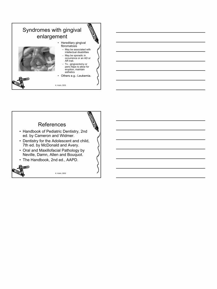
Syndromes with gingival enlargement
• Hereditary gingival fibromatosis– May be associated with
intellectual disabilities– May be sporadic in
occurrence or an AD or AR trait.
– Tx - gingivectomy or perio flaps to allow for eruption, maintain esthetics
• Others e.g.: Leukemia.
K. Kohli, DDS
References• Handbook of Pediatric Dentistry, 2nd
ed. by Cameron and Widmer. • Dentistry for the Adolescent and child,
7th ed. by McDonald and Avery. • Oral and Maxillofacial Pathology by
Neville, Damn, Allen and Bouquot. • The Handbook, 2nd ed., AAPD.