Oral mucous membrane
72
Oral Mucous Membrane Upload Upload By : Ahmed Ali Abbas By : Ahmed Ali Abbas Babylon University College of Dentistry Babylon University College of Dentistry download download this file from Website on this file from Website on Google Google TheOptimalSmile.wix.com TheOptimalSmile.wix.com Then choose Lectures Then choose Lectures Then Second Stage Then Second Stage Then choose the lecture you need Then choose the lecture you need
Transcript of Oral mucous membrane
Oral Mucous Membrane
UploadUpload By : Ahmed Ali Abbas By : Ahmed Ali Abbas
Babylon University College of Dentistry Babylon University College of Dentistry
downloaddownload this file from Website on this file from Website on Google Google
TheOptimalSmile.wix.comTheOptimalSmile.wix.com Then choose Lectures Then choose Lectures Then Second StageThen Second Stage Then choose the lecture you needThen choose the lecture you need

1.Oral Mucosa
The moist lining of the oral cavity is called Oral Mucosa or Oral Mucous Membrane
The function of oral mucosa is protection, sensation and secretion
It is continuous with the skin outside the oral cavity and differs from it in appearance and structure
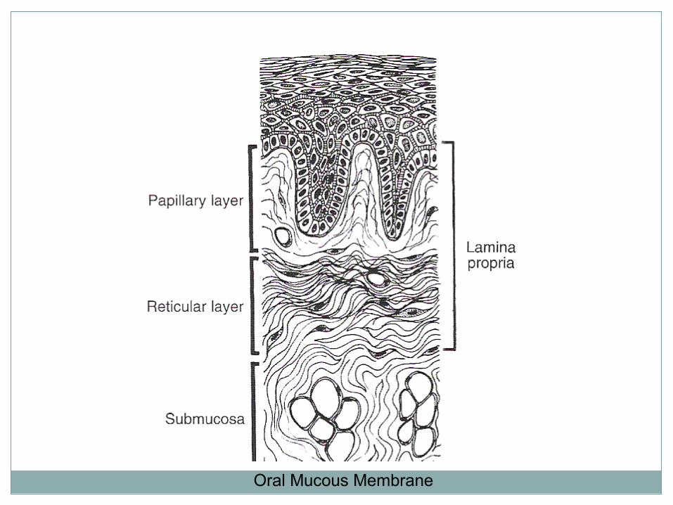
Oral Mucous Membrane

Oral Mucous Membrane
Epithelium
Lamina Propria
Submucosa
PeriosteumBone


Oral Epithelium
Oral epithelium forms the surface of the oral mucosa that forms a barrier between the oral environment and the deeper tissues
It is derived from the embryonic ectoderm
It is stratified squamous epithelium and may or may not be keratinized
Beneath the epithelium lies the connective tissue

Oral Epithelium -Keratinized

CELL LAYERS OF ORTHOKERATINISED EPITHELIUM
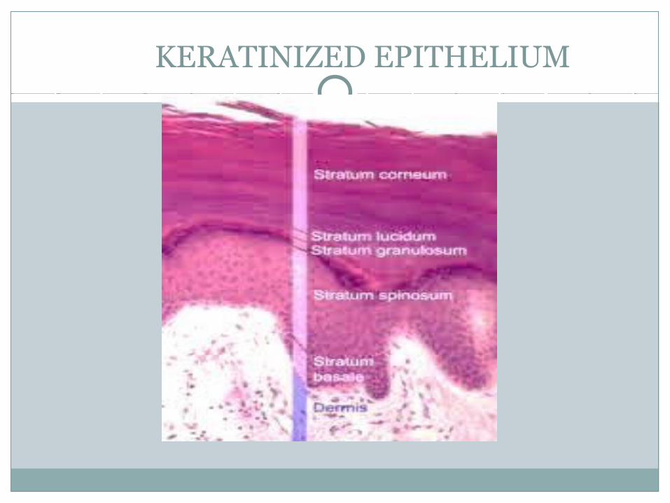
KERATINIZED EPITHELIUM

Keratinized oral epithelium
Most of the oral mucosal surface is lined by nonkeratinized stratified squamous epithelium except gingiva, hard palate and dorsal surface of the tongue where the epithelium is keratinized
The keratinized cells have no nuclei and the cytoplasm is displaced by large numbers of keratin filaments
Keratinized epithelium is associated with masticatory function and have four layers of cells

The four layers are:
1.Stratum Basale
2.Stratum Spinosum
3.Stratum Granulosum
4.Stratum Corneum
Cellular layers of oral epithelium - Keratinized

1. Stratum Basale
The cells of the stratum basale are cuboidal or low columnar and form a single layer resting on the basal lamina
The basal lamina is at the interface of the epithelium and lamina propria
Epithelial cells of the oral mucosa are in a constant state of renewal
The basal cells show the maximum mitotic activity

2. Stratum Spinosum
Stratum spinosum is usually several cells thick
They shaped like polyhedron with short cytoplasmic processes
The stratum basale and the first layers of stratum spinosum are referred to as stratum germinativum because these cells give rise to new epithelial cells
3. Stratum Granulosum
Cells of stratum granulosum are flat and are found in layers of three to five cells thick
This layer is prominent in keratinized epithelium (and absent in nonkeratinized epithelium)
These cells have keratohyaline granules in their cytoplasm
Keratohyaline granules help to form the matrix of the keratin fibres found in the superficial layer

4. Stratum Corneum
Cells of stratum corneum are flat, devoid of nuclei and full of keratin filament surrounded by a matrix
These cells are continuously being sloughed and are replaced by epithelial cells that migrate from the underlying layers
Nonkeratinized oral epithelium
Nonkeratinized epithelial cells in the superfecial layers do not have keratin filaments in the cytoplasm
The surface cells also have nuclei
The stratum corneum and stratum granulosum layers are absent
This epithelium is associated with lining of the oral cavity
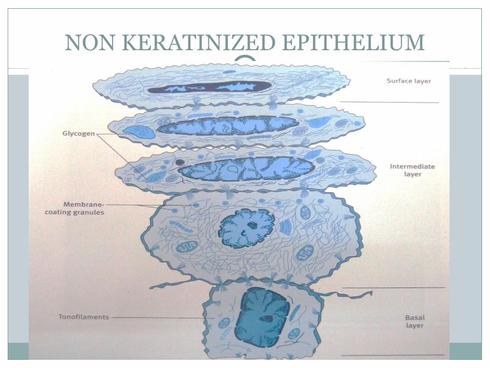
NON KERATINIZED EPITHELIUM
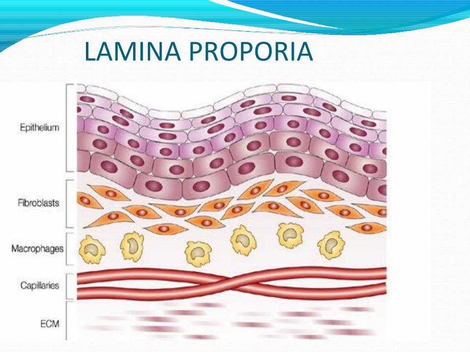
LAMINA PROPORIA
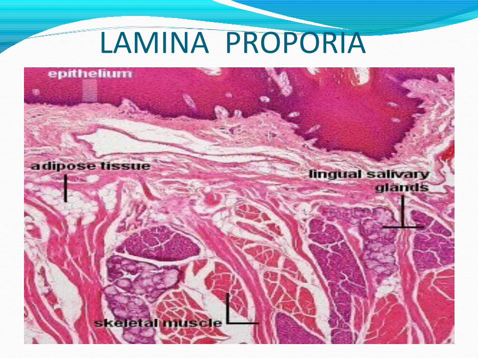
LAMINA PROPORIA
Connective tissue
Connective tissue can be differentiated as Lamina Propria and Submucosa
Lamina propria
Lamina propria is the connective tissue layer immediately below the epithelium
It can be divided into papillary layer and the reticular layer
Papillary layer forms finger like projections of connective tissue that extend deep in the epithelial layers

Papillary layer is prominent in masticatory mucosa and reticular layer is prominent in lining mucosa
Lamina prorpria consists of blood vessels and cells like fibroblasts, cells of blood vessels and lymphatics and nerves
Epithelium is avascular, hence its metabolic needs come via the vessels of the lamina propria

Submucosa
Submucosa lies below the lamina propria and serves as an attachment between lamina propria and bone or skeletal muscle
It is found in the cheeks, lips and parts of the palate
It consists of large blood vessels, nerves and lymphatics and its functions are nutrition and defense
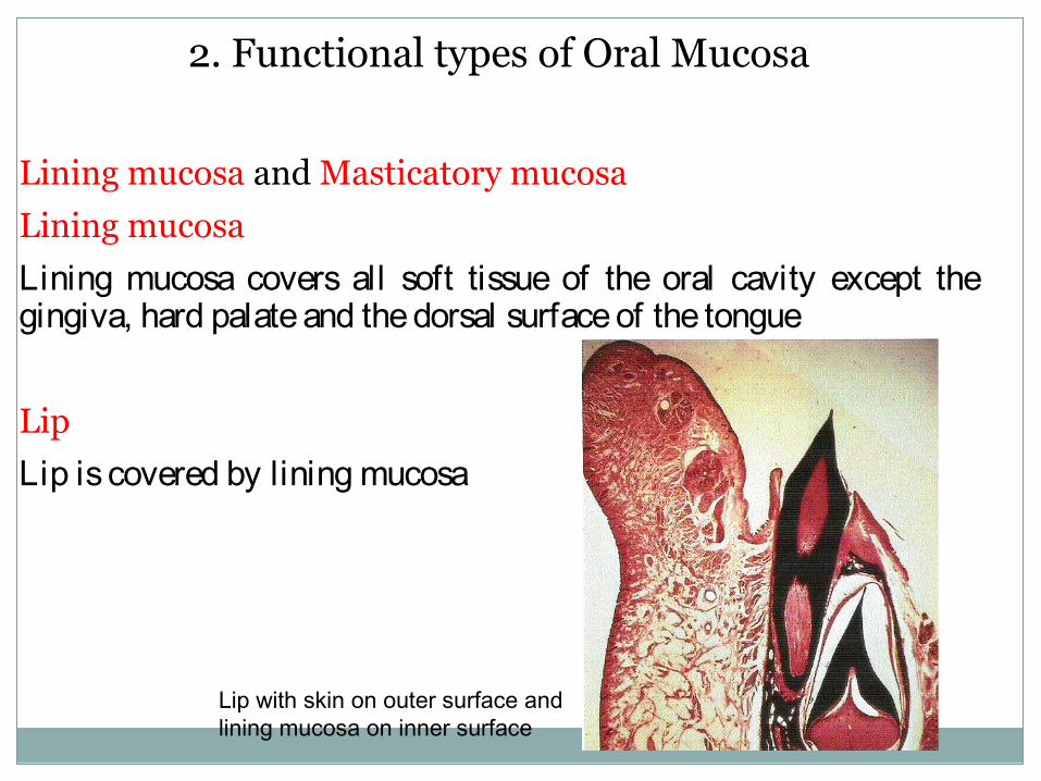
2. Functional types of Oral Mucosa
Lining mucosa and Masticatory mucosa
Lining mucosa
Lining mucosa covers all soft tissue of the oral cavity except the gingiva, hard palate and the dorsal surface of the tongue
Lip
Lip is covered by lining mucosa
Lip with skin on outer surface and lining mucosa on inner surface

Vermilion border
The junction between the skin and mucous membrane is known as the vermilion border
Here the epithelium is thin therefore, the red blood cells in the capillaries show through contributing to the vermilion colour
Vermelion Zone

VERMILLION BORDER OF LIP

Ventral surface of the tongue
The lining mucosa here contains both lamina propria and submucosa
The submucosa merges with the muscle bundles of the ventral surface of the tongue
Ventral surface of tongue
HARD PALATE

SOFT PALATE
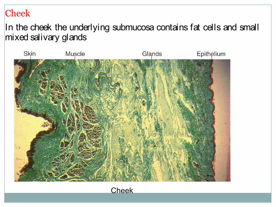
Cheek
In the cheek the underlying submucosa contains fat cells and small mixed salivary glands
Cheek

Floor of the mouth
The mucous membrane of the floor of the mouth is thin and loosely attached to the underlying structures
Floor of the mouth
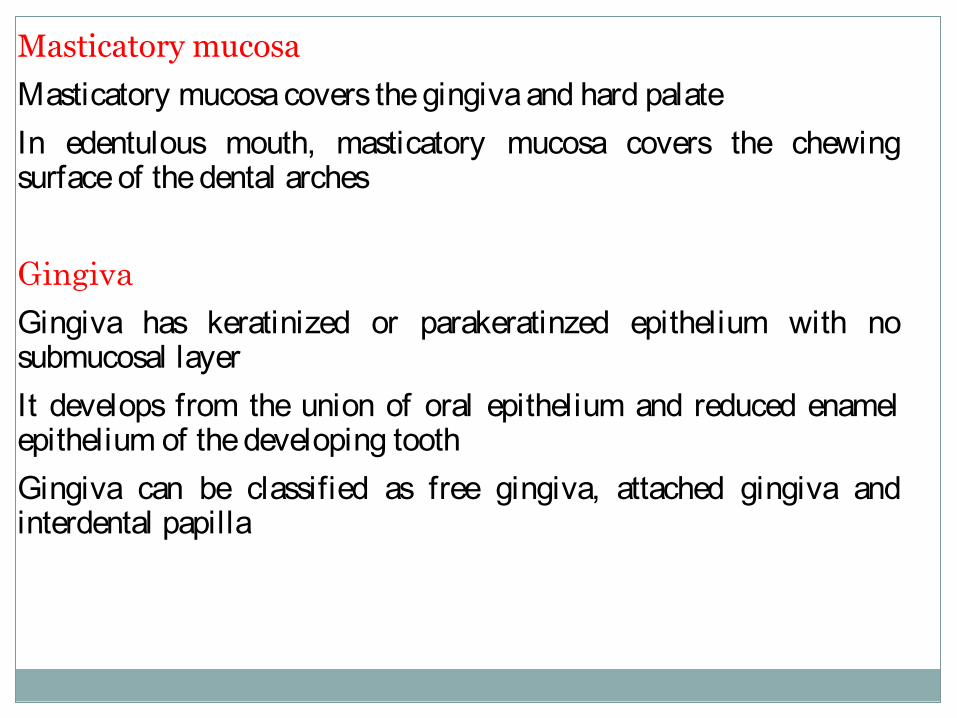
Masticatory mucosa
Masticatory mucosa covers the gingiva and hard palateIn edentulous mouth, masticatory mucosa covers the chewing surface of the dental arches
Gingiva
Gingiva has keratinized or parakeratinzed epithelium with no submucosal layerIt develops from the union of oral epithelium and reduced enamel epithelium of the developing toothGingiva can be classified as free gingiva, attached gingiva and interdental papilla

Normal Gingiva

Development of gingiva from oral epithelium and reduced enamel epithelium
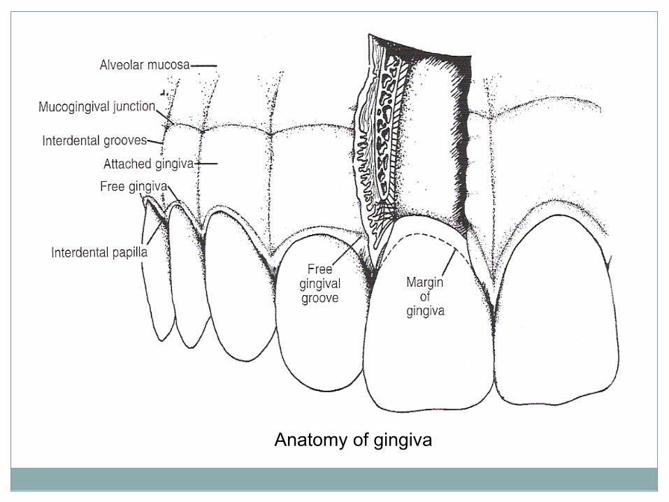
Anatomy of gingiva

Free gingiva (or marginal gingiva)
It is that part of the oral mucosa that surrounds the necks of the teeth and forms the free margin of the gingival tissue
It is differentiated apically from the attached gingival by the free gingival groove
The inner side of it forms the gingival sulcus
The free gingival mucosa is composed of stratified squamous epithelium that may be keratinized, parakeratinzed or sometimes nonkeratinized
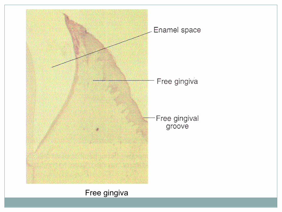
Free gingiva

A- gingiva
B- sulcular epithelium
C- junctional epithelium
D- lamina propria (connective tissue)
E- alveolar process
F- PDL
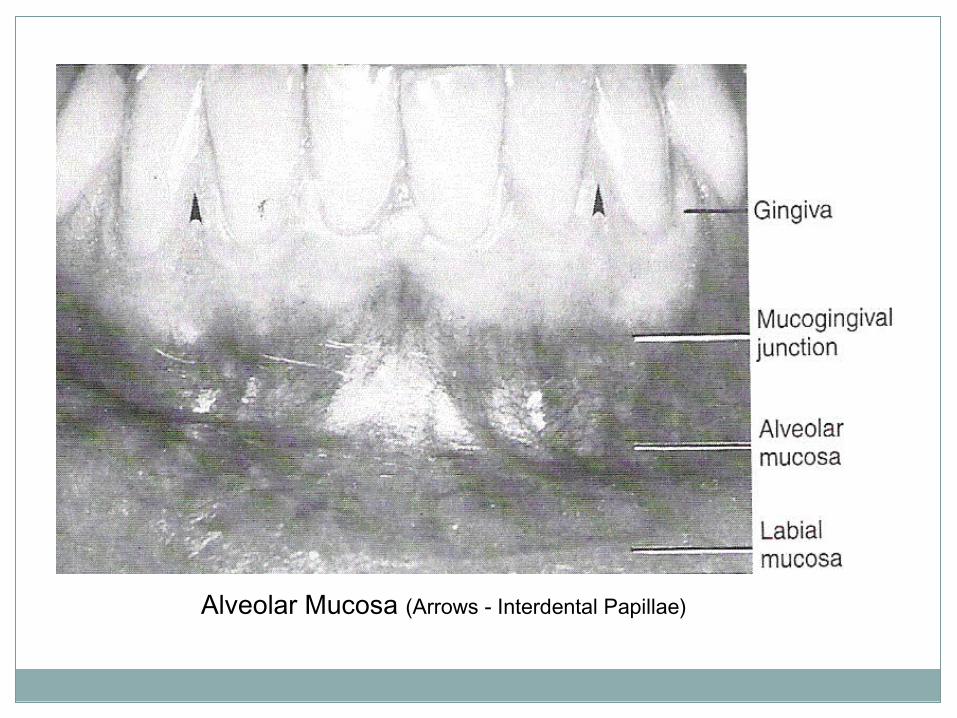
Alveolar Mucosa (Arrows - Interdental Papillae)
Attached gingiva
The attached gingiva lies between the free gingival groove and the alveolar mucosa
The junction of the attached gingiva and the alveolar mucosa is called mucogingival junction
In healthy mouth attached gingiva shows stippling (orange-peel appearance) which is a characteristic of this type of mucosa

Histology of gingiva
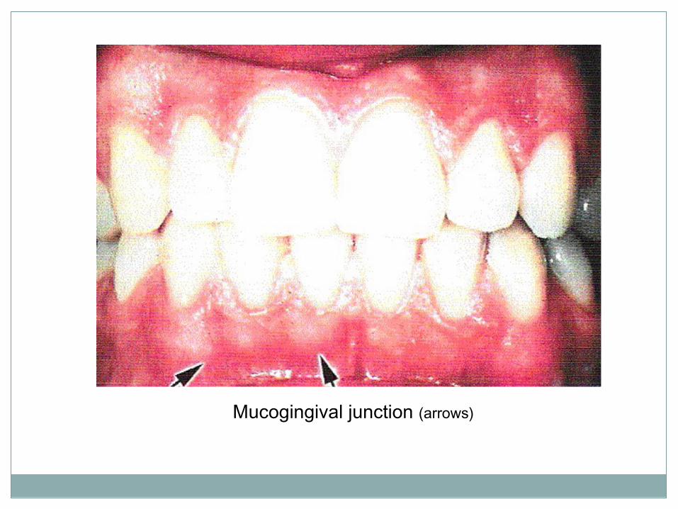
Mucogingival junction (arrows)
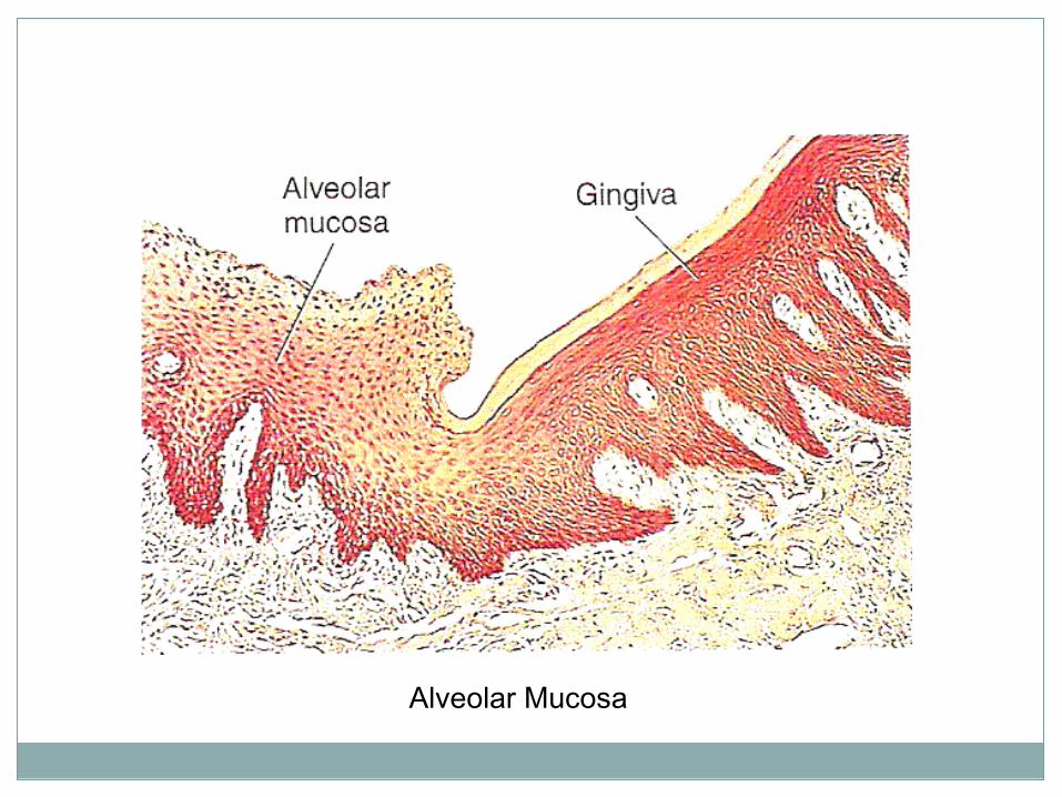
Alveolar Mucosa
Interdental papilla
Interdental papillae are those parts of gingival tissue that appear in-between teeth apical to the contact points
Interdental grooves extend vertically between the interdental papilla corresponding to the depressions between the roots
Confirming to the shape of the interproximal contact area is a valley like depression in the interdental papilla called Col.
This depression lies in the facial and lingual plane

Interdental Papilla (arrow)
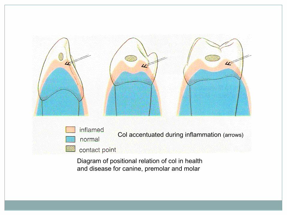
Diagram of positional relation of col in health and disease for canine, premolar and molar
Col accentuated during inflammation (arrows)

Junctional epithelium
Junctional epithelium forms the seal of the gingival epithelium and the tooth
It forms the floor of the gingival sulcus and extends apically to the enamel of the tooth
Disturbances of epithelial attachment results in deepening of the sulcus which is a sign of gingival/periodontal disease

Histology of gingiva

GINGIVA

TYPES OF EPIFHELIUM:
Oral or outer epithelium sulcular epitheliumJunctional epithelium
Sulcular epithelium
Junction epithelium
Oral epithelium

HISTOLOGY OF GINGIVA

GINGIVAL EPITHELIUM
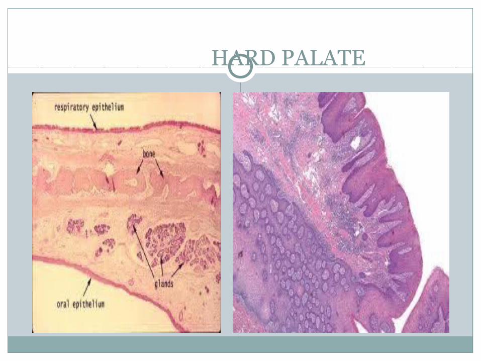
Hard palate
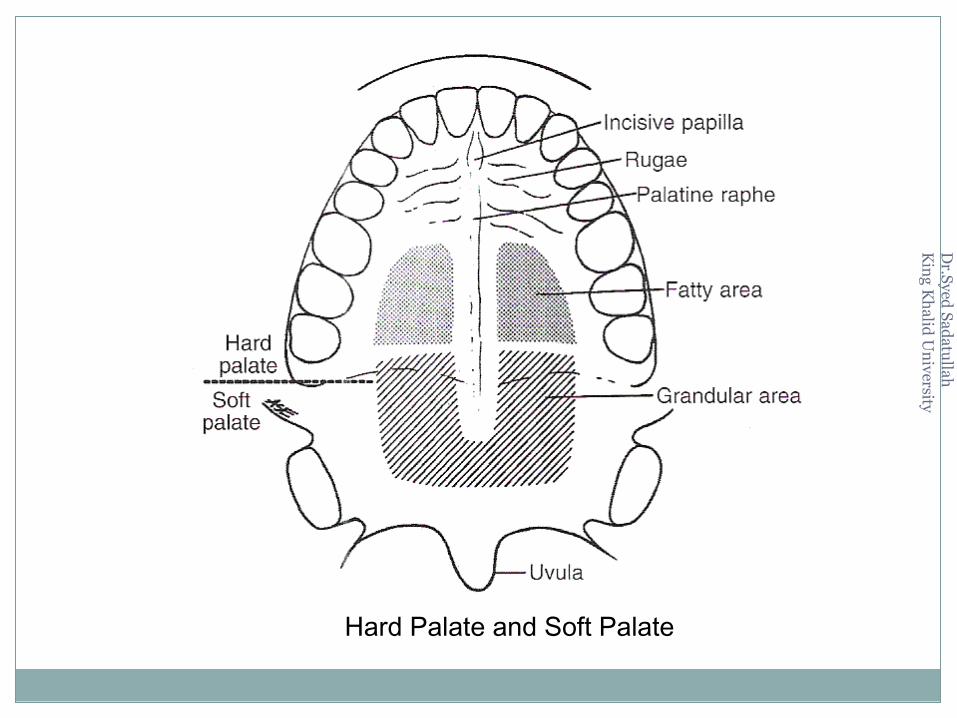
The surface of the hard palate that is visible in the mouth is covered by masticatory mucosa
The lateral regions of the posterior part contains palatine glandsThese glands are purely mucous glands
The midline of the hard palate is called median raphe where there is no submucosa
A series of folds appear in the anterior part of the palate called rugae
Dr.Syed
Sa datu
llah
Kin
g Kh
alid U
niversit y
Hard Palate and Soft Palate

3 Mucosa of the Tongue
Specialized mucosa covers the dorsal surface of the body of the tongue
The connective tissue binds the epithelium to the underlying skeletal muscle
The epithelium is modified, keratinized, stratified covered with papillae, which can be seen by naked eye

The different papillae found on the dorsal surface of the tongue are:1.Filliform papillae
2.Funginform papillae
3.Circumvallate papillae
4.Foliate papillae
Dorsum of Tongue
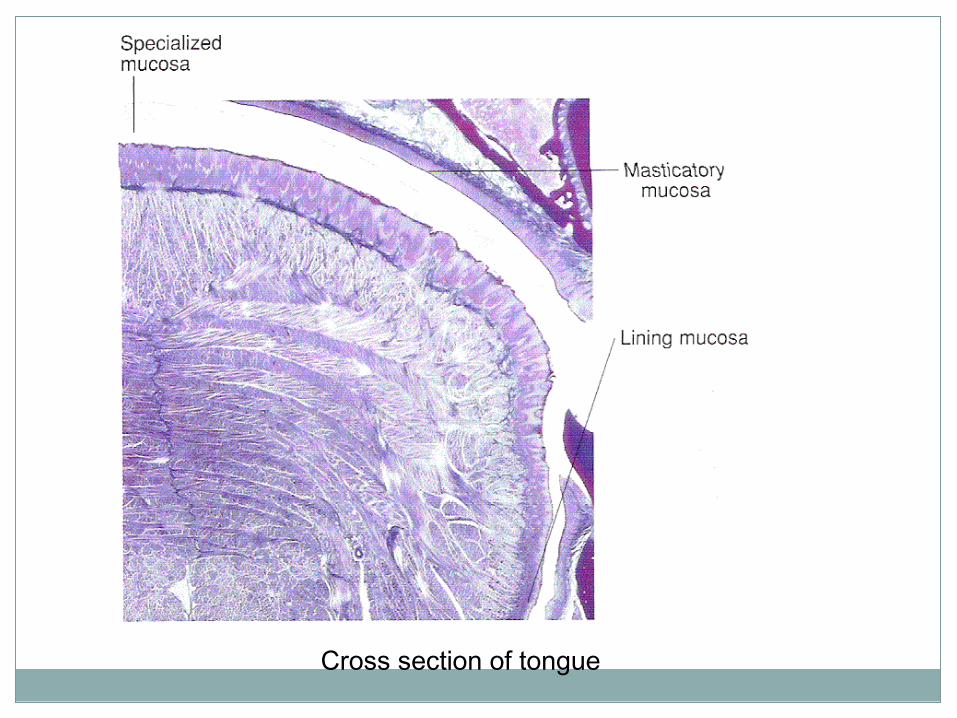
Cross section of tongue
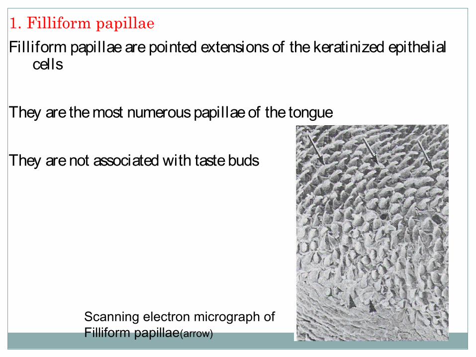
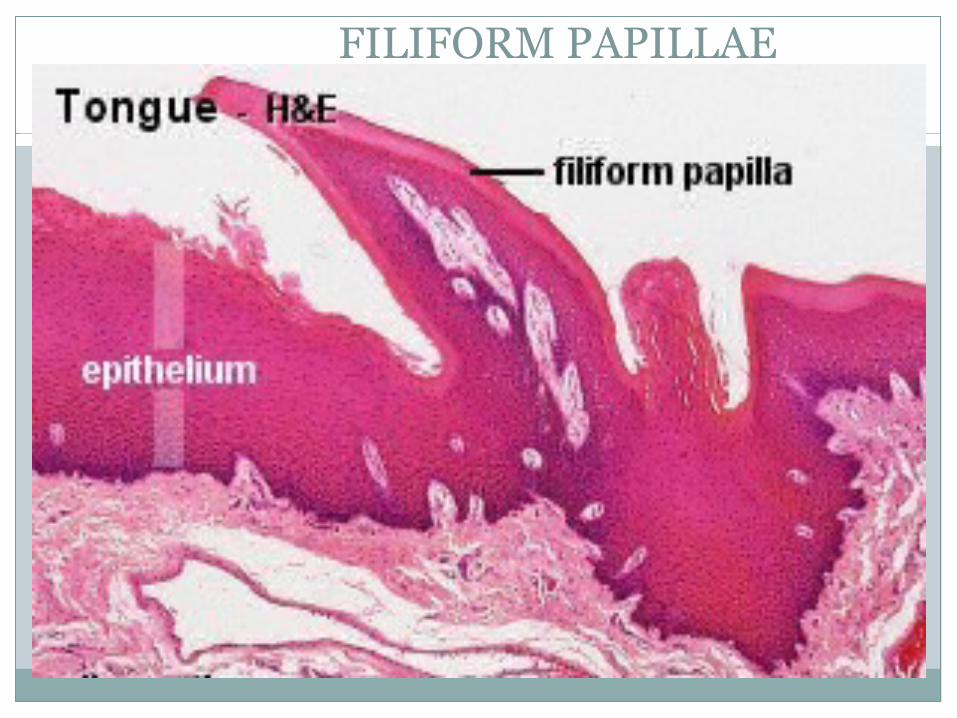
1. Filliform papillae
Filliform papillae are pointed extensions of the keratinized epithelial cells
They are the most numerous papillae of the tongue
They are not associated with taste buds
Scanning electron micrograph of Filliform papillae(arrow)

Filliform Papillae
2. Fungiform papillae
Fungiform papillae are fewer than the filliform papillae and are scattered over the dorsal surface of the tongue
They are rounded elevations above the surface of the tongue
They have taste buds on their superior surfaces
The surface of fungiform papillae is not keratinized

Scanning electron micrograph of Fungiform papillae (arrow) surrounded by Filliform papillae

Fungiform Papillae
3. Circumvallate papillae
The circumvallate papillae are located at the junction of the anterior two thirds (body) and posterior one thirds (base) of the tongue
There are eight to twelve in number and are bigger than fungiform papillae
Circumvallate papillae are lined with taste buds and also openings of serous glands
The secretion from the serous glands washes away food for renewal of taste
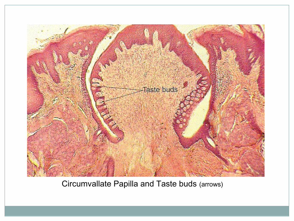
Circumvallate Papilla and Taste buds (arrows)
4. Foliate papillae
Foliate papillae are located in the furrows along the posterior sides of the tongue
They may be lined with taste buds
They are not prominent in human beings

TONGUE

PAPILLAE OF TONGUE
FILIFORM PAPILLAE

FUNGIFORM & CIRCUMVALLATE

TASTE BUDS

TASTE BUDS

TASTE PORES

Thank You