Oral and Maxillofacial Surgeons and ... - Head and Neck Trauma
49
Oral and Maxillofacial Surgeons and the seriously injured patient Barts and The London NHS Trust
Transcript of Oral and Maxillofacial Surgeons and ... - Head and Neck Trauma
Oral and Maxillofacial Surgeons and the seriously
injured patient
Barts and The London NHS Trust
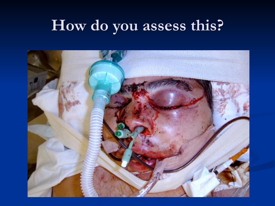
How do you assess this?
Primary Survey
A Airway & Cervical Spine B Breathing & Ventilation C Circulation & Arrest Haemorrhage D Disability E Exposure
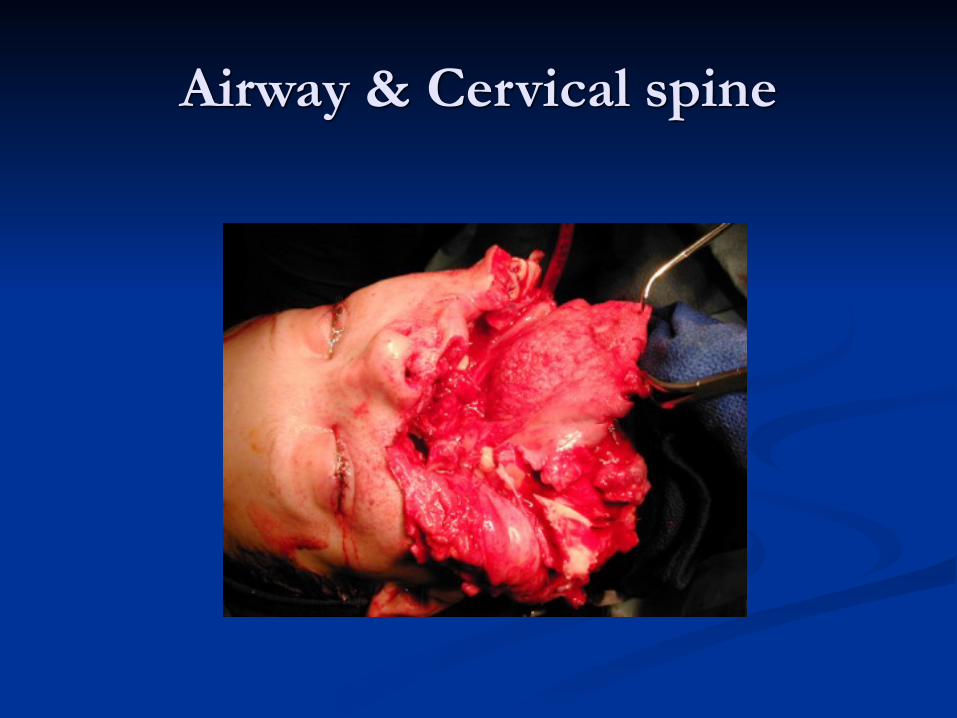
Airway & Cervical spine
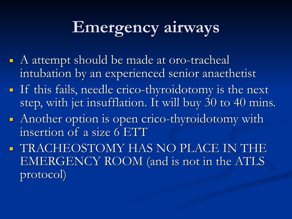
Emergency airways
■ A attempt should be made at oro-tracheal intubation by an experienced senior anaethetist
■ If this fails, needle crico-thyroidotomy is the next step, with jet insufflation. It will buy 30 to 40 mins.
■ Another option is open crico-thyroidotomy with insertion of a size 6 ETT
■ TRACHEOSTOMY HAS NO PLACE IN THE EMERGENCY ROOM (and is not in the ATLS protocol)
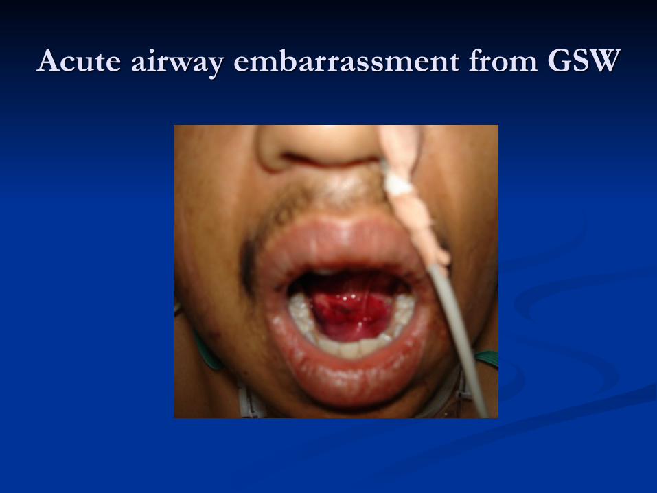
Acute airway embarrassment from GSW
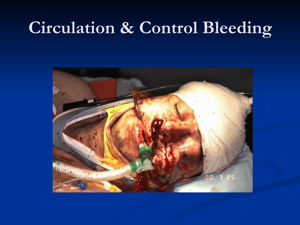
Circulation & Control Bleeding
Controlling acute maxillofacial bleeding
■ Midface fractures bleed profusely. ■ Place a medium mouth prop on each side and
insert an epistat into each nostril ■ Inflate the posterior balloon with saline, tug back
and then inflate the anterior one. ■ If epistats are not available then pack the nose
using posterior nasal packs.
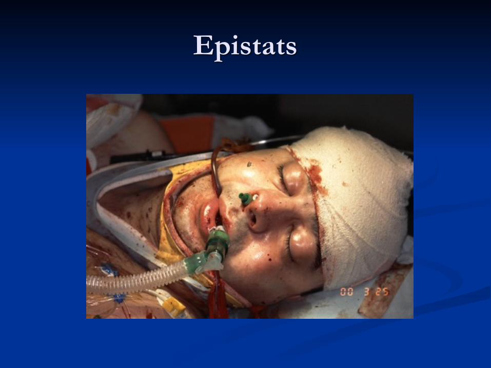
Epistats
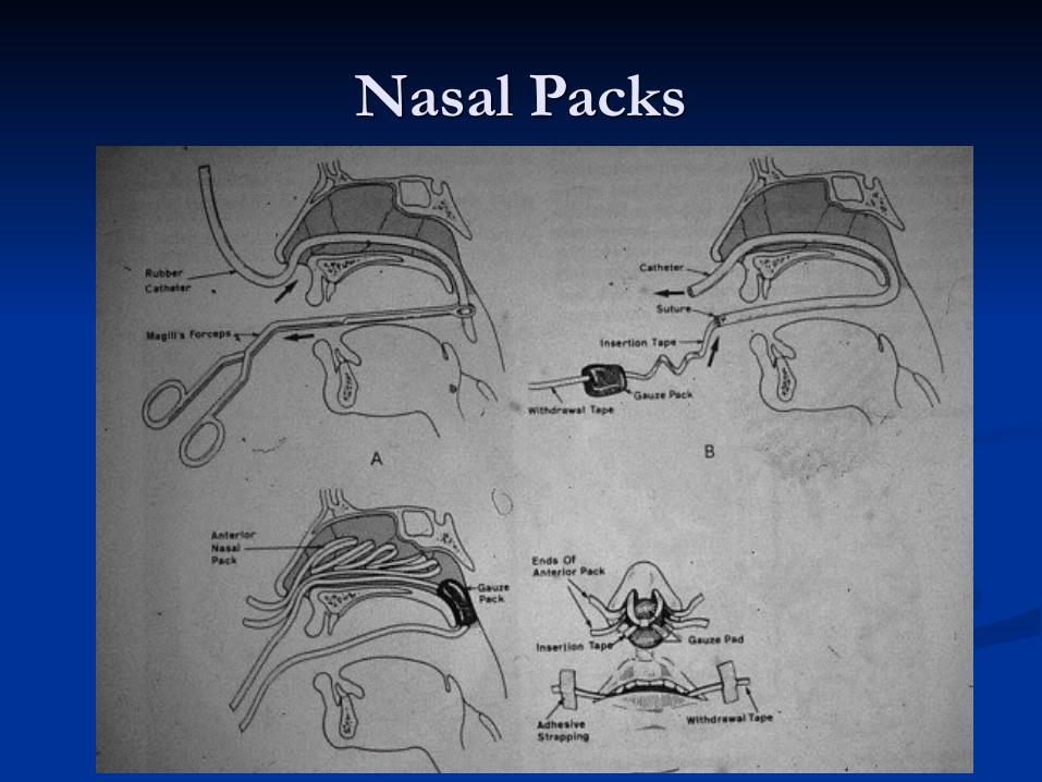
Nasal Packs
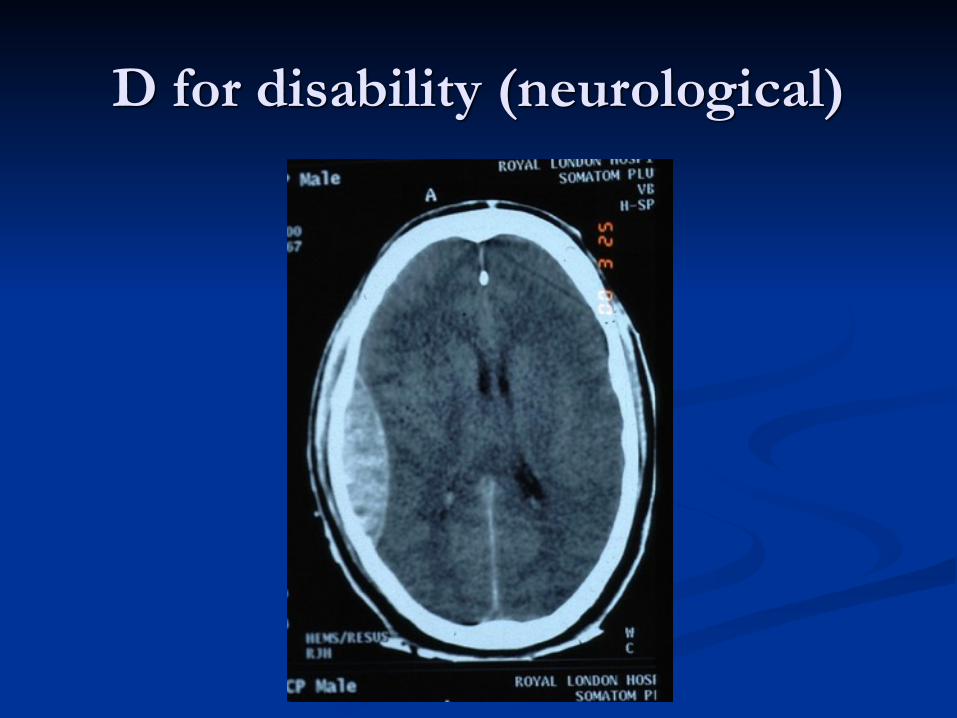
D for disability (neurological)
Secondary Survey
■ A systematic examination of the entire body from head to toe.
■ Carried out only once the primary survey has been completed and the resulting issues dealt with.
■ A thorough Maxillofacial examination is part of the secondary survey, systematically examining all the facial structures to identify potential injuries. Document findings clearly.
Maxillofacial Secondary Survey■ LOOK for swelling, bruising, lacerations, or signs specific
to facial bone fractures (e.g. subconjunctival haemorrhage or Battles sign.
■ FEEL the entire facial skeleton by bilateral palpation to elicit crepitus or steps. If patient conscious test sensation.
■ MOVE the maxilla and mandible to see if there is abnormal mobility indicative of a fracture e.g. lefort 2/3
■ EXAMINE the eyes for globe injury, visual acuity, pupils and orbit disruption. ?Ophthalmology. Auroscope for ears
■ EXAMINE the oral cavity for signs of facial bone fractures, avulsed or fractured teeth and dental occlusion.
■ Reassess once extubated…
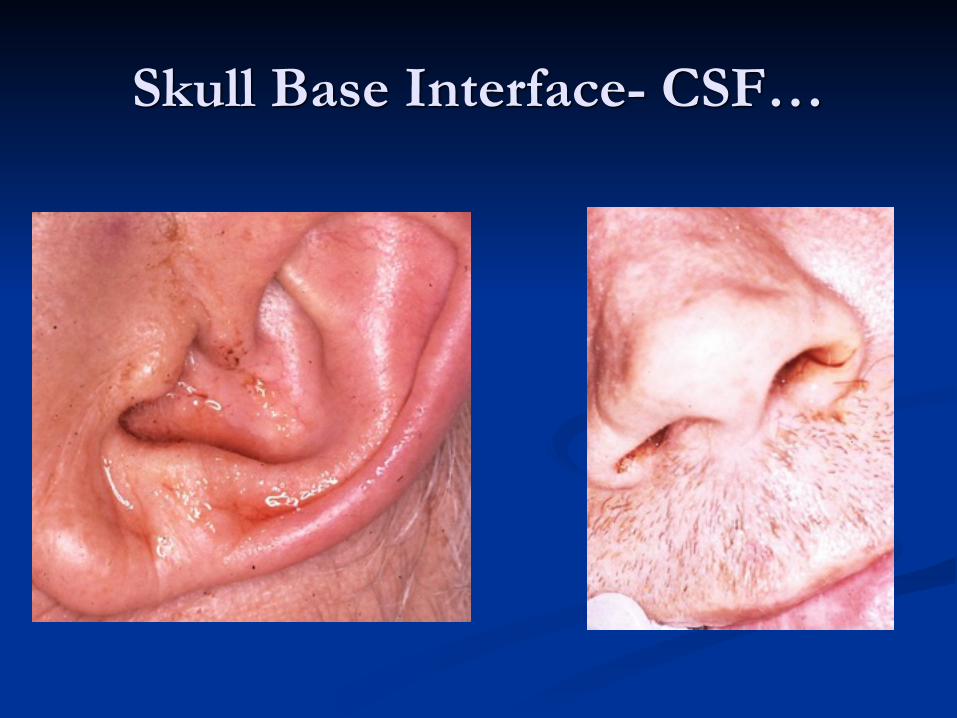
Skull Base Interface- CSF…
Imaging
■ Seriously injured patients usually are going to the CT scanner for their brain, doing axial scans of the facial bones takes minimal extra time. Ask for 3D reformat.
■ Conscious co-operative patients can have a facial bone series (OPG, PA jaws and single Occipitomental).
■ Further imaging is carried out according to the specific injuries identified by the facial bone series or the clinical examination.
■ Chest x-ray if unaccounted missing teeth
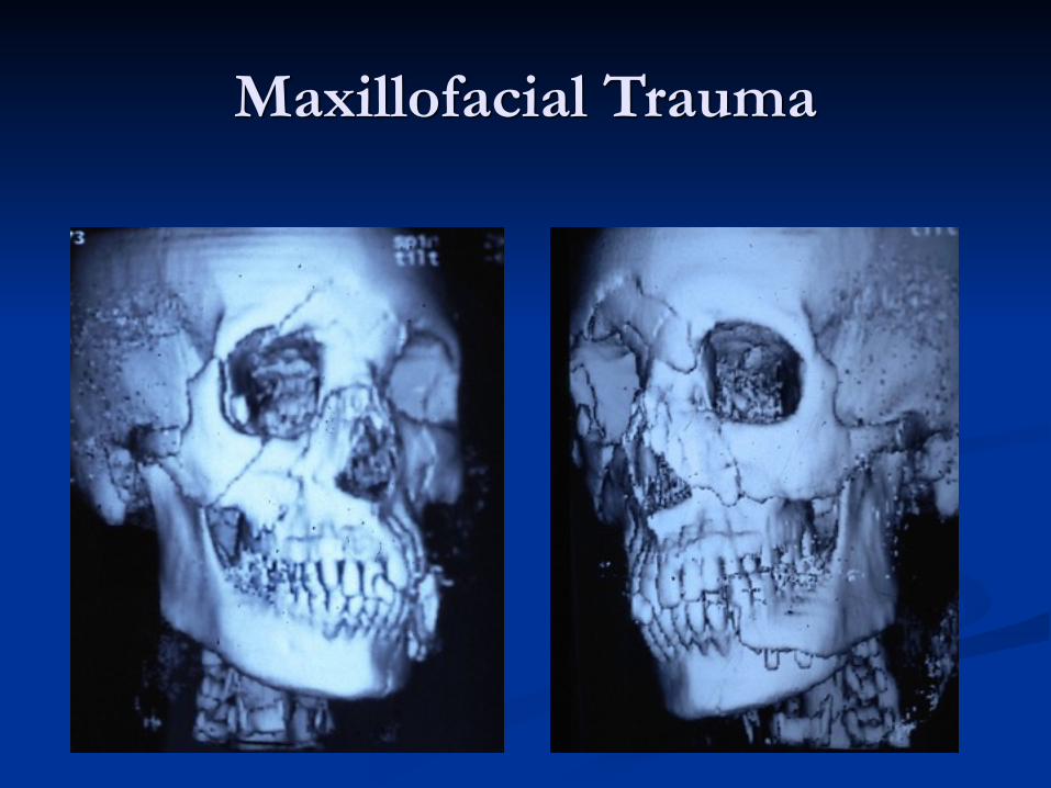
Maxillofacial Trauma
Soft Tissue Trauma
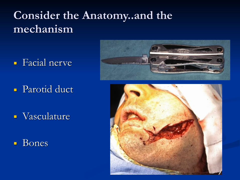
Consider the Anatomy..and the mechanism
■ Facial nerve
■ Parotid duct
■ Vasculature
■ Bones
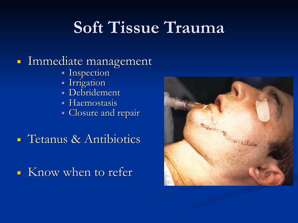
Soft Tissue Trauma
■ Immediate management ■ Inspection ■ Irrigation ■ Debridement ■ Haemostasis ■ Closure and repair
■ Tetanus & Antibiotics
■ Know when to refer
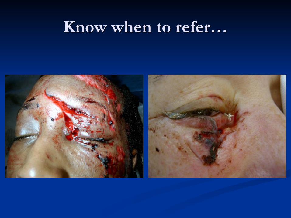
Know when to refer…
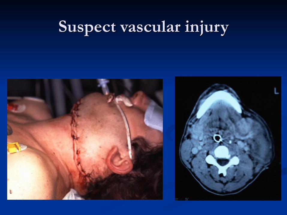
Suspect vascular injury
Beware penetrating neck wounds
■ Size and Level of entry wound is unreliable. ■ Do not explore depth with your finger! Anything deeper
than platysma MUST be formally assessed. ■ Obtain history from ambulance crew - type of weapon,
amount of blood loss at scene, vital signs ■ Haemodynamic status is important (pulse and BP)
■ If unstable at scene and unstable in resus room - need exploration in main theatre with a vascular surgeon and on table angiogram facility
■ If unstable at scene but stable in resus room - investgate further with CT angiogram
■ If stable at scene and stable in resus - explore and close at leisure
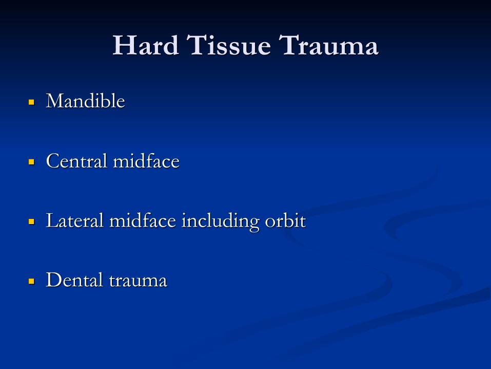
Hard Tissue Trauma
■ Mandible
■ Central midface
■ Lateral midface including orbit
■ Dental trauma
Mandibular Fractures
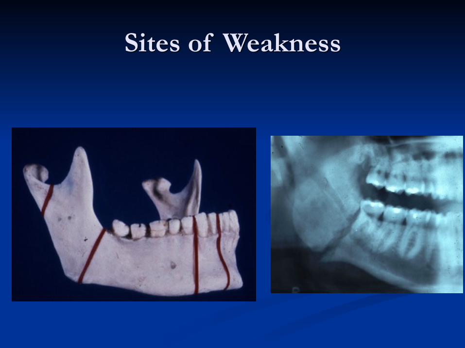
Sites of Weakness
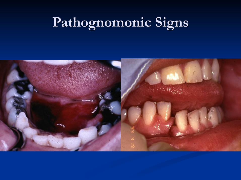
Pathognomonic Signs
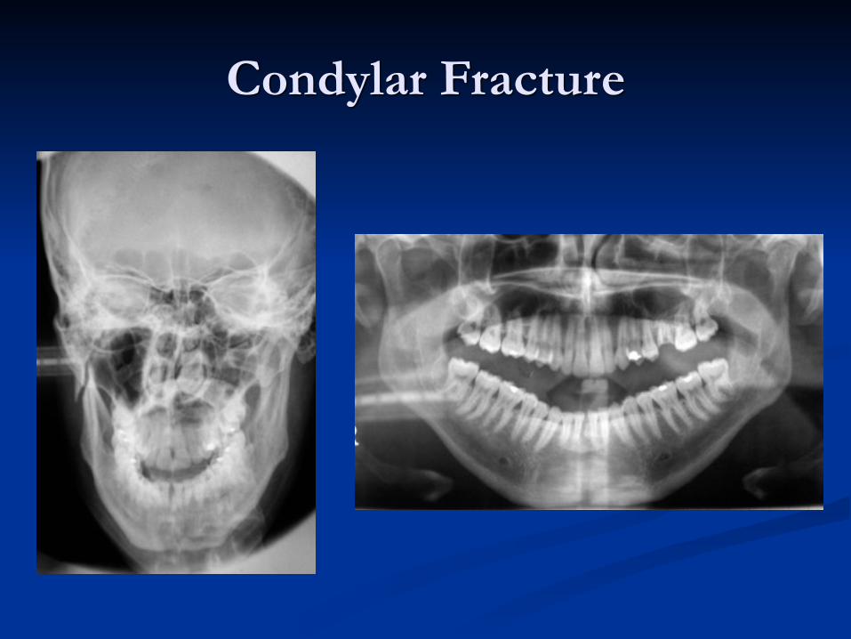
Condylar Fracture
Central Midface Fractures
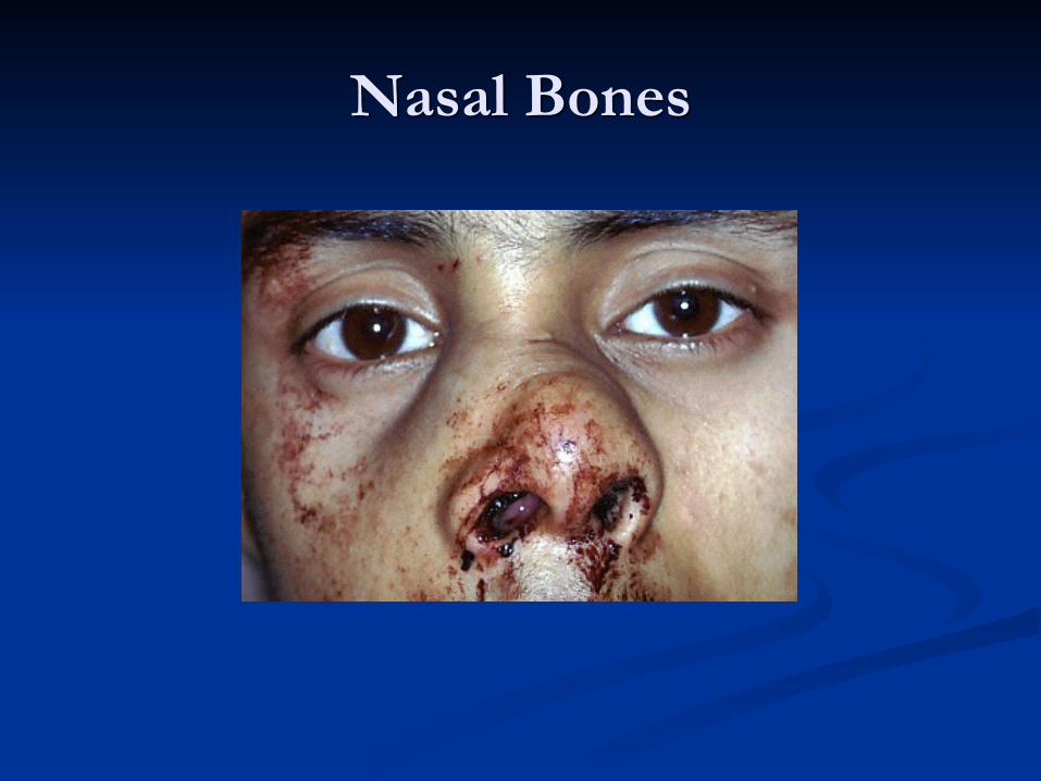
Nasal Bones

Nasoethmoid
■ Telecanthus (>35mm)
■ Blunting of canthal angle
■ Loss of nasal bridge projection
■ Upturned nasal tip
■ Loss of almond shaped eye
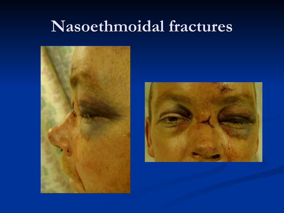
Nasoethmoidal fractures
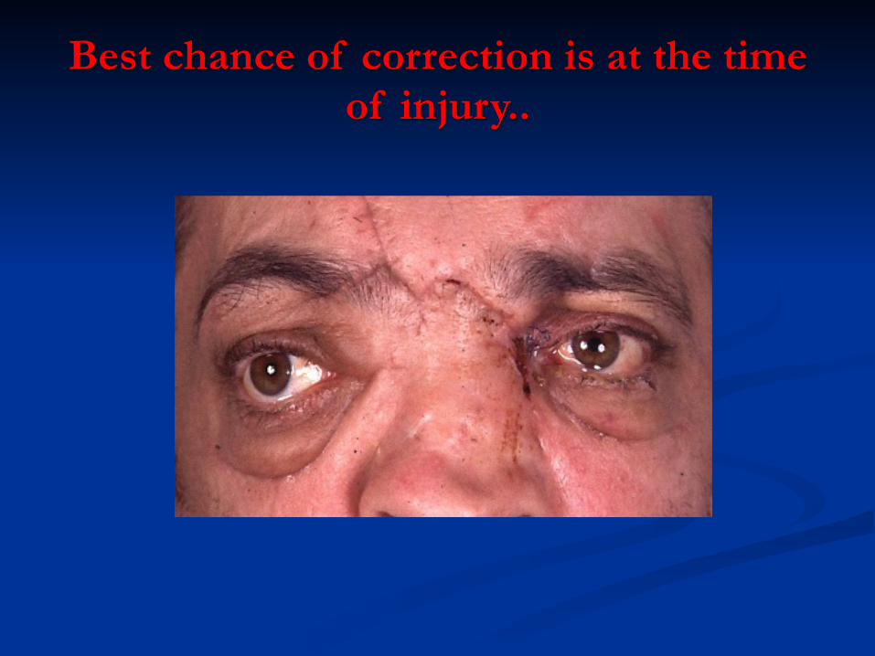
Best chance of correction is at the time of injury..
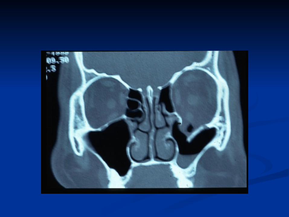
Orbital Fractures
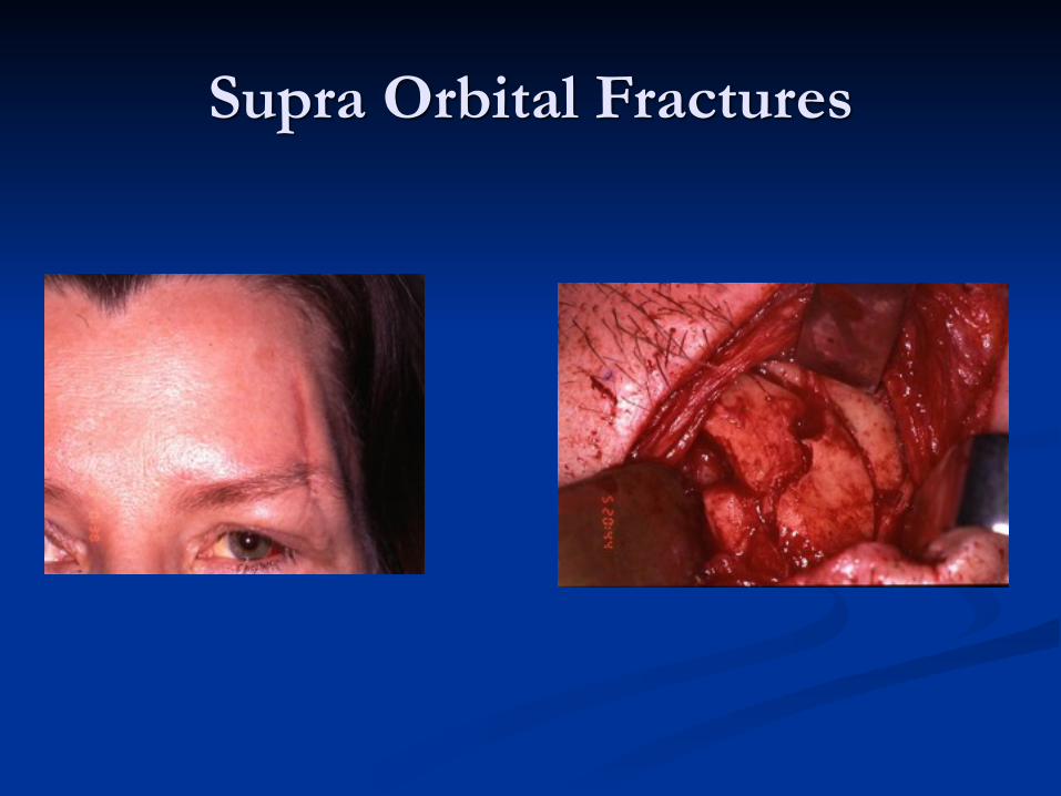
Supra Orbital Fractures
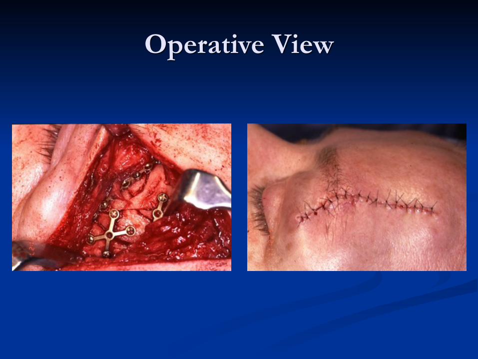
Operative View
Maxillary & Midface Fractures
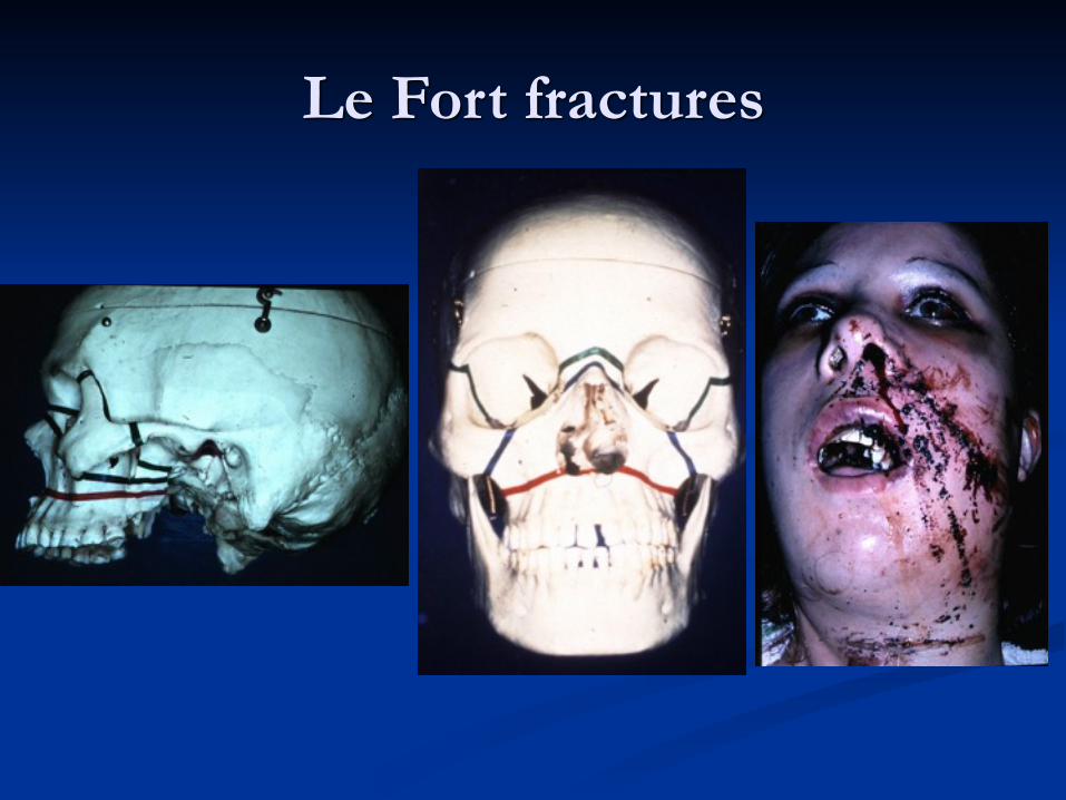
Le Fort fractures
Signs of Le Fort 2/3 injury
■ Severe swelling/bruising (football)
■ Subconjunctival haemorrhage and chemosis
■ Diplopia and limitation of eye movements
■ Panda eyes/Battle sign/CSF leaks = skull base
■ Malocclusion and pathological movement of midface
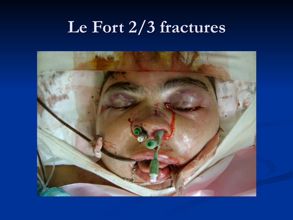
Le Fort 2/3 fractures
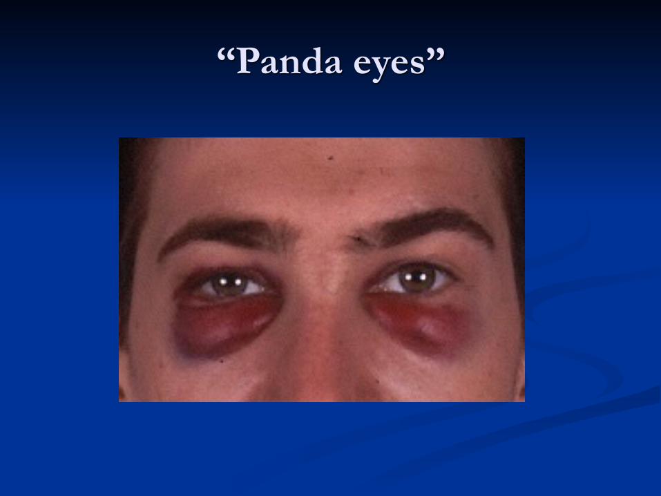
“Panda eyes”
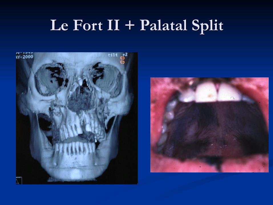
Le Fort II + Palatal Split
Lateral Midface
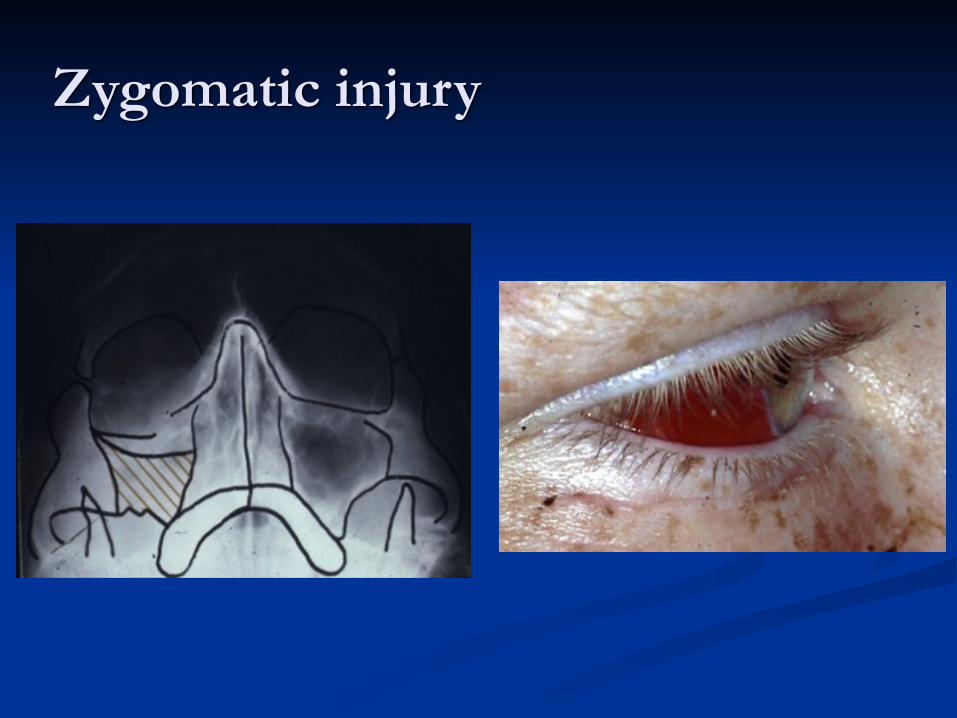
Zygomatic injury
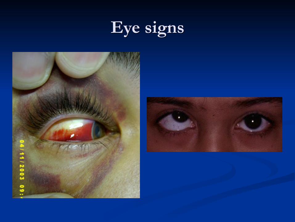
Eye signs
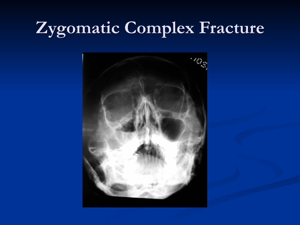
Zygomatic Complex Fracture
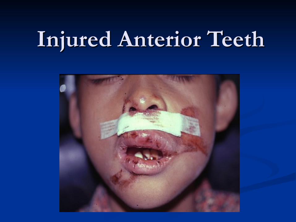
Injured Anterior Teeth
Timing of definitive treatment■ Maxillofacial bleeding or loss of vision requires immediate
intervention ■ Non-brain injured patients can have definitive facial
fracture treatment once swelling has reduced and investigations completed, usually first 2 days for mandibles, and 5-10 days for midface/orbit fractures
■ Brain injured patients need combined care by Neurosurgeon, Maxillofacial Surgeon and Ophthalmic surgeon. Timing of Maxillofacial fracture fixation is a team decision, taking into account the risks of worsening the brain injury against the morbidity of leaving the fractures untreated. Try to fix mandible early, midface can wait 2 weeks, by which time the neurological picture will usually have cleared.
Thank you.