OR Write-up CS
14
TABLE OF CONTENTS I. Patient¶s Profile ««««««««««««««««.1 II. Preparation of Patient «««««««««««««..1 III. Physiology of Pregnancy ««««««««..............2 IV. Pathophysio logy of Meconium Stained««««««.8 V. Discussion of the Procedure ««««««««««..9 VI. Instrumentation ««««««««««««««««10
-
Upload
alextan1981 -
Category
Documents
-
view
219 -
download
0
Transcript of OR Write-up CS

8/8/2019 OR Write-up CS
http://slidepdf.com/reader/full/or-write-up-cs 1/13
0
TABLE OF CONTENTS
I. Patient¶s Profile ««««««««««««««««.1
II. Preparation of Patient «««««««««««««..1
III. Physiology of Pregnancy ««««««««..............2
IV. Pathophysiology of Meconium Stained««««««.8
V. Discussion of the Procedure ««««««««««..9
VI. Instrumentation ««««««««««««««««10

8/8/2019 OR Write-up CS
http://slidepdf.com/reader/full/or-write-up-cs 2/13
1
I. Patient¶s Profile
Case Number: 489223Name: Arcela Botangen Ibag
Age: 22 years old
Gender: Female
Date of Birth: April 24, 1988
Address: Bison, Tuba, Benguet
Date of Operation: June 22, 2010
Pre-op Diagnosis: G (1) P (0) pregnancy Uterine 40 weeks cephalic in Labor
Post-op Diagnosis: Protracted Cervical Dilatation (Thickly meconium Stained
Amniotic Fluid)
Operation Performed: Low Segment Caesarean Section
Surgeon: Dr. Rosario Laranang
Assistant: Dr. Nani Wadingan
Instrument Nurse: Beverly Del Rosario
Sponge Nurse: Alexander Tan
Circulating Nurse: Gerard Cruz
Graduate Nurse: Muffet Calica, RN
Anesthesiologist: Dr. Inocencio Laranang
Anesthesia: Sub Arachnoid Block
Medicine Used: Bupivacaine
Operation Started: 1:28 am
Anesthesia Started: 1:22 am
Operation Ended: 2:05 am
II. Preparation of the Patient
y Transferred to Operating Room table and positioned comfortably in supine position, and
prepared accordingly.
y Sub Arachnoid Block Anesthesia was infiltrated by Dr. Inocencio Laranang.
y Skin Preparation done.

8/8/2019 OR Write-up CS
http://slidepdf.com/reader/full/or-write-up-cs 3/13
2
III. Physiology of Pregnancy
Definition of pregnancy
Pregnancy is defined as the course of embryo and fetal growth and development
in uterine. It begins at the fertilization and end the delivery of the fetal and its
attachment.
Fertilization
Fertilization is the joining of a sperm and an egg. A sperm is a male gamete that
is released into the vagina of a female during intercourse. In order for fertilization to
occur there must be a mature ovum present. Every month one of the ovaries releases
an egg which will meet one of the A 4 million sperm the male ejaculates into the vagina.
The sperm swim through the cervix and into the uterus which lead to the fallopian tubes.
This is where fertilization is most likely to take place. The high amount of sperm in the
ejaculate is needed because only around 100 survive to enter reach the fertilization site.
In order to penetrate the egg the sperm must first break through 2 barriers surrounding
the ovum. The acrosome of sperm comes in contact with the corona radiata and
releases digestive enzymes that break down a gelatinous layer around the egg called,
the zona pellucida. Once a sperm reaches the plasma membrane of the egg it sets off a
reaction that spreads across the membrane of the egg preventing other sperm from
breaking through the egg membrane. Once the sperm reaches the inside of the egg it
sheds its tail and the two nuclei fuse and now the 23 chromosomes from the egg and
the 23 chromosomes of the sperm join and they become a zygote. Chromosomes
contain all the information needed to determine the genetic structure of the new baby.
Normally all human beings have two chromosomes that determine sex: A combination
of X and Y makes a male or a combination of X and X makes a female. All ovum have X
sex chromosomes where as sperm have both X or Y sex chromosomes. Therefore, the
male gametes determine the sex of the baby.
Pre-embryonic Period
After fertilization, the zygote begins a process of dividing by mitosis in a process
called cleavage. It divides until it reaches 16 cells. It is now referred to as a morula. As
the morula floats freely within the uterus, it starts to bring nutrients into the cells. Themorula fills with fluid and the cells inside start to form two separate groups. At this stage
it is now a blastocyst . The inner layer of cells is called the embryoblast, and will become
the fetus. The outer layer is called a trophoblast which will develop into part of the
placenta. At this point the zona pellucida is disintegrating. The trophoblast contains
8/8/2019 OR Write-up CS
http://slidepdf.com/reader/full/or-write-up-cs 4/13
3
specialized cells that become extensions, like fingers, that grow into the endometrium
once in contact with the well thickened endometrium.
Implantation
The blastocyst preserves itself by secreting a hormone that indirectly stops
menstruation. The trophoblast cells secrete hCG hormones that help maintain the
corpus luteum that would normally regress. In turn, the corpus luteum continues to
secrete progesterone, which maintains the endometrium of the uterus in the secretory
phase. This helps the blastocyst to continue to grow and stay embedded within the
endometrium. The fetal life support system and the placenta begin to form, and
eventually the placenta will take over the job of producing progesterone.
Ectoderm
This forms the nervous tissue and the epithelium covering the outer body
surface. Epidermis of skin, including hair and nails, glands of skin, linings of oral cavity,
nasal cavity, anal canal, vagina, brain, spinal cord, sensory organs, lens of eye and
epithelium of conjunctiva (a membrane that covers the sclera and lines the inside of the
eyelids), pituitary gland, adrenal medulla, and enamel of teeth.
Mesoderm
This forms all of the muscle tissue and the connective tissue of the body, as well
as the kidneys and the epithelium of the serous membranes and blood vessels. All
muscle tissue (skeletal, smooth, cardiac), all connective tissue (fibrous connective
tissue, bone, blood, cartilage), dentin of teeth, adrenal cortex, kidneys and ureters,
internal reproductive viscera, epithelium lining vessels, joint cavities, and the serous
body cavities.
Endoderm
Forms the lining epithelium and glands of the visceral body systems. Lining
epithelium and glands of digestive, respiratory, and parts of urogenital systems, thyroid
and parathyroid glands, and thymus.
Formation of Placenta
As changes to the endometrium occur, cellular growth and the accumulation of glycogen cause fetal and maternal tissue to come together. This formation makes the
functional unit called the placenta. The placenta does not mix blood between mother
and fetus, but allows nutrients and waste products to diffuse between the two blood
systems. The placenta provides protection by filtering out many harmful substances that

8/8/2019 OR Write-up CS
http://slidepdf.com/reader/full/or-write-up-cs 5/13
4
the mother comes in contact with. Unfortunately the placenta cannot protect against
some teratogens including but not limited to:
y Thalidomide
y Heroin
y Cocaine
y Aspirin
y Alcohol
y Chemicals in cigarette smoke
y Propecia, also known as Finasteride, which can cause birth defects simply by a woman
handeling a broken pill during pregnancy.
Amniotic Fluid
Attached to placenta is the membranous sac which surrounds and protects the
embryo. This sac is called the amnion. It grows and begins to fill, mainly with water,
around two weeks after fertilization. This liquid is called Amniotic fluid, it allows the fetus
to move freely, without the walls of the uterus being too tight against its body. Buoyancy
is also provided here for comfort. After a further 10 weeks the liquid contains proteins,
carbohydrates, lipids and phospholipids, urea and electrolytes, all which aid in the
growth of the fetus. In the late stages of gestation much of the amniotic fluid consists of
fetal urine. The fetus swallows the fluid and then voids it to prepare its digestive organs
for use after birth. The fetus also "breathes" the fluid to aid in lung growth and
development.
Not enough amniontic fluid, or oligohydramnios, can be a concern during
pregnancy. Oligohydramnios can be caused by infection, kidney dysfunction or
malformation (since much of the late amniotic fluid volume is urine), procedures such as
chorionic villus sampling (CVS), and preterm, premature rupture of membranes
(PPROM). One possible outcome of oligohydramnios can cause is underdeveloped, or
hypoplastic, lungs. This condition is potentially fatal and the baby can die shortly after
birth. Babies with too little amniotic fluid can also develop contractures of the limbs,
including clubbing of the feet and hands.
As with too little fluid, too much fluid or polyhydramnios, can be a cause or an
indicator of problems for the mother and baby. Polyhydramnios is a predisposing risk
factor for cord prolapse and is sometimes a side effect of a macrosomic pregnancy. In
both cases, however, the majority of pregnancies proceed normally and the baby isborn healthy.
Preterm, premature rupture of membranes (PPROM) is a condition where the
amniotic sac leaks fluid before 38 weeks of gestation. This can be caused by a bacterial
infection or by a defect in the structure of the amniotic sac, uterus, or cervix. In some
8/8/2019 OR Write-up CS
http://slidepdf.com/reader/full/or-write-up-cs 6/13
5
cases the leak can spontaneously heal, but in most cases of PPROM, labor begins
within 48 hours of membrane rupture. When this occurs, it is necessary that the mother
receive treatment immediately to postpone labor if the fetus is not viable, for as long as
is safe, and for antibiotic treatments to avoid possible infection in the mother and baby.
If rupture occures too early in pregnancy little can be done to save the fetus.
A very rare and most often fatal obstetric complication is an amniotic fluid
embolism, or leakage of amniotic fluid into the mothers vascular systems causing an
allergic reation. This allergic reaction results in cardiorespiratory (heart and lung)
collapse, developing into a condition known as disseminated intravascular coagulation
in which the mothers blood looses it's ability to clot.
Amniotic band syndrome, or ABS, occurs when the inner fetal membrane
(amnion) ruptures without injury to the outer membrane (chorion). Fibrous bands from
the ruptured amnion float in the amniotic fluid and can entangle the fetus, reducing
blood supply and causing congenital limb abnormalities dysmelia. In some cases a
complete "natural" amputation of a digit(s) or limb may occur before birth or the digit(s)
or limbs may be necrotic (dead) requiring surgical removal.
Endocrine Function of the Placenta
Here are pituitary like hormones and steroid hormones secreted from the
placenta. The pituitary like hormones are hCG and hCS. HCG is similar to LH and helps
maintain the mothers corpus luteum. HCS is like prolactin and growth hormone and help
aid in increasing fat breakdown that spares the use of glucose from the mothers tissues.
This effect leaves more glucose available to the placenta and the fetus for necessary
growth. The steroid hormones are progesterone and estrogen. Progesterone helps
maintain the endrometrium and supports the growth of mammary glands. Estrogen also
helps maintain the endrometrium and growth of mammary glands as well as inhibits
prolactin secretion.
Developing Baby
The womb is expanding, the baby is growing and taking all the nourishment from
the mother. What once started as a microscopic two-celled egg, will be formed into ababy in just 12 weeks. The baby develops from conception to term, in a month-to-month
progress.

8/8/2019 OR Write-up CS
http://slidepdf.com/reader/full/or-write-up-cs 7/13
6
Overview of Developmental Milestones
WEEK CHANGES IN MOTHER DEVELOPMENT OF BABY
Pre-embryonic Development
1 week Ovulation OccursFertilization occurs, cell division begins and
continues, chorion appears
Embryonic Development
2
weeks
Symptoms of early pregnancy
(nausea, breast swelling and
tenderness, fatigue); blood
pregnancy tests may showpositive
Implantation occurs; amnion and yolk sac
appear; embryo has tissue; placenta begins
to form
3
weeks
First period missed; urine
pregnancy test may show
positive; early pregnancy
symptoms continue
Nervous system begins to develop; allantois
and blood vessels are present and placenta
is well formed
4
weeks
Limb buds form; heart is beating; nervous
system further develops; embryo has tail;
other systems are forming
5
weeks
Uterus is the size of a hen's egg;
mother may need to urinate
frequently
Embryo is curved, head is large, limb buds
are showing division, nose, ears and eyes
are noticeable
6
weeksUterus is the size of an orange
Fingers and toes are present and skeleton is
cartilaginous
8
weeks
Uterus can be felt above the
pubic bone
Fetus begins to look human; limbs aredeveloping and major organs forming; facial
features are becoming refined
Fetal Development
12
weeksUterus is the size of a grapefruit
Head grows faster than the rest of the body;
facial features are apparent, but there is no
layer of fat yet and the skin is translucent;
gender can be distinguished via ultrasound;
fingernails appear
16
weeksFetal movement can be felt
Fine hair (lanugo) grows over the body; fetus
resembles a tiny human being; skeleton is
visible

8/8/2019 OR Write-up CS
http://slidepdf.com/reader/full/or-write-up-cs 8/13
7
20-22
weeks
Uterus reaches up to the level of
umbilicus and pregnancy is
obvious
Vernix caseosa, the protective fatty coating,
begins to be deposited; heartbeat can be
heard
24
weeks
Doctor can tell where baby's
head, back and limbs are;
breasts have enlarged and
nipples and areola are darker,
colostrum is produced
Fully formed but still thin; much larger and
very active, all major organs are working, the
lungs and digestive system need more time
to develop; body is covered in fine hair called
lanugo
32
weeks
Uterus reaches halfway between
umbilicus and rib cage
Most babies are in a head down position in
the womb; head is more in proportion to the
body; eyes are open; babies born at this
stage have a good chance of living
36
weeks
Weight gain is averaging about a
pound a week; standing and
walking are becoming very
difficult because the center of
gravity is thrown forward
Body hair begins to disappear, fat is being
deposited
40
weeks
Uterus is up to the rib cage,
causing shortness of breath and
heartburn; sleeping is very
difficult
Not much room to move in the womb; fully
mature, baby moves less, and the
surrounding fluid reduces and the womb
expands its limits

8/8/2019 OR Write-up CS
http://slidepdf.com/reader/full/or-write-up-cs 9/13
8
IV. Pathophysiology of Meconium Stained
Meconium aspiration syndrome is a disease of term and post-term infants and its
severity is linked to co-existing fetal asphyxia.
Aspiration of meconium into the distal airways can occur either antenatally or
postnatally, but in the majority of affected infants the exact timing is not clear.
Aspiration is known to occur prior to delivery, as meconium has been found in the lungs
of stillbirths and in infants delivered pre-labour by caesarean section without evidence of
fetal distress.
Postnatal inhalation can occur late in the second stage or immediately after delivery if
the infant gasps or makes breathing movements while the oropharynx, nasopharynx or
trachea contains meconium-stained liquor.
Meconium has a number of adverse effects on the neonatal lung, which may ultimately
lead to the respiratory failure (and hypoxaemia) which characterizes MAS.
Meconium:
1. Causes mechanical blockage of the airway,
2. Acts as a chemical irritant causing pneumonitis, alveolar collapse and cell necrosis
3. Although initially sterile, predisposes to secondary bacterial infection
8/8/2019 OR Write-up CS
http://slidepdf.com/reader/full/or-write-up-cs 10/13
9
V. Discussion of the Procedure
Into OR with an ongoing IVF of D5LRS at 800cc level with a consent signed for
Caesarian section
Transferred to OR table positioned to supine comfortably and prepared
accordingly
Sub arachnoid block infiltrated by Dr. Inocencio Laranang
Skin preparation done
IFC inserted by NOD
Initial sponge, instruments and needles count done
Drapes applied
Operation started whereby a midline incision down to the peritoneum is made by
Dr. Rosario Laranang
Uterus is identified and incised
Scooping of fetal head
Delivered an alive baby boy attended by Dr. Perez
Placental delivery in Duncan presentation
Uterine incision is closed
Final sponge, instruments and needles count done and complete
The wound is closed in layer
Operation ended, top dressing applied to the wound
To RR with the same IVF of D5LRS of 600cc level , with the urine drainage at
300cc level.
8/8/2019 OR Write-up CS
http://slidepdf.com/reader/full/or-write-up-cs 11/13
10
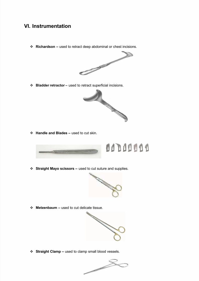
VI. Instrumentation
Richardson ± used to retract deep abdominal or chest incisions.
Bladder retractor ± used to retract superficial incisions.
Handle and Blades ± used to cut skin.
Straight Mayo scissors ± used to cut suture and supplies.
Metzenbaum ± used to cut delicate tissue.
Straight Clamp ± used to clamp small blood vessels.

8/8/2019 OR Write-up CS
http://slidepdf.com/reader/full/or-write-up-cs 12/13
11
Curved Clamp ± used to clamp hard to reach small blood vessels.
Allis forceps ± used to grasp tissue.
Bobcock ± used to grasp delicated tissue.
Thumb forceps ± used to grasp tough tissue.
Tissue forceps ± used to grasp tissue.

8/8/2019 OR Write-up CS
http://slidepdf.com/reader/full/or-write-up-cs 13/13
12
Needle holder ± used to hold needles in place.
Towel clips ± used to hold towels in place.