Or thopaedic Journal of Review Surger y
10
Review Current status of the magnetically controlled growing rod in treatment of early-onset scoliosis: What we know after a decade of experience Jason Pui Yin Cheung and Kenneth MC Cheung Abstract The magnetically controlled growing rod (MCGR) has had approximately 10 years of clinical experience worldwide. Clinical effectiveness to control early-onset scoliosis is consistent even at final surgery. MCGRs have significantly lower relative percentage of infection or wound complications as compared to traditional growing rods. Most common com- plications include foundation failure and failure of distraction. Contouring of the rod especially at the proximal segment while accommodating for the straight actuator remains a difficult task and its failure may lead to proximal junctional kyphosis. Unique complications of MCGR include clunking, temporary diminishing distraction gains, and metallosis. Temporary reductions in distraction gains are observed as the MCGR lengthens but return to normal baseline distraction gains after rod exchange. Lack of standardization for rod configuration, distraction strategies and decisions of whether to keep the rods in situ, remove without fusion surgery or to perform spinal fusion at skeletal maturity will require further study. Keywords actuator, complications, distraction, lengthening, magnetically controlled growing rod Date received: 20 July 2019; Received revised 31 August 2019; accepted: 11 September 2019 Introduction Management of early-onset scoliosis (EOS) is not a simple task. These spinal deformities occur in young children and if left untreated, the curvature may rapidly deteriorate and lead to cosmetic disfigurement and poor pulmonary devel- opment. 1–8 Growing rods are one of the popular treatment options for EOS because it can prevent curve deterioration while allowing spinal growth. 9–11 In a matched cohort study with the Shilla procedure, similar growth and curve correction parameters were observed. 12 Traditionally, growing rods require open manual distractions approxi- mately every 6 months 9,10,13–17 but has increased risk of anesthetic and wound complications. 1,2 Repeated surgery under general anesthesia also has potential deleterious effects on brain development. This is especially important for young children (<3 years) as sedation and anesthetic drugs may lead to poor brain development. 18 Due to these limitations of traditional growing rods (TGRs), the magnetically controlled growing rod (MCGR) system has been developed to allow for gradual lengthen- ing on an outpatient basis. 19,20 The MCGR allows for per- iodical noninvasive spinal lengthening under continuous neurological observation in an awake patient by use of a large external magnet. Hence, unlike TGRs, MCGRs can be distracted during outpatient clinic visits, thereby Department of Orthopaedics and Traumatology, The University of Hong Kong, Pokfulam, Hong Kong SAR, China Corresponding author: Jason Pui Yin Cheung, Department of Orthopaedics and Traumatology, Queen Mary Hospital, The University of Hong Kong Medical Centre, 5/F Professorial Block, Pokfulam, Hong Kong SAR, China. Email: [email protected] Journal of Orthopaedic Surgery 27(3) 1–10 ª The Author(s) 2019 Article reuse guidelines: sagepub.com/journals-permissions DOI: 10.1177/2309499019886945 journals.sagepub.com/home/osj Journal of Or thopaedic Surger y Creative Commons Non Commercial CC BY-NC: This article is distributed under the terms of the Creative Commons Attribution-NonCommercial 4.0 License (http://www.creativecommons.org/licenses/by-nc/4.0/) which permits non-commercial use, reproduction and distribution of the work without further permission provided the original work is attributed as specified on the SAGE and Open Access pages (https://us.sagepub.com/en-us/nam/open-access-at-sage).
Transcript of Or thopaedic Journal of Review Surger y
Review
Current status of the magneticallycontrolled growing rod in treatmentof early-onset scoliosis: What we knowafter a decade of experience
Jason Pui Yin Cheung and Kenneth MC Cheung
AbstractThe magnetically controlled growing rod (MCGR) has had approximately 10 years of clinical experience worldwide.Clinical effectiveness to control early-onset scoliosis is consistent even at final surgery. MCGRs have significantly lowerrelative percentage of infection or wound complications as compared to traditional growing rods. Most common com-plications include foundation failure and failure of distraction. Contouring of the rod especially at the proximal segmentwhile accommodating for the straight actuator remains a difficult task and its failure may lead to proximal junctionalkyphosis. Unique complications of MCGR include clunking, temporary diminishing distraction gains, and metallosis.Temporary reductions in distraction gains are observed as the MCGR lengthens but return to normal baseline distractiongains after rod exchange. Lack of standardization for rod configuration, distraction strategies and decisions of whetherto keep the rods in situ, remove without fusion surgery or to perform spinal fusion at skeletal maturity will requirefurther study.
Keywordsactuator, complications, distraction, lengthening, magnetically controlled growing rod
Date received: 20 July 2019; Received revised 31 August 2019; accepted: 11 September 2019
Introduction
Management of early-onset scoliosis (EOS) is not a simple
task. These spinal deformities occur in young children and
if left untreated, the curvature may rapidly deteriorate and
lead to cosmetic disfigurement and poor pulmonary devel-
opment.1–8 Growing rods are one of the popular treatment
options for EOS because it can prevent curve deterioration
while allowing spinal growth.9–11 In a matched cohort
study with the Shilla procedure, similar growth and curve
correction parameters were observed.12 Traditionally,
growing rods require open manual distractions approxi-
mately every 6 months9,10,13–17 but has increased risk of
anesthetic and wound complications.1,2 Repeated surgery
under general anesthesia also has potential deleterious
effects on brain development. This is especially important
for young children (<3 years) as sedation and anesthetic
drugs may lead to poor brain development.18
Due to these limitations of traditional growing rods
(TGRs), the magnetically controlled growing rod (MCGR)
system has been developed to allow for gradual lengthen-
ing on an outpatient basis.19,20 The MCGR allows for per-
iodical noninvasive spinal lengthening under continuous
neurological observation in an awake patient by use of a
large external magnet. Hence, unlike TGRs, MCGRs can
be distracted during outpatient clinic visits, thereby
Department of Orthopaedics and Traumatology, The University of Hong
Kong, Pokfulam, Hong Kong SAR, China
Corresponding author:
Jason Pui Yin Cheung, Department of Orthopaedics and Traumatology,
Queen Mary Hospital, The University of Hong Kong Medical Centre, 5/F
Professorial Block, Pokfulam, Hong Kong SAR, China.
Email: [email protected]
Journal of Orthopaedic Surgery27(3) 1–10
ª The Author(s) 2019Article reuse guidelines:
sagepub.com/journals-permissionsDOI: 10.1177/2309499019886945
journals.sagepub.com/home/osj
Journal ofOr thopaedicSurger y
Creative Commons Non Commercial CC BY-NC: This article is distributed under the terms of the Creative Commons
Attribution-NonCommercial 4.0 License (http://www.creativecommons.org/licenses/by-nc/4.0/) which permits non-commercial
use, reproduction and distribution of the work without further permission provided the original work is attributed as specified on the SAGE and Open
Access pages (https://us.sagepub.com/en-us/nam/open-access-at-sage).

avoiding the risks of repeated surgical lengthening.2,9,21
There is also the possibility for distractions to be carried
out more frequently to mimic normal physiological growth
more closely.22
This presents a potential huge benefit for children as rod
distractions no longer need to be carried out under general
anesthesia. This may provide additional advantages to
spine length gains by avoiding spine autofusion associated
with sudden and forceful surgical distractions at irregular
intervals.21,23 Furthermore, due to infrequent need for
admissions and general anesthesia, there is potential cost-
saving benefits of the MCGR over the TGR.24,25
In the past few years, our understanding of the
MCGR’s role in the management of EOS has improved.
In this review, we will highlight its history, development,
and future directions with specific emphasis on instru-
mentation strategies and implantation configurations, and
complications.
History of the MCGR
The first report of an MCGR dates back to 2004 with the
Phenix rod developed by Arnaud Souberian, a French aero-
nautical engineer.20 With a large-sized internal magnet and a
permanent external magnet placed on the skin, this device
allowed for lengthening 5 days out of a week and aimed to
focus correction at the curve apex. Unfortunately, this device
fell out of favor with the death of its inventor. The current
iteration of the MCGR is the MAGEC® rod (Figure 1), ini-
tially developed by Ellipse Technologies, Inc. (Irvine, Cali-
fornia, USA) and subsequently acquired by NuVasive, Inc.
(San Diego, California, USA) in 2016. The first tests of this
novel technology were performed by Akbarnia et al.26 in
Yucatan pigs. Initial findings verified the ability of the rod
to safely distract using an external magnet.
Cheung et al.19 described the very first experience with
the MCGR in humans. Of the five patients implanted in that
clinical trial, two patients completed a 2-year follow-up at
the time of reporting. Initial results showed sequential
lengthening of the rods with each distraction, which was
performed at 1.5–2 mm per month. There were no complica-
tions and the functional outcome using the 30-item Scoliosis
Research Society (SRS-30) questionnaire was good. Since
this initial experience, there have been five subsequent
structural modifications of the MCGR.22 Loss of distraction
was encountered between distraction sessions of the first
implanted MCGR in 2009. Because of this, the original rod
design (Figure 2) required the insertion of a “keeper plate” or
stainless steel plate within the rod next to the magnet to
prevent it from rotating on its own without the external
magnet. This “second generation” rod (Figure 3) was intro-
duced in 2010 with an increased rod shaft diameter from 9 to
10.5 mm to house this steel plate. The third modification was
in 2012 to the welding procedure of the junction between the
actuator and the rod shaft. Rod fractures were a concern and
the switch in the welding technique from a pulsed laser to a
continuous laser reduced the occurrence of any weak points
on the rod surface. Essentially, the entire enlarged portion of
the rod became a continuous segment without breaks. Two
other modifications were made since 2015. Firstly, a smaller
sized actuator (70 mm) was made for smaller sized children.
Secondly, the actuator pin has been reinforced to avoid pin
fracture (Figure 4).27
Current MCGR
Currently, these MCGRs come in two configurations: stan-
dard and offset depending on the direction of rod extension
from the housing portion of the rod. These rods house an
actuator with a magnet inside that is driven by an external
magnet, called a remote controller (Figure 5), to distract the
rod. Careful insertion of the MCGR is necessary as the
standard and offset rods extend in different directions. An
arrow is placed on the rod to avoid incorrect rod insertion
and subsequent distraction failure. The actuator cannot be
Figure 1. The MCGR with a thickened actuator portion thathouses the magnet. MCGR: magnetically controlled growing rod.
Figure 2. The first generation MCGR can be identified withoutany expanded portion in the shaft of the rod. MCGR: magneticallycontrolled growing rod.
2 Journal of Orthopaedic Surgery 27(3)

contoured but the rod segment proximal and distal to the
actuator can be contoured to fit the anchors. Two sizes of
actuators (70 and 90 mm) are available depending on
patient size. The rod diameter comes in selection of 4.5,
5.5, and 6 mm.
Case selection
Similar to TGR, MCGRs are used for patients with EOS
and a sizeable Cobb angle (i.e. 50�), large potential for
further spine growth and observed curve progression.
Essentially, any type of scoliosis is amenable to MCGR
treatment with consistent length gains especially with pri-
mary surgery.28 This has also been shown in congenital
scoliosis23,28,29 and fixation to the pelvis,30 whereby dis-
traction gains are similar to that found in idiopathic or
syndromic patients. However, Keskinen et al.31 have
shown that conversion cases do not fare as well in terms
of growth gains. This may be related to the already stiffer
spines contributed by previous TGR surgeries. Larger com-
parative studies at longer follow-up are required to study
this relationship further.
There are several unique applications of the MCGR’s
gradual distraction utility. Thoracic insufficiency syn-
drome is commonly treated by vertical expandable prosthe-
tic titanium rods (VEPTR).32 A hybrid with the MCGR can
help patients wean off ventilator and achieve some spine
length gain. However, these patients do not have enough
growth potential to sustain continuous distractions.33 The
MCGR can also be used to gradually correct severe defor-
mities.34,35 In such situation, it acts like an internalized
halo-gravity traction device to allow for awake correction
and reduces risk of neurological complications and techni-
cal difficulties during the definitive fusion surgery. Skov
et al.36 utilized an interesting construct, whereby the
MCGR was only used on the concave side while a sliding
rod construct was used on the convex side to control the
apical vertebrae similar to the Shilla technique.37 The
short-term results appear to be quite promising and similar
to dual MCGR technique.
The MCGR is not advised if patients require magnetic
resonance imaging (MRI) to assess a pathology such as
syringomyelia. Although no adverse effects have been
shown with using the MRI,38,39 image artifacts of up to
30 cm of image distortion have been observed. Hence, the
spinal cord lesion cannot be monitored adequately.
Figure 3. The second generation MCGR with an increasedactuator diameter due to the keeper plate. The rods are insertedin one standard and one offset configuration. MCGR: magneticallycontrolled growing rod.
Figure 4. Fractured actuator pin.
Figure 5. External remote controller used for outpatientdistractions.
Cheung and Cheung 3

Nevertheless, the efficacy of the distraction mechanism is
maintained without any loss of distraction. There is also no
heating concern with regards to surrounding soft tissue.40
Anchors
Instrumentation levels usually incorporate the end-to-end
vertebrae to maintain control of the major curve. Some
curves with significant shift may require recruitment of
more proximal and distal levels to achieve balance. How-
ever, this should be avoided if possible as reoperation due
to foundation failure (Figure 6) is not uncommon and the
additional revised levels will need eventual fusion as
well.28,41 In the longest follow-up study to date with mul-
tiple graduates, final fusion occurs at 6.5 years after initial
implantation and all patients required extension of fusion
levels 2 levels proximally and distally.28 The position of the
neutral or stable vertebrae governs whether longer fusions
are necessary as the added-on levels will need to be
instrumented.
The choice of instrumentation strategy is especially dif-
ficult for small-sized children. Clinical examination of the
patient’s body size and understanding of the length of the
spinal column is necessary to decide whether MCGRs are
able to fit. Although 70 mm actuators are available, this
may already be too long for smaller sized children. The
surgeon must bear in mind that the 70 mm is only the length
of the actuator. Additional length is reserved for the prox-
imal and distal foundations and also for rod contouring as
the actuator segment is straight and cannot be contoured.
Delaying MCGR surgery with further casting or bracing, or
with a period of halo-traction may be necessary until the
rods can be inserted without sacrificing levels.
With the straight actuator segment and MCGR distrac-
tions, there is a risk for proximal junctional kyphosis (PJK).
Contouring of the rod to the correct sagittal alignment may
be more difficult than TGRs, and thus overbending of the
proximal rod may be necessary. Hence, the prevalence of
PJK is high with up to 30% in some reports but not all
require reoperation (approximately 15%).28,41–44 In gen-
eral, the proximal levels of T1 or T2 should be avoided if
possible as more cranial levels may be needed in the final
fusion surgery to address the PJK. Conversion to rib
anchors may help prevent these complications and has been
well-established in TGR surgery.45,46 However, its role in
MCGR and whether the rate of PJK is lower for rib hooks
as compared to screws require future study.
Pedicle screws are generally preferred by most surgeons
due to their stronger pullout strength as compared to
hooks.22 However, hooks are less stiff and may also allow
more vertebral rotation, which is an advantage during dif-
ferential lengthening. In addition to the advantages of rib
hooks described above for PJK, rib-based implants also
avoid disruption of peri-spinous musculature that is asso-
ciated with spine autofusion.21,47
Rod configuration and implantation
Similar to TGR, dual rods are preferred over single rods for
MCGR surgery.48 Dual rods have been shown to produce
increased distraction forces and to allow for differential cor-
rection.17,19 Single rods may only be used in very thin patients
without adequate soft tissue coverage and if the severe rota-
tional and gibbus deformities preclude rod insertion.
MCGRs can be placed in two standard rod configura-
tions or one standard and one offset configuration. Internal
mechanical testing from manufacturers suggests that
increased distraction forces may be applied if both rods are
placed in standard configuration. While one standard and
one offset configuration may allow for differential correc-
tion, two standard rods have their magnets too close to
each other to allow for differential lengthening. For
Figure 6. A case with intact upgoing rib hooks (left), ploughing of the hooks (middle), and proximal foundation failure with looseningof hook anchors (right).
4 Journal of Orthopaedic Surgery 27(3)

neuromuscular scoliosis where pelvic obliquity is common,
standard and offset configuration may be particularly useful
with this function. Another advantage of standard and offset
configuration as compared to two standard rods is the reduc-
tion of “cross talk,” whereby magnets placed too close to
each other create a stronger internal magnetic field.49 In such
situations, a larger external distraction force is necessary for
distraction and rod slippage or clunking may occur, which
limits the successfulness of lengthening. This clunking event
refers to the inability for the rod’s internal magnet to com-
plete a full revolution and results in stalling and the magnet
flips back to its original position. Clinically, sudden jerky
movements are observed as compared to the normal
“wobble” feeling during a smooth distraction.
Like TGRs, MCGRs should be inserted subfascially
(Figure 7) to prevent skin impingement. For insertion and
exchange of the MCGR through the mini-incisions, a chest
drain can be used to help the MCGR pass through the
subfascial plane.50 Bending of the rod too close to the
motor should be avoided as it may damage it and lead to
distraction problems. Whether intraoperative distractions
should be performed is controversial. The authors’ practice
is to achieve overall balance without loading the implants
to reduce the risk of early clunking, implant failure, and
anchor plowing. In these situations, early clunking is diffi-
cult for the surgeon to distinguish between a genuine rod
problem or due to overloading of the rod.
Distractions
MCGRs typically are distracted 1–2 months after implanta-
tion. The frequency of distractions may vary from once
monthly to 6-monthly, and these decisions are usually influ-
enced by patient convenience (availability of distraction ser-
vices and distance required for patients to travel) rather than
exact science. Those who distract at frequent intervals may
adopt a standard distracted length (approximately 2 mm) per
month while those with 6-monthly intervals may distract
until clunking to achieve maximum lengthening.51
Monitoring the amount of length gain is crucial as this
confirms that the MCGR remains functional. X-rays can be
used to measure the amount of distracted length by the
height of the housing portion of the rod (Figure 8). Since
the maximum length distractible is 4.8 cm, the measured
distance provides indication for when rod exchanges are
required. Ultrasound (Figure 9) is a popular option for mon-
itoring MCGR distractions as it provides similar information
without radiation exposure.52 It has been shown to correlate
well with radiographs with a shallow learning curve.53,54
With the ultrasound, radiographs should only be obtained
occasionally to assess the deformity and rod integrity.
There is no consensus on how distractions are performed
and may vary between patients due to anatomical consid-
erations and if clunking occurs. The authors elect a stan-
dardized procedure during these outpatient distractions for
all patients. Outpatient distractions are performed with
patients in prone position or in sitting. The ultrasound is
first used to measure the current rod length prior to distrac-
tions. Then a small handheld magnet can be used to identify
the internal magnet position over the skin surface and this
position is marked on the skin. The targeted distraction
Figure 7. Subfascial insertion of MCGR rods. MCGR: magneti-cally controlled growing rod.
Figure 8. Radiographic measurement of distracted length.
Cheung and Cheung 5

length input is set in the external remote controller and the
device is placed over this skin marking. The available con-
troller provided by industry is made by two larger magnets
and a central slit between the magnets should be placed
over the skin marking to optimize the effect of the magnets’
magnetic fields. Usually, an alternate distraction technique
is utilized, whereby one rod is distracted to the targeted
length followed by the other rod. Patients with big humps
may only allow access for one magnet. In cases with clunk-
ing, the alternating technique with smaller targeted distrac-
tion lengths (approximately 0.2–0.5 mm) can be applied to
try and reduce the stresses internally.
Complications
Complications of MCGR are not uncommon. At 2-year
follow-up, up to 46.7% of patients may have had an
unplanned operation.41 In a recent systematic review, the
complication rate can reach up to 44.5% with unplanned
revision surgery up to 33% at 29.7 months follow-up.55 The
most common causes of reoperation are failure of rod dis-
traction and proximal foundation failure. Similar to TGRs,
MCGRs still have a high risk of implant-related complica-
tions like rod fractures (Figure 10) and PJK56 but may have
reduced rate of infections.57 Kwan et al.41 suggested that
patients undergoing more frequent distractions (1 week to
2 months interval) may have higher risk of PJK and failure
of rod distractions. Conversely, foundation failures may
occur more frequently in distraction intervals of 3–6 months.
Failure to distract the MCGR can be technical or mechan-
ical in origin. Technical complications are more severe as
they are avoidable and often lead to complete distraction
failure and require revision surgery. This may include inap-
propriate bending of the rod near the expanded portion of the
rod and incorrect rod insertion and configuration leading to
inability to distract along the long axis of the rod. Mechan-
ical failures include spontaneous bone formation (Figure 11)
near the housing unit limiting further distractions, actuator
pin fracture27 which has been rectified with the most recent
rod modification, and clunking. Several risk factors for
clunking include increased body habitus such as older age
and increased body mass index, as well as reduced distance
between the two internal magnets due to cross talk.49
Increased body habitus is associated with a thicker subcuta-
neous layer and this increases the distance between the
external remote controller and the internal magnets, thereby
reducing the amount of force that can be transferred to the
MCGR.58 Hence, lengthening outcomes may be suboptimal
in more obese patients. The ability to lengthen the rod
reduces with 2.1%/mm of tissue depth.58
A unique phenomenon observed from TGR is the “law
of diminishing returns,” whereby decreasing gains in spinal
lengthening is observed and may occur as early as the first
successive lengthening.21,47 This has been contributed by
progressive stiffness of an immature spine with prolonged
instrumentation in situ or autofusion as a result of trauma to
spinal ligaments after forceful distractions at infrequent
intervals.21 The short-term results with MCGRs contradict
in terms of diminishing returns and may be related to the
large variations in distraction techniques and frequen-
cies.23,49,59,60 Ahmad et al.59 suggested in the short-term
follow-up that this rate of decline is gradual as compared to
the rapid decline seen in TGR. There is apparent diver-
gence observed between targeted (intended amount of
lengthened input in the external remote controller) and
Figure 9. Ultrasonic measurement of distracted length.
Figure 10. MCGR fracture. MCGR: magnetically controlledgrowing rod.
Figure 11. Calcified deposits at the extendable portion ofthe rod.
6 Journal of Orthopaedic Surgery 27(3)
achieved (actual length measured on radiographs or ultra-
sound) lengthening with increasing MCGR distraction.58,61
One interesting observation suggests that the reductions in
lengthening are due to reduced distraction forces as the rod
lengthens rather than internal spinal stiffness.49 The ability
to distract returns to normal baseline after rod exchange
with a new MCGR. These findings suggest that transient
diminishing gains are caused by a rod design problem, in
which the mechanical properties prevent achievement of
full 4.8-cm rod lengthening. This is supported by a recent
biomechanical study on gradual reductions in maximum
force output by the MCGR. Poon et al.62 showed that by
using a straight MCGR, the maximum force at 0 mm was
208.0 N, at 25 mm was 199.6 N, and at 40 mm was 192.0 N.
There is a decrease in maximal force generated as the rod is
lengthening, which is a possible rationale for these transient
reductions in distraction length gains. It is important to note
that this study used a straight rod rather than a bent rod
which does not match the clinical scenario. The change in
force output may be more pronounced in a bent rod. Rush-
ton et al.63 corroborated with these findings using force
testing of explanted MCGRs showing that the duration of
rod implantation is inversely proportional to the reduction
in force generated by the rods. In their study, no force can
be generated if the MCGR has been implanted for over
38 months. The effect of wear at the distractable portion of
the rod on force output should also be studied in the future.
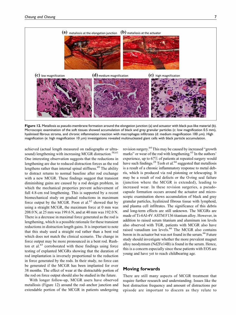
With longer follow-up, MCGR users have observed
metallosis (Figure 12) around the rod–anchor junction and
extendable portion of the MCGR in patients undergoing
revision surgery.64 This may be caused by increased “growth
marks” or wear of the rod with lengthening.65 In the authors’
experience, up to 67% of patients at repeated surgery would
have such findings.28 Teoh et al.64 suggested that metallosis
is a result of a chronic inflammatory response to metal deb-
ris, which is produced via rod pistoning or telescoping. It
may be a result of rod defects or the O-ring seal failure
(junction where the MCGR is extended), leading to
increased wear. In these revision surgeries, a pseudo-
capsule formation occurs around the actuator and micro-
scopic examination shows accumulation of black and gray
granular particles, hyalinized fibrous tissue with lymphoid,
and plasma cell infiltrates. The significance of this debris
and long-term effects are still unknown. The MCGRs are
made of Ti-6Al-4V ASTM F136 titanium alloy. However, in
addition to raised serum titanium and aluminum ion levels
also observed with TGR, patients with MCGR also have
raised vanadium ion levels.66 The MCGR also contains
boron in its actuator but was not found in the serum.66 Future
study should investigate whether the more prevalent magnet
alloy neodymium (Nd2Fe14B) is found in the serum. Hence,
this is a concern especially since these patients with EOS are
young and have yet to reach childbearing age.
Moving forwards
There are still many aspects of MCGR treatment that
require further research and understanding. Issues like the
best distraction frequency and amount of distractions per
episode are important to discern as they relate to
Figure 12. Metallosis as pseudo-membrane formation around the elongation junction (a) and actuator with black pus-like material (b).Microscopic examination of the soft tissues showed accumulation of black and grey granular particles (c: low magnification 0.5 mm),hyalinised fibrous stroma, and chronic inflammation reaction with macrophages infiltrates (d: medium magnification 100 mm). Highmagnification (e: high magnification 10 mm) investigations revealed multinucleated giant cells with black particle accumulation.
Cheung and Cheung 7

lengthening outcomes. Changes that occur in the sagittal
and axial plane with successive distractions also require
further study. Most utilize X-rays to observe for any
implant fracture or loosening. The role of other methods
like low-dose CT to determine spontaneous fusion and
migration of foundation anchors have yet to be determined.
The effects of MCGR on lung function and vertebral remo-
deling with growth should be determined. The actual ben-
efits of MCGR over TGR in health-related quality of life
measures are unclear in the early follow-up.67–69 Similarly,
short-term follow-up studies have observed similar results
in terms of spinal growth with the MCGRs compared to
TGR, VEPTR, and Shilla and Luque trolley systems.70 This
will need to be revisited at graduation surgery. The
decision-making at the time of MCGR graduation is also
uncertain. For TGRs, one study suggested that only 38%(10/26) patients at skeletal maturity are suitable for implant
removal without spinal fusion.71 However, nine of these
patients had progression of the deformity after implant
removal resulting in an additional surgery for fusion.
Whether patients can or should remove their MCGRs with-
out fusion or whether every patient needs final fusion
should be studied. These are all pertinent questions that
hopefully combined multicenter databases such as the
Pediatric Spine Study Group (PSSG) will answer in the
coming decade.
Conclusions
MCGR is a significant advancement in the treatment of
EOS. It provides benefits of noninvasive outpatient length-
ening and thus allows for more frequent distractions with
smaller lengthening increments. There is still high preva-
lence for complications and revision surgery similar to
TGRs but has reported reduced risk for infection. Despite
this, MCGR reduces the need for regular surgery to distract
the rods and may have significant benefits in terms of social
and psychological aspects including patient satisfaction.
Declaration of conflicting interests
The author(s) declared no potential conflicts of interest with
respect to the research, authorship, and/or publication of this
article.
Funding
The author(s) received no financial support for the research,
authorship, and/or publication of this article.
ORCID iD
Jason Pui Yin Cheung https://orcid.org/0000-0002-7052-0875
References
1. Akbarnia BA and Emans JB. Complications of growth-
sparing surgery in early onset scoliosis. Spine (Phila Pa
1976) 2010; 35: 2193–2204.
2. Bess S, Akbarnia BA, Thompson GH, et al. Complications of
growing-rod treatment for early-onset scoliosis: analysis of
one hundred and forty patients. J Bone Joint Surg Am 2010;
92: 2533–2543.
3. Campbell RM Jr, Smith MD, Mayes TC, et al. The charac-
teristics of thoracic insufficiency syndrome associated with
fused ribs and congenital scoliosis. J Bone Joint Surg Am
2003; 85: 399–408.
4. Cheung JP, Samartzis D, and Cheung KM. Management of
early-onset scoliosis, http://www.boneandjoint.org.uk/con
tent/focus/management-early-onset-scoliosis (2013, accessed
01 June 2019).
5. Goldberg CJ, Gillic I, Connaughton O, et al. Respiratory
function and cosmesis at maturity in infantile-onset scoliosis.
Spine (Phila Pa 1976) 2003; 28: 2397–2406.
6. James JI. Idiopathic scoliosis; the prognosis, diagnosis, and
operative indications related to curve patterns and the age at
onset. J Bone Joint Surg Br 1954; 36: 36–49.
7. James JI, Lloyd-Roberts GC, and Pilcher MF. Infantile struc-
tural scoliosis. J Bone Joint Surg Br 1959; 41: 719–735.
8. Redding GJ and Mayer OH. Structure-respiration function
relationships before and after surgical treatment of early-
onset scoliosis. Clin Orthop Relat Res 2011; 469:
1330–1334.
9. Akbarnia BA, Breakwell LM, Marks DS, et al. Dual growing
rod technique followed for three to eleven years until final
fusion: the effect of frequency of lengthening. Spine (Phila
Pa 1976) 2008; 33: 984–990.
10. Akbarnia BA, Marks DS, Boachie-Adjei O, et al. Dual grow-
ing rod technique for the treatment of progressive early-onset
scoliosis: a multicenter study. Spine (Phila Pa 1976) 2005;
30: 46–57.
11. Winter RB, Moe JH, and Lonstein JE. Posterior spinal
arthrodesis for congenital scoliosis. An analysis of the cases
of two hundred and ninety patients, five to nineteen years old.
J Bone Joint Surg Am 1984; 66: 1188–1197.
12. Luhmann SJ, Smith JC, McClung A, et al. Radiographic out-
comes of Shilla growth guidance system and traditional
growing rods through definitive treatment. Spine Deform
2017; 5: 277–282.
13. Elsebai HB, Yazici M, Thompson GH, et al. Safety and effi-
cacy of growing rod technique for pediatric congenital spinal
deformities. J Pediatr Orthop 2011; 31: 1–5.
14. Sponseller PD, Thompson GH, Akbarnia BA, et al. Growing
rods for infantile scoliosis in Marfan syndrome. Spine (Phila
Pa 1976) 2009; 34: 1711–1715.
15. Sponseller PD, Yazici M, Demetracopoulos C, et al. Evi-
dence basis for management of spine and chest wall defor-
mities in children. Spine (Phila Pa 1976) 2007; 32: S81–S90.
16. Thompson GH, Akbarnia BA, and Campbell RM Jr. Growing
rod techniques in early-onset scoliosis. J Pediatr Orthop
2007; 27: 354–361.
17. Thompson GH, Akbarnia BA, Kostial P, et al. Comparison of
single and dual growing rod techniques followed through
definitive surgery: a preliminary study. Spine (Phila Pa
1976) 2005; 30: 2039–2044.
8 Journal of Orthopaedic Surgery 27(3)

18. US Food & Drug Administration. FDA Drug Safety Commu-
nication: FDA review results in new warnings about using
general anesthetics and sedation drugs in young children and
pregnant women, https://www.fda.gov/Drugs/DrugSafety/
ucm532356.htm (2016, accessed 04 December 2018).
19. Cheung KM, Cheung JP, Samartzis D, et al. Magnetically
controlled growing rods for severe spinal curvature in young
children: a prospective case series. Lancet 2012; 379:
1967–1974.
20. Wick JM and Konze J. A magnetic approach to treating pro-
gressive early-onset scoliosis. AORN J 2012; 96: 163–173.
21. Sankar WN, Skaggs DL, Yazici M, et al. Lengthening of dual
growing rods and the law of diminishing returns. Spine (Phila
Pa 1976) 2011; 36: 806–809.
22. Cheung JP, Cahill P, Yaszay B, et al. Special article: update
on the magnetically controlled growing rod: tips and pitfalls.
J Orthop Surg (Hong Kong) 2015; 23: 383–390.
23. Cheung JP, Bow C, Samartzis D, et al. Frequent small dis-
tractions with a magnetically controlled growing rod for
early-onset scoliosis and avoidance of the law of diminishing
returns. J Orthop Surg (Hong Kong) 2016; 24: 332–337.
24. Charroin C, Abelin-Genevois K, Cunin V, et al. Direct costs
associated with the management of progressive early onset
scoliosis: estimations based on gold standard technique or
with magnetically controlled growing rods. Orthop Trauma-
tol Surg Res 2014; 100: 469–474.
25. Wong CKH, Cheung JPY, Cheung PWH, et al. Traditional
growing rod versus magnetically controlled growing rod for
treatment of early onset scoliosis: cost analysis from implan-
tation till skeletal maturity. J Orthop Surg (Hong Kong) 2017;
25: 2309499017705022.
26. Akbarnia BA, Mundis GM Jr., Salari P, et al. Innovation in
growing rod technique: a study of safety and efficacy of a
magnetically controlled growing rod in a porcine model.
Spine (Phila Pa 1976) 2012; 37: 1109–1114.
27. Jones CS, Stokes OM, Patel SB, et al. Actuator pin fracture in
magnetically controlled growing rods: two cases. Spine J
2016; 16: 287–291.
28. Cheung JPY, Yiu K, Kwan K, et al. Mean 6-year follow-up of
magnetically controlled growing rod patients with early onset
scoliosis: a glimpse of what happens to graduates. Neurosur-
gery 2019; 84: 1112–1123.
29. Lampe LP, Bovingloh AS, Gosheger G, et al. Magnetically
controlled growing rods in treatment of early-onset scoliosis:
a single center study with a minimum of 2-year-follow up and
preliminary results after converting surgery. Spine (Phila Pa
1976) 2019; 44: 1201–1210.
30. Lorenz HM, Braunschweig L, Badwan B, et al. High correla-
tion between achieved and expected distraction using magne-
tically controlled growth rods (MCGR) with rib to pelvis
fixation in pediatric spine deformity. J Pediatr Orthop
2019; 39: 334–338.
31. Keskinen H, Helenius I, Nnadi C, et al. Preliminary compar-
ison of primary and conversion surgery with magnetically
controlled growing rods in children with early onset scoliosis.
Eur Spine J 2016; 25: 3294–3300.
32. Campbell RM Jr. VEPTR: past experience and the future of
VEPTR principles. Eur Spine J 2013; 22(2): 106–117.
33. Kwan KYH, Cheung JPY, Yiu KKL, et al. Ten year follow-
up of Jarcho-Levin syndrome with thoracic insufficiency
treated by VEPTR and MCGR VEPTR hybrid. Eur Spine J
2018; 27: 287–291.
34. Cheung JP, Samartzis D, and Cheung KM. A novel approach
to gradual correction of severe spinal deformity in a pediatric
patient using the magnetically-controlled growing rod.
Spine J 2014; 14: 7–13.
35. Welborn MC, Krajbich JI, and D’Amato C. Use of magnetic
spinal growth rods (MCGR) with and without preoperative
halo-gravity traction (HGT) for the treatment of severe early-
onset scoliosis (EOS). J Pediatr Orthop 2019; 39: 293–297.
36. Skov ST, Wijdicks SPJ, Bunger C, et al. Treatment of early-
onset scoliosis with a hybrid of a concave magnetic driver
(magnetic controlled growth rod) and a contralateral passive
sliding rod construct with apical control: preliminary report
on 17 cases. Spine J 2018; 18: 122–129.
37. McCarthy RE, Luhmann S, Lenke L, et al. The Shilla growth
guidance technique for early-onset spinal deformities at
2-year follow-up: a preliminary report. J Pediatr Orthop
2014; 34: 1–7.
38. Budd HR, Stokes OM, Meakin J, et al. Safety and compat-
ibility of magnetic-controlled growing rods and magnetic
resonance imaging. Eur Spine J 2016; 25: 578–582.
39. Woon RP, Andras LM, Noordeen H, et al. Surgeon survey
shows no adverse events with MRI in patients with magneti-
cally controlled growing rods (MCGRs). Spine Deform 2018;
6: 299–302.
40. Poon S, Nixon R, Wendolowski S, et al. A pilot cadaveric
study of temperature and adjacent tissue changes after
exposure of magnetic-controlled growing rods to MRI. Eur
Spine J 2017; 26: 1618–1623.
41. Kwan KYH, Alanay A, Yazici M, et al. Unplanned reopera-
tions in magnetically controlled growing rod surgery for early
onset scoliosis with a minimum of two-year follow-up. Spine
(Phila Pa 1976) 2017; 42: 1410–1414.
42. Lebon J, Batailler C, Wargny M, et al. Magnetically con-
trolled growing rod in early onset scoliosis: a 30-case multi-
center study. Eur Spine J 2017; 26: 1567–1576.
43. Obid P, Yiu KKL, Cheung KM, et al. Reliability of rod
lengthening, thoracic, and spino-pelvic measurements on
biplanar stereoradiography in patients treated with magneti-
cally controlled growing rods. Spine (Phila Pa 1976) 2018;
43: 1579–1585.
44. Ridderbusch K, Rupprecht M, Kunkel P, et al. Preliminary
results of magnetically controlled growing rods for early
onset scoliosis. J Pediatr Orthop 2017; 37: 575–580.
45. Akbarnia BA, Yaszay B, Yazici M, et al. Biomechanical
evaluation of 4 different foundation constructs commonly
used in growing spine surgery: Are rib anchors comparable
to spine anchors? Spine Deform 2014; 2: 437–443.
46. Heflin J, Welborn M, Ramirez-Lluch N, et al. Parallel prox-
imal fixation in rib-based growing rod system: a novel
Cheung and Cheung 9
approach to deal with proximal anchor migration. Spine
(Phila Pa 1976) 2018; 43: 855–858.
47. Cahill PJ, Marvil S, Cuddihy L, et al. Autofusion in the
immature spine treated with growing rods. Spine (Phila Pa
1976) 2010; 35: 1199–1203.
48. Subramanian T, Ahmad A, Mardare DM, et al. A six-year
observational study of 31 children with early-onset scoliosis
treated using magnetically controlled growing rods with a
minimum follow-up of two years. Bone Joint J 2018; 100:
1187–1200.
49. Cheung JPY, Yiu KKL, Samartzis D, et al. Rod lengthening
with the magnetically controlled growing rod: factors influ-
encing rod slippage and reduced gains during distractions.
Spine (Phila Pa 1976) 2018; 43: 399–405.
50. Munigangaiah S, Brown P, Mohamed M, et al. A novel tech-
nique for the subfascial insertion of magnetically controlled
growing rods—the Alder Hey technique. J Craniovertebr
Junction Spine 2018; 9: 250–253.
51. Mardare M, Kieser DC, Ahmad A, et al. Targeted distraction:
spinal growth in children with early-onset scoliosis treated
with a tail-gating technique for magnetically controlled grow-
ing rods. Spine (Phila Pa 1976) 2018; 43: 1225–1231.
52. Stokes OM, O’Donovan EJ, Samartzis D, et al. Reducing
radiation exposure in early-onset scoliosis surgery patients:
novel use of ultrasonography to measure lengthening in
magnetically-controlled growing rods. Spine J 2014; 14:
2397–2404.
53. Cheung JP, Bow C, Samartzis D, et al. Clinical utility of
ultrasound to prospectively monitor distraction of magneti-
cally controlled growing rods. Spine J 2016; 16: 204–209.
54. Cheung JPY, Yiu KKL, Bow C, et al. Learning curve in
monitoring magnetically controlled growing rod distractions
with ultrasound. Spine (Phila Pa 1976) 2017; 42: 1289–1294.
55. Thakar C, Kieser DC, Mardare M, et al. Systematic review of
the complications associated with magnetically controlled
growing rods for the treatment of early onset scoliosis. Eur
Spine J 2018; 27: 2062–2071.
56. Beaven A, Gardner AC, Marks DS, et al. Magnetically con-
trolled growing rods: the experience of mechanical failure
from a single center consecutive series of 28 children with
a minimum follow-up of 2 years. Asian Spine J 2018; 12:
794–802.
57. Teoh KH, Winson DM, James SH, et al. Do magnetic grow-
ing rods have lower complication rates compared with con-
ventional growing rods? Spine J 2016; 16: 40–44.
58. Gilday SE, Schwartz MS, Bylski-Austrow DI, et al. Observed
length increases of magnetically controlled growing rods are
lower than programmed. J Pediatr Orthop 2018; 38:
133–137.
59. Ahmad A, Subramanian T, Panteliadis P, et al. Quantifying
the “law of diminishing returns” in magnetically controlled
growing rods. Bone Joint J 2017; 99: 1658–1664.
60. Gardner A, Beaven A, Marks D, et al. Does the law of dimin-
ishing returns apply to the lengthening of the MCGR rod in
early onset scoliosis with reference to growth velocity?
J Spine Surg 2017; 3: 525–530.
61. Cobanoglu M, Shah SA, Gabos P, et al. Comparison of
intended lengthening of magnetically controlled growing
rods: ultrasound versus X-ray. J Pediatr Orthop 2019; 39:
141–146.
62. Poon S, Spencer HT, Fayssoux RS, et al. Maximal force
generated by magnetically controlled growing rods decreases
with rod lengthening. Spine Deform 2018; 6: 787–790.
63. Rushton PRP, Smith SL, Forbes L, et al. Force testing of
explanted magnetically controlled growing rods. Spine (Phila
Pa 1976) 2019; 44: 233–239.
64. Teoh KH, von Ruhland C, Evans SL, et al. Metallosis fol-
lowing implantation of magnetically controlled growing rods
in the treatment of scoliosis: a case series. Bone Joint J 2016;
98: 1662–1667.
65. Joyce TJ, Smith SL, Rushton PRP, et al. Analysis of
explanted magnetically controlled growing rods from seven
UK spinal centers. Spine (Phila Pa 1976) 2018; 43: 16–22.
66. Yilgor C, Efendiyev A, Akbiyik F, et al. Metal ion release
during growth-friendly instrumentation for early-onset sco-
liosis: a preliminary study. Spine Deform 2018; 6: 48–53.
67. Aslan C, Olgun ZD, Ayik G, et al. Does decreased surgical
stress really improve the psychosocial health of early-onset
scoliosis patients? A comparison of traditional growing rods
and magnetically-controlled growing rods patients reveals
disappointing results. Spine (Phila Pa 1976) 2019; 44:
656–663.
68. Bekmez S, Afandiyev A, Dede O, et al. Is magnetically con-
trolled growing rod the game changer in early-onset scolio-
sis? A preliminary report. J Pediatr Orthop 2019; 39:
195–200.
69. Doany ME, Olgun ZD, Kinikli GI, et al. Health-related qual-
ity of life in early-onset scoliosis patients treated surgically:
EOSQ scores in traditional growing rod versus magnetically
controlled growing rods. Spine (Phila Pa 1976) 2018; 43:
148–153.
70. Wijdicks SPJ, Tromp IN, Yazici M, et al. A comparison of
growth among growth-friendly systems for scoliosis: a sys-
tematic review. Spine J 2019; 19: 789–799.
71. Kocyigit IA, Olgun ZD, Demirkiran HG, et al. Graduation
protocol after growing-rod treatment: removal of implants
without new instrumentation is not a realistic approach.
J Bone Joint Surg Am 2017; 99: 1554–1564.
10 Journal of Orthopaedic Surgery 27(3)