Optum Learning: Comprehensive Anatomy and Physiology for · 2014-02-27 · Optum Learning:...
16
2015 Optum Learning: Comprehensive Anatomy and Physiology for ICD-10-CM and ICD-10-PCS Coding
Transcript of Optum Learning: Comprehensive Anatomy and Physiology for · 2014-02-27 · Optum Learning:...
2015
Optum Learning:
Comprehensive Anatomy and Physiology for ICD-10-CM and ICD-10-PCS Coding
© 2014 OptumInsight, Inc. i
ContentsIntroduction ................................................................................................................1
Welcome to Comprehensive Anatomy and Physiology for ICD-10-CM and ICD-10-PCS Coding .....................................................................1
Summary ....................................................................................................11
Chapter 1. Introduction to the Human Body ........................................................13Anatomy Overview .....................................................................................13
Figure 1.1: Tissue ...................................................................................14Figure 1.2: Anatomical Position .............................................................17Figure 1.3: Body Planes ..........................................................................19Figure 1.4: Motion .................................................................................20
Summary ....................................................................................................21Knowledge Assessment Questions ..............................................................22
Chapter 2. ICD-10-CM: Integumentary System ....................................................25Anatomic Overview ....................................................................................25
Figure 2.1: Skin .....................................................................................25Figure 2.2: Hair and Glands ...................................................................26Figure 2.3: Nail ......................................................................................27
Anatomy and Physiology and the ICD-10-CM Code Set ...........................27Figure 2.4: Surface and Solid Lesions .....................................................32Figure 2.5: Sac Lesions ...........................................................................38Figure 2.6: Four Stages of Pressure Ulcers ..............................................42Figure 2.7: Degrees of Burns ..................................................................46
Summary ....................................................................................................47Knowledge Assessment Questions ..............................................................48
Chapter 3. ICD-10-CM: Skeletal System and Articulations ..................................51Anatomic Overview ....................................................................................51
Figure 3.1: Cranium ..............................................................................52Figure 3.2: Facial Bones .........................................................................53Figure 3.3: Vertebrae ..............................................................................54Figure 3.4: Spine ....................................................................................55Figure 3.5: Ribs and Sternum ................................................................56Figure 3.6: Shoulder/Upper Extremity ...................................................56Figure 3.7: Hand ...................................................................................57Figure 3.8: Right Coxal Bone .................................................................58Figure 3.9: Right Tibia and Fibula, Anterior View .................................58Figure 3.10: Right Foot, Dorsal .............................................................59Figure 3.11: Synovial Joint Structures ....................................................60
Anatomy and Physiology and the ICD-10-CM Code Set ...........................61Figure 3.12: Tears of Meniscus ..............................................................68Figure 3.13: Kyphosis and Lordosis ........................................................81Figure 3.14: Scoliosis and Kyphoscoliosis ...............................................81
Summary ....................................................................................................83Knowledge Assessment Questions ..............................................................84
Chapter 4. ICD-10-CM: Muscular System ...............................................................87Anatomic Overview ....................................................................................87
Figure 4.1: Joint Structures ....................................................................89Figure 4.2: Muscles of the Face ..............................................................91Figure 4.3: Eye Musculature ..................................................................92Figure 4.4: Associated Regions of Pain due to Neck Injuries ..................93
Comprehensive Anatomy and Physiology for ICD-10-CM and ICD-10-PCS Coding
ii © 2014 OptumInsight, Inc.
Figure 4.5: Muscles of Erector Spine ...................................................... 94Figure 4.6: Diaphragm ........................................................................... 95Figure 4.7: Pectoralis Major ................................................................... 96Figure 4.8: Posterior Thorax .................................................................. 97Figure 4.9: Shoulder Muscles ................................................................. 98Figure 4.10: Upper Arm ........................................................................ 98Figure 4.11: Forearm ............................................................................. 99Figure 4.12: Elbow .............................................................................. 100Figure 4.13: Thumb and Palm ............................................................. 100Figure 4.14: Hand ............................................................................... 101Figure 4.15: Anatomy of Anterior Abdominal Wall ............................. 101Figure 4.16: Male Pelvic Floor ............................................................. 102Figure 4.17: Female Pelvic Floor .......................................................... 102Figure 4.18: Anterior View of Hip ....................................................... 103Figure 4.19: Gluteal Muscles ................................................................ 103Figure 4.20: Anterior View of Thigh .................................................... 104Figure 4.21: Gluteus Maximus ............................................................. 104Figure 4.22: Lower Leg ........................................................................ 105Figure 4.23: Foot ................................................................................. 106Figure 4.24: Foot ................................................................................. 106
Anatomy and Physiology and the ICD-10-CM Code Set ......................... 107Summary ................................................................................................. 117Knowledge Assessment Questions ............................................................ 118
Chapter 5. ICD-10-CM: Nervous System ............................................................. 121Anatomic Overview .................................................................................. 121
Figure 5.1: Brain .................................................................................. 121Figure 5.2: Spinal Column ................................................................... 122Figure 5.3: Peripheral Nervous System ................................................ 124Figure 5.4: Adnexa ............................................................................... 126Figure 5.5: Eye Musculature ................................................................ 126Figure 5.6: Ear Anatomy ...................................................................... 127
Anatomy and Physiology and the ICD-10-CM Code Set ......................... 128Figure 5.7: Neuromuscular Junction .................................................... 133Figure 5.8: Parkinson’s Disease ............................................................ 134Figure 5.9: Pyramidal Pathways ........................................................... 136Figure 5.10: Myelin and Nerve Structure ............................................. 138Figure 5.11: Circle of Willis ................................................................. 142Figure 5.12: Cerebrovascular Arteries ................................................... 143Figure 5.13: Trigeminal and Facial Nerve Branches ............................. 144Figure 5.14: Facial Nerves .................................................................... 145Figure 5.15: Anterior Chamber ............................................................ 146Figure 5.16: Posterior Segment of Eye ................................................. 151Figure 5.17: Glaucoma ........................................................................ 154Figure 5.18: Cataract ........................................................................... 155Figure 5.19: Arcus Senilis ..................................................................... 157Figure 5.20: External Ear ..................................................................... 161Figure 5.21: Middle and Inner Ear ...................................................... 162Figure 5.22: Schematic of Labyrinth and Semicircular Ducts ............... 164
Summary ................................................................................................. 168Knowledge Assessment Questions ............................................................ 169
Chapter 6. ICD-10-CM: Endocrine System .......................................................... 171Anatomic Overview .................................................................................. 171
Figure 6.1: Endocrine System .............................................................. 173Figure 6.2: Pancreas ............................................................................. 173Figure 6.3: Pituitary Gland .................................................................. 174Figure 6.4: Thyroid ............................................................................. 176Figure 6.5: Dorsal View of Parathyroid Glands .................................... 177
Contents
© 2014 OptumInsight, Inc. iii
Figure 6.6: Uterus and Ovaries ............................................................178Figure 6.7: Male Pelvic Organs ............................................................178Figure 6.8: Thymus Gland ...................................................................179
Anatomy and Physiology and the ICD-10-CM Code Set .........................180Figure 6.9: Goiter ................................................................................180Figure 6.10: Cataract ...........................................................................186
Summary ..................................................................................................200Knowledge Assessment Questions ............................................................201
Chapter 7. ICD-10-CM: Cardiovascular System .................................................. 203Anatomic Overview ..................................................................................203
Figure 7.1: Sections of Heart Muscle ...................................................203Figure 7.2: Anatomy ............................................................................204Figure 7.3: Blood Flow ........................................................................205Figure 7.4: Conduction System of the Heart ........................................205Figure 7.5: Arteries of the Heart ...........................................................207Figure 7.6: Capillary Bed .....................................................................208Figure 7.7: Cerebrovascular Arteries .....................................................209Figure 7.8: Head Veins ........................................................................210Figure 7.9: Arterial System ...................................................................211Figure 7.10: Portal System ...................................................................212
Anatomy and Physiology and the ICD-10-CM Code Set .........................212Figure 7.11: Valvular Function ............................................................213Figure 7.12: Acute Myocardial Infarction .............................................216Figure 7.13: Thoracic and Abdominal Aortic Aneurysm ......................224Figure 7.14: Venous System .................................................................225Figure 7.15: Map of Major Veins .........................................................227Figure 7.16: Veins of Lower Extremities ..............................................228
Summary ..................................................................................................237Knowledge Assessment Questions ............................................................238
Chapter 8. ICD-10-CM: Blood and Blood-Forming Organs ............................... 241Anatomic Overview ..................................................................................241
Figure 8.1: Red Blood Cells .................................................................242Figure 8.2: Blood Types .......................................................................243Figure 8.3: White Blood Cells ..............................................................244Figure 8.4: Platelets ..............................................................................246Figure 8.5: Blood Coagulation .............................................................247
Anatomy and Physiology and the ICD-10-CM Code Set .........................247Figure 8.6: Thalassemia ........................................................................249Figure 8.7: Normal and Sickle Red Blood Cells ...................................250
Summary ..................................................................................................254Knowledge Assessment Questions ............................................................255
Chapter 9. ICD-10-CM: Lymphatic System ......................................................... 257Anatomic Overview ..................................................................................257
Figure 9.1: Lymphatic Capillaries .........................................................257Figure 9.2: Lymphatic System ..............................................................258Figure 9.3: Thyroid Gland ...................................................................259Figure 9.4: Lymph Node ......................................................................260Figure 9.5: Axllary Lymph Nodes ........................................................260Figure 9.6: Lymph Nodes of Trunk .....................................................261Figure 9.7: Lymphatic Drainage ...........................................................261Figure 9.8: Tongue ..............................................................................262
Anatomy and Physiology and the ICD-10-CM Code Set .........................262Summary ..................................................................................................272Knowledge Assessment Questions ............................................................273
Comprehensive Anatomy and Physiology for ICD-10-CM and ICD-10-PCS Coding
iv © 2014 OptumInsight, Inc.
Chapter 10. ICD-10-CM: Respiratory System ..................................................... 275Anatomic Overview .................................................................................. 275
Figure 10.1: Lower Respiratory System ................................................ 276Figure 10.2: Larynx .............................................................................. 277Figure 10.3: Lungs ............................................................................... 278Figure 10.4: Upper Respiratory System ................................................ 279
Anatomy and Physiology and the ICD-10-CM Code Set ......................... 280Figure 10.5: Bronchoscopy .................................................................. 282Figure 10.6: Bronchioli and Alveoli ..................................................... 283Figure 10.7: Emphysema ..................................................................... 287Figure 10.8: Alveoli, Asbestoses, Air Sacs ............................................. 290Figure 10.9: Pleural Effusion ............................................................... 294Figure 10.10: Firearm Injury, X-ray ..................................................... 299Figure 10.11: Upper Respiratory System .............................................. 301
Summary ................................................................................................. 302Knowledge Assessment Questions ............................................................ 303
Chapter 11. ICD-10-CM: Digestive System ......................................................... 307Anatomic Overview .................................................................................. 307
Figure 11.1: Digestive System .............................................................. 307Figure 11.2: Stomach and Pylorus ....................................................... 308Figure 11.3: Duodenum ...................................................................... 309
Anatomy and Physiology and the ICD-10-CM Code Set ......................... 310Figure 11.4: Teeth ............................................................................... 310Figure 11.5: Saliva Glands ................................................................... 312Figure 11.6: Liver ................................................................................ 313Figure 11.7: Pancreas ........................................................................... 316Figure 11.8: Esophagus ........................................................................ 317Figure 11.9: Stomach ........................................................................... 319Figure 11.10: Volvulus and Diverticulitis ............................................ 321Figure 11.11: Large Intestine ............................................................... 323Figure 11.12: Colostomy ..................................................................... 326
Summary ................................................................................................. 330Knowledge Assessment Questions ............................................................ 331
Chapter 12. ICD-10-CM: Urinary System ............................................................. 335Anatomic Overview .................................................................................. 335
Figure 12.1: Urinary System ................................................................ 335Figure 12.2: Kidney ............................................................................. 336Figure 12.3: Nephron ......................................................................... 336Figure 12.4: Bladder ............................................................................ 337
Anatomy and Physiology and the ICD-10-CM Code Set ......................... 338Summary ................................................................................................. 353Knowledge Assessment Questions ............................................................ 354
Chapter 13. ICD-10-CM: Reproductive Systems ................................................ 357Anatomic Overview: Male Reproductive System ...................................... 357
Figure 13.1: Male Genitalia ................................................................. 357Figure 13.2: Glans Penis ...................................................................... 358Figure 13.3: Testis and Sperm Generation ........................................... 358Figure 13.4: Prostate and Seminal Vesicles ........................................... 360
Anatomy and Physiology and the ICD-10-CM Code Set: Male Reproductive System ......................................................................... 360
Figure 13.5: Male Urinary and Reproductive Systems .......................... 361Figure 13.6: Slitting of Prepuce ........................................................... 362Figure 13.7: Penis ................................................................................ 365
Summary: Male Reproductive System ...................................................... 366Anatomic Overview: Female Reproductive System ................................... 367
Contents
© 2014 OptumInsight, Inc. v
Figure 13.8: Female External Genitalia ................................................367Figure 13.9: Female Reproductive System ............................................368
Anatomy and Physiology and the ICD-10-CM Code Set: Female Reproductive System .........................................................................370
Figure 13.10: Ovary and Fallopian Tube .............................................371Summary: Female Reproductive System ...................................................374Anatomic Overview: Pregnancy, Childbirth, and the Puerperium ............374Anatomy and Physiology and the ICD-10-CM Code Set: Pregnancy,
Childbirth, and the Puerperium ........................................................374Summary: Pregnancy, Childbirth, and the Puerperium ............................380Knowledge Assessment Questions ............................................................381
Chapter 14. Anatomy and Physiology and the ICD-10-PCS Code Set: Nervous and Circulatory Systems ....................................................................... 387
Overview ..................................................................................................387Central Nervous System (0) ......................................................................388
Figure 14.1: Brain Sections ..................................................................390Figure 14.2: Skull Layers ......................................................................391Figure 14.3: Cranial Nerves .................................................................393
Peripheral Nervous System (1) .................................................................397Figure 14.4: Peripheral Nervous System ...............................................398Figure 14.5: Brachial Plexus .................................................................399Figure 14.6: Median and Ulnar Nerves ................................................400Figure 14.7: Sympathetic Nervous System ...........................................402
Heart and Great Vessels (2) ......................................................................405Figure 14.8: Coronary Arteries .............................................................406Figure 14.9: Heart Anatomy ................................................................407
Upper Arteries (3) ....................................................................................411Figure 14.10: Map of Upper Arteries ...................................................412Figure 14.11: Cerebrovascular Arteries .................................................414Figure 14.12: Circle of Willis ...............................................................414Figure 14.13: External Carotid .............................................................415
Lower Arteries (4) .....................................................................................417Figure 14.14: Lower Arteries ................................................................418
Upper Veins (5) .......................................................................................423Figure 14.15: Map of Upper Veins ......................................................424Figure 14.16: Head and Neck Veins ....................................................426
Lower Veins (6) ........................................................................................428Figure 14.17: Map of Lower Veins .......................................................429
Summary ..................................................................................................432Knowledge Assessment Questions ............................................................433
Chapter 15. Anatomy and Physiology and the ICD-10-PCS Code Set: Lymphatic, Sense Organ, and Respiratory Systems .......................................... 435
Lymphatic and Hemic Systems (7) ...........................................................435Figure 15.1: Lymphatic System ............................................................437
Eye (8) .....................................................................................................441Figure 15.2: Eye ...................................................................................443Figure 15.3: Retina (E, F) ....................................................................443Figure 15.4: Eye Musculature ..............................................................444Figure 15.5: Lacrimal System ...............................................................445
Ear, Nose, and Sinus (9) ...........................................................................447Figure 15.6: Ear Anatomy ....................................................................448Figure 15.7: Middle and Inner Ear .......................................................449Figure 15.8: Nasal Turbinates ..............................................................450Figure 15.9: Paranasal sinuses ..............................................................451
Comprehensive Anatomy and Physiology for ICD-10-CM and ICD-10-PCS Coding
vi © 2015 OptumInsight, Inc.
Respiratory System (B) ............................................................................. 453Figure 15.10: Bronchi .......................................................................... 454Figure 15.11: Lungs ............................................................................. 455
Summary ................................................................................................. 456Knowledge Assessment Questions ............................................................ 457
Chapter 16. Anatomy and Physiology and the ICD-10-PCS Code Set: Digestive and Endocrine Systems ....................................................................... 459
Mouth and Throat (C) ............................................................................. 459Figure 16.1: Mouth Frontal View (Upper) .......................................... 460Figure 16.2: Mouth Frontal View (Lower) ........................................... 460Figure 16.3: Oral Anatomy .................................................................. 461Figure 16.4: Mandibular Glands .......................................................... 462Figure 16.5: Pharynx ........................................................................... 462
Gastrointestinal System (D) ..................................................................... 465Figure 16.6: Upper GI ......................................................................... 466Figure 16.7: Stomach and Pylorus ....................................................... 467Figure 16.8: Lower GI ......................................................................... 468Figure 16.9: Rectum and Anus ............................................................ 469
Hepatobiliary System and Pancreas (F) .................................................... 471Figure 16.10: Liver .............................................................................. 472Figure 16.11: Gallbladder and Ducts ................................................... 473Figure 16.12: Pancreas ......................................................................... 473
Endocrine System (G) .............................................................................. 475Figure 16.13: Endocrine System .......................................................... 476Figure 16.14: Adrenal Glands .............................................................. 477Figure 16.15: Thyroid and Parathyroid Glands .................................... 478Figure 16.16: Thyroid (Frontal) ........................................................... 478
Summary ................................................................................................. 479Knowledge Assessment Questions ............................................................ 480
Chapter 17. Anatomy and Physiology and the ICD-10-PCS Code Set: Skin, Subcutaneous Tissue, and Musculoskeletal Systems .............................. 483
Skin and Breast (H) ................................................................................. 483Figure 17.1: Integumentary Anatomy (0-9, A-P) ................................. 484Figure 17.2: Nail Anatomy (Q, R) ....................................................... 485Figure 17.3: Breast (T-X) ..................................................................... 485
Subcutaneous Tissue and Fascia (J) .......................................................... 488Muscles (K) .............................................................................................. 492
Figure 17.4: Muscles ............................................................................ 493Tendons (L) ............................................................................................. 498
Figure 17.5: Wrist and Forearm Cross-section ..................................... 499Figure 17.6: Tendons of the Wrist and Hand ...................................... 500Figure 17.7: Shoulder Tendons ............................................................ 500Figure 17.8: Leg Muscles and Tendons ................................................ 501Figure 17.9: Lower Leg Tendons (N, P) .............................................. 501
Bursae and Ligaments (M) ....................................................................... 503Figure 17.10: Shoulder Anatomy ......................................................... 504Figure 17.11: Knee Bursae ................................................................... 506
Head and Facial Bones (N) ...................................................................... 508Figure 17.12: Head and Facial Bones ................................................... 509Figure 17.13: Skull Bones .................................................................... 510
Upper Bones (P) ...................................................................................... 512Figure 17.14: Humerus and Scapula .................................................... 514Figure 17.15: Radius and Ulna ............................................................ 515Figure 17.16: Hand Bones ................................................................... 516
Contents
© 2014 OptumInsight, Inc. vii
Lower Bones (Q) ......................................................................................517Figure 17.17: Hip Bone Anatomy ........................................................518Figure 17.18: Pelvic and Lower Bones ..................................................519Figure 17.19: Foot Bones .....................................................................520
Upper Joints (R) .......................................................................................522Figure 17.20: Vertebral Joint ...............................................................524Figure 17.21: Shoulder Joint ................................................................524Figure 17.22: Hand Joints ....................................................................525
Lower Joints (S) ........................................................................................527Figure 17.23: Hip Joint ........................................................................528Figure 17.24: Knee Joint (C, D) ..........................................................529Figure 17.25: Lateral View, Knee (C, D) ..............................................529Figure 17.26: Foot ...............................................................................530
Summary ..................................................................................................531Knowledge Assessment Questions ............................................................532
Chapter 18. Anatomy and Physiology and the ICD-10-PCS Code Set: Genitourinary Systems ......................................................................................... 535
Urinary System (T) ..................................................................................535Figure 18.1: Genitourinary System ......................................................536Figure 18.2: Kidney (0, 1, 2, 5) ............................................................537Figure 18.3: Bladder ............................................................................537
Female Reproductive System (U) .............................................................539Figure 18.4: Female Reproductive System ............................................540Figure 18.5: Female External Structures ...............................................541
Male Reproductive System (V) .................................................................543Summary ..................................................................................................545Knowledge Assessment Questions ............................................................546
Chapter 19. Anatomy and Physiology and the ICD-10-PCS Code Set: Anatomical Region Systems ................................................................................ 549
Anatomical Regions ..................................................................................549Summary ..................................................................................................551Knowledge Assessment Questions ............................................................552
Appendix A. Knowledge Review Answers .......................................................... 553Chapter 1. Introduction to the Human Body ...........................................553Chapter 2. ICD-10-CM: Integumentary System ......................................555Chapter 3. ICD-10-CM: Skeletal Systems and Articulations ....................557Chapter 4. ICD-10-CM: Muscular System ..............................................560Chapter 5. ICD-10-CM: Nervous System ................................................563Chapter 6. ICD-10-CM: Endocrine System .............................................566Chapter 7. ICD-10-CM: Cardiovascular System ......................................568Chapter 8. ICD-10-CM: Blood and Blood-Forming Organs ...................573Chapter 9. ICD-10-CM: Lymphatic System ............................................576Chapter 10. ICD-10-CM: Respiratory System .........................................580Chapter 11. ICD-10-CM: Digestive System .............................................584Chapter 12. ICD-10-CM: Urinary System ...............................................588Chapter 13. ICD-10-CM: Reproductive Systems .....................................591Chapter 14. Anatomy and Physiology and the ICD-10-PCS Code
Set: Nervous and Circulatory Systems ...............................................599Chapter 15. Anatomy and Physiology and the ICD-10-PCS Code
Set: Lymphatic, Sense Organ, and Respiratory Systems .....................602Chapter 16. Anatomy and Physiology and the ICD-10-PCS Code
Set: Digestive and Endocrine Systems ...............................................605
Comprehensive Anatomy and Physiology for ICD-10-CM and ICD-10-PCS Coding
viii © 2014 OptumInsight, Inc.
Chapter 17. Anatomy and Physiology and the ICD-10-PCS Code Set: Skin, Subcutaneous Tissue, and Musculoskeletal Systems .......... 608
Chapter 18. Anatomy and Physiology and the ICD-10-PCS Code Set: Genitourinary Systems ............................................................... 611
Chapter 19. Anatomy and Physiology and the ICD-10-PCS Code Set: Anatomical Region Systems ....................................................... 614
Appendix B. Body Part Key .................................................................................. 615
Appendix C. Root Operation Conversion Table ................................................. 649
Comprehensive Anatomy and Physiology for ICD-10-CM and ICD-10-PCS Coding
58 © 2014 OptumInsight, Inc.
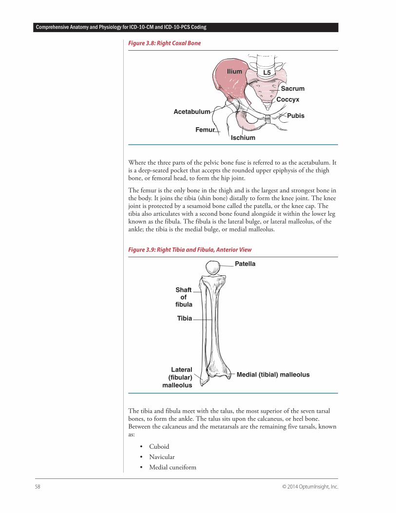
Figure 3.8: Right Coxal Bone
Where the three parts of the pelvic bone fuse is referred to as the acetabulum. It is a deep-seated pocket that accepts the rounded upper epiphysis of the thigh bone, or femoral head, to form the hip joint.
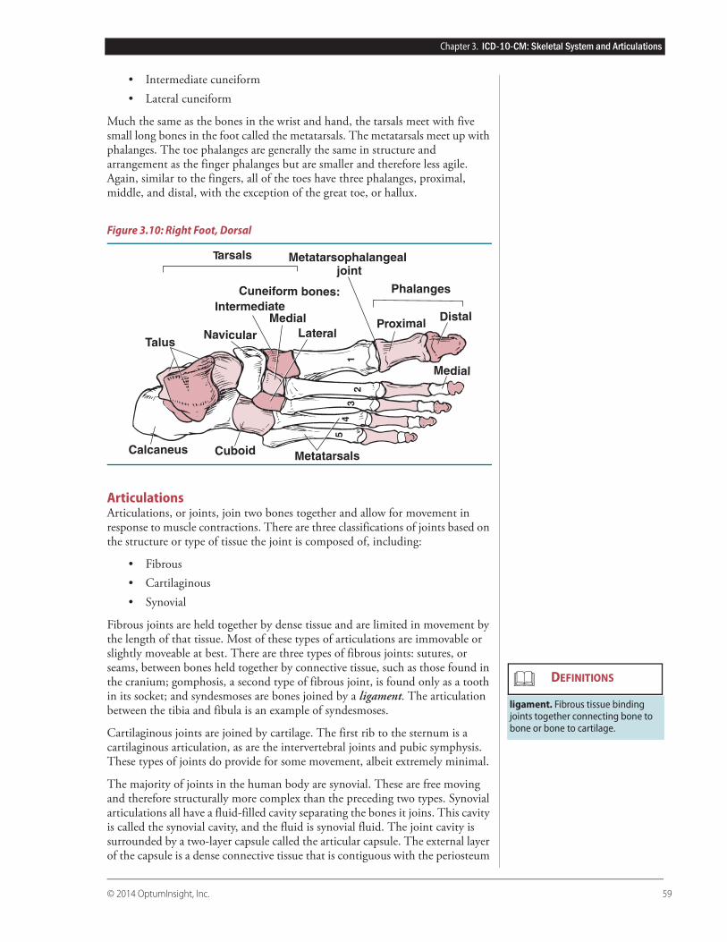
The femur is the only bone in the thigh and is the largest and strongest bone in the body. It joins the tibia (shin bone) distally to form the knee joint. The knee joint is protected by a sesamoid bone called the patella, or the knee cap. The tibia also articulates with a second bone found alongside it within the lower leg known as the fibula. The fibula is the lateral bulge, or lateral malleolus, of the ankle; the tibia is the medial bulge, or medial malleolus.
Figure 3.9: Right Tibia and Fibula, Anterior View
The tibia and fibula meet with the talus, the most superior of the seven tarsal bones, to form the ankle. The talus sits upon the calcaneus, or heel bone. Between the calcaneus and the metatarsals are the remaining five tarsals, known as:
• Cuboid• Navicular• Medial cuneiform
Femur
Ischium
Coccyx
Pubis
Sacrum
L5Ilium
Acetabulum
Ilium
Medial (tibial) malleolus
Shaftof
fibula
Tibia
Patella
Lateral
(fibular)
malleolus
Chapter 3. ICD-10-CM: Skeletal System and Articulations
© 2014 OptumInsight, Inc. 59
• Intermediate cuneiform• Lateral cuneiform
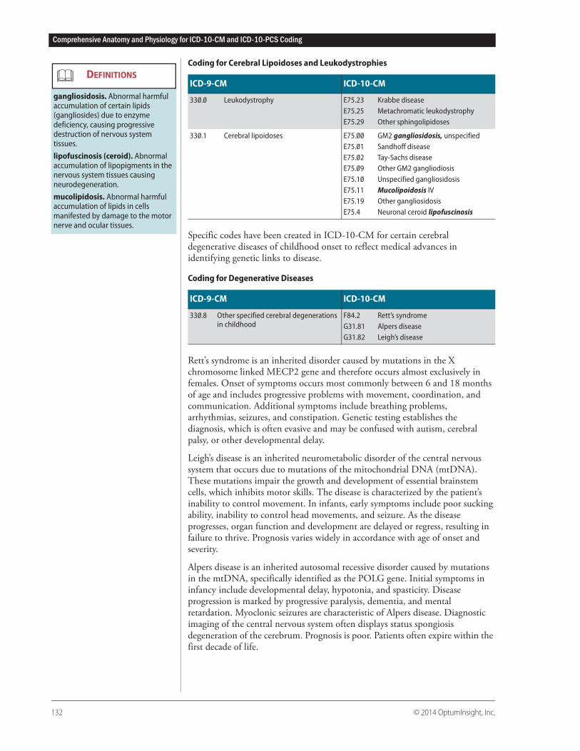
Much the same as the bones in the wrist and hand, the tarsals meet with five small long bones in the foot called the metatarsals. The metatarsals meet up with phalanges. The toe phalanges are generally the same in structure and arrangement as the finger phalanges but are smaller and therefore less agile. Again, similar to the fingers, all of the toes have three phalanges, proximal, middle, and distal, with the exception of the great toe, or hallux.
Figure 3.10: Right Foot, Dorsal
ArticulationsArticulations, or joints, join two bones together and allow for movement in response to muscle contractions. There are three classifications of joints based on the structure or type of tissue the joint is composed of, including:
• Fibrous• Cartilaginous• Synovial
Fibrous joints are held together by dense tissue and are limited in movement by the length of that tissue. Most of these types of articulations are immovable or slightly moveable at best. There are three types of fibrous joints: sutures, or seams, between bones held together by connective tissue, such as those found in the cranium; gomphosis, a second type of fibrous joint, is found only as a tooth in its socket; and syndesmoses are bones joined by a ligament. The articulation between the tibia and fibula is an example of syndesmoses.
Cartilaginous joints are joined by cartilage. The first rib to the sternum is a cartilaginous articulation, as are the intervertebral joints and pubic symphysis. These types of joints do provide for some movement, albeit extremely minimal.
The majority of joints in the human body are synovial. These are free moving and therefore structurally more complex than the preceding two types. Synovial articulations all have a fluid-filled cavity separating the bones it joins. This cavity is called the synovial cavity, and the fluid is synovial fluid. The joint cavity is surrounded by a two-layer capsule called the articular capsule. The external layer of the capsule is a dense connective tissue that is contiguous with the periosteum
TalusNavicular
Cuneiform bones:
MedialIntermediate
LateralProximal
Distal
Phalanges
Tarsals
Medial
MetatarsalsCuboidCalcaneus
12
34
5
Metatarsophalangeal joint
ligament. Fibrous tissue binding joints together connecting bone to bone or bone to cartilage.
DEFINITIONS
Comprehensive Anatomy and Physiology for ICD-10-CM and ICD-10-PCS Coding
132 © 2014 OptumInsight, Inc.
Coding for Cerebral Lipoidoses and Leukodystrophies
Specific codes have been created in ICD-10-CM for certain cerebral degenerative diseases of childhood onset to reflect medical advances in identifying genetic links to disease.
Coding for Degenerative Diseases
Rett’s syndrome is an inherited disorder caused by mutations in the X chromosome linked MECP2 gene and therefore occurs almost exclusively in females. Onset of symptoms occurs most commonly between 6 and 18 months of age and includes progressive problems with movement, coordination, and communication. Additional symptoms include breathing problems, arrhythmias, seizures, and constipation. Genetic testing establishes the diagnosis, which is often evasive and may be confused with autism, cerebral palsy, or other developmental delay.
Leigh’s disease is an inherited neurometabolic disorder of the central nervous system that occurs due to mutations of the mitochondrial DNA (mtDNA). These mutations impair the growth and development of essential brainstem cells, which inhibits motor skills. The disease is characterized by the patient’s inability to control movement. In infants, early symptoms include poor sucking ability, inability to control head movements, and seizure. As the disease progresses, organ function and development are delayed or regress, resulting in failure to thrive. Prognosis varies widely in accordance with age of onset and severity.
Alpers disease is an inherited autosomal recessive disorder caused by mutations in the mtDNA, specifically identified as the POLG gene. Initial symptoms in infancy include developmental delay, hypotonia, and spasticity. Disease progression is marked by progressive paralysis, dementia, and mental retardation. Myoclonic seizures are characteristic of Alpers disease. Diagnostic imaging of the central nervous system often displays status spongiosis degeneration of the cerebrum. Prognosis is poor. Patients often expire within the first decade of life.
ICD-9-CM ICD-10-CM
330.0 Leukodystrophy E75.23 Krabbe diseaseE75.25 Metachromatic leukodystrophyE75.29 Other sphingolipidoses
330.1 Cerebral lipoidoses E75.00 GM2 gangliosidosis, unspecifiedE75.01 Sandhoff diseaseE75.02 Tay-Sachs diseaseE75.09 Other GM2 gangliodiosisE75.10 Unspecified gangliosidosisE75.11 Mucolipoidosis IVE75.19 Other gangliosidosisE75.4 Neuronal ceroid lipofuscinosis
ICD-9-CM ICD-10-CM
330.8 Other specified cerebral degenerations in childhood
F84.2 Rett’s syndromeG31.81 Alpers diseaseG31.82 Leigh’s disease
gangliosidosis. Abnormal harmful accumulation of certain lipids (gangliosides) due to enzyme deficiency, causing progressive destruction of nervous system tissues.
lipofuscinosis (ceroid). Abnormal accumulation of lipopigments in the nervous system tissues causing neurodegeneration.
mucolipidosis. Abnormal harmful accumulation of lipids in cells manifested by damage to the motor nerve and ocular tissues.
DEFINITIONS
Chapter 5. ICD-10-CM: Nervous System
© 2014 OptumInsight, Inc. 133
Alzheimer’s and Parkinson’s Disease and Related SyndromesICD-9-CM contains a specific code for Alzheimer’s disease; ICD-10-CM has been expanded to provide information regarding onset within the code set itself.
Coding for Alzheimer’s Disease
Alzheimer’s disease is a chronic, progressive form of dementia caused by the destruction of subcortical white matter in the brain with plaque formations. Diagnostic imaging typically reveals shrinkage of white matter and an increase in the size of the ventricles.
In a normally functioning process, the ends of nerve cells (synapses) release neurotransmitter molecules that bind to neurotransmitter receptors, thus converting a nerve impulse into a chemical signal. The neurotransmitter receptor receives the transmitter signal and, in turn, generates a responsive nerve impulse. In Alzheimer's disease, there is a deficit in the levels of acetylcholine and degeneration of cholinergic neurons, neurotransmitter proteins, and cells within the brain. The debilitating and ultimately fatal disease is manifested by cognitive and memory decline and other neuropsychiatric symptoms and behavioral dysfunctions. Patients exhibit progressive behavioral changes, including loss of interest and memory problems. The associated frustration and aggressiveness can be treated with sedative or neuroleptic therapies.
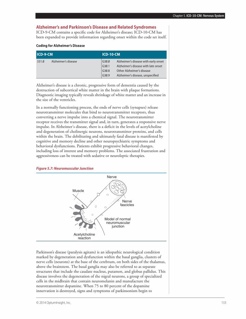
Figure 5.7: Neuromuscular Junction
Parkinson’s disease (paralysis agitans) is an idiopathic neurological condition marked by degeneration and dysfunction within the basal ganglia, clusters of nerve cells (neurons) at the base of the cerebrum, on both sides of the thalamus, above the brainstem. The basal ganglia may also be referred to as separate structures that include the caudate nucleus, putamen, and globus pallidus. This disease involves the degeneration of the nigral neurons, a group of specialized cells in the midbrain that contain neuromelanin and manufacture the neurotransmitter dopamine. When 75 to 80 percent of the dopamine innervation is destroyed, signs and symptoms of parkinsonism begin to
ICD-9-CM ICD-10-CM
331.0 Alzheimer’s disease G30.0 Alzheimer’s disease with early onsetG30.1 Alzheimer’s disease with late onsetG30.8 Other Alzheimer’s diseaseG30.9 Alzheimer’s disease, unspecified
Nerve
Nervefascicles
Muscle
Model of normal neuromuscular
junction
Acetylcholinereaction
Appendix A. Knowledge Review Answers
© 2014 OptumInsight, Inc. 573
Chapter 8. ICD-10-CM: Blood and Blood-Forming Organs
1. What are the three main functions of blood?a. Transportation
b. Regulation
c. Protection
Rationale: Blood serves many purposes that can be divided into three main functions: transportation, regulation, protection.
2. What are the formed elements found in blood?a. Red blood cells
b. White blood cells
c. Platelets
Rationale: There are formed elements, consisting of red blood cells, white blood cells, and platelets, and plasma, in which the formed elements “float.”
3. The plasma is the liquid that suspends the formed elements.Rationale: There are formed elements, consisting of red blood cells, white blood cells, and platelets, and plasma, in which the formed elements “float.”
4. Erythrocytes are also known as red blood cells. Rationale: Red blood cells, or erythrocytes, make up more than 99 percent of the formed elements.
5. The red blood cells’ main function is to carry oxygen to cells and transport some carbon dioxide away.Rationale: Red blood cells travel throughout the body delivering oxygen and removing some of the carbon dioxide the cells release.
6. Hemoglobin is the protein responsible for the red color of blood.Rationale: The protein molecules, known as hemoglobin, are responsible for the blood’s color. When the RBCs are carrying oxygen, the blood appears bright red; when the hemoglobin is de-oxygenated, the blood appears blue when viewed through blood vessel walls.
7. What are the four blood types?a. A
b. B
c. AB
d. O
Rationale: The blood type group is determined by identifying up to two antigens on the surface of an erythrocyte. These antigens are known as antigen A and antigen B. The absence or presence of these determine the four blood types:• A: Antigen A is present.• B: Antigen B is present.• AB: Both antigens are present.
Appendix B. Body Part Key
© 2014 OptumInsight, Inc. 625
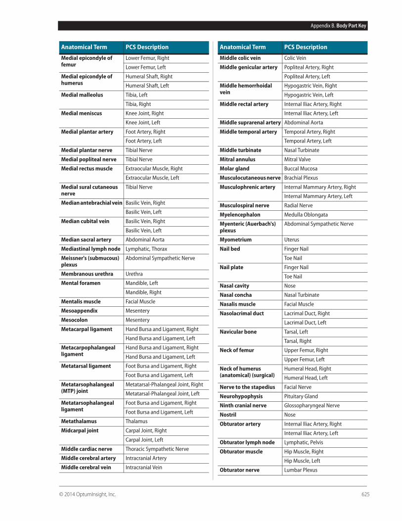
Medial epicondyle of femur
Lower Femur, Right
Lower Femur, Left
Medial epicondyle of humerus
Humeral Shaft, Right
Humeral Shaft, Left
Medial malleolus Tibia, Left
Tibia, Right
Medial meniscus Knee Joint, Right
Knee Joint, Left
Medial plantar artery Foot Artery, Right
Foot Artery, Left
Medial plantar nerve Tibial Nerve
Medial popliteal nerve Tibial Nerve
Medial rectus muscle Extraocular Muscle, Right
Extraocular Muscle, Left
Medial sural cutaneous nerve
Tibial Nerve
Median antebrachial vein Basilic Vein, Right
Basilic Vein, Left
Median cubital vein Basilic Vein, Right
Basilic Vein, Left
Median sacral artery Abdominal Aorta
Mediastinal lymph node Lymphatic, Thorax
Meissner's (submucous) plexus
Abdominal Sympathetic Nerve
Membranous urethra Urethra
Mental foramen Mandible, Left
Mandible, Right
Mentalis muscle Facial Muscle
Mesoappendix Mesentery
Mesocolon Mesentery
Metacarpal ligament Hand Bursa and Ligament, Right
Hand Bursa and Ligament, Left
Metacarpophalangeal ligament
Hand Bursa and Ligament, Right
Hand Bursa and Ligament, Left
Metatarsal ligament Foot Bursa and Ligament, Right
Foot Bursa and Ligament, Left
Metatarsophalangeal (MTP) joint
Metatarsal-Phalangeal Joint, Right
Metatarsal-Phalangeal Joint, Left
Metatarsophalangeal ligament
Foot Bursa and Ligament, Right
Foot Bursa and Ligament, Left
Metathalamus Thalamus
Midcarpal joint Carpal Joint, Right
Carpal Joint, Left
Middle cardiac nerve Thoracic Sympathetic Nerve
Middle cerebral artery Intracranial Artery
Middle cerebral vein Intracranial Vein
Anatomical Term PCS Description
Middle colic vein Colic Vein
Middle genicular artery Popliteal Artery, Right
Popliteal Artery, Left
Middle hemorrhoidal vein
Hypogastric Vein, Right
Hypogastric Vein, Left
Middle rectal artery Internal Iliac Artery, Right
Internal Iliac Artery, Left
Middle suprarenal artery Abdominal Aorta
Middle temporal artery Temporal Artery, Right
Temporal Artery, Left
Middle turbinate Nasal Turbinate
Mitral annulus Mitral Valve
Molar gland Buccal Mucosa
Musculocutaneous nerve Brachial Plexus
Musculophrenic artery Internal Mammary Artery, Right
Internal Mammary Artery, Left
Musculospiral nerve Radial Nerve
Myelencephalon Medulla Oblongata
Myenteric (Auerbach's) plexus
Abdominal Sympathetic Nerve
Myometrium Uterus
Nail bed Finger Nail
Toe Nail
Nail plate Finger Nail
Toe Nail
Nasal cavity Nose
Nasal concha Nasal Turbinate
Nasalis muscle Facial Muscle
Nasolacrimal duct Lacrimal Duct, Right
Lacrimal Duct, Left
Navicular bone Tarsal, Left
Tarsal, Right
Neck of femur Upper Femur, Right
Upper Femur, Left
Neck of humerus (anatomical) (surgical)
Humeral Head, Right
Humeral Head, Left
Nerve to the stapedius Facial Nerve
Neurohypophysis Pituitary Gland
Ninth cranial nerve Glossopharyngeal Nerve
Nostril Nose
Obturator artery Internal Iliac Artery, Right
Internal Iliac Artery, Left
Obturator lymph node Lymphatic, Pelvis
Obturator muscle Hip Muscle, Right
Hip Muscle, Left
Obturator nerve Lumbar Plexus
Anatomical Term PCS Description
© 2014 OptumInsight, Inc. 649
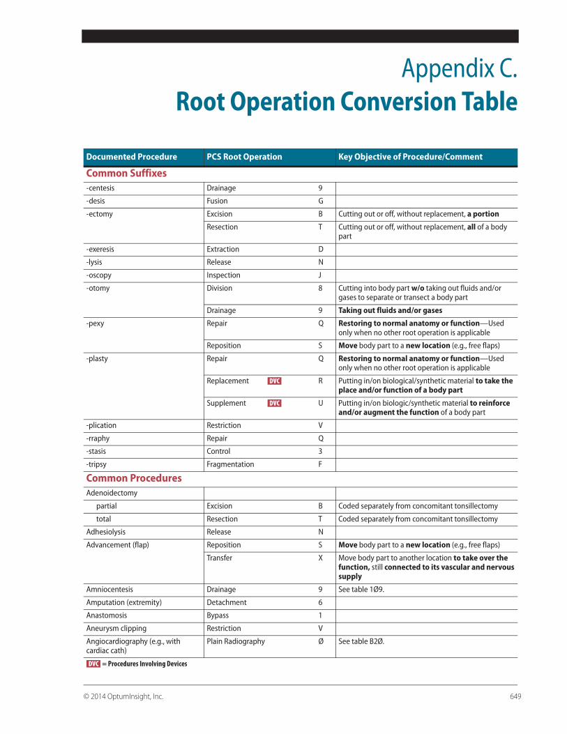
Appendix C.Root Operation Conversion Table
Documented Procedure PCS Root Operation Key Objective of Procedure/Comment
Common Suffixes-centesis Drainage 9
-desis Fusion G
-ectomy Excision B Cutting out or off, without replacement, a portion
Resection T Cutting out or off, without replacement, all of a body part
-exeresis Extraction D
-lysis Release N
-oscopy Inspection J
-otomy Division 8 Cutting into body part w/o taking out fluids and/or gases to separate or transect a body part
Drainage 9 Taking out fluids and/or gases
-pexy Repair Q Restoring to normal anatomy or function—Used only when no other root operation is applicable
Reposition S Move body part to a new location (e.g., free flaps)
-plasty Repair Q Restoring to normal anatomy or function—Used only when no other root operation is applicable
Replacement R Putting in/on biological/synthetic material to take the place and/or function of a body part
Supplement U Putting in/on biologic/synthetic material to reinforce and/or augment the function of a body part
-plication Restriction V
-rraphy Repair Q
-stasis Control 3
-tripsy Fragmentation F
Common ProceduresAdenoidectomy
partial Excision B Coded separately from concomitant tonsillectomy
total Resection T Coded separately from concomitant tonsillectomy
Adhesiolysis Release N
Advancement (flap) Reposition S Move body part to a new location (e.g., free flaps)
Transfer X Move body part to another location to take over the function, still connected to its vascular and nervous supply
Amniocentesis Drainage 9 See table 1Ø9.
Amputation (extremity) Detachment 6
Anastomosis Bypass 1
Aneurysm clipping Restriction V
Angiocardiography (e.g., with cardiac cath)
Plain Radiography Ø See table B2Ø.
= Procedures Involving Devices
DVC
DVC
DVC