Optimizing Intracellular Flow Cytometry: Simultaneous ... · For Research Use Only. Not for use in...
34
For Research Use Only. Not for use in diagnostic or therapeutic procedures. Simultaneous Detection of Cytokines and Transcription Factors Presented by Jurg Rohrer, PhD, BD Biosciences Optimizing Intracellular Flow Cytometry: Optimizing Intracellular Flow Cytometry: 23-10780-00
Transcript of Optimizing Intracellular Flow Cytometry: Simultaneous ... · For Research Use Only. Not for use in...
For Research Use Only. Not for use in diagnostic or therapeutic procedures.
Simultaneous Detection of Cytokines and Transcription Factors
Presented by Jurg Rohrer, PhD, BD Biosciences
Optimizing Intracellular Flow Cytometry:
Optimizing Intracellular Flow Cytometry:
23-10780-00
For Research Use Only. Not for use in diagnostic or therapeutic procedures.
Outline
• Introduction– Cytokines– Transcription factors
• Basic concepts of intracellular flow cytometry– Optimization examples
• Treg/Th17 cell analysis– Considerations– Examples
For Research Use Only. Not for use in diagnostic or therapeutic procedures.
Cytokines
• Soluble polypeptides produced by most nucleated cells in the body
• Some potent producers include endothelial and epithelial cells and resident macrophages, especially near the interface with the external environment
• Critical to the development and functioning of both the innate and adaptive immune responses
• Promote cellular differentiation and proliferation– Example: IL-2 involved in T cell activation and maintenance
of a Th1 response• Work in either an autocrine or paracrine manner
For Research Use Only. Not for use in diagnostic or therapeutic procedures.
Th17 Cells
• A subset of CD4+ T helper cells• Developmentally distinct from Th1 and Th2 cells• Immunity against bacterial and fungal infectious• Play a key role in autoimmune diseases (tissue
injury)• Controlling Th17 activity could aid in the treatment of
autoimmune diseases• TGF-β, IL-6, IL-21, IL-1β,
and IL-23 appear to drive
Th17 development• Produce IL-17A, IL-17F; also IL-21, IL-22, IL-26, and
less TNF and IL-6
For Research Use Only. Not for use in diagnostic or therapeutic procedures.
Transcription Factors
• Proteins that bind to specific DNA sequences• Control the transfer of genetic information
from DNA to RNA• Regulators of gene expression• A single transcription factor can bind
hundreds of promoters
For Research Use Only. Not for use in diagnostic or therapeutic procedures.
Regulatory T Cells• Tregs = CD4+ T regulatory cells• Comprise ~ 1–3% of human PBMCs and ~ 4–8% of
mouse spleen• Actively suppress T cell proliferation• Play a crucial role in T cell homeostasis• nTreg develop in the thymus, iTreg require TGFβ, IL-
2 and RA• FoxP3, a forkhead family transcription factor, is a
specific marker for Tregs• FoxP3 is necessary for the development and function
of Tregs
For Research Use Only. Not for use in diagnostic or therapeutic procedures.
Regulatory T Cells, cont’d
• Produce TGFβ
and IL-10 and express high levels of CD25 and low levels of CD127
• Diminish immune responses against cancers, allogeneic transplants, and infectious pathogens
• Dampening Treg activity could improve anti-tumor responses and responses to vaccinations and chronic infections
• Deficiencies contribute to the development of autoimmune diseases
• Boosting Treg activity could be useful in the treatment of T cell induced diseases
For Research Use Only. Not for use in diagnostic or therapeutic procedures.
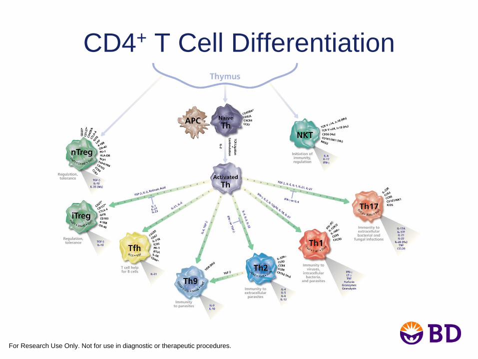
CD4+ T Cell Differentiation
For Research Use Only. Not for use in diagnostic or therapeutic procedures.
What is Intracellular Flow Cytometry?
• Detection of:– Transcription factors– Intracellular signaling molecules– Cytokines– Structural proteins– Scaffold proteins– Pan and phospho-specific antigens
For Research Use Only. Not for use in diagnostic or therapeutic procedures.
Considerations for Intracellular Flow Cytometry
• Must permeabilize a cell to access cell contents
• If a cell is permeabilized, then contents could “leak” out and the protein of interest could be lost
• Therefore, cells are fixed first, followed by permeabilization
• To detect secreted proteins, they must be “trapped” within the cell prior to fixation and permeabilization to increase the likelihood of detection
For Research Use Only. Not for use in diagnostic or therapeutic procedures.
Considerations for Intracellular Flow, cont’d
• Protein transport inhibition– Monensin vs Brefeldin A (BD GolgiStop™ vs
BD GolgiPlug™ inhibitor)– Optimal time for inhibition– Optimal concentration of inhibitor
• Fixation– Concentration (paraformaldehyde)– Time– Temperature– Compatibility with fluorochromes– Compatibility of cell surface markers
For Research Use Only. Not for use in diagnostic or therapeutic procedures.
Considerations for Intracellular Flow, cont’d.
• Permeabilization– Perm agent (saponin, methanol, Tween® 20, Triton X-100TM)– Concentration– Time– Temperature– Compatibility with fluorochromes– Compatibility of cell surface markers
• Different locations in cells are more difficult to access• Types of proteins being identified, single or in a
complex?
For Research Use Only. Not for use in diagnostic or therapeutic procedures.
Considerations for Intracellular Flow, cont’d.
• Antibody staining– Order– Concentration– Time– Temperature– Fluorochromes
• Storage conditions– Buffer– Time
• Matching one antibody protocol with another antibody protocol
For Research Use Only. Not for use in diagnostic or therapeutic procedures.
Buffer Choices
• Fixation buffer• BD Cytofix/Cytoperm™ and BD™ Perm/Wash
buffer• BD Pharmingen™ FoxP3 buffer set (mouse or
human)• BD™ Phosflow Perm Buffer II• BD™ Phosflow Perm Buffer III• BD IntraSure™ kit
• BD FastImmune™ kits
For Research Use Only. Not for use in diagnostic or therapeutic procedures.
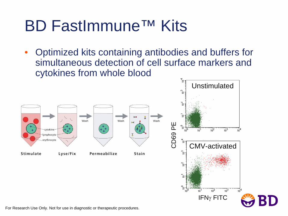
BD FastImmune™ Kits• Optimized kits containing antibodies and buffers for
simultaneous detection of cell surface markers and cytokines from whole blood
CMV-activated
Unstimulated
IFNγ
FITC
CD
69 P
E
For Research Use Only. Not for use in diagnostic or therapeutic procedures.
Case Study• The study of Treg and Th17 cells• Requires the need to detect both FoxP3 and IL-17 in the
same sample• Unique protocols for both mouse and human FoxP3
staining • Questions are:
– How well does IL-17 staining work in the FoxP3 buffer system?– How well do other intracellular and surface markers work with
the FoxP3 buffer system?
• Examples of FoxP3 optimization followed by addition of other markers
For Research Use Only. Not for use in diagnostic or therapeutic procedures.
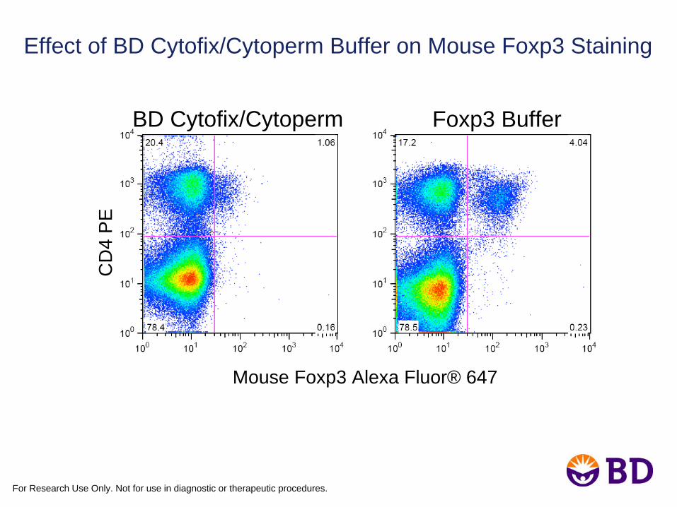
Effect of BD Cytofix/Cytoperm Buffer on Mouse Foxp3 Staining
Mouse Foxp3 Alexa Fluor® 647
Foxp3 BufferC
D4
PE
BD Cytofix/Cytoperm
For Research Use Only. Not for use in diagnostic or therapeutic procedures.
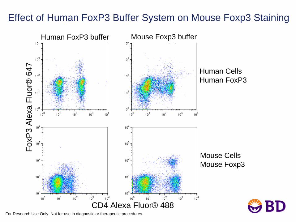
Effect of Human FoxP3 Buffer System on Mouse Foxp3 Staining
Human CellsHuman FoxP3
Mouse CellsMouse Foxp3
FoxP
3 A
lexa
Flu
or®
647
CD4 Alexa Fluor® 488
Mouse Foxp3 bufferHuman FoxP3 buffer
For Research Use Only. Not for use in diagnostic or therapeutic procedures.
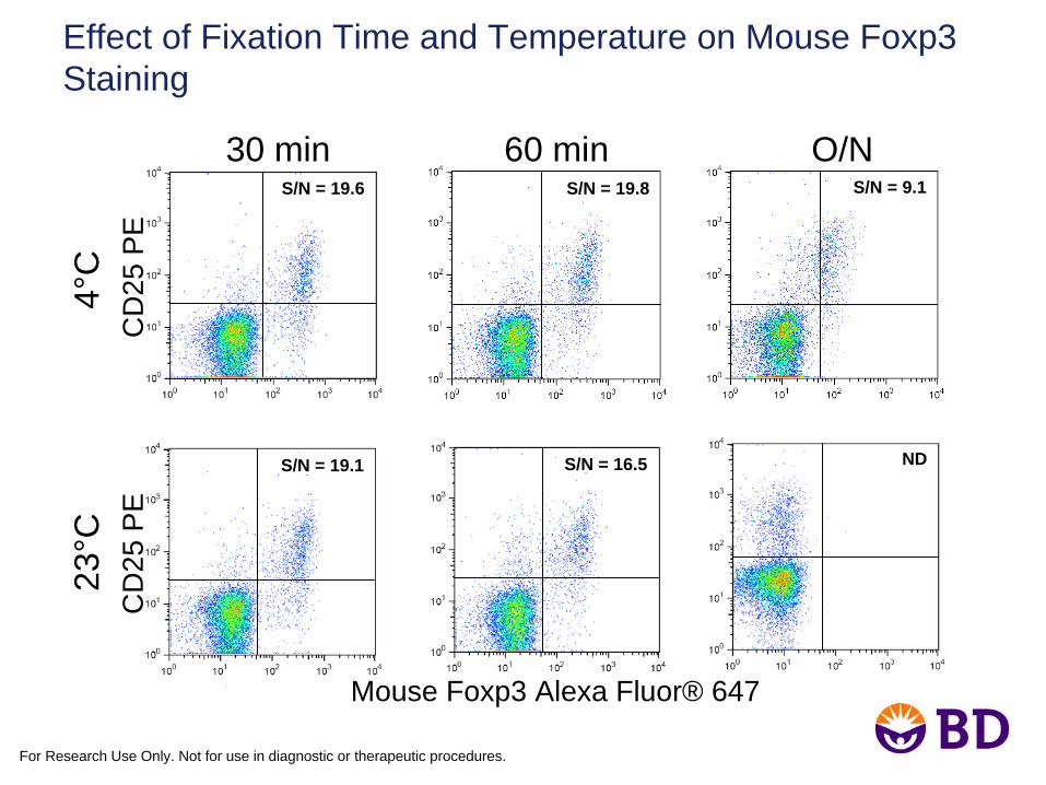
Effect of Fixation Time and Temperature on Mouse Foxp3 Staining
30 min 60 min O/NS/N = 19.6 S/N = 19.8 S/N = 9.1
CD
25 P
E4°
C
S/N = 19.1 S/N = 16.5 ND
23°C
Mouse Foxp3 Alexa Fluor® 647
CD
25 P
E
For Research Use Only. Not for use in diagnostic or therapeutic procedures.
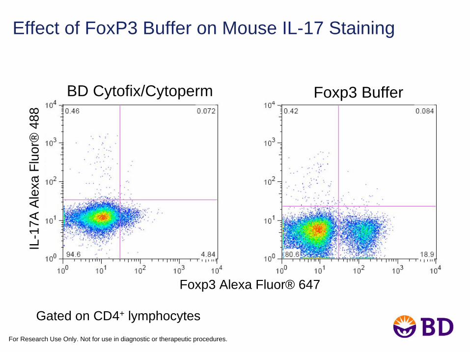
Effect of FoxP3 Buffer on Mouse IL-17 Staining
Gated on CD4+ lymphocytes
Foxp3 Alexa Fluor® 647
IL-1
7A A
lexa
Flu
or®
488
Foxp3 BufferBD Cytofix/Cytoperm
For Research Use Only. Not for use in diagnostic or therapeutic procedures.
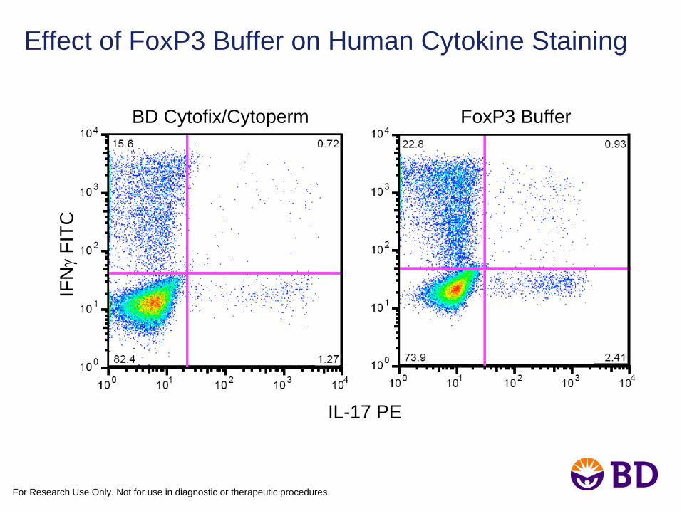
Effect of FoxP3 Buffer on Human Cytokine Staining
BD Cytofix/Cytoperm FoxP3 Buffer
IFNγ
FITC
IL-17 PE
For Research Use Only. Not for use in diagnostic or therapeutic procedures.
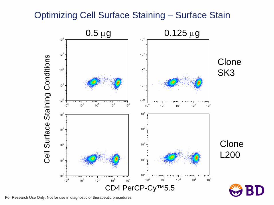
Optimizing Cell Surface Staining – Surface Stain
CloneSK3
CloneL200
0.5 μg 0.125 μg
CD4 PerCP-Cy™5.5
Cel
l Sur
face
Sta
inin
g C
ondi
tions
For Research Use Only. Not for use in diagnostic or therapeutic procedures.
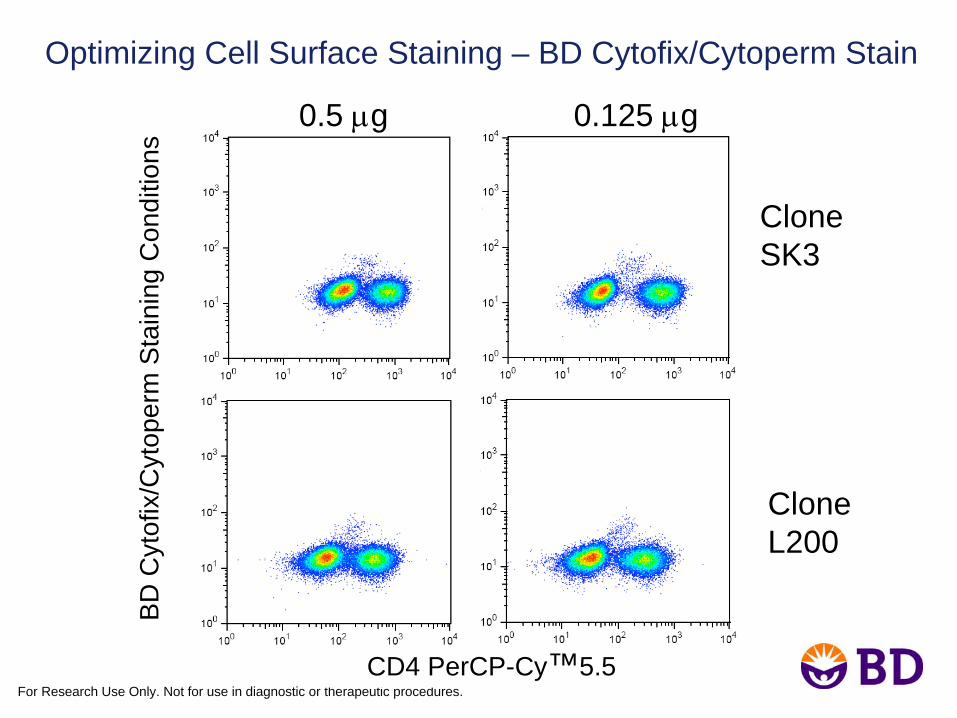
Optimizing Cell Surface Staining – BD Cytofix/Cytoperm Stain
CloneSK3
CloneL200
CD4 PerCP-Cy™5.5
BD
Cyt
ofix
/Cyt
oper
m S
tain
ing
Con
ditio
ns0.5 μg 0.125 μg
For Research Use Only. Not for use in diagnostic or therapeutic procedures.
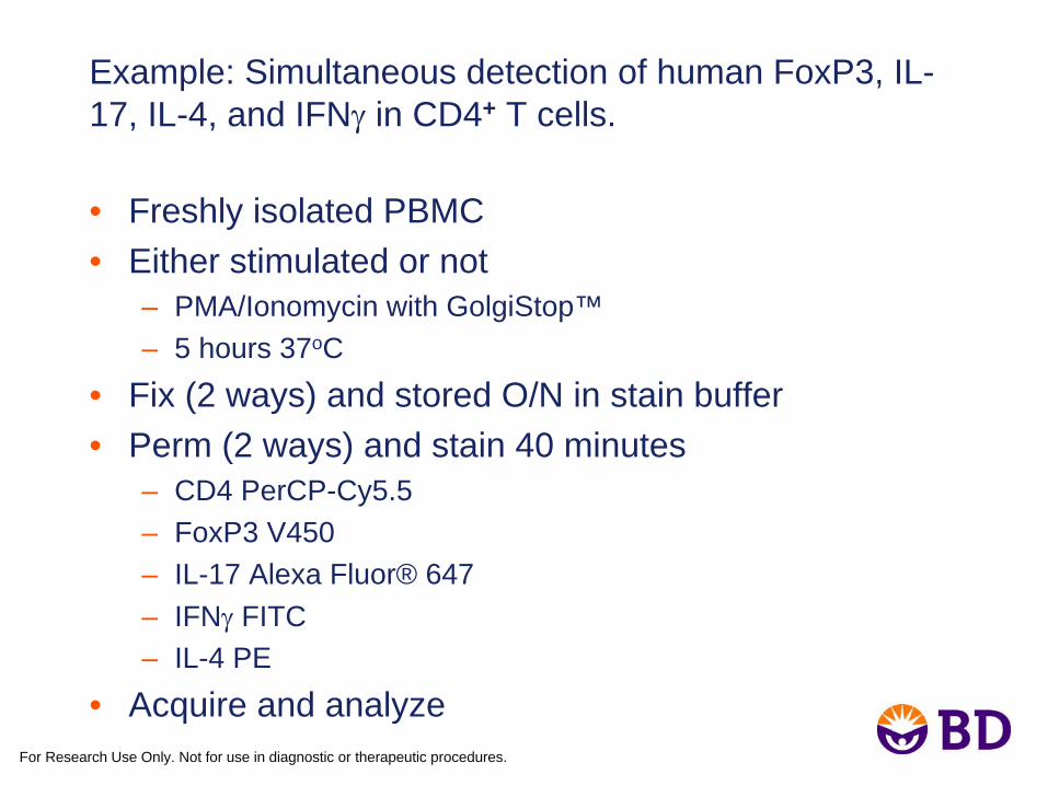
Example: Simultaneous detection of human FoxP3, IL- 17, IL-4, and IFNγ
in CD4+ T cells.
• Freshly isolated PBMC• Either stimulated or not
– PMA/Ionomycin with GolgiStop™– 5 hours 37oC
• Fix (2 ways) and stored O/N in stain buffer• Perm (2 ways) and stain 40 minutes
– CD4 PerCP-Cy5.5– FoxP3 V450– IL-17 Alexa Fluor® 647– IFNγ
FITC– IL-4 PE
• Acquire and analyze
For Research Use Only. Not for use in diagnostic or therapeutic procedures.
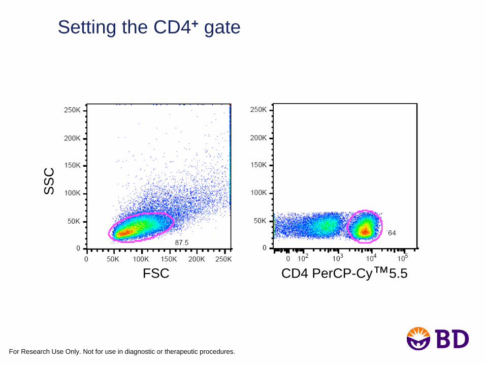
Setting the CD4+ gate
FSC CD4 PerCP-Cy™5.5
SS
C
For Research Use Only. Not for use in diagnostic or therapeutic procedures.
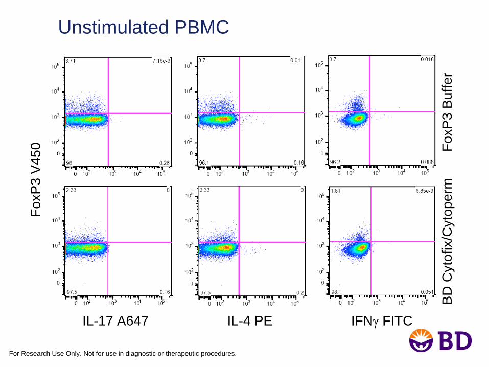
Unstimulated PBMCFo
xP3
V45
0
IL-17 A647 IL-4 PE IFNγ
FITC
BD
Cyt
ofix
/Cyt
oper
m
F
oxP
3 B
uffe
r

For Research Use Only. Not for use in diagnostic or therapeutic procedures.
Stimulated PBMCFo
xP3
V45
0
IL-17 A647 IL-4 PE IFNγ
FITC
BD
Cyt
ofix
/Cyt
oper
m
F
oxP
3 B
uffe
r
For Research Use Only. Not for use in diagnostic or therapeutic procedures.
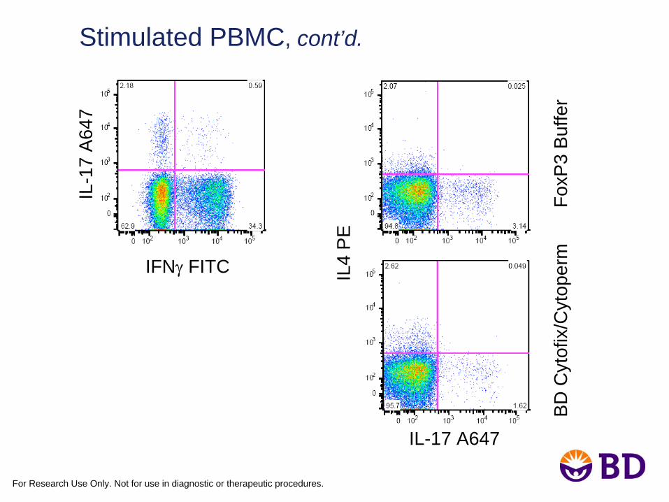
Stimulated PBMC, cont’d.
IL-1
7 A
647
IL-17 A647
BD
Cyt
ofix
/Cyt
oper
m
F
oxP
3 B
uffe
r
IL4
PE
IFNγ
FITC
For Research Use Only. Not for use in diagnostic or therapeutic procedures.
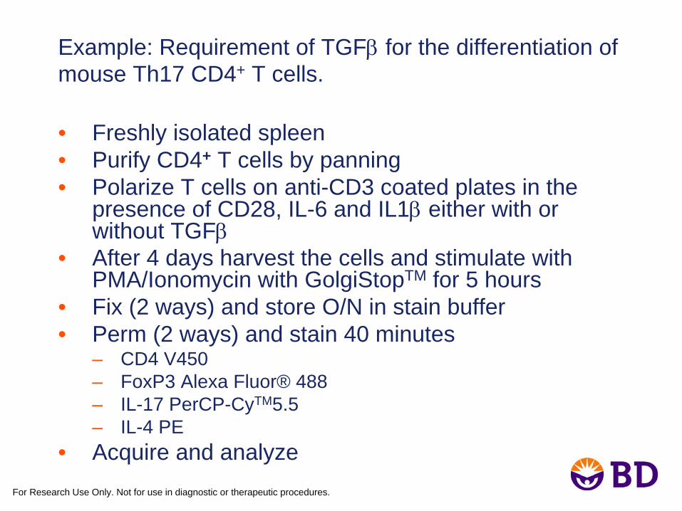
Example: Requirement of TGFβ
for the differentiation of mouse Th17 CD4+ T cells.
• Freshly isolated spleen• Purify CD4+ T cells by panning• Polarize T cells on anti-CD3 coated plates in the
presence of CD28, IL-6 and IL1β
either with or without TGFβ
• After 4 days harvest the cells and stimulate with PMA/Ionomycin with GolgiStopTM for 5 hours
• Fix (2 ways) and store O/N in stain buffer• Perm (2 ways) and stain 40 minutes
– CD4 V450– FoxP3 Alexa Fluor® 488– IL-17 PerCP-CyTM5.5– IL-4 PE
• Acquire and analyze
For Research Use Only. Not for use in diagnostic or therapeutic procedures.
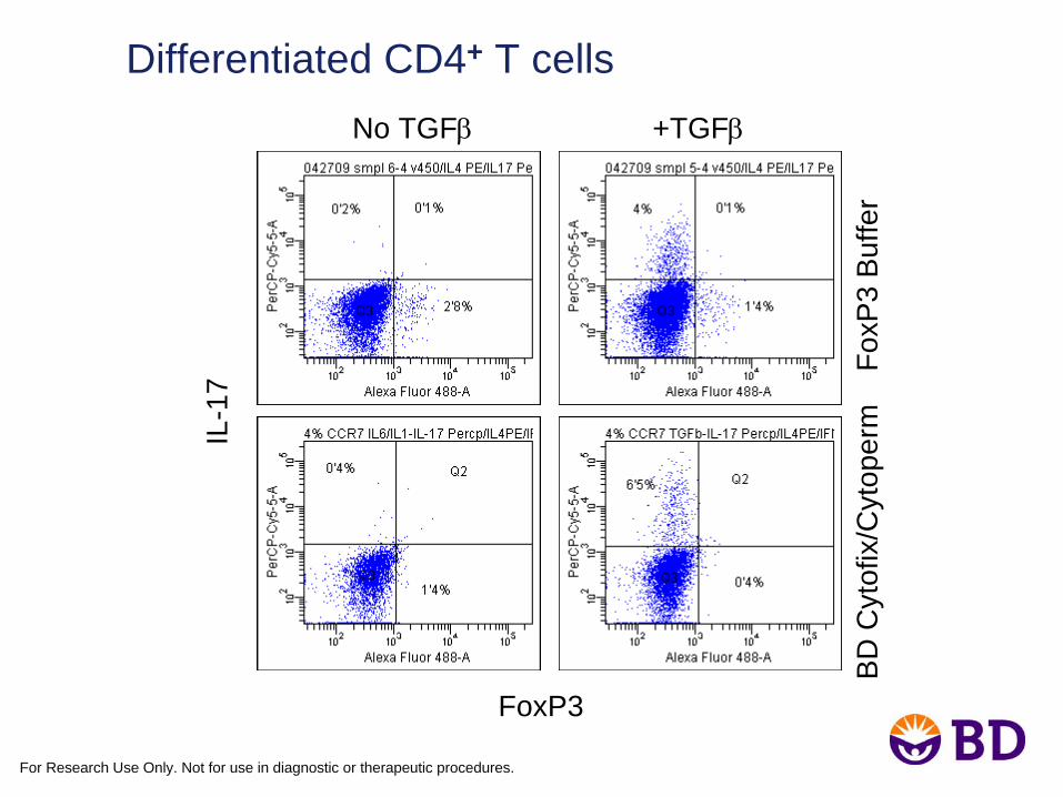
Differentiated CD4+ T cells
IL-1
7
FoxP3
BD
Cyt
ofix
/Cyt
oper
m
Fox
P3
Buf
fer
No TGFβ
+TGFβ
For Research Use Only. Not for use in diagnostic or therapeutic procedures.
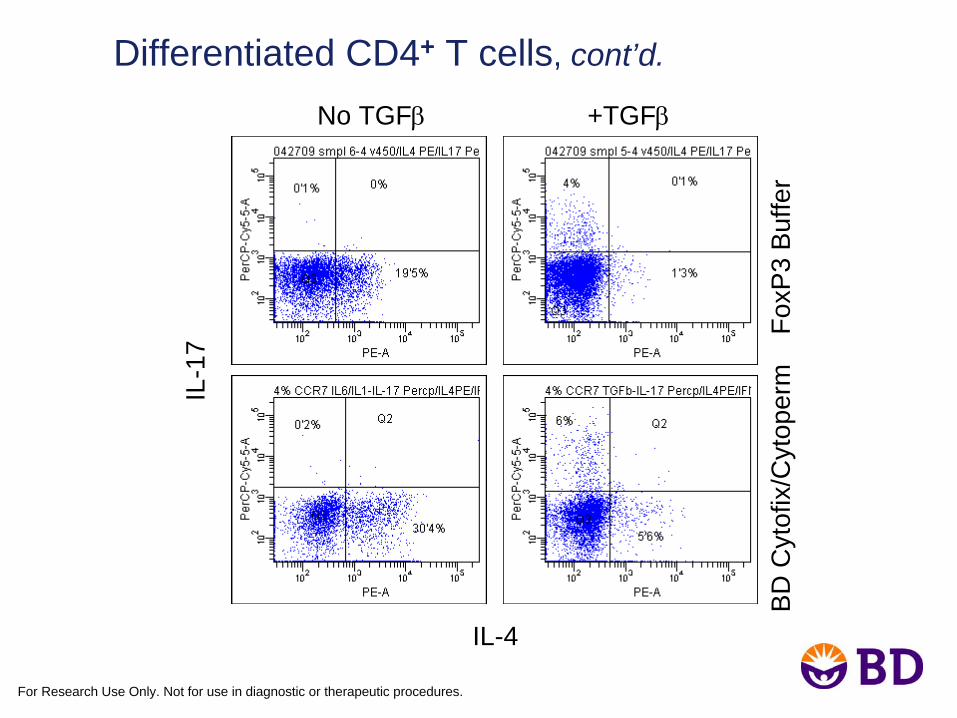
Differentiated CD4+ T cells, cont’d.
IL-1
7
IL-4
No TGFβ
+TGFβ
BD
Cyt
ofix
/Cyt
oper
m
Fox
P3
Buf
fer
For Research Use Only. Not for use in diagnostic or therapeutic procedures.
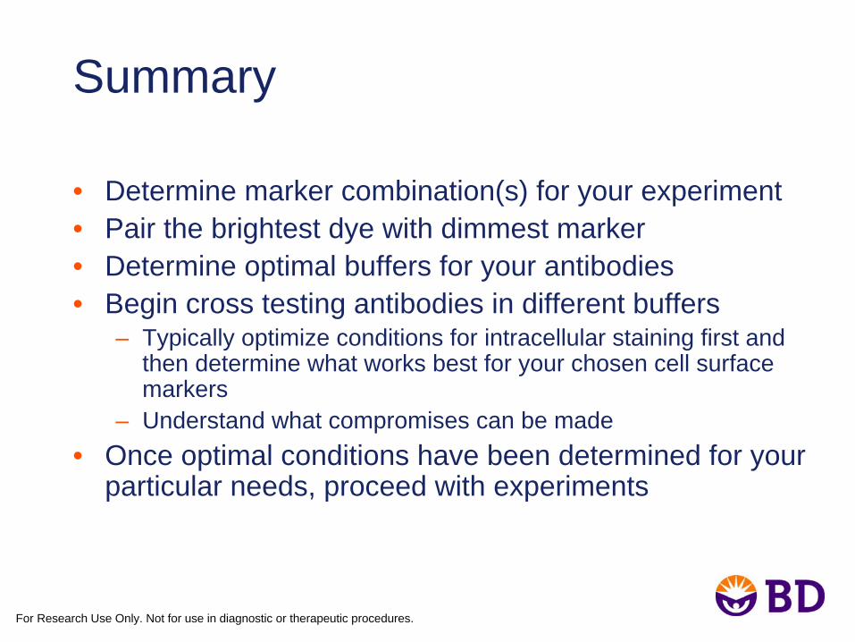
Summary
• Determine marker combination(s) for your experiment• Pair the brightest dye with dimmest marker• Determine optimal buffers for your antibodies• Begin cross testing antibodies in different buffers
– Typically optimize conditions for intracellular staining first and then determine what works best for your chosen cell surface markers
– Understand what compromises can be made• Once optimal conditions have been determined for your
particular needs, proceed with experiments
For Research Use Only. Not for use in diagnostic or therapeutic procedures.
Acknowledgements
• Xiao-Wei Wu• Ai-Li Wei• Li Li• Ravi Hingorani• Jeanne Elia• Christopher Boyce
For Research Use Only. Not for use in diagnostic or therapeutic procedures.
If you have further questions:
Contact your US Reagent Sales Repor e-mail: [email protected]
Alexa Fluor®
is a registered trademark of Molecular Probes, Inc.
Cy™ is a trademark of Amersham Biosciences.