Optimizing Intracellular Flow Cytometry: Detection of Cytokines ...
27
Detection of Cytokines, Transcription Factors, and Phosphoprotein by Flow Cytometry Presented by Erika O’Donnell, PhD, BD Biosciences Optimizing Intracellular Flow Cytometry 23-14876-00
Transcript of Optimizing Intracellular Flow Cytometry: Detection of Cytokines ...

Detection of Cytokines, Transcription Factors, and Phosphoprotein by Flow Cytometry
Presented by
Erika O’Donnell, PhD, BD Biosciences
Optimizing Intracellular
Flow Cytometry
23-14876-00

Outline
• Basic principles of intracellular flow cytometry
• Detection of cytokines
• Detection of transcription factors
• Detection of phosphoprotein
• Combining techniques
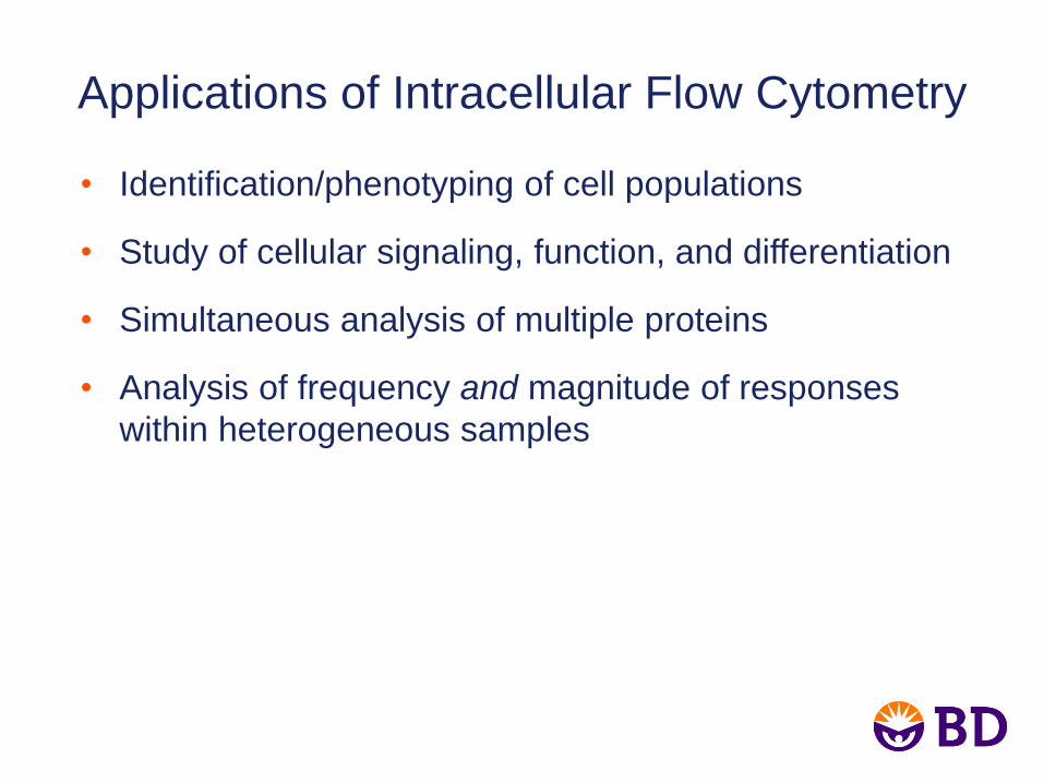
Applications of Intracellular Flow Cytometry
• Identification/phenotyping of cell populations
• Study of cellular signaling, function, and differentiation
• Simultaneous analysis of multiple proteins
• Analysis of frequency and magnitude of responses
within heterogeneous samples

Applications of Intracellular Flow Cytometry
• Human whole blood was
stimulated with staphylococcal
enterotoxin B or cytomegalovirus
pp65 for 6 hours in the presence
of Brefeldin A.
• Cells were fixed, permeabilized,
and stained using the BD
FastImmune™ 3-color CD4
intracellular cytokine detection kit.
• Cells were analyzed on a BD
FACSVerse™ flow cytometer.
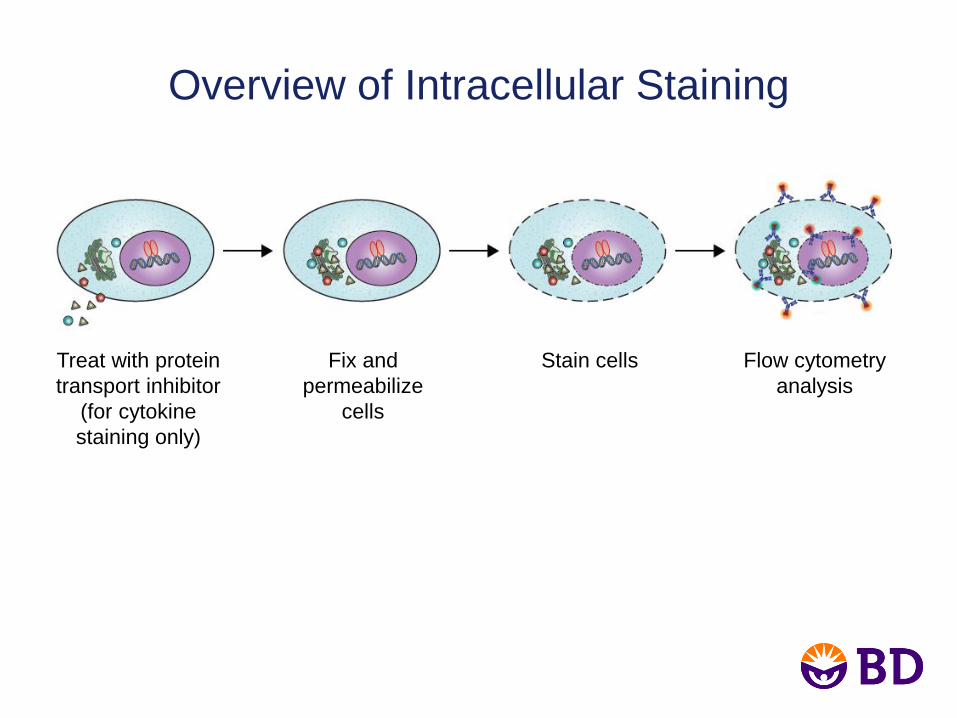
Overview of Intracellular Staining
Treat with protein
transport inhibitor
(for cytokine
staining only)
Fix and
permeabilize
cells
Stain cells Flow cytometry
analysis

• To access intracellular antigens, cells must be fixed
and permeabilized.
• Different permeabilization conditions favor the
detection of different types of epitopes.
Optimal Conditions for Intracellular Staining
Depend on Epitope Accessibility
– Cytokines (once trapped inside the cell) are accessible using gentle
conditions.
– Transcription factors and phosphoproteins often require stronger
permeabilization buffers.
– Cellular fixation and permeabilization conditions can have adverse
effects on surface antigens or fluorochromes.

Optimal Conditions for Intracellular Staining
Depend on Epitope Accessibility
• Human PBMCs were left untreated (–) or were
activated (+) with human IFN- (Stat1 pY701) or
PMA (Stat1 pS727).
• Cells were fixed using BD Cytofix™ fixation buffer
and permeabilized using BD Phosflow™ perm
buffer I, II, III, or IV prior to staining.
– +
– +
– +
– +
Stat1 (pS727) PE Stat1 (pY701) PE
Mild Detergent (Saponin)
Perm/Wash Buffer I
Methanol: Low Conc.
Perm Buffer II
Methanol: High Conc.
Perm Buffer III
Harsh Detergent
Perm Buffer IV (1X)
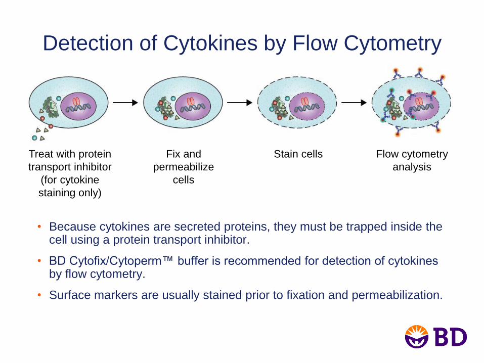
Detection of Cytokines by Flow Cytometry
• Because cytokines are secreted proteins, they must be trapped inside the cell using a protein transport inhibitor.
• BD Cytofix/Cytoperm™ buffer is recommended for detection of cytokines by flow cytometry.
• Surface markers are usually stained prior to fixation and permeabilization.
Treat with protein
transport inhibitor
(for cytokine
staining only)
Fix and
permeabilize
cells
Stain cells Flow cytometry
analysis

Protein Transport Inhibitors for Cytokine
Detection by Flow Cytometry
Monensin (BD GolgiStop™) and
Brefeldin A (BD GolgiPlug™) inhibitors
are commonly used to trap cytokines
inside the cell for analysis.
• Work by slightly different mechanisms
– Monensin prevents protein
secretion by interacting with the
Golgi transmembrane Na++/H+
transport.
– Brefeldin A redistributes
intracellularly produced proteins
from the cis/medial Golgi complex
to the endoplasmic reticulum.
• Different inhibitors may work better for
detection of different cytokines.
Species Cytokines Transport
Inhibitor
Human IL-1, IL-6, IL-8,
TNF- Monensin
Human
IFN-, IL-2, IL-10,
IL-12, MCP-1,
MCP-3, MIG,
MIP-1, RANTES
Either
monensin or
brefeldin A
Mouse IL-6, IL-12, TNF- Brefeldin A
Mouse GM-CSF, IL-3,
IL-4, IL-5, IL-10 Monensin
Mouse IFN-, IL-2
Either
monensin or
brefeldin A

Example 1: IFN- and IL-2 Production in
CD8+ Cells
• Human PBMCs were stimulated with staphylococcal enterotoxin B for 6 hours
in the presence of Brefeldin A.
• Cells were fixed and permeabilized using the BD Cytofix/Cytoperm buffer
system.
• Cells were stained with CD3 FITC, CD4 PerCP-Cy™5.5, CD8 BD Horizon™
Brilliant Violet™ 421, IFN- PE, and IL-2 APC.
• Cells were analyzed on a BD FACSVerse flow cytometer.
IFN- PE-A IL-2 APC-A
CD
8 B
rill
ian
t V
iole
t 4
21
-A
CD
8 B
rill
ian
t V
iole
t 42
1-A

Detection of Transcription Factors by
Flow Cytometry
• Transcription factors are proteins that bind to specific DNA sequences
and regulate gene expression.
• BD Pharmingen™ transcription factor buffer is the recommended
starting buffer.
– Compatible with staining of most surface markers (stained before or
after cellular permeabilization) and cytokines
Fix and
permeabilize
cells
Stain cells Flow cytometry
analysis

Example 2: Detection of FoxP3
Regulatory T Cells (Tregs)
• Tregs are a subset of T cells that regulate
the immune response by suppressing the
activity of other T cells.
• Human PBMCs were stained for surface
markers CD4 FITC, CD25 Brilliant Violet
421, and CD127 Alexa Fluor® 647.
• After washing, cells were fixed and
permeabilized with the BD Pharmingen
transcription factor buffer set and stained
with FoxP3 PE-CF594.
• Data was acquired on a BD FACSVerse
flow cytometer.
102 103 104 105 0
10
2
10
3
10
4
10
5
0
CD25 Brilliant Violet 421
CD
12
7 A
lex
a F
luo
r® 6
47
-90
-74
102 103 104 105 0
20
30
40
50
1
0
FoxP3 PE-CF594
Co
un
t
-221
0
-102

Detection of Phosphoprotein by Flow
Cytometry (BD Phosflow)
• Proteins are phosphorylated in response to
many types of stimuli including cytokines and
small molecules.
• Protein phosphorylation is transient; cells
must be fixed quickly to maintain
phosphoepitopes.
• Perm buffer III is the recommended starting
buffer for most BD Phosflow applications.
– Perm buffer III is a harsh denaturing
buffer.
– Other perm buffers are available.

Considerations for Phosphospecific Flow
Cytometry
• Stimulation kinetics: most phosphorylation events occur very rapidly
• Controls: Unlike isotype controls, unstimulated cells take into account
basal phosphorylation and the unique background characteristics of
each antibody
• Expression level of signaling protein of interest
• Perm buffer III can impact surface marker staining performed before
or after fixation and permeabilization
– The BD FACSelect™ buffer compatibility resource lists buffer
compatibility for many popular markers.
(http://www.cytobank.org/facselect/)

Example 3: Enhanced IL-2 Sensitivity
of Tregs
• Stimulation by IL-2 leads to Stat5 (pY694)
phosphorylation in most human T cells.
• Tregs express large amounts of the IL-2
receptor alpha chain (CD25). Do they respond
differently to treatment with IL-2?
CD4 T cells CD8 T cells
IL-2
No Stim
Stat5 (pY694) Alexa Fluor® 647
Human whole blood was
stimulated with 1, 10, or 100
ng/mL of IL-2 for 15 min prior to
fixation, permeabilization, and
staining with the BD Phosflow™
T-cell activation kit. 102 103 104 105 0
10
2
10
3
10
4
10
5
0
CD25 Brilliant Violet 421
CD
12
7 A
lex
a F
luo
r® 6
47
-90
-74

Determination of Buffer Compatibility
• T-cell subsets were identified using CD4 PerCP-Cy5.5, CD8 APC-Cy™7,
CD25 Brilliant Violet 421, and CD127 Alexa Fluor® 647.
• To determine compatibility and recommended staining conditions for perm
buffer III, the BD FACSelect buffer compatibility resource was used.

Determination of Buffer Compatibility
(continued)
• CD4 is compatible with perm buffer III and other buffers.
• CD127 is not compatible with post-permeabilization staining.
– Use an alternative protocol with CD127.

Example 3: Enhanced IL-2 Sensitivity of
Tregs (continued)
• Human PBMCs were stained with CD127 Alexa Fluor® 647 during a
15-minute stimulation with 0-, 0.01-, 0.1-, 1-, 10-, or 100-ng/mL doses
of recombinant IL-2.
• Cells were fixed using BD Cytofix fixation buffer and permeabilized
using perm buffer III.
• Cells were then stained with Stat5 (pY694) Alexa Fluor® 488, CD4
PerCP-Cy5.5, CD8 APC-Cy7, and CD25 Brilliant Violet 421.
• Samples were acquired using a BD LSRFortessa™ flow cytometer and
analyzed using Cytobank software.

Example 3: Results

Considerations when Combining Different
Intracellular Techniques
• Timing of signaling responses
– Signaling responses such as protein phosphorylation may
have ended before others such as cytokine expression
begin.
• Buffer selection
– Need to select markers and fluorochromes compatible with
the permeabilization method needed.
– May need to try multiple buffers.
• Staining protocols
– Staining surface markers prior to cell permeabilization may
be necessary.

Example 4: IL-2 Response in Th1-Like and
Non-Th1 Effector Memory CD4+ T Cells
• In this experiment, T-bet was used to identify Th1-like cells.
– T-bet is a transcription factor that controls the expression of IFN-.
• The T-bet antibody is compatible with perm buffer III.

Example 4, continued
• Human whole blood was stimulated with various concentrations
of IL-2 (0.05–100 ng/mL) for 15 min.
• Cells were fixed with BD Phosflow™ lyse/fix buffer and
permeabilized with perm buffer III.
• Cells were stained with CD3 Alexa Fluor® 488, CD4 PE-Cy7,
CD45RA V450, T-bet PE, and Stat5 (pY694) Alexa Fluor® 647.
• Samples were acquired using a BD™ LSR II flow cytometer and
analyzed with Cytobank software.

Example 4: Results
A B
C

Example 5: Phenotypic Analysis of Th17
Cells from Mouse Spleen and Thymus
• RORT is important for the secretion of IL-17 and the maintenance
of CD4+CD8+ thymocytes.
• Cells isolated from BALB/c thymus and spleen were surface stained
with fluorescently labeled antibodies to surface markers CD44,
CD62L, CD196, and appropriate isotype controls.
• Cells were fixed and permeabilized with the BD Pharmingen
transcription factor buffer set.
• Cells were then stained with antibodies to transcription factors
RORT and Foxp3 as well as cytokines IL-17A and IFN-.

Example 5: Results

Summary and Conclusions
• Intracellular flow cytometry is a powerful technique for the
study of cellular signaling, function, and differentiation
within subpopulations of cells.
• Different buffers work best for particular applications due
to the biochemistry and cellular localization of the antigen.
– BD Cytofix/Cytoperm (Cat. No. 554722) for cytokines
– BD Pharmingen transcription factor buffer set (Cat. No. 562574)
for transcription factors as well as transcription factors combined
with cytokines
– BD Phosflow™ perm buffer III (Cat. No. 558050) for
phosphoprotein detection

If you have further questions:
Contact your US Reagent Sales Rep
or e-mail: [email protected]
For Research Use Only. Not for use in diagnostic or therapeutic procedures.
Alexa Fluor is a registered trademark of Life Technologies Corporation.
CF is a trademark of Biotium, Inc.
Cy™ is a trademark of Amersham Biosciences Corp.