Optimal Transfection Conditions and the Safety Profiles … · Optimal Transfection Conditions and...
21
116 Med J Malaysia Vol 65 October 2010 Optimal Transfection Conditions and the Safety Profiles of Dextran- Spermine/Plasmid DNA as a Potential Gene Transfer Vector to Mouse Airway W Y Wendy-Yeo *,** , R Rosli *, ** , A Veerakumarasivam ** , S A Rahman * , A J. Domb *** , S Abdullah *, ** * UPM-MAKNA Cancer Research Laboratory, Institute of Bioscience, Universiti Putra Malaysia, 43400 UPM Serdang,Selangor, Malaysia, ** Medical Genetics Laboratory, Faculty of Medicine and Health Sciences, Universiti Putra Malaysia, 43400 UPM Serdang, Selangor, Malaysia, *** Department of Medicinal Chemistry and Natural Products, School of Pharmacy, The Hebrew University-Hadassah Medical School,Jerusalem 91120, Israel. SUMMARY The emergence of gene therapy offers a new paradigm to the field of molecular medicine. However, current viral and non-viral gene transfer vectors are not efficient and often restricted by dose- limiting toxicity. Thus, generation of a new gene delivery vector, which is efficient and with good safety profile is highly required. In this study, optimal transfection conditions and safety profile a novel biodegradable cationic polymerdextran-spermine (D- SPM) in mouse airways were ascertained. The highest level of gene expression in the lungs of BALB/c mice was detected at D-SPM to plasmid DNA (pDNA) weight ratio (w/w) of 16, with 13.5 μg pDNA. No significant induction of pro-inflammatory cytokines in the broncho alveolar lavage fluids was observed, which implies no overt toxicity occurred in the mouse lungs. In short, these results demonstrate that D-SPM has moderate gene transfer efficiency but with acceptable safety profile in the mouse airways. INTRODUCTION The advancement in the gene transfer technology has made possible for the development of gene therapy. However, the main challenge for a successful gene therapy is the development of a safe and effective gene transfer vector.The viral vectors have been extensively used in gene therapy clinical trials, as they are able to deliver gene efficiently to target cells.Yet, the risk of insertional mutagenesis by the provirus poses a serious safety concern (Hacein-Bey- Albinaet al., 2003). Due to this reason, non-viral vectors are currently emerging as a favorable option to viral vectors.Hosseinkhaniet al.,(2004) has shown that a new biodegradable polycation,dextran- spermine (D-SPM), was efficient attransfecting cells and tissues in vitro and in vivo.However, no study has been performed to ascertain the effectiveness and the safety of this gene transfer agent exclusively in the lung of mouse via intranasal delivery. Therefore, this study aims:(1) to determine the optimum weight- mixing ratio of D-SPM to pDNA and the loading capacity ofpDNA in the complexthat can give the maximum level of gene expression, and (2)to measure the induction of pro-inflammatory cytokines in the mouse airway. MATERIALS AND METHODS Plasmid pCIKLux expressing luciferase reporter gene was kindly provided by the Gene Medicine Research Group, Oxford University, UK. QIAGEN EndoFree ® Plasmid Mega kit (Qiagen, Germany) was used to prepare a large-scale quantity of plasmid.Complexes of D-SPM and pDNA were prepared at various D- SPM/pDNA weight-mixing ratios of 13 to 18 (μg D- SPM/ μg pDNA) (w/w). To determine the most optimal amount of pDNA, increasing amount of pDNA(9.5 to 17.5 μg) was added to D-SPM with the w/w kept at a constant ratio. Female BALB/c mice, 6 to 8 weeks of age were used in the experiment, with n=6 for each experimental group. All animal experiments were carried out in accordance to the Guidelines for Animal Experiments of Universiti Putra Malaysia.Mice were euthanized by neck- dislocation. The lung and trachea of the mice were harvested for reporter gene expression analysis. The Luciferase Assay System (Promega, USA) was used for luciferase reporter quantification on a GloMax TM 20/20 luminometer (Promega, USA).The obtained relative light units (RLUs) from luciferase assay were normalized against total protein, which was quantified using a Bio-Rad DC Protein Assay (Bio- Rad Laboratories, USA).The levels IFN-γand TNF-α in the bronchio alveolar lavage fluids (BALF) were quantified using Duoset® ELISA Development System kits (R&D Systems, USA). RESULTS The D-SPM/pDNA complex ranging from ratio 13 to 18 showed almost similar levels of gene expression to
Transcript of Optimal Transfection Conditions and the Safety Profiles … · Optimal Transfection Conditions and...

116
Med J Malaysia Vol 65 October 2010
Optimal Transfection Conditions and the Safety Profiles of Dextran-
Spermine/Plasmid DNA as a Potential Gene Transfer Vector to
Mouse Airway
W Y Wendy-Yeo*,**
, R Rosli*, **
, A Veerakumarasivam**
, S A Rahman*, A J. Domb
*** , S
Abdullah*, **
*UPM-MAKNA Cancer Research Laboratory, Institute of Bioscience, Universiti Putra Malaysia, 43400 UPM
Serdang,Selangor, Malaysia,**
Medical Genetics Laboratory, Faculty of Medicine and Health Sciences, Universiti
Putra Malaysia, 43400 UPM Serdang, Selangor, Malaysia,***
Department of Medicinal Chemistry and Natural
Products, School of Pharmacy, The Hebrew University-Hadassah Medical School,Jerusalem 91120, Israel.
SUMMARY The emergence of gene therapy offers a new
paradigm to the field of molecular medicine.
However, current viral and non-viral gene transfer
vectors are not efficient and often restricted by dose-
limiting toxicity. Thus, generation of a new gene
delivery vector, which is efficient and with good
safety profile is highly required. In this study, optimal
transfection conditions and safety profile a novel
biodegradable cationic polymerdextran-spermine (D-
SPM) in mouse airways were ascertained. The
highest level of gene expression in the lungs of
BALB/c mice was detected at D-SPM to plasmid
DNA (pDNA) weight ratio (w/w) of 16, with 13.5 µg
pDNA. No significant induction of pro-inflammatory
cytokines in the broncho alveolar lavage fluids was
observed, which implies no overt toxicity occurred in
the mouse lungs. In short, these results demonstrate
that D-SPM has moderate gene transfer efficiency but
with acceptable safety profile in the mouse airways.
INTRODUCTION
The advancement in the gene transfer technology has
made possible for the development of gene therapy.
However, the main challenge for a successful gene
therapy is the development of a safe and effective
gene transfer vector.The viral vectors have been
extensively used in gene therapy clinical trials, as
they are able to deliver gene efficiently to target
cells.Yet, the risk of insertional mutagenesis by the
provirus poses a serious safety concern (Hacein-Bey-
Albinaet al., 2003). Due to this reason, non-viral
vectors are currently emerging as a favorable option
to viral vectors.Hosseinkhaniet al.,(2004) has shown
that a new biodegradable polycation,dextran-
spermine (D-SPM), was efficient attransfecting cells
and tissues in vitro and in vivo.However, no study has
been performed to ascertain the effectiveness and the
safety of this gene transfer agent exclusively in the
lung of mouse via intranasal delivery. Therefore, this
study aims:(1) to determine the optimum weight-
mixing ratio of D-SPM to pDNA and the loading
capacity ofpDNA in the complexthat can give the
maximum level of gene expression, and (2)to
measure the induction of pro-inflammatory cytokines
in the mouse airway.
MATERIALS AND METHODS
Plasmid pCIKLux expressing luciferase reporter gene
was kindly provided by the Gene Medicine Research
Group, Oxford University, UK. QIAGEN EndoFree®
Plasmid Mega kit (Qiagen, Germany) was used to
prepare a large-scale quantity of plasmid.Complexes
of D-SPM and pDNA were prepared at various D-
SPM/pDNA weight-mixing ratios of 13 to 18 (µg D-
SPM/ µg pDNA) (w/w). To determine the most
optimal amount of pDNA, increasing amount of
pDNA(9.5 to 17.5 µg) was added to D-SPM with the
w/w kept at a constant ratio. Female BALB/c mice, 6
to 8 weeks of age were used in the experiment, with
n=6 for each experimental group. All animal
experiments were carried out in accordance to the
Guidelines for Animal Experiments of Universiti
Putra Malaysia.Mice were euthanized by neck-
dislocation. The lung and trachea of the mice were
harvested for reporter gene expression analysis. The
Luciferase Assay System (Promega, USA) was used
for luciferase reporter quantification on a GloMaxTM
20/20 luminometer (Promega, USA).The obtained
relative light units (RLUs) from luciferase assay were
normalized against total protein, which was
quantified using a Bio-Rad DC Protein Assay (Bio-
Rad Laboratories, USA).The levels IFN-γand TNF-α
in the bronchio alveolar lavage fluids (BALF) were
quantified using Duoset® ELISA Development
System kits (R&D Systems, USA).
RESULTS
The D-SPM/pDNA complex ranging from ratio 13 to
18 showed almost similar levels of gene expression to

117
Med J Malaysia Vol 65 October 2010
the untreated group. The highest reporter gene
expression was observed at weight-mixing ratio of
16, where 91.23 ± 30.18 RLU/mg protein was
obtained (Fig. 1a). For the optimal amount of pDNA,
the highest gene expression level was detected from
the D-SPM/pDNA complex containing 13.5 µg of
pDNA, accounting for 158.1 ± 79.6 RLU/mg protein.
This reading was approximately 3-fold higher
compared to the untreated group, although it was not
statistically significant (Fig. 1b). Mice were weighed
prior to the gene delivery and after they were
sacrificedat day 1 post-treatment. Although the D-
SPM and D-SPM/pDNA treated mice showed a
massive reduction of weight (results not shown), the
mice appeared healthy with no physical sign of
illness. For pro-inflammatory cytokines assay,
theadministration of D-SPM/pDNA resulted in no
significant difference in the levels of IFN-γ and TNF-
α between the D-SPM/pDNA treated group and the
control untreated group (Fig. 2).
Fig. 1a.Luciferase expression in the trachea and lung
of BALB/c mice following the administration of
various weight-mixing ratios of D-SPM/pCIKlux
complexes day 2 post-treatment.Fig. 1b. Luciferase
expression in the trachea and lung of BALB/c mice
following the administration of D-SPM with different
amount of pCIKLux at day 2 post-treatment.
Fig. 2.Pulmonary inflammatory indicators, IFN-γ and
TNF-α concentrations in the BALF of D-SPM/pDNA
in comparison to naked pDNA and dextran-spermine
only (D-SPM).
DISCUSSION A modest increase of reporter gene expression by D-
SPM/pDNA was observed at w/w of 16containing
13.5 µg of pDNA compared to the untreated group.
No improvement of reporter gene expression was
seen as the w/w or pDNA was increased. A possible
reason to this is that increasing the w/w or pDNA
may increase the cationic dextran moiety, resulting in
the increase of the size of the complex. This
eventually impedes its uptake once it reaches certain
size due to the steric effect of the random charge
distribution surface of the cationic carrier (Eliyahuet
al., 2007). Although there was a massive reduction of
mice weight seen in the D-SPM and D-SPM/pDNA
treated groups, the doses were well tolerated and none
of the mice died during the course of the experiment.
No increase in the levels of TNF-αand IFN-γ was
seen, implying no overt toxicity occurred in the lung
of mouse following the delivery of the D-
SPM/pDNA.These results suggest that although D-
SPM/pDNA has moderate gene delivery ability, it
was well tolerated and was relatively safe for gene
transfer to the lung of mouse.
REFERENCES
1. Eliyahu, H., Joseph, A., Schillemans, J.P.,et
al., 2007. Characterization and in vivo
performance of dextran–sperminepolyplexes
and DOTAP/cholesterol lipoplexes
administeredlocally and systemically.
Biomaterials 28(14): 2339-2349.
2. Hacein-Bey-Abina S, Von Kalle C, Schmidt
M, et al. 2003. LMO2-associated clonal T
cell proliferation in two patients after gene
therapy for SCID-X1. Science. 302:415-419.
3. Hosseinkhani, H., Azzam, T., Tabata, Y.,
Domb, A.J. 2004.Dextran-
sperminepolycation: an efficient nonviral
vector in vitro and in vivo gene transfection.
Gene Therapy, 11: 194-203
1a
1

118
Med J Malaysia Vol 65 October 2010
Delivery of Plasmid Expressing Green Fluorescent Protein by
PEGylated Dextran-Spermine to Acute Myeloid Leukemic Cells
R Amini*, R Rosli
*,**, S Abdullah
*,**, H Hosseinkhani
***, A Veerakumarasivam
*
*Medical Genetics Laboratory, Faculty of Medicine and Health Sciences, Universiti Putra Malaysia, 43400 UPM
Serdang, Selangor, Malaysia,**
UPM-MAKNA Cancer Research Laboratory, Institute of Bioscience, Universiti Putra
Malaysia, 43400 UPM Serdang, Selangor, Malaysia, ***
Associate Professor School of Biomedical Engineering,
National Yang Ming University No 155, Sec.2, LiNong St., Taipei 112, Taiwan
SUMMARY
Vascular endothelial growth factor (VEGF) is a
potent angiogenic molecule and is overexpressed in
most tumours and haematological malignancies. It is
closely associated with tumor growth and metastasis.
By exploiting the biological utilities of nanoparticles,
we evaluated the potential of a cationic nano-sized
PEGylated Dextran–Spermine (PEG-DPSM) in
delivering plasmid carrying green fluorescent protein
(GFP) to acute myeloid leukaemic cells. We
evaluated the transfection efficiency of the PEG-
DSPM/phMGFP nanoparticle in K562 and HL60
cells. The results suggest that improvements in the
physiochemical property of the PEG-DSPM have to
be made before it can be used to efficiently deliver
therapeutic gene, such as VEGF inhibitors, to target
acute myeloid leukemia.
INTRODUCTION
Angiogenesis is the formation of new blood vessels
that sprout from the existing vasculature. One of most
important angiogenic factors is the vascular
endothelial factor (VEGF). VEGF has a central role
in endothelial cell proliferation and differentiation.
The VEGF-A165 isoform plays a pivotal role in
tumour angiogenesis1. To date, 4 VEGF receptors
have been identified; Flt-1 (VEGFR-1), Flk-1/KDR
(VEGFR-2), Flt-4 (VEGFR-3), and neuropilin-1
(NRP- 1). The binding of VEGF-A to its receptors
induces mitogenesis and chemotaxis of normal
endothelial cells and increases vascular permeability,
all of which contribute to new vessel formation and
tumour growth2. The vital function of VEGF in
malignant promotion suggests a potential avenue in
cancer therapy by the blocking of VEGF3. Cationic
molecular carriers are promising gene delivery
systems that can be used to transport therapeutic gene
which can subsequently increase the biological
activity of VEGF inhibitors. They condense large
genes into compact structures and mask the negative
DNA charges to facilitate transfection into most cell
types. A nano-sized cationic polysaccharide, Dextran-
spermine (DSPM), has been shown to be highly
efficient in transfecting cells in vitro and in vivo4. In
addition, the neovasculature targeting can be
improved by surface modification of the nanoparticles
to incorporate a specific ligand for endothelial
receptors5. In this study, the delivery efficacy of
PEGylated dextran-spermine in acute leukemic cells
was examined.
MATERIALS AND METHODS The pcDNA3.1expressing hMGFP was generated.
The hMGFP was isolated from phMGFP and the
hMGFP fragment was inserted into pcDNA3.1.
Sequencing was performed to confirm the sequence
and orientation of the newly developed plasmid,
phMGFP. GFP expression of recombinant plasmid
was confirmed by transfection of K562 cells using
LipofectamineTM
2000 transfection reagent. The GFP
expression was observed using fluorescence
microscopy. The PEGylated DSPM/phMGFP
complexes were constructed in different weight ratios
(w/w) of PEG-DSPM to phMGFP. Agarose gel
electrophoresis was used to determine the optimum
ratio of the complex. The cytotoxic effects of these
complexes were evaluated by performing the MTS
assay on K562 and HL60 cells. The size and
distribution of the complexes were measured by TEM
and particle size analyzer. The transfection efficiency
of the construct was evaluated based on GFP
expression using flow cytometry.

119
Med J Malaysia Vol 65 October 2010
RESULTS AND DISCUSSION
Fig 1: (A) Confirmation of GFP expression in cells
transfected with phMGFP. (B) Agarose gel
electrophoresis of different concentrations of PEG-
DSPM/phMGFP (Lane 1-6: 500-400-200-100-50-
10ug / 10ug of phMGFP, Lane 7: Uncut phMGFP).
GFP expression was confirmed in transfected K562
cells at 72 hours post-transfection with
lipofectamine/phMGFP complex. Agarose gel
electrophoresis confirmed that the optimum
ratio of the PEG-DSPM/phMGFP complex was
at the ratio of 5.
Fig 2: MTS assay analysis of cytotoxic effects
of the transfection of PEG-DSPM/phMGFP
complex on K562 and HL60 cells at 4 hours (A)
and 48 hours (B)
The IC50 of the PEG-DSPM/phMGFP complex
was 50.1187 ug/ml at 4 hours, and 28.183 ug/ml
at 48 hours.
Fig3: TEM analysis of size and distribution of PEG-
DSPM/phMGFP nanoparticle.
By using the TEM, we estimated the size of PEG-
DSPM/phMGFP complex to be between 31.19 nm
and 61.31nm.
Cytotoxicity effect of Ds on leukemic cell line (4 hr)
0
20
40
60
80
100
120
140
160
2.699 2.398 2.097 1.796 1.495 1.194 0.892 0.591 0.29
Concentration of Ds (ug/ml)
% C
ell v
iability
Hl60
K562
M 1 2 3 4 5 6 7 M A B
cytotoxicity effect of Ds onleukemic cells (48 hr)
0
20
40
60
80
100
120
140
160
2.699 2.3979 2.0969 1.7959 1.4949 1.1937 0.8927 0.5911 0.29
Concentration of DS (mg/ml)
% C
ell v
iability
HL60
K562
A
B
A
B
B

120
Med J Malaysia Vol 65 October 2010
Fig 4: GFP expression analysis by flowcytometry
of K562 transfected by (A)
Lipofectamine/phMGFP and (B) PEG-
DSPM/phMGFP
PEG-DSPM only yielded < 10% of GFP positive
K562 cells as compared to >30% when
Lipofectamine was used.
CONCLUSION
Our results demonstrate that although we have
characterized and determined the optimum ratio of
PEG-DSPM and phMGFP, the transfection efficiency
of the complex is not ideal. Further modifications on
the physiochemical properties of the PEG-DSPM,
such as increasing the polymer length or masking the
anionic charges of the complex, are required before
the GFP gene can be substituted with VEGF
inhibitors to target acute myeloid leukemia.
REFERENCES
1. Taniguchi, E., Sakisaka, S., Matsuo, K., et
al. (2001). Expression and Role of Vascular
Endothelial Growth Factor in Liver
Regeneration After Partial Hepatectomy in
Rats. Histochemistry & Cytochemistry., 49;
1: 121–129.
2. Ueda, Y., Yamagishi, T., Samata, K., et al.
(2003). A novel low molecular weight
antagonist of vascular endothelial growth
factor receptor binding: VGA1155.
Molecular Cancer Therapeutics., 1105-
1111.
3. He, Y., Smith, S.K., Day, K.A., et al. (1999).
Alternative Splicing of Vascular Endothelial
Growth Factor (VEGF) - R1 (FLT-1) pre-
mRNA Is Important for the Regulation of
VEGF Activity. MOL ENDO., 13; 4:537-
544.
4. Hosseinkhani, H., Azzam, T., Tabata, Y.,
and Domb, A.J. (2000). Dextran-spermine
polycation: An efficient none –viral vector
for in vitro and in vivo gene transfection.
5. Koomagi, R., Zintl, F., Sauerbrey, A., and
Volm, M. (2001). Vascular Endothelial
Growth Factor in Newly Diagnosed and
Recurrent Childhood Acute Lymphoblastic
Leukemia as Measured by Real-Time
Quantitative Polymerase Chain Reaction.
Clinical Cancer Research. 7: 3381–3384.

121
Med J Malaysia Vol 65 October 2010
Calreticulin: A Novel Biomarker of Invasive Breast Cancer
M R Zamanian*, R Rosli
*,**, S A Rahman
**,***, S Abdullah
*,**, A Veerakumarasivam
*
*Medical Genetics Laboratory, Faculty of Medicine and Health Sciences, Universiti Putra Malaysia, 43400 UPM
Serdang, Selangor, Malaysia,**
UPM-MAKNA Cancer Research Laboratory, Institute Bioscience, Universiti Putra
Malaysia, 43400 UPM Serdang,Selangor, Malaysia, ***
Department of Pathology, Faculty of Medicine and Health
Sciences, Universiti Putra Malaysia, 43400 UPM Serdang, Selangor, Malaysia
SUMMARY
Calreticulin (CRT) is a multifunction endoplasmic
reticulum protein. We investigated the expression
pattern of CRT in a cohort of breast cancer tissues
and through siRNA modulation of gene expression,
attempted to delineate the genotype-invasive
phenotype correlation. Specific overexpression of
CRT in the stromal compartments of malignant
tissues was confirmed by immunohistochemistry. Our
CRT-knockdown cell line model implicates that
breast cancer cells’ migratory potential is CRT-
dosage dependent.
INTRODUCTION
Breast cancer is the second leading cause of cancer
deaths 1. Invasion is an important hallmark of cancer
that confers the ability of cells tometastasize 2. Hence,
finding new molecular predictors of invasion and
metastasis is the key for the improvement of cancer
management3. CRT is an endoplasmic reticulum
protein. CRT is a major player in intracellular
calcium storage and transfer 4. It also plays a role in
other cellular functions, namely chaperoning and
adhesion5,6
. Some studies have tried to correlate
overexpression of CRT in tumourigenesis 7,8,9
. For
example, CRT has been postulated as a contributing
factor in thrombospondin-1’s role in invasion 10
. CRT
has been shown to interact with Estrogen Receptor- α
and reverts hormone independent inhibition of breast
cancer cell invasion 8.
MATERIALS AND METHODS To confirm the CRT-associated breast cancer pattern
of expression, immunohistochemistry analysis of
paraffin-embedded breast cancer tissues of different
invasive states was conducted. Using siRNA
technology, we then developed a CRT-knockdown
model of MCF7, breast cancer cell line and evaluated
the relationship between CRT expression and the
migratory potential using migration assays. Western
blotting and quantitative-real time polymerase chain
reaction (qRT-PCR) was used to evaluate the level of
CRT expression. ß-actin was used as the house
keeping gene.
RESULTS
Immunohistochemistry and meta-analysis of the
histopathological results (Figure 1) confirmed that the
expression of CRT was significantly higher in the
stromal parts of malignant tissues when compared to
those from the non-malignant samples (Table 1).
Subsequently, successful siRNA-mediated CRT gene
silencing in MCF7 cells was confirmed both atmRNA
and protein levels by qRT-PCR and western blot,
respectively. Consequently, using a migration assay,
the migratory potential of CRT-deficient cells were
compared with CRT-expressing cells (control)
(Figure 2). The results demonstrated a significant loss
in the migratory potential of CRT-deficient cells
(p<0.05).
Stromal compartment Glandular compartment Invasive region
Fig 1: CRT expression and localization in a spectrum of breast cancer
tissues. High-intensity CRT expression was found in the glandular and
stromal compartments of breast cancer tissues. While expression was
detectable in the nucleus, the majority of expression was cytoplasmic.
Table 1: One-way ANOVA of immunohistochemistry analysis of CRT
expression. The analysis showed significant higher CRT expression
in stromal parts of cancer samples compared to non-malignant
samples (p<0.05).
122
Med J Malaysia Vol 65 October 2010
DISCUSSION AND CONCLUSION Our results demonstrate that CRT, is involved
(in)directly in conferring an invasive phenotype. We
confirmed a higher expression of CRT in malignant
tissues. By knocking down CRT in vitro, we were
able to reduce the migratory potential of breast cancer
cells. Future work will focus on defining the
mechanistic role of invasion and identifying possible
CRT-dependent pro-invasive targets as diagnostic,
prognostic and therapeutic biomarkers of breast
cancer.
REFERENCES
1. Ghafoor A, Jemal A, Ward E, Cokkinides V,
Smith R, Thun M, (2004). Trends in breast
cancer by race and ethnicity. CA Cancer J
Clin. 2003 Nov-Dec;53(6):342-55. Erratum
in: CA Cancer J Clin.54(3):181.
2. Van't Veer LJ, Paik S, Hayes DF, (2005).
Gene expression profiling of breast cancer: a
new tumor marker. J Clin Oncol.
10;23(8):1631-5.
3. Wittekind C, Neid M. (2005) Cancer
invasion and metastasis. Oncology. 69 Suppl
1:14-6.
4. Michalak M, Corbett EF, Mesaeli N,
Nakamura K, Opas M,.(1999) Calreticulin:
one protein, one gene, many functions.
Biochemistry Journal. 1;344 Pt 2:281-92.
5. Michalak M, Milner RE, Burns K, Opas M.,
(1992) Calreticulin. Biochemistry Journal.
1;285 ( Pt 3):681-92.
6. Kageyama S, Isono T, Iwaki H,
Wakabayashi Y, Okada Y, Kontani K,
Yoshimura K, Terai A, Arai Y, Yoshiki T.
(2004) Identification by proteomic analysis
7. of calreticulin as a marker for bladder cancer
and evaluation of the diagnostic accuracy of
its detection in urine. Clincal Chemistry.
50(5): 857-66.
8. Yoon GS, Lee H, Jung Y, Yu E, Moon HB,
Song K, Lee I. (2000) Nuclear matrix of
calreticulin in hepatocellular carcinoma.
Cancer Research. 15;60(4):1117-20.
9. Platet N, Cunat S, Chalbos D, Rochefort H,
Garcia M,. (2000) Unliganded and liganded
estrogen receptors protect against cancer
invasion via different mechanisms.
Molecular Endocrinology.14(7):999-1009.
10. Du XL, Yang H, Liu SG, Luo ML, Hao JJ,
Zhang Y, Lin DC, Xu X, Cai Y, Zhan QM,
Wang MR, (2009). Calreticulin promotes
cell motility and enhances resistance to
anoikis through STAT3-CTTN-Akt pathway
in esophageal squamous cell carcinoma.
Oncogene. (42):3714-22.
11. Dai E, Stewart M, Ritchie B, Mesaeli N,
Raha S, Kolodziejczyk D, Hobman ML, Liu
LY, Etches W, Nation N, Michalak M,
Lucas A, (1997) Calreticulin, a potential
vascular regulatory protein, reduces intimal
hyperplasia after arterial injury.
Arteriosclerosis, Thrombo Vascular
Biolology. 17(11): 2359-68.
Control MCF7 CRT-knock down MCF7 Control MCF7 CRT-knock down MCF7
0 hour 0 hour 20 hours post scratching 20 hours post scratching
Figure 2 – Migration assay. The assay showed significant higher migratory potentials in
control MCF7 cells as compared to CRT-knockdown cells
123
Med J Malaysia Vol 65 October 2010
Gender Differences in T Cell Stimulation by Mitogen Detected using
Flowcytometry
C Y Loh*, M Kandiah**, E R Mohd Tohit*, R Ramasamy*, M Abdullah*
*Department of Pathology, Faculty of Medicine and Health Sciences, UPM.
**Department of Nutrition and Dietetics,
Faculty of Medicine and Health Sciences, UPM.
SUMMARY
Lineage identification of blood cells proliferated in
culture is possible on flow cytometry unlike
conventional method using labeled thymidine. We
determined the percentage of proliferated T cells
(CD3), monocytes (CD14) and granulocytes (CD16)
after stimulation with PHA, LPS and PMA using flow
cytometry. Increased proliferation of immune cells
was observed after mitogen stimulation. We also
observed gender differences in the percentage of
proliferated cells. Flow cytometry may be useful as a
tool to study functional assays of immune cells.
INTRODUCTION
The protective function of the immune system may be
compromised by various diseases and conditions.
The many components of the immune system and
heterogeneity in cell lineages and function make it
necessary to identify the particular arm that is
defective. The functional aspect of immune response
may be determined by an increase in cell proliferation
after stimulation with mitogens. Mitogens such as
PHA (phytohemagglutinin), LPS (lipopolysaccharide)
and PMA (phorbol 12-myristate 13-acetate) are used
to stimulate different population of immune cells.
The conventional method to measure cell
proliferation involves the incorporation of isotopic
thymidine into cultured PBMC isolated from blood.
The drawbacks of this method include the use of
radioisotopes, the need to isolate PBMC and the
inability to identify the cell lineage stimulated.
MATERIALS AND METHOD We tested an alternative method which uses a small
volume of whole blood and utilizes the flow
cytometry detection of surface marker CD3, CD14
and CD16 to identify T cells, monocytes and
granulocytes, respectively. Cells were collected from
six male and six female apparently healthy donors.
Three different concentrations of PHA, LPS and
PMA were incubated with cells for three days.
Analysis was performed based on modified version as
described by Haines and Gunner (2000). Cell
proliferation was determined by the increase in the
number of events of proliferated cells identified by
specific CD marker after stimulation with mitogen
(Figure 1).
RESULTS
We observed PHA significantly increased the
percentage of T cells, monocytes and granulocytes.
LPS and PMA significantly increased the percentage
of T cells, suppressed monocytes but had no effect on
granulocytes. Furthermore, we also found
stimulation of T cells by PHA was significantly
higher among male than female samples (Figure 2)
Figure 1: Flowcytometric analysis of a blood sample
A) before and B) after 3-days mitogen stimulation. i)
Scatter plots of whole sample. Cell events positive
for ii) CD3 (y-axis) and CD14 (x-axis) and iii) CD16
were detrmined from the respective quadrants.
1086
931
448
449
902 2784
A) Before Stimulation B) After Stimulatiom

124
Med J Malaysia Vol 65 October 2010
Figure 2: Percentage increase in A) CD3 B) CD14
and C) CD16 cells after stimulation with various
concentration of PHA, LPS and PMA among male
and female healthy donors. * Cell percentages
significantly different between genders (p<0.05).
DISCUSSION AND CONCLUSION The result here showed an increase in the percentage
of the CD3+ after stimulation with PHA and PMA
from both female and male donors. It has also been
shown that monocytic cells showed a dose-dependent
reduction to LPS stimulation. In the results
suppression of CD14+ cells were also observed after
stimulation with LPS and the decrease in cell
percentage was direct proportional to the
concentration of LPS used. Thus, the flow cytometry
method successfully showed increased proliferation
of various immune cell lineages after mitogen
stimulation. Furthermore, it identified gender
differences in immune cell response to mitogen. This
method also minimized the risk of contamination,
reduced the number of steps required and allowed the
inclusion of the granulocyte population unlike the
conventional method utilizing PBMC isolation.
REFERENCES
1. A. Sinistro, C. Ciaprini, S. Natoli, et al.
(2007). Lipopolysaccharide desensitizes
monocytes-macrophages to CD40 ligand
stimulation. In Immunology Original Article.
Vol.122. pg. 362-370.
2. H. Gaines and G. Biberfeld. (2000).
Measurement of Lymphoproliferation at the
Single-Cell Level by Flow Cytometry. In
Methods of Molecular Biology. Ed. Kearse,
K.P. Humana Press Inc., Totowa, New
Jersey. Vol.134.pg.243-255.
*
*
A)
B)
C)
Mitogen
Mitogen
Mitogen

125
Med J Malaysia Vol 65 October 2010
Haematological Parameters of Leukaemic Rats Supplemented with
Morinda Citrifolia
H. Hazilawati*, A.H. Hutheyfa
*, S.M. Rosly
**, S. Jasni
*, M.M. Noordin
* , S. Shanmugavelu
**
*Department of Veterinary Pathology and Microbiology, Faculty of Veterinary Medicine, Universiti Putra Malaysia,
43400 Serdang, Selangor, Malaysia. **
Strategic Livestock Research Centre, Malaysian Agricultural Research and
Development Institute, 43400 Serdang, Selangor, Malaysia
SUMMARY Morinda citrifolia (mengkudu) had been reported to
have anti-tumor activity, which has been researched
widely in several animal models. Sixty four eight-
week-old male Sprague Dawley rats were divided
into four groups of 16 rats per group namely control,
MNU, Morinda citrifolia (MC), and MC+MNU
group. The MNU and MC+MNU groups received
four consecutive intraperitoneal (i.p) injections of N-
methyl-N-nitrosourea (MNU) at a dose of 60 mg/kg
for induction of leukaemia. Rats in the MC and
MC+MNU groups were fed daily with a ration mixed
with M. citrifolia at a dose of 5000 mg/kg body
weight. The peripheral blood samples were collected
at 20 weeks post MNU administration into EDTA
tubes and analysed for a complete blood count. Blood
smears stained with Wright’s stain were prepared for
a manual differential leukocyte count and
examination of the leukaemic cells. The results were
analysed using a one-way ANOVA. Results in this
study showed that MNU group had significant
lymphocytosis (66.9±98.14) compared to the other
groups. The morphology of the lymphocytes in the
MNU and MC+MNU groups showed a typical
morphology of leukaemic cell, while the other groups
had normal lymphocyte morphology. Rats in the
MNU group also had anaemia with significant
reduction in total erythrocyte number (6.11±2.73 x
1012
/L), haemoglobin concentration (131.64±21.32
g/L) and packed cell volume (37.92±8.50 L/L). The
erythron parameters of MC+MNU group were
comparable to the control and MC groups. In
conclusion, daily supplementation of M. citrifolia
reduced the proliferation of circulating leukaemic
cells.
INTRODUCTION
Morinda citrifolia (mengkudu) had been reported to
have anti-microbial and anti-tumor activities, which
has been researched widely in several animal models 1,2
.N-methyl-N-nitrosourea (MNU) is a model of
carcinogenic chemicals based on sufficient evidence
of carcinogenicity in experimental studies 3,4
. It
induced both benign and malignant tumors in various
organs depending on the species and the routes of
administration 5. The administration of MNU at high
sub-lethal doses characteristically induced early and
high incidence of leukaemia and/or malignant
lymphomas in young rats 3,4
. The present study
investigated the anti-tumour effects of M. citrifolia on
experimental-induced leukaemia in male Sprague
Dawley rats using the MNU.
MATERIALS AND METHODS
A total of 64 eight-week-old male Sprague Dawley
rats were housed in an animal room with controlled
conditions at the Animal House, Malaysian
Agriculture Research and Development Institute,
Serdang and the rats were provided with tap water
and fed with commercial chow daily. The rats were
divided into 4 groups of 16 rats per group namely
control, MNU, MC and MC+MNU group. Leukaemia
was induced by administrating intraperitoneally (i.p)
freshly prepared MNU (Sigma-Aldrich, N4766-25G)
at a dose of 60 mg/kg body weight for two
consecutive weeks (total dose of 240 mg/kg body
weight). Dried M. citrifolia fruits were ground into
powder and mixed into ration. The ration was fed
daily to the rats with a dose of 5000 mg/kg body
weight. The rats were euthanised at 20 weeks post i.p
injections of MNU by bleeding under anaesthesia.
The peripheral blood samples were collected into
EDTA tubes and analysed for a complete blood count
using an Automated Haematology Analyser (Cell-
Dyn3700®, Abott, USA). Blood smears were
prepared and stained with Wright’s stain for a manual
differential leukocyte count and examination of
leukaemic cells. The results were analysed
statistically using one way-ANOVA (SPSS version
17).
RESULTS
Results in this study showed MNU group had
significant lymphocytosis compared to the control,
MC and MC+MNU groups (Table 1). The
morphology of the lymphocytes in the MNU and
MC+MNU groups showed a typical morphology of
leukaemic cell, while the other groups had normal

126
Med J Malaysia Vol 65 October 2010
lymphocyte morphology. Rats in the MNU group also
had anaemia with significant reduction in total
erythrocyte number, haemoglobin concentration and
packed cell volume (PCV) (Table 2). The erythron
parameters of MC+MNU group were comparable to
the control and MC groups.
Table 1: Total leukocyte and differential leukocyte numbers (Mean±SD)
Group
Total
leukocyte
(x 109/L)
Lymphocyte
(x 109/L)
Neutrophil
(x 109/L)
Monocyte
(x 109/L)
Eosinophil
(x 109/L)
Basophil
(x 109/L)
Control 11.4±2.0 8.8±1.6 2.0±0.5 0.36±0.13 0.21±0.10 0
MNU 70.3±98.9* 66.9±98.1* 3.1±1.6 0.20±0.25 0.10±0.10 0.01±0.04
MC 12.3±1.3 9.6±1.3 2.0±0.6 0.37±0.13 0.24±0.14 0
MC+MNU 16.1±10.5 13.4±10.6 2.3±1.1 0.28±0.32 0.17±0.13 0
*Significantly different at p<0.05
Table 2: Total erythrocyte number, haemoglobin concentration and PCV (Mean±SD)
Group Erythrocyte
(x 1012
/L)
Haemoglobin
(g/L)
PCV
(L/L)
Control 8.51±0.47 150.12±5.00 45.31±1.57
MNU 6.11±2.73* 131.64±21.32* 37.92±8.50*
MC 8.80±0.51 151.43±4.70 45.31±1.85
MC+MNU 7.62±1.77 142.47±18.34 42.18±4.24
*Significantly different at p<0.05
DISCUSSION
Chemical compounds extracted from M. citrifolia have
been shown to inhibit the activity of oncogenes
associated with various tumors [6]
. The extract from the
M. citrifolia fruit juice for example can prolong the life
of C57 B1/6 mice implanted with lung carcinoma [7]
and
prevent mammary gland cancer induced by 7, 12-
dimethylbenz(a)anthracene (DMBA) in female Sprague
Dawley rats [8]
. Similar to those findings, results in this
study showed supplementation of M. citrifolia dried fruit
reduce the proliferation of circulating leukaemic cells in
rats.
REFERENCES
1. Locher CP, Burchb MT, Mowerb HF, et al. Anti-
microbial activity and anti-complement activity of
extracts obtained from selected Hawaiian
medicinal plants. Journal of Ethnopharmacology
1995; 49: 23-32.
2. Wang MY and Su C. Cancer preventive effect of
Morinda citrifolia (Noni). Annals of New York
Academy Science 2001; 952: 161-169.
3. Uwagawa S, Tsuda H, Inoue T, et al. Enhancing
potential of 6 different carcinogens on multi-organ
tumorigenesis after initial treatment with N-
methyl-N-nitrosourea in rats. Japanese Journal of
Cancer Research 1991; 82: 1397-1405.
4. Locher CP, Burchb MT, Mowerb HF, et al. Anti-
microbial activity and anti-complement activity of
extracts obtained from selected Hawaiian
medicinal plants. Journal of Ethnopharmacology
1995; 49: 23-32.
5. Wang MY and Su C. Cancer preventive effect of
Morinda citrifolia (Noni). Annals of New York
Academy Science 2001; 952: 161-169.
6. Uwagawa S, Tsuda H, Inoue T, et al. Enhancing
potential of 6 different carcinogens on multi-organ
tumorigenesis after initial treatment with N-
methyl-N-nitrosourea in rats. Japanese Journal of
Cancer Research 1991; 82: 1397-1405.
7. Locher CP, Burchb MT, Mowerb HF, et al. Anti-
microbial activity and anti-complement activity of
extracts obtained from selected Hawaiian
medicinal plants. Journal of Ethnopharmacology
1995; 49: 23-32.
8. Wang MY and Su C. Cancer preventive effect of
Morinda citrifolia (Noni). Annals of New York
Academy Science 2001; 952: 161-169.

127
Med J Malaysia Vol 65 October 2010
Toxicological Evaluation of Phyllanthus amarus in Rats:
Haematological Profiles
S M Rosly*, S Shanmugavelu
*, J Indu Bala
**, H Hadijah
***, S Ahmad Tarmizi,
***, Y Nor
Idayusni*, K Subramaniam*
*Strategic Livestock Research Centre,
**Biotechnology Research Centre,
***Food Technology Research Centre,
Malaysia Agricultural Research and Development Institute, GPO Box 12301, 50774 Kuala Lumpur, Malaysia.
SUMMARY
A toxicity study of Phyllanthus amarus was
conducted on female Sprague-Dawley (SD) rats in
accordance to the OECD Guideline 407 for repeated
dose toxicity. A total of 30 rats (10 per group) were
fed crude freeze dried Phyllanthus amarus at a dose
of 0, 2000 and 5000 mg/kg body weight/day for 28
days. Clinical observations were recorded and body
weight and feed consumption measured throughout
the study. At the end of the study all rats were
subjected to a full necropsy and blood samples were
collected for clinical pathology. Total leucocyte
count (5.98 x10³/µl) and lymphocyte count (4.60
x10³/µl) were found to be significantly (p<0.05)
lower in rats of the low dose group. Nevertheless, the
differences observed were within normal range of
normal healthy rats and were considered to be of no
toxicological significance. It was concluded that
Phyllanthus amarus was not toxic at the highest
inclusion rate of 5000 mg/kg body weight.
INTRODUCTION Phyllanthus amarus locally known as “dukung anak”
has been used in Malaysian traditional medicine
(MTM) and other tropical countries for the treatment
of various diseases and illnesses. Several in vitro and
in vivo studies demonstrate a range of potentially
beneficial effects against jaundice, constipation,
diarrhoea, kidney ailments, ringworm, ulcers, malaria,
genito-urinary infections, hemorrhoids and gonorrhea 1. Phyllanthus amarus was also observed to
demonstrate anti-inflammatory 2 and anti-viral
properties 3. The active components of Phyllanthus
amarus such as lignans, flavonoids, alkaloids,
ellagitannins, terpenes, and phenylpropanoids are
believed to contribute to these medicinal effects of
this plant 4. The increased use of this plant has
resulted in concern about both its efficacy and safety.
Despite wide spread use of Phyllanthus amarus in
MTM, a literature survey revealed the lack of proper
toxicological evaluation of these local varieties. It is
particularly important to detect toxicity occurring
either after a short or prolonged exposure to
Phyllanthus amarus. The toxicity studies also
provide a preclinical safety evaluation standard
expected to be performed before Phyllanthus amarus
can be evaluated in human. This study was therefore
carried out with the objective of investigating the
safety of Phyllanthus amarus through a 28-day
subacute toxicity study in Sprague Dawley (SD) rats.
MATERIALS AND METHODS
A total of 30 female Sprague-Dawley (SD) rats at 4
weeks of age, with an average body weight of 100-
120 grams were used in the study. The rats were
acclimatized to the housing conditions for a period of
2 weeks and the treatment commenced at the age of 6
weeks. All the rats were individually housed in
polycarbonate mesh bottom cage during
acclimatization period and thereafter, kept in a room
maintained at a temperature of 25-27°C with a
relative humidity of 40-70% in a 12-h light/dark cycle
regime. The rats were weighed and randomly
assigned to three groups (10 per group) namely
control, low dose and high dose according to
randomized complete block design (RCBD). Crude
freeze dried Phyllanthus amarus was incorporated
into rats diet at dose levels of 0 (control), 2000 (low
dose) and 5000 (high dose) mg/kg body weight/ day
for 28 days. Feed were given ad libitum and all rats
had free access to water. The amounts of supplied
and residual diet were weighed twice a week in order
to calculate the daily food consumption. The rats
were observed daily for clinical signs and mortality.
At the end of the study, all the rats were subjected to
a full necropsy. Blood samples were collected for
clinical pathology, selected organs were weighed and
tissues were preserved from all animals for
histopathology. The following haematological
components were analyzed: total and differential
white blood cells (WBC), red blood cells (RBC),
haematocrit/packed cell volume (HCT/PCV),
haemoglobin (Hb), mean corpuscular volume (MCV),
mean corpuscular haemoglobin (MCH), mean
corpuscular haemoglobin concentration (MCHC) and
platelets using an automated blood analyzer (Cell
Dyn® 3700, Abbott Diagnostic, USA). The mean
values and standard errors were calculated from the

128
Med J Malaysia Vol 65 October 2010
data obtained and then statistically analyzed using
SAS version 9.1. Duncan’s multiple range analysis
was employed to determine the differences in
parameters between treatments group.
RESULTS
There was no significant difference that was
attributed to the administration of the test substance
between groups (Table 1) except the total leucocyte
count and lymphocyte count which were significantly
(p<0.05) lower in low dose group compared to
control.
DISCUSSION AND CONCLUSION
The present study was conducted to evaluate the
safety of Phyllanthus amarus included daily in a diet
of female Sprague-Dawley rats. Phyllanthus amarus
was well tolerated and did not produce any signs of
toxicity when fed at dose level 2000 and 5000
mg/kg/day. There were no adverse haematologic
changes related to the treatment doses. Nevertheless,
the lower leucocyte count in low dose group could
not be ascertained but the value was within normal
range of rats of this strain and age. In this study,
significantly decreased leucocytes were contributed
by the decrease in lymphocytes, nevertheless, the
lymphocytes were still within the normal range of
normal healthy rat. Lymphocytes are a type of white
blood cell that is responsible for protecting the body
against bacterial and viral infections. One of the most
common causes of clinically low WBC is an
underlying viral infection which can cause a
temporary drop in lymphocytes as more of them are
drawn away to fight infection [5]
. Based on the
observation made throughout the experimental
period, all the rats were clinically healthy and did not
show any signs of infection. Therefore, the low
leucocyte and lymphocyte counts were considered to
be no toxicological significance. It was concluded
that Phyllanthus amarus was not toxic, even at the
highest inclusion rate of 5000 mg/kg body weight.
Table 1: The means haematology value of treatment groups in female rats fed Phyllanthus amarus (mean ± S.E.M).
Parameters Treatment Group
Control Low Dose High Dose p-
value
Normal range
WBC (x10³/µl) 8.91 ± 0.95a 5.97 ± 0.58
b 7.69 ± 0.82
a,b 0.04 (2.9-20.9)
Neu (x10³/µl) 1.41 ± 0.33 0.88 ± 0.33 0.87 ± 0.07 0.13 (0.15-7.69)
Lymph (x10³/µl) 6.73 ± 0.61a 4.59 ± 0.58
b 6.19 ± 0.73
a,b 0.05 (1.64-19.5)
Mono (x10³/µl) 0.43 ± 0.06 0.21 ± 0.05 0.39 ± 0.31 0.17 (0.00-1.61)
Eosin (x10³/µl) 0.18 ± 0.04 0.12 ± 0.03 0.21 ± 0.08 0.48 (0.00-0.71)
Baso (x10³/µl) 0.15 ± 0.06 0.17 ± 0.10 0.09 ± 0.05 0.76 (0.00-0.18)
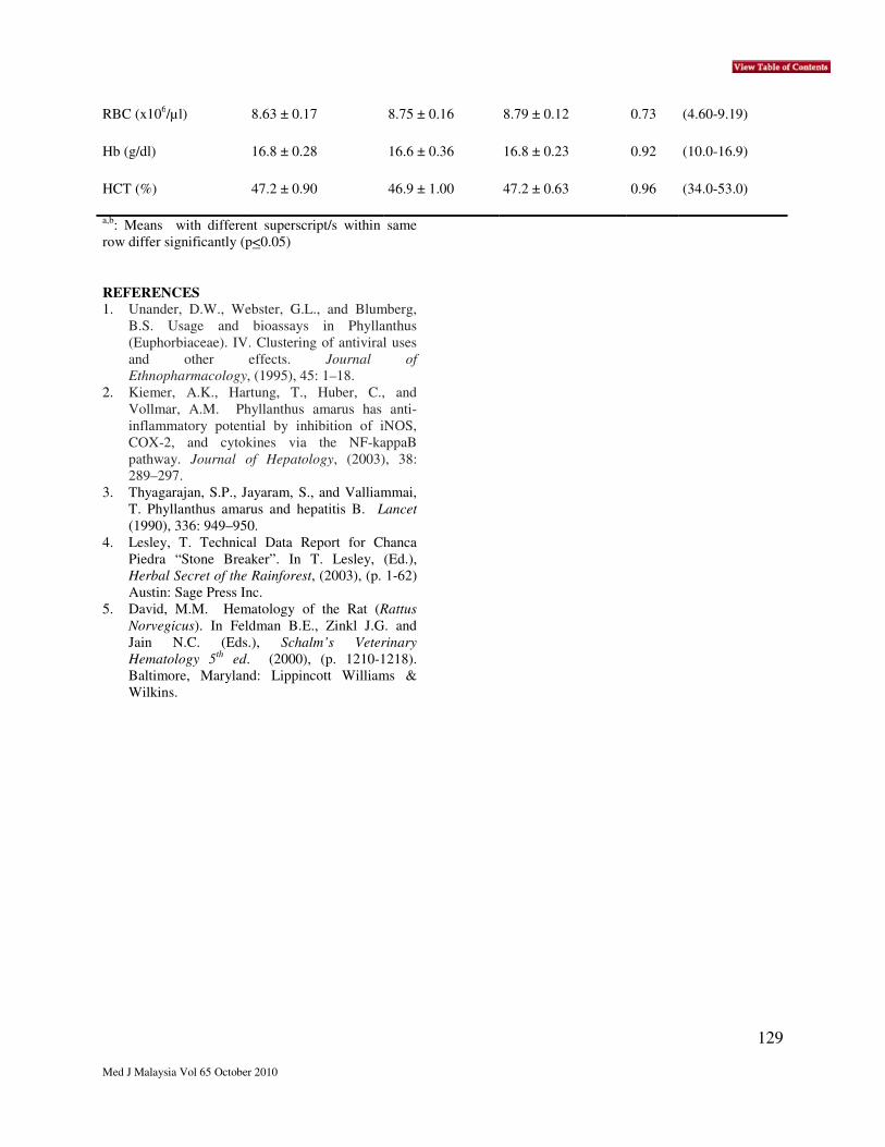
129
Med J Malaysia Vol 65 October 2010
RBC (x106/µl) 8.63 ± 0.17 8.75 ± 0.16 8.79 ± 0.12 0.73 (4.60-9.19)
Hb (g/dl) 16.8 ± 0.28 16.6 ± 0.36 16.8 ± 0.23 0.92 (10.0-16.9)
HCT (%) 47.2 ± 0.90 46.9 ± 1.00 47.2 ± 0.63 0.96 (34.0-53.0)
a,b: Means with different superscript/s within same
row differ significantly (p<0.05)
REFERENCES
1. Unander, D.W., Webster, G.L., and Blumberg,
B.S. Usage and bioassays in Phyllanthus
(Euphorbiaceae). IV. Clustering of antiviral uses
and other effects. Journal of
Ethnopharmacology, (1995), 45: 1–18.
2. Kiemer, A.K., Hartung, T., Huber, C., and
Vollmar, A.M. Phyllanthus amarus has anti-
inflammatory potential by inhibition of iNOS,
COX-2, and cytokines via the NF-kappaB
pathway. Journal of Hepatology, (2003), 38:
289–297.
3. Thyagarajan, S.P., Jayaram, S., and Valliammai,
T. Phyllanthus amarus and hepatitis B. Lancet
(1990), 336: 949–950.
4. Lesley, T. Technical Data Report for Chanca
Piedra “Stone Breaker”. In T. Lesley, (Ed.),
Herbal Secret of the Rainforest, (2003), (p. 1-62)
Austin: Sage Press Inc.
5. David, M.M. Hematology of the Rat (Rattus
Norvegicus). In Feldman B.E., Zinkl J.G. and
Jain N.C. (Eds.), Schalm’s Veterinary
Hematology 5th
ed. (2000), (p. 1210-1218).
Baltimore, Maryland: Lippincott Williams &
Wilkins.

130
Med J Malaysia Vol 65 October 2010
In Vitro Anti-cancer Activity of Ficus deltoidea Ethanolic Extract on
Human Ovarian Carcinoma Cell Line
M A Nor Azurah*, M R Sarmidi*, F A Abdul Majid**, K Chobotova*
*Chemical Engineering Pilot Plant, Universiti Teknologi Malaysia, Department of **Bioprocess Engineering,
Faculty of Chemical Engineering and Natural Resources, Universiti Teknologi Malaysia.
SUMMARY Ficus deltoidea is a Malaysian traditional herb that is
known for its therapeutic value. The aim of this study
is to examine the effect of ethanolic extract towards
human ovarian carcinoma cell line or A2780. MTT
assay was performed to see the growth pattern of this
cell. The measurement of glucose uptake and LDH
release has been done for further conformation. The
fluorochrome staining by fluorescence microscopy
using acridine orange and ethidium bromide double
staining was also been performed in this study to
examine the presence of apoptotic body.
INTRODUCTION
Ficus deltoidea or Mas Cotek is from Moraceae
family also known as serapat, sempit-sempit, and
agoluran in Sabah and tabat barito in Indonesia (Wan
Hasan, 2007). The fruits are chewed to relieve
headache, toothache and cold; powdered root and
leaves of the plant has been applied externally to
wounds and sores and around the joints to relief of
rheumatism (Sulaiman et al., 2008). Some local
people also believe that this herb can actually cure
cancer.
MATERIALS AND METHODS
F. deltoidea was collected from Muadzam, Pahang.
The ethanolic extract was prepared according to the
methods by Song et al., 2007 with a slight
modification. A2780 cell line was used and cultured
in RPMI 1640 medium, FBS and Glutamine. Cell
proliferation assay was done using slightly modified
protocol by Feshney, 2005. Glucose uptake was
measured by C111 Cobas according to the
manufacturer’s instructions. The activity of lactate
dehydrogenase was measured using Biochemistry
Analyzer according to manufacturer’s instructions.
Determination of apoptotic body was determined
using fluorescence microscope and the protocols were
followed as described previously by Cohen, 1993.
Cisplatin was used as posititve control.
RESULTS
The IC50 value observed is 195.166 + 20.21 µg/ml as
shown in Figure 1. The percentage of glucose uptake
shows that this extract has the lowest uptake at
1000µg/ml of concentration as shown in Figure 2.
Intra-cellular and extra-cellular activity might have
been blocked and the cells stop proliferating thus they
stop consuming glucose. Figure 3 shows the result of
LDH release after treatment with extract. The result
shows that there is no significant difference as
compared to negative and positive controls. Figure 4
shows the apoptotic body observation of cell line
after treatment with extract. The figure shows the
presence of apoptotic body. Different mechanism can
be concluded, cells without any treatment release
almost the same amount of LDH as cisplatin as a
negative control due to 100% of glucose
consumption. However, for cisplatin cells treatment,
the excessive amount of LDH is caused by cell
toxicity and cell lysis. The effect of 1000µg/ml on
LDH release result could be signified as the cisplatin
mechanism.
IC50 for ethanolic extract
0
20
40
60
80
100
120
140
160
100050025012562.531.2515.67.813.9control
Concentration (ug/ml)
% o
f c
on
tro
l
Fig 1. The percentage survival of control after
treatment with extract.

131
Med J Malaysia Vol 65 October 2010
92
.25
93
.08
93
.40
93
.22
93
.53
88
.22
0.00
20.00
40.00
60.00
80.00
100.00
120.00
1000500250125controlcisplatin
Concentration (ug/ml)
Pe
rce
nta
ge
(%
)
Fig 2. The percentage of glucose uptake of F.
deltoidea extract on A2780 cell line.
78
.21
99
.04
10
0
10
0
10
0
38
.46
0.00
20.00
40.00
60.00
80.00
100.00
120.00
1000500250125controlcisplatin
Concentration (ug/ml)
Pe
rce
nta
ge
(%
)
Fig 3. The percentage of LDH release of F.
deltoidea extract on A2780 cell line
A
B
Fig 4. A comparison of apoptosis staining between
negative control (A) and cells treated with 250 ug/ml
ethanolic extract (B).
DISCUSSION The result indicated that this extract may be a
promising source for anticancer drug development.
The least amount of glucose uptake was observed at
the highest concentration of extract is supported by
the fact that many types of cancer including ovarian
cancer have increased glycolytic activities compared
to non-malignant cells (warbug, 1956). The normal
plasma membrane is impermeable to LDH, but
damage to the cell membrane results in a change in
the membrane permeability and subsequent leakage
of LDH into the extracellular fluid (Rae, T, 1997).
The presence of apoptotic body after treatment with
extract indicates that cell death was cause through
apoptotic pathway.
REFERENCE
1. Rae, T. (1977). Tolerance of mouse
macrophages in vitro to barium sulphate
used in orthopaedic bone cement. Journal of
Biomedical materials and research. Volume
11, issue 6, pages 839-846, November 1977.
132
Med J Malaysia Vol 65 October 2010
Phyllanthus niruri Reduces Renal Azotaemia in Rats Induced with
Chronic Renal Damage
H Hazilawati*, P Farah Dina
*, S M Rosly
**, S Shanmugavelu
**, M M Noordin
*
*Department of Pathology and Microbiology, Faculty of Veterinary Medicine,
Universiti Putra Malaysia, 43400 Serdang, Selangor, Malaysia, **Strategic Livestock Research Centre, Malaysian
Agricultural Research and Development Institute, 43400 Serdang, Selangor, Malaysia.
SUMMARY
Phyllanthus niruri is commonly used for treatment of
jaundice, hepatitis, kidney stones, diuretics, influenza
and antibacterial agent. This study was conducted to
investigate the effects of P. niruri in reducing the
severity of azotaemia in rats induced with chronic
renal damage. A total of 32 male Sprague Dawley rats
were randomly assigned into four treatment groups;
Group A (control), Group B (adenine), Group C (P.
niruri) and Group D (adenine+P. niruri), consisting of
8 rats each. Chronic renal damage was induced using
0.75% adenine mixed with rat diet (daily feeding for 6
weeks). P. niruri was supplemented daily at a dose of
3000 mg/kg body weight throughout the 10 weeks of
the experimental period. Results showed that P. niruri
reduced the severity of azotaemia in rats induced with
chronic renal damage.
INTRODUCTION
Phyllanthus niruri which is locally known as Dokong
anak, is a tropical herb tree that can easily be found in
Malaysia. The plant is highly valued for its
hepatoprotective, antidiabetic, antihypertensive,
analgesic, anti-inflammatory and antimicrobial
properties 1,2
. The objective of this study was to
examine the effects of P. niruri in reducing the
severity of renal azotaemia in rats induced with
chronic renal damage.
MATERIALS AND METHODS
Thirty-two male Sprague Dawley rats were used in
this study. The rats were randomly divided into four
groups of 8 animals each; Group A (control), Group
B (adenine), Group C (P. niruri) and Group D
(adenine+P. niruri). Group B and D were induced
with chronic renal damage by daily feeding diet
containing 0.75% adenine (Sigma-Aldrich, USA) for
6 weeks. The rats were then fed with normal rat diet
for another 2 weeks before they were sacrificed.
Group C and D were supplemented daily with P.
niruri (obtained from a local supplier) at a dose 3000
mg/kg body weight for 10 weeks. Blood samples
were collected fortnightly (week 0, 2, 4, 6, 8 and 10)
from tail vein for creatinine (Cr) and blood urea
nitrogen (BUN) levels. The samples were analysed
using a Dry Chemistry Analyser (scil Reflovet® Plus,
Roche Diagnostics) followed as per manufacturer’s
instruction. At the end of the experiment (week 10),
24-hour urine samples were collected. Urine volume
was recorded, urine creatinine and protein
concentrations were determined using an Automated
Biochemistry Analyser (Biorex, TRX7010, Manheim,
Japan). Creatinine clearance (CrCl) was then
calculated. Blood samples were also collected via
portal vein after the rats were humanely sacrificed by
bleeding under anaesthesia. Serum creatinine (Cr)
concentration was determined using the Automated
Biochemistry Analyser and urine protein to creatinine
ratio (UPC) was calculated. Data obtained were
calculated for mean values and standard deviation
using one-way ANOVA (SPSS version 17.0 for
windows). The differences between groups were
determined by Duncan’s multiple range analysis.
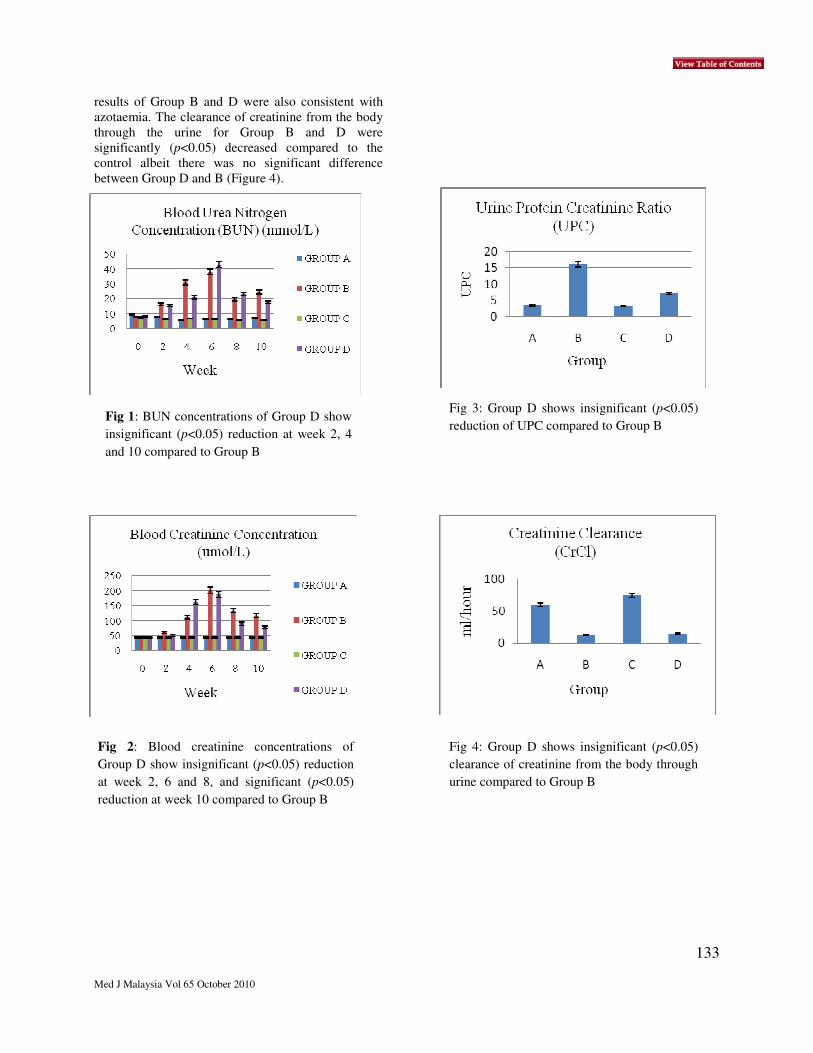
RESULTS
The BUN and blood Cr concentrations of all groups
are shown in Figure 1 and 2, respectively. Group A
and C had normal concentrations of BUN and blood
Cr throughout the experimental period. Gradual
increases in BUN and blood Cr concentrations (renal
azotaemia) were observed in both Group B and D
during the adenine treatment. The concentrations
were significantly (p<0.05) decreased after
elimination of adenine from the diet at week 8 and 10
although they were still significantly (p<0.05) higher
than the control. Group D had insignificant (p<0.05)
reduction in BUN concentrations at week 2, 4 and 10
compared to the Group B. It also had insignificant
(p<0.05) reduction in blood creatinine concentrations
at week 2, 6 and 8, and significant reduction (p<0.05)
at week 10 of the experiment compared to the Group
B. Consistent with azotaemia, Group B and D showed
significant increase in the UPC compared to the
control (Figure 3). Group B had UPC 5 times higher
than the control while Group D had UPC 2 times
higher than the control. Although Group D had lower
UPC compared to Group B, statistically it was not
significant (p<0.05). Similar to the UPC, the CrCl
133
Med J Malaysia Vol 65 October 2010
results of Group B and D were also consistent with
azotaemia. The clearance of creatinine from the body
through the urine for Group B and D were
significantly (p<0.05) decreased compared to the
control albeit there was no significant difference
between Group D and B (Figure 4).
Fig 1: BUN concentrations of Group D show
insignificant (p<0.05) reduction at week 2, 4
and 10 compared to Group B
Fig 2: Blood creatinine concentrations of
Group D show insignificant (p<0.05) reduction
at week 2, 6 and 8, and significant (p<0.05)
reduction at week 10 compared to Group B
Fig 3: Group D shows insignificant (p<0.05)
reduction of UPC compared to Group B
Fig 4: Group D shows insignificant (p<0.05)
clearance of creatinine from the body through
urine compared to Group B

134
Med J Malaysia Vol 65 October 2010
DISCUSSION
P. niruri has shown protective effects on different
organs in the body especially liver and kidneys 3,4
. It
is used to treat a lot of conditions including kidney
stones (Chopra et al., 1986). One of the active
ingredients found in another species of this herb (P.
amarus), quercetin (one of the bioflavonoids), has
been reported can inhibit xenobiotic-induced
nephrotoxicity 5,6
. Results in this study showed that
daily supplementation of P. niruri at 3000 mg/kg
body weight had shown nephroprotective activity.
Generally it only mildly reduced the severity of
azotaemia. Further studies should be conducted by
using higher doses of dried P. niruri and/or using its
extract.
REFERENCES
1. Ramanam MV and Sainani GS. Drugs in
infective hepatitis. Punjab Medical Journal,
1962; 10: 607-712.
2. Adeneye AA and Arogundade MO.
Immunostimulatory and haematopoietic activities
of the leaf and seed aqueous extract of
Phyllanthus amarus in normal and
cyclophosphamide-treated mice. Nigerian
Journal of Health and Biomedical Sciences,
2007; 6:53-57.
3. Tabasum N, Chatterrredi S, Grawal SS and
Ahmed N. Hepatoprotective studies of
Phyllanthus niruri on paracetamol-induced liver
cell damage in albino mice. Journal of
Experimental Medicine, 2005; 12: 211-213.
4. Nishiura J, Campos A, Boim M and Schor N.
Effect of Phyllanthus niruri on urinary calcium
levels in calcium stone forming patients. Journal
of Clinical and Laboratory Investigation of
Urolithies and Related Areas, 2005; 32, 362-366.
5. Devipriya S and Shyamaladevim CS. Protective
effect of quercetin in cisplatin induced cell injury
in the rat kidney. Indian Journal of
Pharmacology, 1999; 31: 422.
6. Annie S, Rajagopal PL and Malini S. Effect of
Cassia auriculata Linn. root extract on cisplatin
and gentamicin-induced renal injury.
Phytomedicine, 2005; 12: 555-560.
135
Med J Malaysia Vol 65 October 2010
Effects of Morinda citrifolia on Early Stage of Leukaemia in Rats
*H Hazilawati, *H Nursyuhada , **S M Rosly, **S Shanmugavellu, *M M Noordin
*Department of Pathology and Microbiology, Faculty of Veterinary Medicine,
Universiti Putra Malaysia, 43400 Serdang, Selangor,
Malaysia. **Strategic Livestock Research Centre,
Malaysian Agricultural Research and Development
Institute, 43400 Serdang, Selangor, Malaysia
SUMMARY
We have studied the effects of Morinda citrifolia on
haemogram parameters in rats with early stage of
leukaemia. A total of 32 male Sprague Dawley rats
were divided into four groups (n=8) namely control,
Morinda citrifolia (MC), N-methyl-N-nitrosourea
(MNU) and MC+MNU group. Leukaemia induced
rats were provided with died M. citrifolia fruits
incorporated into rat diet at a dose of 3000 mg/kg
body weight were given daily to the MC and
MC+MNU groups. Results showed there were no
significant differences in the leukon and erythron
parameters in all groups. Although lymphocytosis
was not observed in the MNU and MC+MNU groups,
examination of the blood morphology showed a
typical morphology of leukaemic cell. Seventy-five
and sixty percent of rats in the MNU and MC+MNU
groups, respectively, had leukaemia. Our results
indicate that examination of blood smear provides an
important tool for diagnosis of early stage of
leukaemia and daily supplementation of M. citrifolia
at the dose of 3000 mg/kg reduced the incidence of
early stage of leukaemia in rats.
INTRODUCTION Morinda citrifolia offers a potential pharmaceutical
values. The entire plant was used by the Polynesians
as folk medicine to treat common illnesses. It was
reported that the fruit juice is able to stimulate
significant body’s immune responses 1 and therefore
was mainly consumed for health maintenance 2. One
of the carcinogenic chemical that able to induce
lymphoma or leukaemia in rats is N-methyl-N-
nitrosourea (MNU) 3, 4
. This study was conducted to
examine the effects of M. citrifolia on early stage of
leukaemia in rats induced using MNU.
MATERIALS AND METHODS
Thirty-two, 8-week old male Sprague Dawley rats
were placed under controlled conditions and
randomly divided into four groups (n=8) namely
control, N-methyl-N-nitrosourea (MNU), M. citrifolia
(MC) and MC+MNU group. The control and MC
groups were injected with normal saline. The MNU
and MC+MNU groups were injected intraperitoneally
(i.p) with freshly prepared MNU (Sigma-Aldrich,
USA), bi-weekly for 2 consecutive weeks at a dose of
60 mg/kg body weight per administration. The MC
and MC+MNU groups were fed daily with ground
dried M. citrifolia fruit at a dose of 3000mg/kg body
weight mixed with commercial ground rat diet. At 12
week of post MNU administration, bloods were
collected via cardiac puncture and analysed for a
complete blood count using an Automated
Haematology Analyser (Cell-Dyn® 3700, Abbott
Diagnostics, USA). Blood smear was prepared and
stained with Wright’s stain for a manual differential
leukocyte count and examination for the presence of
leukaemic cells. Packed cell volume (PCV) was
determined manually using a standard method.
Results were analysed using SPSS version 17.0 for
windows and differences were considered significant
at p<0.05.
RESULTS
Results for the haematology are shown in Table 1 and
Table 2. The leukon and erythron parameters showed
no significant differences in all the groups.
Examination of the leukocyte morphology revealed
75% and 60% of rats in the MNU and MC+MNU
groups, respectively, had leukaemia (Table 3).
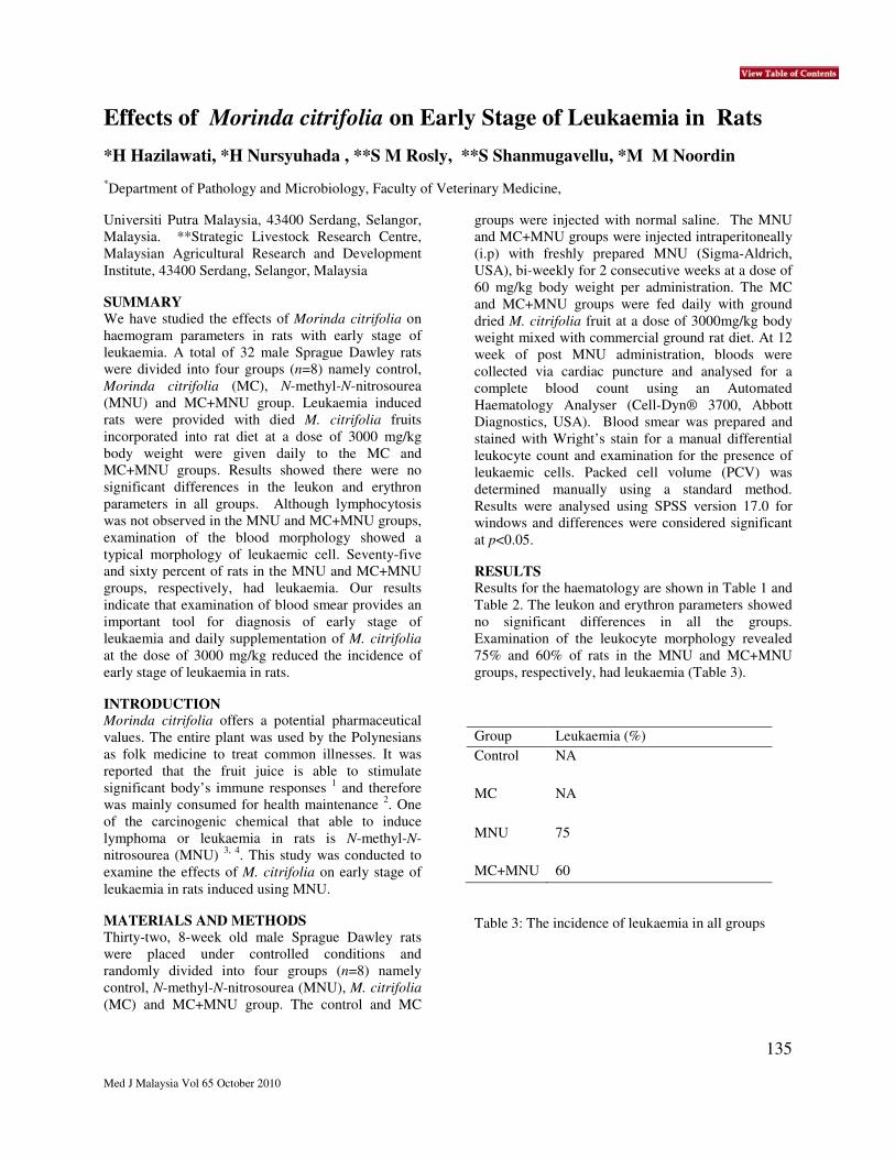
Group Leukaemia (%)
Control NA
MC NA
MNU 75
MC+MNU 60
Table 3: The incidence of leukaemia in all groups

136
Med J Malaysia Vol 65 October 2010
Table 1: The leukon parameters in all groups (Mean±SD)
Group Leukocytes
(x109/L)
Lymphocyte
(x109/L)
Neutrophil
(x109/L)
Monocyte
(x109/L)
Eosinophil
(x109/L)
Basophil
(x109/L)
Control
10.1±2.7
7.73±2.20
0.60±0.81
0.25±0.14
0.05±0.03
0.31±0.06
MC
10.0±4.0
7.30±4.37
0.21±0.03
0.34±0.32
0.04±0.01
0.36±0.09
MNU
8.4±1.1
6.53±0.82
0.20±0.02
0.27±0.25
0.05±0.05
0.20±0.16
MC+
MNU
9.7±1.2
7.41±1.49
0.18±0.04
0.28±0.30
0.07±0.06
0.24±0.18
Table 2: The erythron parameters in all groups (Mean±SD)
Group PCV (L/L) RBC
(x 1012
/L)
Haemoglobin
(g/L)
Control 52.5±4.1 9.90±1.10 184.8±20.55
MC
49.3±0.6
8.72±0.27
163.0±10.66
MNU
51.3±2.5
8.73±0.50
167.3±4.65
MC+MNU
51.8±2.1
8.83±0.65
167.5±10.66
DISCUSSION
According to Hutheyfa et al. 3,4
, administration of
MNU at the dose of 60 mg/kg body weight per
injection twice a week for two consecutive weeks
(with a total dose of 240 mg/kg body weight)
successfully induced 30% leukaemia with marked
lymphocytosis and 100% lymphoma in young male
Sprague Dawley rats after 20 weeks of injections. Our
results showed 75% of male Sprague Dawley rats
administered with MNU using similar method
described by Hutheyfa et al. had leukaemia without
lymphocytosis after 12 weeks of injections. It is
suggested that examination of blood smear is
important for diagnosis of early stage of leukaemia.
Daily supplementation of M. citrifolia to the rats at
the dose of 3000 mg/kg body weight reduced the
incidence of early stage of leukaemia to 60%.
Administration of M. citrifolia at a higher dose (5000
mg/kg body weight) has significantly reduced the
incidence of leukaemia at 20 week post MNU
administration 5.
REFERENCES
1. Hirazumi AY. Antitumor Studies of a
Traditional Hawaiian Medicinal Plant, Morinda
citrifolia (noni), in vitro and in vivo [doctoral
dissertation]. University of Hawai‘I, Honolulu,
Hawaii. 1990.
2. Wang MY, Brett JW, Jarakae J, Diane N and
Gary A. Morinda citrifolia (Noni): A Literature
Review and Recent Advances in Noni Research.
Acta Pharmacology Sinaca 2002, 12: 1127-
1141.
3. Hutheyfa AH, Hazilawati H, Rosly SM, Jasni S,
Noordin, MM and Shanmugavelu S. N-methyl-
N-nitrosourea induced leukaemia in male
Sprague-Dawley rats. Proceedings of the
International Conference on Animal Health and
Human Safety, 2009 December 06-08;
Putrajaya, Malaysia. Faculty of Veterinary
Medicine, Universiti Putra Malaysia-Airlangga
University, Indonesia-Department of Veterinary
Services, Malaysia 2009a, 210-213.
4. Hutheyfa AH, Hazilawati H, Rosly SM, Jasni S,
Noordin M M and Shanmugavelu S.
Development of lymphomas in male Sprague-
Dawley rats exposed to N-methyl-N-nitrosourea.
Proceedings of the International Conference on
Animal Health and Human Safety, 2009
December 06-08; Putrajaya, Malaysia. Faculty
of Veterinary Medicine, Universiti Putra
Malaysia-Airlangga University, Indonesia-
Department of Veterinary Services, Malaysia
2009b, 214-218.