
Optical coherence tomography guided microinjections in...
8
Optical coherence tomography guided microinjections in live mouse embryos: high-resolution targeted manipulation for mouse embryonic research Saba H. Syed Andrew J. Coughlin Monica D. Garcia Shang Wang Jennifer L. West Kirill V. Larin Irina V. Larina
Transcript of Optical coherence tomography guided microinjections in...
Optical coherence tomography guidedmicroinjections in live mouseembryos: high-resolution targetedmanipulation for mouse embryonicresearch
Saba H. SyedAndrew J. CoughlinMonica D. GarciaShang WangJennifer L. WestKirill V. LarinIrina V. Larina
Optical coherence tomographyguidedmicroinjectionsin live mouse embryos: high-resolution targetedmanipulation for mouse embryonic research
Saba H. Syed,a Andrew J. Coughlin,b Monica D. Garcia,a Shang Wang,a Jennifer L. West,bKirill V. Larin,a,c and Irina V. Larinaa,*aBaylor College of Medicine, Department of Molecular Physiology and Biophysics, One Baylor Plaza, Houston, Texas 77030, United StatesbDuke University, Department of Biomedical Engineering, Hudson Hall, Durham, North Carolina 27708, United StatescUniversity of Houston, Department of Biomedical Engineering, 4605 Cullen Boulevard, Houston, Texas 77204, United States
Abstract. The ability to conduct highly localized delivery of contrast agents, viral vectors, therapeutic or phar-macological agents, and signaling molecules or dyes to live mammalian embryos is greatly desired to enable avariety of studies in the field of developmental biology, such as investigating the molecular regulation of cardio-vascular morphogenesis. To meet such a demand, we introduce, for the first time, the concept of employingoptical coherence tomography (OCT)-guide microinjections in live mouse embryos, which provides preciselytargeted manipulation with spatial resolution at the micrometer scale. The feasibility demonstration is performedwith experimental studies on cultured live mouse embryos at E8.5 and E9.5. Additionally, we investigate theOCT-guided microinjection of gold–silica nanoshells to the yolk sac vasculature of live cultured mouse embryosat the stage when the heart just starts to beat, as a potential approach for dynamic assessment of cardiovascularform and function before the onset of blood cell circulation. Also, the capability of OCT to quantitatively monitorand measure injection volume is presented. Our results indicate that OCT-guided microinjection could be a use-ful tool for mouse embryonic research. © 2015 Society of Photo-Optical Instrumentation Engineers (SPIE) [DOI: 10.1117/1.JBO.20.5
.051020]
Keywords: optical coherence tomography; mouse embryo; microinjection; real-time imaging; cardiovascular development; goldnanoshells.
Paper 140625SSR received Sep. 29, 2014; accepted for publication Dec. 2, 2014; published online Jan. 12, 2015.
1 Introduction and BackgroundThe capability of injecting cells, contrast agents, dyes, or otheragents into specific regions in the mouse embryo is of greatimportance for biomedical studies of developmental proc-esses.1–3 Unlike early microinjection approaches1,4 that areessentially blind procedures with no real-time feedback onthe location of needle and the status of the injection, image-guided microinjection has been an attractive approach for con-ducting targeted delivery in mouse embryos, which brings sig-nificant improvement to the molecular and cellular manipulationof embryos at various stages of gestation.2,3,5 Efforts regardingthe development of image-guided microinjection have beenmainly focused on employing reflected light microscopy3,6,7
and ultrasonic imaging techniques2,5,8–10 for mouse embryosat very early and midgestational stages, respectively. Specifi-cally, Wefers et al. utilize an optical stereomicroscope to monitorthe microinjection of mRNA into single-cell mouse embryos forthe generation of targeted mouse mutants.3 The whole injectionprocess can be clearly visualized through the microscope; how-ever, due to a lack of depth resolving capability, this approachhas very limited applications for microinjections to the mouseembryos at later gestational stages. High-frequency ultrasonicimaging techniques, such as the ultrasound backscatter micro-scope,11 have been used to guide in utero microinjections tothe mouse embryos from E6.5 to E11.5.5,9,10 With a spatialresolution that is close to 50 μm, the delivery of cells, contrast
agents or virus to the brain,5,9 the ectoplacental cone,10 theamniotic cavity,10 and the anterior limb bud5 can be real-timemonitored with a relatively high precision. Targeting and micro-manipulation of the cardiovascular system, the first organ sys-tem to fully develop in the embryo, provides a straightforwardmeans to deliver an injectable molecule to embryonic tissue viathe vasculature. Furthermore, the early development of theembryonic cardiovascular system is a critical time point indevelopment when embryo viability is highly dependent oncardiovascular form and function. Microinjections to the embry-onic cardiovascular system could enable various importantapplications such as studying the molecular regulation of cardio-vascular morphogenesis, or the manipulation and rescue of earlyembryonic cardiovascular defects. However, such procedureshave not been realized (to the best of our knowledge) andcould be very challenging with currently available imagingmodalities, mainly because the spatial resolution is insufficientto resolve the blood vessels on the embryonic yolk sac. Thus, adepth-resolved imaging technique with better spatial resolutionthat is suitable for live mouse embryonic imaging is needed forguiding microinjections with small-volume delivery to preciselocations within embryonic structures.
Optical coherence tomography (OCT) is a low-coherencethree-dimensional (3-D) imaging modality that has the spatialresolution of ∼1 to 10 μm with an imaging depth of 1 to3 mm in most highly scattering tissues,12–14 which couldpossibly be used to meet the demand for high-resolution
*Address all correspondence to: Irina V. Larina, E-mail: [email protected] 0091-3286/2015/$25.00 © 2015 SPIE
Journal of Biomedical Optics 051020-1 May 2015 • Vol. 20(5)
Journal of Biomedical Optics 20(5), 051020 (May 2015)

image-guided microinjection. Besides the major applications inthe fields of ophthalmology15 and cardiology,16 OCT has beenextensively used for embryonic imaging in several animalmodel systems such as chick,17–20 quail,21,22 Xenopus,23,24 zebra-fish,25,26 mouse,27–31 and rat.32 Among these, mouse is an idealmammalian model for studying the physiological function andpathological process of human organ systems, and variousmouse mutants are available to help improve understandingthe genetic basis of development and diseases.33 ApplyingOCT to mouse embryonic imaging represents an importantstep for advanced phenotyping of the developmental status.34–36 Recently, with a primary focus on mouse embryonic cardio-vascular development, our group has combined OCT imagingwith embryo culturing techniques,37 and developed methodsand protocols for 3-D high-resolution imaging of live mouseembryos during the embryonic days of 7.5 to 10.28,29,38–40
Specifically, four-dimensional (4-D) OCT imaging of embry-onic cardiodynamics has been achieved with nongated dataacquisition approaches and computational reconstruction algo-rithms.41,42 Also, the employment of Doppler OCT has enabledquantitative hemodynamic analysis in the developing mouseembryo with a spatial resolving ability reaching single movingblood cells.28,29 Such dynamic imaging capabilities of OCTwithboth structural and functional information suggest its potentialfor precise manipulation of mouse embryos.
In this study, we introduce, for the first time, the concept ofusing OCT for high-resolution image guidance of microinjec-tions in live mouse embryos. We present the procedure and dem-onstrate the feasibility of OCT-guided microinjections intoblood vessels of the yolk sac in the mouse embryos at E8.5and E9.5. With pilot experiments, we further investigate the pos-sibility of delivering gold–silica nanoshells into the cardio-vascular system of mouse embryos under the guidance ofOCT, which could be utilized as an approach for studyingflow dynamics in the vascular network in response to the initialembryonic heartbeat. We also show the capability of OCT toquantitatively measure injection volume with high sensitivity.Our experimental results indicate that the proposed OCT-guidedmicroinjection in live mouse embryos could be a useful tool forprecisely targeted manipulation in mouse embryonic research.
2 Materials and Methods
2.1 Mouse Embryo Preparation
Timed matings of CD-1 mice are set overnight and checked forvaginal plugs daily. The day of the observed vaginal plug iscounted as E0.5. Embryos are dissected at E8.5 and E9.5with the yolk sac remaining intact in the culture medium thatcontains 89% DMEM/F12, 1% Pen-strep solution, and 10%FBS (Life Technologies, Thermo Fisher Scientific Corp.).The dissection station is maintained at 37°C. After dissection,embryos are transferred into a humidified incubator maintainedat 37°C, 5% CO2 and allowed to recover for at least 30 min. Thedetailed procedure regarding the preparation of mouse embryoscan be found in our previous work.43 To immobilize the embryosduring OCT imaging and microinjection, a 2% agarose gel pre-pared with the culture medium is utilized. Melted gel is pouredin a 35-mm diameter culture dish and allowed to solidify with 1to 2 mm thickness. Prior to transferring the embryo into the cul-ture dish, a small well of about the same size as the embryo ismade in the gel and the whole dish is filled with the culturemedium. For the OCT-guided microinjection, the embryo withinthe culture dish is placed in an incubator (humidified, 37°C, 5%CO2), where the OCT imaging probe and the microinjectionsetup are located, as shown in Fig. 1.
2.2 Gold–Silica Nanoshells
Gold–silica shell-core nanoshells with near-infrared extinctionare synthesized via a four-step process as previouslydescribed.44,45 The nanoshells have a diameter of approximately150 nm with the diameter of the silica core ∼120 nm. The sizesof the nanoshells are verified with high contrast-transmissionelectron microscope imaging, and their optical properties areevaluated using ultraviolet-visible spectroscopy (Varian Cary50 Bio UV/Visible Spectrophotometer, McKinley Scientific,LLC.). To prevent particle flocculation upon injection in themouse embryo, the surfaces of the nanoshells are passivatedwith thiol-terminated poly(ethylene glycol) (PEG-SH, LaysanBio, MW ¼ 5000 kDa).
Fig. 1 System setup of OCT-guided microinjection in live mouse embryos. L: laser; M: mirror; AL: ach-romatic lens; FA: fiber adaptor; TG: transmission gratings; FOC: fiber optic coupler; PC: polarizationcontroller; C: collimator; A: aperture; GM: galvanometer mirror; SL: scan lens; S: sample; MS: microinjec-tion system; FS: foot switch; AT: air tube; PH: pipette holder; N: needle; LS: linear stage; RS: rotationalstage.
Journal of Biomedical Optics 051020-2 May 2015 • Vol. 20(5)
Syed et al.: Optical coherence tomography guided microinjections in live mouse embryos. . .

2.3 Microinjection Setup
Quartz capillary pipettes (Sutter Instrument Co.) are pulled on aP-2000 micropipette laser-based puller system (SutterInstrument Co.). The injection material (3 μl) is loaded intothe capillary pipette and the tip of the pipette is then carefullybroken using dissecting forceps. The resulted microinjectionneedle is attached to the pipette holder of the microinjection sys-tem (Pico-Injector, Harvard Apparatus Co.) that applies com-pressed gas for a wide range of delivery volumes of theloaded material. A home-built micromanipulator is used tofix the pipette holder and precisely control the position of theneedle with multi-dimensional adjustment (Fig. 1).
2.4 Spectral Domain Optical CoherenceTomography System and Experimental Methods
A home-built spectral domain OCT system46 is utilized forimage-guided microinjection. The schematic of the systemsetup is shown in Fig. 1. The OCT system employs a low-coher-ence Titanium–Sapphire laser source (Micra-5, Coherence, Inc.)that has a central wavelength of ∼808 nm and a bandwidth of∼110 nm. The output of the laser with the collimated beam iscoupled into a single-mode fiber and directed to a 50∶50 fiberoptic coupler. A neutral density filter is utilized to adjust thepower that is delivered to the fiber. The light reflected fromthe reference arm and backscattered from the sample armforms interference fringes which are detected and spatiallyresolved by a high-resolution spectrometer with a complemen-tary metal oxide semiconductor line-scanning camera (spL4096-140 k, Basler, Inc.). The OCT system is set to operate at a 50-kHz A-line acquisition speed during the experiments. The axialresolution of the system is measured to be ∼5 μm in tissue, andthe full width at half maximum of the sample-arm imaging beamat the focal plane is measured to be ∼4 μm. Such spatial reso-lution provides superior resolving capability of the detailedstructures inside the mouse embryos. Also, the ∼5.5 mm avail-able imaging depth in air enables the capture of most organ andfunctional systems in the embryos.43
For conducting microinjection with the guidance of OCT im-aging, the OCT system is operated in the imaging mode of arepeated B-scan that covers a depth-resolved two-dimensional(2-D) field of view (FOV) from the mouse embryo. The positionof the injection needle is carefully adjusted to place it within theOCT B-Scan image for real-time continuous visualization of thewhole process, including needle insertion and material delivery.As an incubator is utilized tomaintain the proper environment forculturing the mouse embryo, during and after the OCT-guidedmicroinjection, the embryo continues its growth. In the pilotexperiments for the feasibility studies, saline, fluorescent dextran(Oregon Green®514, 70000 MW, Life Technologies, ThermoFisher Scientific Corp.), and gold–silica nanoshells have beenutilized as the delivery material to the embryonic yolk sacvasculature.
3 Results and DiscussionsAn example of the OCT-guided microinjection of saline into theextraembryonic vasculature of an E8.5 mouse embryo is pre-sented in Fig. 2. The mouse embryo is dissected, cultured, posi-tioned, and imaged using OCT, where the beating heart and thecirculation of blood within both the embryo and the yolk sacvasculature can be clearly observed. The microinjection needleis filled with saline and aligned to appear in the upper left corner
of the OCT B-scan image with the tip close to the blood vesselon the yolk sac, as shown in Fig. 2(a). Upon the alignment of theneedle, the real-time OCT imaging is used to guide the insertionof the needle into the yolk sac vasculature without inhibiting theblood circulation, as shown in Fig. 2(b). The injection of saline,which is nonscattering, can be observed from the OCT B-scanimage with the dark spot in the targeted vessel and close to thetip of the injection needle, as shown in Fig. 2(c). Monitored byOCT imaging, the removal of the microinjection needle from theyolk sac vasculature reveals no significant leak of blood cells, asshown in Fig. 2(d).
As a further feasibility demonstration of the proposed OCT-guided microinjection, a small volume of fluorescent dextran isinjected into the blood circulation of a cultured mouse embryo atE9.5. A 3-D OCT structural image of the embryonic yolk sac isshown in Fig. 3(a), which is characterized by a hierarchical net-work of large vessels branching out to smaller capillary beds.The dotted line indicates the position of where repeated OCTB-scan images are acquired that are used to guide the micro-injection of the florescent dextran into the large vitelline artery.
Fig. 2 OCT-guidedmicroinjection of saline in the yolk sac vasculatureof a cultured live mouse embryo at E8.5. Real-time continuous opticalcoherence topography (OCT) B-scan images clearly show the wholemicroinjection process (Video 1, MOV, 10.1 MB) [URL: http://dx.doi.org/10.1117/1.JBO.20.5.051020.1], including (a) the alignment ofthe injection needle, (b) the insertion of the needle to a blood vesselof the yolk sac, (c) the injection of saline to the vasculature and (d) theremoval of the injection needle. The scale bar corresponds to 300 μmand applies to all figures.
Journal of Biomedical Optics 051020-3 May 2015 • Vol. 20(5)
Syed et al.: Optical coherence tomography guided microinjections in live mouse embryos. . .

Similar procedures, such as the injection of saline, are conductedin this experiment. Based on the 2-D depth-resolved OCTimage, the vitelline artery of the yolk sac is precisely targeted.Upon the alignment of the dextran-loaded needle with the OCT
B-scan image, the insertion of the needle into the targeted vitel-line artery is performed with real-time guidance from OCT, asshown in Fig. 3(b). Once the needle tip is placed at a desiredposition inside the vessel, injections of fluorescent dextran sol-ution mixed with gold–silica nanoshells are conducted, and thenthe needle is removed from the yolk sac vasculature. Aftermicroinjection, the cultured embryo is immediately imagedusing a fluorescent microscope. The resulting fluorescenceimage presented in Fig. 3(c) shows that the injected fluorescentdextran has filled the whole vasculature network of the yolk sacand the embryo proper due to active circulation, demonstratingsuccessful OCT-guided microinjection. No deleterious influenceon blood flow or heart contraction in the cultured mouse embryohas been observed.
Gold–silica nanoshells44,45 have been identified as a novelcontrast agent to enhance OCT imaging. Figure 4(c) showsthe normalized extinction spectrum of the nanoshells utilizedin this study with a peak position at ∼800 nm. For themouse embryo at early E8.5, the heart has just begun tobeat. At this time, only plasma fills the nascent vasculature,
Fig. 3 OCT-guided microinjection of fluorescent dextran mixed withgold–silica nanoshells in the yolk sac vasculature of a culturedlive mouse embryo at E9.5 (Video 2, MOV, 8.73 MB) [URL: http://dx.doi.org/10.1117/1.JBO.20.5.051020.2]. (a) Three-dimensionalOCT structural image showing the vascular network of the embryonicyolk sac. (b) OCT depth-resolved B-scan image showing the micro-injection of fluorescent dextran with the tip of the injection needleinside the targeted vitelline artery. (c) Fluorescent microscopicimage of the embryo after microinjection showing fluorescencefrom dextran labeling the whole vascular network of the embryo,which indicates successful OCT-microinjection with no observable in-fluence on blood flow or heart contraction in the cultured mouseembryo. The scale bars correspond to 300 μm.
Fig. 4 OCT-guided microinjection of gold–silica nanoshells in the yolksac vasculature of a cultured live mouse embryo at early E8.5, whenthe heart just starts to beat. (a) Precise positioning of the injectionneedle into a vessel of the yolk sac that is only filled with plasma.(b) Microinjection of gold–silica nanoshells into the yolk sac vascula-ture as a contrast agent for the potential dynamic study of the cardio-vascular formation and function before the onset of blood cellcirculation. (c) Normalized extinction spectrum of the nanoshellsused in this study showing the peak around 800 nm. The scale barcorresponds to 300 μm and also applies for (b).
Journal of Biomedical Optics 051020-4 May 2015 • Vol. 20(5)
Syed et al.: Optical coherence tomography guided microinjections in live mouse embryos. . .
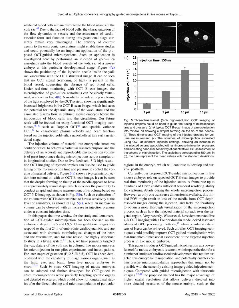
while red blood cells remain restricted to the blood islands of theyolk sac.47 Due to the lack of blood cells, the characterization ofthe flow dynamics in vessels and the assessment of cardio-vascular form and function during this gestational stage cur-rently remain very challenging. The delivery of contrastagents to the embryonic vasculature might enable these studiesand could potentially be an important application of the pro-posed OCT-guided microinjections. Such an application isinvestigated here by performing an injection of gold–silicananoshells into the blood vessels of the yolk sac of a mouseembryo at this particular developmental stage. Figure 4(a)shows the positioning of the injection needle inside the yolksac vasculature with the OCT structural image. It can be seenthat no OCT signal (scattering of light) is present in theblood vessel, suggesting the absence of red blood cells.Under real-time monitoring with OCT B-scan images, themicroinjection of gold–silica nanoshells can be clearly visual-ized, as shown in Fig. 4(b). Nanoshells provide strong scatteringof the light employed by the OCT system, showing significantlyincreased brightness in the OCT B-scan image, which indicatesthe potential for the dynamic study of the vasculature and theassociated plasma flow in cultured mouse embryos before theintroduction of blood cells into the circulation. Our futurework will be focused on using functional OCT imaging tech-niques,48–50 such as Doppler OCT28 and speckle varianceOCT,39 to characterize plasma velocity and heart functionbased on the injected gold–silica nanoshells at this early gesta-tional stage.
The injection volume of material into embryonic structurescould be critical to achieve a particular research purpose, and thedelivery of an accurate and reproducible microinjection volumeis of great importance during microinjections across samples orin longitudinal studies. Due to live feedback, 3-D high-resolu-tion OCT imaging of injected droplets can also be used to guidetuning of the microinjection time and pressure to control the vol-ume of material delivery. Figure 5(a) shows a typical microinjec-tion into mineral oil with an OCT B-scan image. It can be seenthat the droplet forming on the tip of the needle appears to havean approximately round shape, which indicates the possibility toconduct a rapid and simple measurement of its volume based onOCT 3-D imaging, as shown in Fig. 5(b). Such an estimation ofthe volume with OCT is demonstrated to have a sensitivity at thelevel of nanoliters, as shown in Fig. 5(c), where an increase involume can be observed with an increase in injection pressureunder a constant injection time.
In this paper, the time window for the study and demonstra-tion of OCT-guided microinjection has been focused on theembryonic days of E8.5 and E9.5. These gestational stages cor-respond to the first 24 h of embryonic cardiodynamics, and areassociated with dramatic morphological changes of the heartand the vasculature, and for that reason, are highly importantto study in a living system.51 Thus, we have primarily targetedthe vasculature of the yolk sac in cultured live mouse embryosfor microinjection in our pilot experiments and investigations.For later stages of gestation (E12.5-E18.5), OCT has been dem-onstrated with the capability to image various organs, such asthe limb, eye, and brain, from live mouse embryos inutero.27,52 Such in utero OCT imaging of mouse embryoscan be adopted and further developed for OCT-guided inutero microinjections while precisely targeting specific organsand detailed structures, which could allow for longitudinal stud-ies after the direct labeling and micromanipulation of particular
regions in the embryo, which will continue to develop and sur-vive postbirth.
Currently, our proposed OCT-guided microinjections in livemouse embryos rely on repeated OCT B-scan images to providereal-time monitoring of the injection status. A frame rate up tohundreds of Hertz enables sufficient temporal resolving abilityfor capturing details during the whole microinjection process.However, as only one transverse dimension is available, this lim-ited FOV might result in loss of the needle from OCT depth-resolved images during the injection, and lacks the feasibilityto obtain a more thorough visualization of the microinjectionprocess, such as how the injected material spreads over the tar-geted region. Very recently, Wieser et al. have demonstrated live4-D OCT imaging with a Fourier domain mode locked laser andadvanced GPU processing methods,53 where a volume rate attens of Hertz can be achieved. Such ultrafast OCT imaging tech-niques could possibly improve OCT-guided microinjection withreal-time three-dimensional assessment of the targeted injectionprocess in live mouse embryos.
This paper introduces OCT-guidedmicroinjection as a power-ful tool formouse embryonic research, which opens the door for anumber of studies of cardiovascular development that require tar-geted live embryonic manipulation, and potentially enables cer-tain precise micromanipulation approaches that might not bepossible with the currently existing image-guided injection tech-niques. Compared with guided microinjection with ultrasonicimaging,5,9,10 the proposed method has the major advantage ofhigher spatial resolution that allows delivery directed intomore detailed structures of the mouse embryo, such as the
Fig. 5 Three-dimensional (3-D) high-resolution OCT imaging ofinjected droplets could be used to guide the tuning of microinjectiontime and pressure. (a) A typical OCT B-scan image of a microinjectioninto mineral oil showing a droplet forming on the tip of the needle.(b) Three-dimensional OCT imaging of the injected droplets for vol-ume measurement. (c) The volumes of microinjection estimatedusing OCT at different injection settings, showing an increase inthe injected volume associated with an increase in injection pressure,and indicating nano-liter sensitivity of quantitative OCT assessment ofthe volume of microinjection. The scale bars correspond to 300 μm. In(c), the bars represent the mean values with the standard deviations.
Journal of Biomedical Optics 051020-5 May 2015 • Vol. 20(5)
Syed et al.: Optical coherence tomography guided microinjections in live mouse embryos. . .

vascular network. Also, the superior capability of OCT imagingin live mouse embryos28,29,40 could provide direct characteriza-tion of postinjected embryos with both structural and functionalinformation. Such features of OCT-guided microinjection indi-cate its great potential to complement ultrasound-guided injec-tion and act as an essential approach in mouse embryonicresearch.
4 ConclusionsA new microinjection approach with guidance from OCT imag-ing in live mouse embryos is introduced and demonstrated forthe first time. We present pilot experiments of OCT-guidedmicroinjections of saline, fluorescent dextran, and gold–silicananoshells to the yolk sac vasculature of cultured live mouseembryos at E8.5 and E9.5, and investigate the possible applica-tion regarding the assessment of cardiovascular form and func-tion before blood cells start to circulate. As a 3-D imagingmodality, OCT could also be used to directly characterize micro-injection volume, which can help to tune the injection time andpressure for delivering specific volumes of material to targetedregions inside live mouse embryos. The feature of precisemanipulation with microscale spatial resolution makes OCT-guided microinjection a useful tool in mouse embryonicresearch.
AcknowledgmentsThis work is supported by the National Institutes of Health(R01HL120140 and U54HG006348) as well as by theOptical Imaging and Vital Microscopy core at BaylorCollege of Medicine.
References1. R. Jaenisch, “Mammalian neural crest cells participate in normal embry-
onic development on microinjection into post-implantation mouseembryos,” Nature 318(6042), 181–183 (1985).
2. M. Olsson, K. Campbell, and D. H. Turnbull, “Specification of Mousetelencephalic and mid-hindbrain progenitors following heterotopicultrasound-guided embryonic transplantation,” Neuron 19(4), 761–772 (1997).
3. B. Wefers et al., “Generation of targeted mouse mutants by embryomicroinjection of TALEN mRNA,” Nat. Protocols 8(12), 2355–2379(2013).
4. R. Jaenisch, “Retroviruses and embryogenesis: microinjection ofMoloney leukemia virus into midgestation mouse embryos,” Cell19(1), 181–188 (1980).
5. A. Liu, A. L. Joyner, and D. H. Turnbull, “Alteration of limb and brainpatterning in early mouse embryos by ultrasound-guided injection ofShh-expressing cells,” Mech. Dev. 75(1–2), 107–115 (1998).
6. B. Wefers et al., “Direct production of mouse disease models by embryomicroinjection of TALENs and oligodeoxynucleotides,” Proc. Natl.Acad. Sci. 110(10), 3782–3787 (2013).
7. B. Wefers et al., “Generation of targeted mouse mutants by embryomicroinjection of TALENs,” Methods 69(1), 94–101 (2014).
8. J. C. Slevin et al., “High resolution ultrasound-guided microinjection forinterventional studies of early embryonic and placental development invivo in mice,” BMC Dev. Biol. 6(10), (2006).
9. T. J. Pierfelice and N. Gaiano, “Ultrasound-guided microinjection intothe mouse forebrain in utero at E9.5,” J. Vis. Exp. 45, 1–5 (2010).
10. C. Punzo and C. L. Cepko, “Ultrasound-guided in utero injections allowstudies of the development and function of the eye,” Dev. Dyn. 237(4),1034–1042 (2008).
11. D. H. Turnbull et al., “Ultrasound backscatter microscope analysis ofearly mouse embryonic brain development,” Proc. Natl. Acad. Sci.U. S. A. 92(6), 2239–2243 (1995).
12. D. Huang et al., “Optical coherence tomography,” Science 254(5035),1178–1181 (1991).
13. M. E. Brezinski and J. G. Fujimoto, “Optical coherence tomography:high-resolution imaging in nontransparent tissue,” IEEE J. Sel.Topics Quantum Electron. 5(4), 1185–1192 (1999).
14. A. M. Zysk et al., “Optical coherence tomography: a review of clinicaldevelopment from bench to bedside,” J. Biomed. Opt. 12(5), 051403(2007).
15. J. S. Schuman et al., Optical Coherence Tomography of OcularDiseases, SLACK Incorporated, Thorofare, New Jersey (2013).
16. H. G. Bezerra et al., “Intracoronary optical coherence tomography: acomprehensive review: clinical and research applications,” JACCCardiovasc. Interv. 2(11), 1035–1046 (2009).
17. M. Midgett, S. Goenezen, and S. Rugonyi, “Blood flow dynamicsreflect degree of outflow tract banding in Hamburger–Hamilton stage18 chicken embryos,” J. R. Soc. Interface 11(100) (2014).
18. T. M. Yelbuz et al., “Optical coherence tomography: a new high-reso-lution imaging technology to study cardiac development in chickembryos,” Circulation 106(22), 2771–2774 (2002).
19. M. W. Jenkins, M. Watanabe, and A. M. Rollins, “Longitudinal imagingof heart development with optical coherence tomography,” IEEE J. Sel.Topics Quantum Electron. 18(3), 1166–1175 (2012).
20. P. Li et al., “In vivo functional imaging of blood flow and wall strain ratein outflow tract of embryonic chick heart using ultrafast spectraldomain optical coherence tomography,” J. Biomed. Opt. 17(9), 096006(2012).
21. S. Gu et al., “Optical coherence tomography imaging of earlyquail embryos,” Cold Spring Harb. Protoc. 2011(2), pdb.prot5564(2011).
22. L. M. Peterson et al., “4D shear stress maps of the developing heartusing Doppler optical coherence tomography,” Biomed. Opt. Express3(11), 3022–3032 (2012).
23. S. A. Boppart et al., “Noninvasive assessment of the developingXenopus cardiovascular system using optical coherence tomography,”Proc. Natl. Acad. Sci. U. S. A. 94(9), 4256–4261 (1997).
24. V. X. D. Yang et al., “High speed, wide velocity dynamic rangeDoppler optical coherence tomography (Part II): imaging in vivo car-diac dynamics of Xenopus laevis,” Opt. Express 11(14), 1650–1658(2003).
25. K. Divakar Rao et al., “Noninvasive imaging of ethanol-induced devel-opmental defects in zebrafish embryos using optical coherence tomog-raphy,” Birth Defects Res., Part B 95(1), 7–11 (2012).
26. L. Kagemann et al., “Repeated, noninvasive, high resolution spectraldomain optical coherence tomography imaging of zebrafish embryos,”Mol. Vis. 14, 2157–2170 (2008).
27. S. H. Syed et al., “Optical coherence tomography for high-resolutionimaging of mouse development in utero,” J. Biomed. Opt. 16(4),046004 (2011).
28. I. V. Larina et al., “Live imaging of blood flow in mammalian embryosusing Doppler swept-source optical coherence tomography,” J. Biomed.Opt. 13(6), 060506 (2008).
29. I. V. Larina et al., “Hemodynamic measurements from individual bloodcells in early mammalian embryos with Doppler swept source OCT,”Opt. Lett. 34(7), 986–988 (2009).
30. M. W. Jenkins et al., “Phenotyping transgenic embryonic murine heartsusing optical coherence tomography,” Appl. Opt. 46(10), 1776–1781(2007).
31. W. Luo et al., “Three-dimensional optical coherence tomography of theembryonic murine cardiovascular system,” J. Biomed. Opt. 11(2),021014 (2006).
32. I. V. Larina et al., “Live imaging of rat embryos with Doppler swept-source optical coherence tomography,” J. Biomed. Opt. 14(5), 050506(2009).
33. N. Rosenthal and S. Brown, “The mouse ascending: perspectives forhuman-disease models,” Nat. Cell Biol. 9(9), 993–999 (2007).
34. I. V. Larina et al., “Optical coherence tomography for live imaging ofmammalian development,” Curr. Opin. Genet. Dev. 21(5), 579–584(2011).
35. I. V. Larina, “Refining imaging strategies to enhance understanding ofcongenital anomalies,” in SPIE Newsroom (28 January 2014).
36. I. V. Larina et al., Eds., Imaging Mouse Embryonic Cardiac Function,pp. 1035–1043, Cold Spring Harbor Protocols (2012).
37. M. D. Garcia et al., “Live imaging of mouse embryos,” Cold SpringHarb. Protoc. 2011(4), pdb.top104 (2011).
Journal of Biomedical Optics 051020-6 May 2015 • Vol. 20(5)
Syed et al.: Optical coherence tomography guided microinjections in live mouse embryos. . .
38. I. V. Larina et al., “Imaging mouse embryonic cardiovascular develop-ment,” Cold Spring Harb. Protoc. 2012(10), pdb.top071498 (2012).
39. N. Sudheendran et al., “Speckle variance OCT imaging of the vascula-ture in live mammalian embryos,” Laser Phys. Lett. 8(3), 247–252(2011).
40. K. V. Larin et al., “Live imaging of early developmental processes inmammalian embryos with optical coherence tomography,” J. Innov.Opt. Health Sci. 02(03), 253–259 (2009).
41. I. V. Larina et al., “Sequential turning acquisition and reconstruction(STAR) method for four-dimensional imaging of cyclically movingstructures,” Biomed. Opt. Express 3(3), 650–660 (2012).
42. S. Bhat et al., “4D reconstruction of the beating embryonic heartfrom two orthogonal sets of parallel optical coherence tomographyslice-sequences,” IEEE Trans. Med. Imaging 32(3), 578–588 (2013).
43. M. D. Garcia et al., “Imaging of cardiovascular development in mam-malian embryos using optical coherence tomography,” Methods Mol.Biol. 1214, 151–161 (2015).
44. A. M. Gobin et al., “Near-infrared resonant nanoshells for combinedoptical imaging and photothermal cancer therapy,” Nano Lett. 7(7),1929–1934 (2007).
45. A. J. Coughlin et al., “Gadolinium-conjugated gold nanoshells for mul-timodal diagnostic imaging and photothermal cancer therapy,” Small 10(3), 556–565 (2014).
46. S. Wang et al., “Noncontact quantitative biomechanical characterizationof cardiac muscle using shear wave imaging optical coherence tomog-raphy,” Biomed. Opt. Express 5(7), 1980–1992 (2014).
47. K. E. McGrath et al., “Circulation is established in a stepwise pattern inthe mammalian embryo,” Blood 101(5), 1669–1676 (2003).
48. R. K. Wang et al., “Depth-resolved imaging of capillary networks inretina and choroid using ultrahigh sensitive optical microangiography,”Opt. Lett. 35(9), 1467–1469 (2010).
49. E. Jonathan, J. Enfield, and M. J. Leahy, “Correlation mapping methodfor generating microcirculation morphology from optical coherencetomography (OCT) intensity images,” J. Biophotonics 4(9), 583–587(2011).
50. A. Doronin and I. Meglinski, “Imaging of subcutaneous microcircula-tion vascular network by double correlation optical coherence tomog-raphy,” Laser Photon. Rev. 7(5), 797–800 (2013).
51. M. D. Garcia and I. Larina, “Vascular development and hemodynamicforce in the mouse yolk sac,” Front. Physiol. 5 (2014).
52. I. V. Larina et al., “Optical coherence tomography for live phenotypicanalysis of embryonic ocular structures in mouse models,” J. Biomed.Opt. 17(8), 081410 (2012).
53. W. Wieser et al., “High definition live 3D-OCT in vivo: design andevaluation of a 4D OCT engine with 1 GVoxel∕s,” Biomed. Opt.Express 5(9), 2963–2977 (2014).
Saba H. Syed received her bachelor's degree in biology from theUniversity of Texas at Arlington in 2007. She completed her master's
in biomedical engineering from the University of Houston in 2010. Herresearch work includes live imaging of mouse embryos in utero and inculture using high resolution optical coherence tomography (OCT).
Andrew J. Coughlin is a senior formulation development chemist atSyngenta. Prior to his current role, he studied the design and appli-cation of gold-based nanoparticles for diagnostics and therapeutics inDr. Jennifer West's Laboratory for Biofunctional Materials at DukeUniversity. He received his PhD degree in bioengineering fromRice University. He has BS degrees in biomedical engineering andtextile engineering from North Carolina State University.
Monica D. Garcia is currently a postdoctoral associate at BaylorCollege of Medicine. She received her BS degree in bioengineeringfrom Rice University in 2004 and her PhD degree in molecular physi-ology and biophysics from Baylor College of Medicine in 2013. Herresearch interests include the use of optical coherence tomographyfor the analysis of cardiogenesis and vascular morphogenesis inmouse embryos.
Shang Wang is currently a postdoctoral associate at Baylor Collegeof Medicine. He received his BS degree in optoelectronic informationengineering from Harbin Institute of Technology, China, in 2010 andhis PhD degree in biomedical engineering from the University ofHouston in 2014. His research interests include the developmentof noninvasive low-coherence optical techniques for functional imag-ing of tissue, elastography of ocular and cardiac tissues, and assess-ment of cilia and embryonic heart dynamics.
Jennifer L. West is a Fitzpatrick Family University professor of engi-neering in the Department of Biomedical Engineering at DukeUniversity. She received her PhD degree in biomedical engineeringfrom the University of Texas at Austin in 1996. Her research interestsinclude biomaterials, nanotechnology, and tissue engineering.
Kirill V. Larin is an associate professor in the Department ofBiomedical Engineering at the University of Houston. He receivedhis PhD degree in biomedical sciences and biomedical engineeringfrom the University of Texas Medical Branch in Galveston in 2002.His research interests include biomedical engineering/optics, with aparticular emphasis on diagnostic imaging, biosensing, microscopy,and classification of tissues.
Irina V. Larina is an assistant professor in the Department ofMolecular Physiology and Biophysics at Baylor College ofMedicine. She received her PhD degree in physiology and biophysicsfrom the University of TexasMedical Branch in Galveston in 2005. Herresearch interests include the development and application of inno-vative methods for live dynamic imaging and analysis of mammalianembryonic development.
Journal of Biomedical Optics 051020-7 May 2015 • Vol. 20(5)
Syed et al.: Optical coherence tomography guided microinjections in live mouse embryos. . .