Optic neuritis following diphtheria, tetanus, pertussis ... · Background: Diphtheria, tetanus,...
4
CASE REPORT Open Access Optic neuritis following diphtheria, tetanus, pertussis, and inactivated poliovirus combined vaccination: a case report Preston O’Brien 1* and Robert W. Wong 1,2* Abstract Background: Diphtheria, tetanus, pertussis, and inactivated poliovirus combined vaccine is widely used in young children as part of a series of immunizations before they start attending school. Case studies of demyelinating conditions following administration of diphtheria, tetanus, pertussis, and polio vaccine have been reported, but none so far resulting in optic neuritis. This report further contributes to the database of central nervous system demyelinating conditions affiliated with receipt of vaccines. Case presentation: A previously healthy 27-year-old Hispanic man presented to an emergency department with headache, periorbital pressure, pain with ocular movements, and intermittent blurred vision that developed 1 day after administration of the diphtheria, tetanus, pertussis, and inactivated poliovirus combined vaccine. A diagnosis of optic neuritis was made via ophthalmic examination with fundus photography and automated Humphrey visual field analysis. His vision recovered following treatment with high-dose intravenously administered methylprednisolone followed by a tapered dose of orally administered prednisolone. Conclusions: Although the association between immunizations and the onset of central nervous system demyelinating conditions is well documented, this report, to the best of our knowledge, is the first case of optic neuritis following diphtheria, tetanus, pertussis, and inactivated poliovirus combined vaccination. Inclusion of this case report in the medical community will allow for broader understanding of possible conditions that may present shortly after receipt of vaccination. Keywords: Diphtheria, Tetanus, Pertussis, Virus, Optic neuritis Background Diphtheria, tetanus, pertussis, and inactivated polio- virus combined vaccine (DTaP-IPV) is widely used in young children as part of a series of immunizations before they start attending school. Although clinical trials have shown an excellent safety profile [1], there have been reports of encephalitis, angioneurotic edema, seizures, and serious local reactions following its administration [1, 2]. Although cases of central nervous system (CNS) demyelinating conditions fol- lowing DTaP-IPV vaccine have been reported [3], to the best of our knowledge, we present the first case of optic neuritis. Case presentation A 27-year-old Hispanic man with no significant past medical history presented to an emergency depart- ment with a 5-day history of headache, pain with ocular movements, and intermittent blurred vision starting 1 day after being immunized with DTaP-IPV. Magnetic resonance imaging and a magnetic reson- ance venogram of his brain were unremarkable. A lumbar puncture revealed a normal opening pressure and cerebrospinal fluid studies were positive for * Correspondence: [email protected]; [email protected] It is our aim with the submission of this case report to the Journal of Medical Case Reports to present a new association between receipt of the diphtheria, tetanus, pertussis, and inactivated poliovirus combined vaccine by our patient and his presenting with optic neuritis. It is important that we make efforts to ensure that the medical community is aware of potential central nervous system demyelinating conditions coinciding with receipt of vaccines so that they can follow the observations, treatment, and precautions in dealing with similar circumstances. 1 Austin Retina Associates, 801 W. 38th St, Suite 200, Austin, TX 78705, USA Full list of author information is available at the end of the article © The Author(s). 2018 Open Access This article is distributed under the terms of the Creative Commons Attribution 4.0 International License (http://creativecommons.org/licenses/by/4.0/), which permits unrestricted use, distribution, and reproduction in any medium, provided you give appropriate credit to the original author(s) and the source, provide a link to the Creative Commons license, and indicate if changes were made. The Creative Commons Public Domain Dedication waiver (http://creativecommons.org/publicdomain/zero/1.0/) applies to the data made available in this article, unless otherwise stated. O’Brien and Wong Journal of Medical Case Reports (2018) 12:356 https://doi.org/10.1186/s13256-018-1903-9
Transcript of Optic neuritis following diphtheria, tetanus, pertussis ... · Background: Diphtheria, tetanus,...
CASE REPORT Open Access
Optic neuritis following diphtheria, tetanus,pertussis, and inactivated polioviruscombined vaccination: a case reportPreston O’Brien1* and Robert W. Wong1,2*
Abstract
Background: Diphtheria, tetanus, pertussis, and inactivated poliovirus combined vaccine is widely used in youngchildren as part of a series of immunizations before they start attending school. Case studies of demyelinatingconditions following administration of diphtheria, tetanus, pertussis, and polio vaccine have been reported, butnone so far resulting in optic neuritis. This report further contributes to the database of central nervous systemdemyelinating conditions affiliated with receipt of vaccines.
Case presentation: A previously healthy 27-year-old Hispanic man presented to an emergency department withheadache, periorbital pressure, pain with ocular movements, and intermittent blurred vision that developed 1 dayafter administration of the diphtheria, tetanus, pertussis, and inactivated poliovirus combined vaccine. A diagnosisof optic neuritis was made via ophthalmic examination with fundus photography and automated Humphrey visualfield analysis. His vision recovered following treatment with high-dose intravenously administeredmethylprednisolone followed by a tapered dose of orally administered prednisolone.
Conclusions: Although the association between immunizations and the onset of central nervous systemdemyelinating conditions is well documented, this report, to the best of our knowledge, is the first case of opticneuritis following diphtheria, tetanus, pertussis, and inactivated poliovirus combined vaccination. Inclusion of thiscase report in the medical community will allow for broader understanding of possible conditions that may presentshortly after receipt of vaccination.
Keywords: Diphtheria, Tetanus, Pertussis, Virus, Optic neuritis
BackgroundDiphtheria, tetanus, pertussis, and inactivated polio-virus combined vaccine (DTaP-IPV) is widely used inyoung children as part of a series of immunizationsbefore they start attending school. Although clinicaltrials have shown an excellent safety profile [1], therehave been reports of encephalitis, angioneurotic
edema, seizures, and serious local reactions followingits administration [1, 2]. Although cases of centralnervous system (CNS) demyelinating conditions fol-lowing DTaP-IPV vaccine have been reported [3], tothe best of our knowledge, we present the first caseof optic neuritis.
Case presentationA 27-year-old Hispanic man with no significant pastmedical history presented to an emergency depart-ment with a 5-day history of headache, pain withocular movements, and intermittent blurred visionstarting 1 day after being immunized with DTaP-IPV.Magnetic resonance imaging and a magnetic reson-ance venogram of his brain were unremarkable. Alumbar puncture revealed a normal opening pressureand cerebrospinal fluid studies were positive for
* Correspondence: [email protected]; [email protected] is our aim with the submission of this case report to the Journal of MedicalCase Reports to present a new association between receipt of the diphtheria,tetanus, pertussis, and inactivated poliovirus combined vaccine by ourpatient and his presenting with optic neuritis. It is important that we makeefforts to ensure that the medical community is aware of potential centralnervous system demyelinating conditions coinciding with receipt of vaccinesso that they can follow the observations, treatment, and precautions indealing with similar circumstances.1Austin Retina Associates, 801 W. 38th St, Suite 200, Austin, TX 78705, USAFull list of author information is available at the end of the article
© The Author(s). 2018 Open Access This article is distributed under the terms of the Creative Commons Attribution 4.0International License (http://creativecommons.org/licenses/by/4.0/), which permits unrestricted use, distribution, andreproduction in any medium, provided you give appropriate credit to the original author(s) and the source, provide a link tothe Creative Commons license, and indicate if changes were made. The Creative Commons Public Domain Dedication waiver(http://creativecommons.org/publicdomain/zero/1.0/) applies to the data made available in this article, unless otherwise stated.
O’Brien and Wong Journal of Medical Case Reports (2018) 12:356 https://doi.org/10.1186/s13256-018-1903-9
Inclusion of this case report in the medical community will allow for broader understanding of possible conditions that may present shortly after receipt of vaccination.
A previously healthy 27-year-old Hispanic man presented to an emergency department with headache, periorbital pressure, pain with ocular movements, and intermittent blurred vision that developed 1 day after administration of the diphtheria, tetanus, pertussis, and inactivated poliovirus combined vaccine.
Case studies of demyelinating conditions following administration of diphtheria, tetanus, pertussis, and polio vaccine have been reported
This report further contributes to the database of central nervous system demyelinating conditions affiliated with receipt of vaccines.
there have been reports of encephalitis, angioneurotic
edema, seizures, and serious local reactions following its administration
cases of central nervous system (CNS) demyelinating conditions fol- lowing DTaP-IPV vaccine have been reported
myelin basic protein but negative for oligoclonalbands and neuromyelitis optica autoantibody serology.On examination, his best corrected vision was 20/100
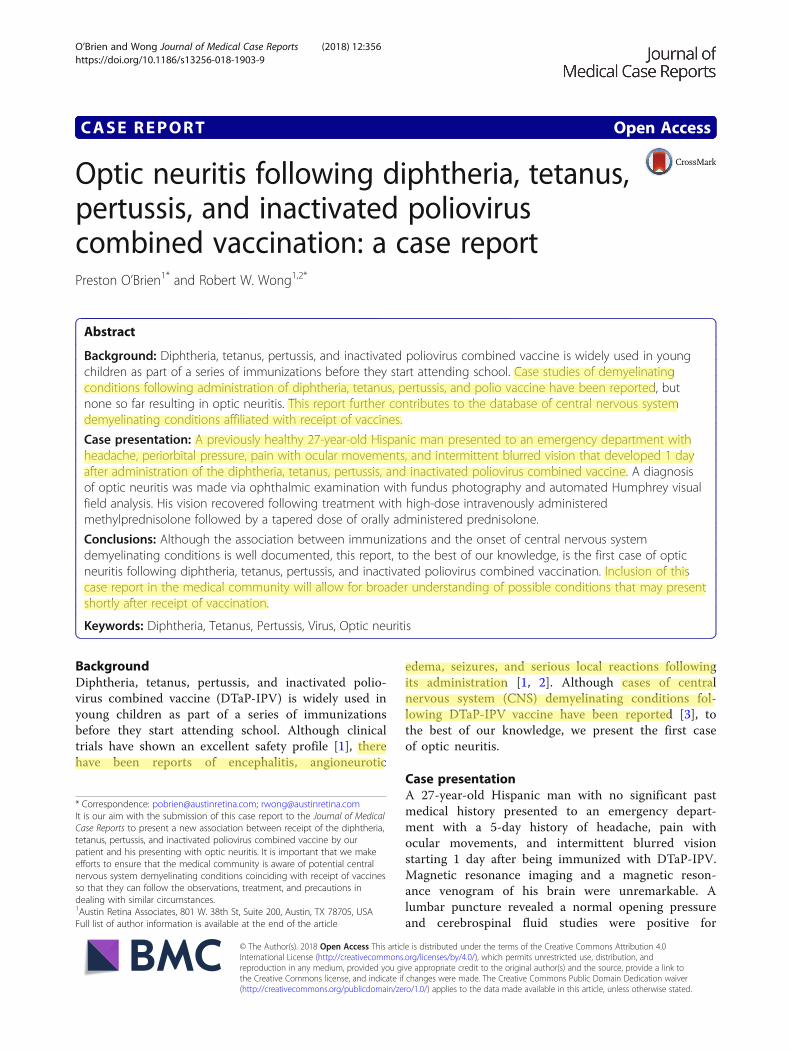
in his right eye and 20/70 in his left eye. Intraocularpressures, pupil examination, ocular alignment, andextraocular movements were normal. Confrontationalvisual fields were restricted in both eyes. Posterior seg-ment examination showed optic nerve swelling andhyperemia in both eyes (Fig. 1) and two microaneurysmsin the mid periphery of his left eye. No evidence of vitri-tis, retinal vasculitis, or choroiditis was seen in eithereye.Serum laboratory testing showed elevated glycated
hemoglobin (A1C) at 6.9%, aspartate aminotransferase(AST), and alanine aminotransferase (ALT). Otherliver tests including bilirubin, alkaline phosphatase,and hepatitis serologies were normal. Tests for infectiousand inflammatory etiologies including angiotensin-con-verting enzyme (ACE), lysozyme, antinuclear antibody(ANA), cytoplasmic antineutrophil cytoplasmic antibodies(c-ANCA), perinuclear antineutrophil cytoplasmic anti-bodies (p-ANCA), lupus panel, rapid plasma reagin (RPR),fluorescent treponemal antibody absorption (FTA-ABS),chest X-ray, and QuantiFERON Gold assay, which were
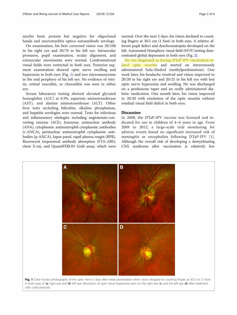
normal. Over the next 5 days, his vision declined to count-ing fingers at 30.5 cm (1 foot) in both eyes. A relative af-ferent pupil defect and dyschromatopsia developed on theleft. Automated Humphrey visual field (HVF) testing dem-onstrated global depression in both eyes (Fig. 2).He was diagnosed as having DTaP-IPV vaccination-re-
lated optic neuritis and started on intravenouslyadministered Solu-Medrol (methylprednisolone). Oneweek later, his headache resolved and vision improved to20/20 in his right eye and 20/25 in his left eye with lessoptic nerve hyperemia and swelling. He was dischargedon a prednisone taper and an orally administered dia-betic medication. One month later, his vision improvedto 20/20 with resolution of the optic neuritis withoutresidual visual field deficit in both eyes.
DiscussionIn 2008, the DTaP-IPV vaccine was licensed and in-dicated for use in children of 4–6 years in age. From2009 to 2012, a large-scale trial monitoring foradverse events found no significant increased risk ofmeningitis or encephalitis following DTaP-IPV [1].Although the overall risk of developing a demyelinatingCNS syndrome after vaccination is relatively low
Fig. 1 Color fundus photography of the optic nerve 5 days after initial presentation when vision dropped to counting fingers at 30.5 cm (1 foot)in both eyes of (a) right eye and (b) left eye. Resolution of optic nerve hyperemia seen on the right eye (c) and the left eye (d) after treatmentwith corticosteroids
O’Brien and Wong Journal of Medical Case Reports (2018) 12:356 Page 2 of 4
He was diagnosed as having DTaP-IPV vaccination-re- lated optic neuritis
(estimated to be 0.1%), it is not negligible [3]. Molecularmimicry from the viral proteins or the adjuvants used inthe preparation of the vaccine have been suspected in thedevelopment of demyelinating disease following vaccin-ation [3, 4]. Molecular mimicry occurs when similaritiesexist between proteins of viruses used in vaccinations andthe components of CNS myelin which may disruptself-tolerance and cause production of autoantibodiesresulting in CNS inflammation including optic neur-itis [3, 5]. Our case is consistent with other cases ofpost-vaccination optic neuritis, most of which develop1–3 weeks after vaccination, typical of an immune-triggered mechanism [3].In most cases, symptoms of optic neuritis were mostly
resolved after treatment with steroids such as intraven-ously administered methylprednisolone followed by ta-pered oral prednisolone for several weeks [3, 5]. Earlyrecognition of ocular signs and symptoms of optic neur-itis following DTaP-IPV vaccination may lead to prompttreatment and preserved vision.
ConclusionsAlthough the association between immunizations andthe onset of CNS demyelinating conditions is well docu-mented, this report, to the best of our knowledge, is thefirst case of optic neuritis following DTaP-IPV vaccin-ation. Inclusion of this case report in the medical com-munity will allow for broader understanding of possibleconditions that may present shortly after receipt ofvaccination.
AbbreviationsA1C: Glycated hemoglobin; ACE: Angiotensin-converting enzyme;ALT: Alanine aminotransferase; ANA: Antinuclear antibody; AST: Aspartateaminotransferase; c-ANCA: Cytoplasmic antineutrophil cytoplasmicantibodies; CNS: Central nervous system; DTaP-IPV: Diphtheria, tetanus,pertussis and inactivated poliovirus combined vaccine; FTA-ABS: Fluorescenttreponemal antibody absorption; p-ANCA: Perinuclear antineutrophilcytoplasmic antibodies; RPR: Rapid plasma reagin
AcknowledgementsNot applicable.
Fig. 2 Automated 30–2 Humphrey visual field testing using Swedish Interactive Testing Algorithm-Fast protocol of (a) the right eye and (b) theleft eye showing gray tone and pattern deviation. The patient was started on intravenously administered Solu-Medrol (methylprednisolone) onSeptember 11, 2013 (top line). Follow-up perimetry 1 week (middle line) and 3 weeks (bottom line) after the initiation of systemic corticosteroidsshowing improvement and resolution of the visual field deficits. MD mean deviation, SITA Swedish Interactive Testing Algorithm
O’Brien and Wong Journal of Medical Case Reports (2018) 12:356 Page 3 of 4
similarities exist between proteins of viruses used in vaccinations and the components of CNS myelin which may disrupt self-tolerance and cause production of autoantibodies resulting in CNS inflammation
Our case is consistent with other cases of post-vaccination optic neuritis, most of which develop 1–3 weeks after vaccination, typical of an immune- triggered mechanism
FundingThere are no other sources of funding.
Availability of data and materialsThe authors agree to making the images and data described in themanuscript freely available for use.
Authors’ contributionsBoth PO and RW contributed equally to the design, drafting, and editing ofthis manuscript. Both authors read and approved the final manuscript.
Ethics approval and consent to participateThe study was sent to the University of Texas at Austin Institutional ReviewBoard and need for further approval was waived.
Consent for publicationWritten and informed consent was obtained from the patient for publicationof the case report and the accompanying images. Copies of the writtenconsent forms are available for review by the Editor-in-Chief of this journal.
Competing interestsThe authors declare that they have no competing interests.
Publisher’s NoteSpringer Nature remains neutral with regard to jurisdictional claims inpublished maps and institutional affiliations.
Author details1Austin Retina Associates, 801 W. 38th St, Suite 200, Austin, TX 78705, USA.2Department of Surgery and Perioperative Services, Dell Medical School,Austin, TX, USA.
Received: 2 April 2018 Accepted: 29 October 2018
References1. Daley MF, Yih WK, Glanz JM, et al. Safety of diphtheria, tetanus, acellular
pertussis and inactivated poliovirus (DTaP-IPV) vaccine. Vaccine. 2014;32(25):3019–24.
2. Aydin H, Ozgul E, Agildere AM. Acute necrotizing encephalopathysecondary to diphtheria, tetanus toxoid and whole-cell pertussisvaccination: diffusion-weighted imaging and proton MR spectroscopyfindings. Pediatr Radiol. 2010;40(7):1281–4.
3. Karussis D, Petrou P. The spectrum of post-vaccination inflammatory CNSdemyelinating syndromes. Autoimmun Rev. 2014;13(3):215–24.
4. Reeves WH, Lee PY, Weinstein JS, Satoh M, Lu L. Induction of autoimmunityby pristane and other naturally occurring hydrocarbons. Trends Immunol.2009;30(9):455–64.
5. O'Dowd S, Bafiq R, Ryan A, Cullinane A, Costello D. Severe bilateral opticneuritis post hepatitis A virus (HAV) and typhoid fever vaccination. J NeurolSci. 2015;357(1–2):300–1.
O’Brien and Wong Journal of Medical Case Reports (2018) 12:356 Page 4 of 4