ophthalmology Systematic approaches for reviewing neuro ... · Review Systematic approaches for...
10
Review Systematic approaches for reviewing neuro-imaging scans in ophthalmology Joshua M. Kruger, MD, PhD, a Dean M. Cestari, MD, b and Mary Beth Cunnane, MD c Author affiliations: a Neuro-Ophthalmology Service, Hadassah Medical Center, Kiryat Hadassah, Jerusalem, Israel b Neuro-Ophthalmology Service, Massachusetts Eye and Ear Infirmary, Harvard Medical School, Boston, Massachusetts; c Department of Radiology, Massachusetts Eye and Ear Infirmary, Harvard Medical School, Boston, Massachusetts Abstract Neuroimaging is an important tool in ophthalmology, but many ophthalmologists are uncomfortable evalu- ating actual scans. Unfortunately, exclusive reliance on a radiologist’s report can lead to diagnostic and management errors. We outline a methodology for equipping ophthalmologists with the skills necessary to read neuroimaging studies with respect to specific clinical questions. Introduction Neuro-ophthalmic diseases can be divided broadly into disorders that cause afferent or efferent dysfunction. On the afferent side, patients can experience vision loss due to disease affecting the optic nerve, chiasm, optic tract, thalamus, optic radiations, or cerebral cortex. On the efferent side, patients can develop ocular motility abnor- malities resulting from lesions anywhere from the cortex to the nerves that supply the extraocular muscles or even from the muscles themselves. Advances in imaging now allow clinicians to identify the locus and often the nature of the lesions responsible for afferent or efferent dys- function. It is critical for the general ophthalmologist to know which neuro-imaging study to order for each clini- cal scenario—an issue that we have addressed previ- ously. 1 It is also important for the ophthalmologist to review the study rather than simply to rely on the offi- cial radiology report. Oversights can be made by the radiologist. This may occur because the radiologist has limited experience in orbital/neuro-imaging or because they lack the clinical information necessary to identify the specific region requiring attention. We believe that it is possible to train ophthalmologists to be able to read neuro-imaging studies with approaches focused on spe- cific clinical questions. We do not intend to imply that expert radiological evaluation is unnecessary; rather, we hope to provide ophthalmologists with tools to deepen comprehension of basic neuroimaging techniques to per- mit more informed diagnoses and treatment strategies. The present review aims to provide focused systematic approaches for reviewing various scans and identifying the most common pathologies. The protocols provided in this manuscript require the clinician to be familiar with the various forms of magnetic resonance imaging (MRI; eg, T1, T2, FLAIR). Each sequence highlights various features of neuroanatomy (Table 1; also see Fig- ure 1). Suspected Optic Neuropathy Patients with decreased visual acuity, dyschromatopsia, a relative afferent pupillary defect, and a visual field defect should be presumed to have an optic neuropathy. If the patient is >50 years of age, and there is associated optic nerve swelling, then a diagnosis of anterior ische- mic optic neuropathy can be presumed, and there is no indication for neuroimaging, provided that the swelling resolves within 2 months. 2 For all other presentations, an immediate MRI of the orbits with contrast is indica- ted. When optic neuritis is suspected (eg, associated pain with eye movement), then an MRI of the brain with con- trast is additionally desirable to assess for radiological evidence of multiple sclerosis. 3,4 Review of the MRI scan should begin with the coronal short inversion recovery (STIR) or fat-saturated T2- Published September 19, 2017. Copyright ©2017. All rights reserved. Reproduction in whole or in part in any form or medium without expressed written permission of the Digital Journal of Ophthalmology is prohibited. doi:10.5693/djo.03.2016.05.001 Correspondence: Mary Beth Cunnane, MD, Department of Radiology, Massachusetts Eye and Ear Infirmary, Harvard Medical School, Boston, Massachusetts, 243 Charles Street, Boston, MA 02114 (email: [email protected]). digital journal of ophthalmology, vol. 23 digital journal of ophthalmology, vol. 23
Transcript of ophthalmology Systematic approaches for reviewing neuro ... · Review Systematic approaches for...

ReviewSystematic approaches for reviewing neuro-imaging scans inophthalmologyJoshua M. Kruger, MD, PhD,
a Dean M. Cestari, MD,
b and Mary Beth Cunnane, MDc
Author affiliations: aNeuro-Ophthalmology Service, Hadassah Medical Center, Kiryat Hadassah, Jerusalem, IsraelbNeuro-Ophthalmology Service, Massachusetts Eye and Ear Infirmary, Harvard Medical School, Boston, Massachusetts;cDepartment of Radiology, Massachusetts Eye and Ear Infirmary, Harvard Medical School, Boston, Massachusetts
AbstractNeuroimaging is an important tool in ophthalmology, but many ophthalmologists are uncomfortable evalu-ating actual scans. Unfortunately, exclusive reliance on a radiologist’s report can lead to diagnostic andmanagement errors. We outline a methodology for equipping ophthalmologists with the skills necessary toread neuroimaging studies with respect to specific clinical questions.
IntroductionNeuro-ophthalmic diseases can be divided broadly intodisorders that cause afferent or efferent dysfunction. Onthe afferent side, patients can experience vision loss dueto disease affecting the optic nerve, chiasm, optic tract,thalamus, optic radiations, or cerebral cortex. On theefferent side, patients can develop ocular motility abnor-malities resulting from lesions anywhere from the cortexto the nerves that supply the extraocular muscles or evenfrom the muscles themselves. Advances in imaging nowallow clinicians to identify the locus and often the natureof the lesions responsible for afferent or efferent dys-function. It is critical for the general ophthalmologist toknow which neuro-imaging study to order for each clini-cal scenario—an issue that we have addressed previ-ously.1 It is also important for the ophthalmologist toreview the study rather than simply to rely on the offi-cial radiology report. Oversights can be made by theradiologist. This may occur because the radiologist haslimited experience in orbital/neuro-imaging or becausethey lack the clinical information necessary to identifythe specific region requiring attention. We believe that itis possible to train ophthalmologists to be able to readneuro-imaging studies with approaches focused on spe-cific clinical questions. We do not intend to imply thatexpert radiological evaluation is unnecessary; rather, wehope to provide ophthalmologists with tools to deepencomprehension of basic neuroimaging techniques to per-
mit more informed diagnoses and treatment strategies.The present review aims to provide focused systematicapproaches for reviewing various scans and identifyingthe most common pathologies. The protocols providedin this manuscript require the clinician to be familiarwith the various forms of magnetic resonance imaging(MRI; eg, T1, T2, FLAIR). Each sequence highlightsvarious features of neuroanatomy (Table 1; also see Fig-ure 1).
Suspected Optic NeuropathyPatients with decreased visual acuity, dyschromatopsia,a relative afferent pupillary defect, and a visual fielddefect should be presumed to have an optic neuropathy.If the patient is >50 years of age, and there is associatedoptic nerve swelling, then a diagnosis of anterior ische-mic optic neuropathy can be presumed, and there is noindication for neuroimaging, provided that the swellingresolves within 2 months.2 For all other presentations,an immediate MRI of the orbits with contrast is indica-ted. When optic neuritis is suspected (eg, associated painwith eye movement), then an MRI of the brain with con-trast is additionally desirable to assess for radiologicalevidence of multiple sclerosis.3,4
Review of the MRI scan should begin with the coronalshort inversion recovery (STIR) or fat-saturated T2-
Published September 19, 2017.Copyright ©2017. All rights reserved. Reproduction in whole or in part in any form or medium without expressed written permission of theDigital Journal of Ophthalmology is prohibited.doi:10.5693/djo.03.2016.05.001Correspondence: Mary Beth Cunnane, MD, Department of Radiology, Massachusetts Eye and Ear Infirmary, Harvard Medical School, Boston,Massachusetts, 243 Charles Street, Boston, MA 02114 (email: [email protected]).
digital journal of ophthalmology, vol. 23
digital journal of ophthalmology, vol. 23

weighted sequences immediately posterior to the globes.The optic nerve can be found at the center of an imagi-nary ring formed by the extraocular muscles. It is sur-rounded by a bright cuff of cerebrospinal fluid (CSF).The nerve should be isointense to the subcortical white
matter. The clinician should follow the course of theoptic nerves all the way to the chiasm, and if possible,into the optic tracts, to ensure that there is symmetry,and isointensity to the subcortical white matter through-out the course (Video 1). The finding of increased
Table 1. Comparison of MRI sequences
Figure 1. Axial images through the lateral ventricles demonstrate differences in the appearance of T1-weighted, T2-weighted and FLAIRimages. In each image, a W is placed over the corpus callosum, a white matter tract, and a G is placed over the cortex of the left frontal lobe.A, On this T1-weighted image, the white matter is hyperintense, and the cerebrospinal fluid (CSF) in the ventricles is hypointense comparedto the gray matter. B, On this T2-weighted image, the white matter is hypointense, and the CSF in the ventricles is hyperintense compared tothe gray matter. C, A FLAIR image is a T2-weighted image with suppression of the bright CSF signal. On these images, the white matter ishypointense (like a T2-weighted image), but the CSF in the ventricles is hypointense in comparison to the gray matter.
Kruger et al. 51
digital journal of ophthalmology, vol. 23
digital journal of ophthalmology, vol. 23

brightness (relative to the subcortical white matter) is anonspecific finding suggestive of injury to the affectedsegment. The timing cannot be determined, because bothacute and chronic injury will cause T2 hyperintensity.Proceed to the coronal T1 post-contrast sequence, pref-erably with fat saturation. Once again, the optic nerves,chiasm, and tracts should be isointense to the subcorticalwhite matter. The presence of increased brightness(referred to as “enhancement” on a postcontrast image)indicates breakdown of the blood-brain barrier, whichmay be due to inflammation or tumor. If the nerve doesnot appear enlarged, then the cause is more likelyinflammatory. If optic neuritis is suspected, then onemust assess for intracranial evidence of multiple sclero-sis, specifically T2 hyperintensities, known as “whitespots,” in the brain. Scroll through the axial fluid attenu-ated inversion recovery (FLAIR) sequence, with particu-lar attention to the periventricular areas. The brainstemis best viewed with T2-weighted imaging. The sagittalFLAIR sequence should also be reviewed to assess forthe presence of T2 hyperintense lesions radiating fromthe corpus callosum, known as Dawson’s fingers (seeend of Video 8).
Bitemporal HemianopsiaA chiasmal disorder is typified by a bitemporal hemia-nopsia, due to the compression of the crossing fibersfrom the nasal aspect of each retina. The most commonlesion causing a chiasmopathy is a pituitary adenoma,5which is typically benign, but the differential diagnosisincludes disorders such as an aneurysm6 and pituitaryapoplexy.7 The chiasm can be imaged through either anMRI orbit sequence or an MRI brain scan. Many institu-tions also offer the option of a specific pituitary scan-ning protocol.
Video 1. Normal optic nerve.
Review of the scan (Video 2) should begin with a sagit-tal image, preferably T1. The pituitary gland can beidentified as an area of soft tissue within the bony sella,which lies along the superior-posterior aspect of the darksphenoid sinus. The posterior segment of the pituitarygland is particularly bright, likely due to the presence oflipid in the posterior pituicytes.8 The normal height ofthe pituitary gland is approximately 5 mm,9 but it can beas much as 9 mm.10 Immediately superior to the pitui-tary gland is the suprasellar cistern, which will appeardark on T1 coronal imaging. Above the suprasellar cis-tern is the optic chiasm, which appears as a bright, linearstructure oriented at approximately 45 degrees on T1imaging. The presence of a complete suprasellar cisternindicates that the pituitary cannot be exerting a masseffect on the chiasm.
Next, review the coronal T1 sequence. If it is postcon-trast, then the pituitary will be diffusely bright because itis a highly vascularized structure. A T-shaped structureextends upward from the center of the gland—the verti-
Video 8. Abducens nerve.
Video 2. Sella turcica and pituitary.
52
digital journal of ophthalmology, vol. 23
digital journal of ophthalmology, vol. 23

cal component is the pituitary stalk, and the horizontalcomponent is the optic chiasm. Once again ensure thatCSF clearly separates the chiasm from the pituitarygland. Video 3 demonstrates findings for a pituitary ade-noma. Note that the described approach has not includedreview of the axial scan because the chiasm is poorlyvisualized in that plane.
Homonymous HemianopsiaA visual field defect that occurs right or left of the verti-cal midline in both eyes is referred to as “homonymous”and is associated with a retrochiasmal disorder. Thesecases are best imaged through an MRI of the brain. Ifthe visual field loss has an acute onset in an elderlypatient, then the mechanism likely is due to a stroke, andit is reasonable to forgo contrast because this is not use-ful for demonstrating ischemia. The critical sequence isdiffusion weighted imaging (DWI), which is often per-formed as part of a standard MRI but should be explic-itly requested in these circumstances. If it is a case ofhomonymous visual defect in a younger patient, a massor demyelinating lesion is more likely and contrastshould be included.
The visual pathway extending from the origin of theoptic tracts to the occipital cortex must be inspected.The optic tracts can be identified in both axial and coro-nal images. They extend posteriorly from the opticchiasm and course around the midbrain to the lateralgeniculate body of the thalamus. The optic radiationsrun from the lateral geniculate body to the occipital cor-tex. En route, they spread diffusely within the whitematter of the temporal and parietal lobes. The optic radi-ations are best examined with axial images. The DWI,FLAIR, and T1 pre- and postcontrast images should beinspected for hyperintense lesions in the areas described.
Video 3. Pituitary adenoma.
A hyperintense lesion in DWI can represent either cyto-toxic edema (ie, stroke) or vasogenic edema. If theedema is cytotoxic, then the same region will appearhypointense on the corresponding apparent diffusioncoefficient (ADC) images. An area that is hyperintenseon DWI, hypointense on ADC, and hyperintense on T2-weighted imaging (eg, FLAIR) most likely represents asubacute infarction (Video 4). The additional presenceof hyperintensity on noncontrast T1-weighted images isconcerning for an intracerebral hemorrhage. The pres-ence of hyperintensity specifically on the postcontrastimages only (and not present on the T1 precontrastimages) is suggestive of a neoplastic lesion, vascularlesion, inflammation, or infection.
Oculomotor Nerve PalsyThe third cranial nerve innervates the superior rectus,inferior rectus, medial rectus, and inferior oblique mus-cles as well as the levator palpebrae superioris. It alsocarries the parasympathetic pupillary constrictor fibers.In the most extreme form of an oculomotor nerve palsy,there is complete ptosis, a dilated pupil, and the globewill assume a down-and-out position. It is critical, how-ever, to keep in mind that many presentations of an ocu-lomotor nerve palsy can affect only some of these mus-cles, often in a very mild way. The index of suspicionmust be very high in any case of diplopia with a verticalcomponent. Because the oculomotor nerve enters thesubarachnoid space, it comes into close proximity withthe posterior communicating artery. An aneurysm of thisartery may compress the oculomotor nerve, resulting ina palsy. Rupture of an aneurysm is a life-threateningevent.11 Therefore, any oculomotor nerve palsy must bepresumed to be caused by a posterior communicatinganeurysm until proven otherwise. Computed tomogra-phy angiography (CTA) can be just as sensitive as mag-
Video 4. Occipital lobe infarction.
Kruger et al. 53
digital journal of ophthalmology, vol. 23
digital journal of ophthalmology, vol. 23

netic resonance angiography (MRA) in detecting theseaneurysms12 and delaying imaging to obtain an MRAversus a CTA is not justified. Communication with theneuroradiologist is best to establish the preferred modal-ity for diagnosis of these aneurysms.
Aneurysms are most commonly located at sites of vesselbifurcation; therefore, examination of the angiogramshould be focused on the circle of Willis (Video 5). Theroutes of all major vessels of the anterior and posteriorcirculation should be inspected, with particular attentionto the posterior communicating artery (PCOM). Anyoutpouching of contrast that extends off a vessel butdoes not extend into another known vessel is suspiciousfor an aneurysm. Follow the route of each internal caro-tid artery (ICA) looking carefully at the take-off for thePCOM, a branch of the supraclinoid internal carotidartery. Continue to follow the ICA as it bifurcates intothe anterior and middle cerebral arteries. Follow thecourse of each middle cerebral artery through the bifur-cations into the superior and inferior segments. Followthe course of each anterior cerebral artery, attempting toidentify the anterior communicating artery, a tiny vesselwhich connects the right and left anterior cerebral artery(ACA). Proceed to the posterior circulation, beginningwith the vertebral arteries at the level of the foramenmagnum. As one scrolls caudally, one should note theorigin of the posterior inferior cerebellar arteries(PICAs) off each vertebral artery. The vertebral arteriesthen merge into the basilar artery. Areas of contrast willthen extend off the basilar artery toward the cerebellum,corresponding to the anterior inferior cerebellar arteries(AICA) and the superior cerebellar arteries, respectively.Finally, the basilar artery will bifurcate into left andright posterior cerebral arteries. Volume-renderingreconstruction technologies, such as coronal maximum
Video 5. Computed tomography (CT) angiography for an oculo-motor nerve palsy.
intensity projection (MIP) sequences or 3-D reconstruc-tions may allow for better appreciation of any aneur-ysms. Keep in mind that patients may have multipleaneurysms, and that a complete search of the circle ofWillis should be performed even in patients with aneur-ysms large enough to be appreciated on the initial cur-sory review.
If there is radiological evidence of an aneurysm in thesetting of a headache, then the computed tomography(CT) scan should be reviewed to rule out the presence ofa subarachnoid hemorrhage (ie, a ruptured aneurysm).Particular attention should be paid for any hyperdensityin the basilar cisterns and ventricles.
Review of an MRI scan for an oculomotor nerve palsybegins with the axial T2-weighted sequence at the levelof the midbrain (Video 6). The third cranial nerve islocated posterior to the red nucleus, just lateral to thesagittal midline. Ensure that there is no focal hyperinten-sity in the area. This same region should also be viewedthrough the DWI sequence to rule out any focus ofhyperintensity, which might indicate an acute infarction.The third nerve exits the brainstem anteriorly, just lateralto the interpeduncular cistern, and travels anteriorlytoward the cavernous sinus. This segment is best viewedwith heavily T2-weighted images (eg, FIESTA, CISS,3D DRIVE). Note that the third cranial nerve runs paral-lel to the posterior communicating artery. Coronal post-contrast images throughout the cavernous sinus shouldthen be viewed. The oculomotor nerve is located at thesuperior lateral aspect of the cavernous sinus. It shouldbe isointense to the subcortical white matter. Symmetryshould be confirmed.
Trochlear Nerve PalsyIn the case of an isolated trochlear nerve palsy (or abdu-cens nerve palsy) in a patient >50 years of age who is
Video 6. Oculomotor nerve.
54
digital journal of ophthalmology, vol. 23
digital journal of ophthalmology, vol. 23

diabetic or vasculopathic, the most likely etiology ismicrovascular ischemia, in which case neuroimagingwill be unrevealing. In these circumstances, neuroimag-ing can be deferred as long as there is no worsening ofthe strabismus,13 and the strabismus resolves within 90days. If either of these conditions cannot be met, then anMRI of the brain should be performed with contrast. Inthe case of a young, nondiabetic patient, a structuralcause must be presumed, and an MRI of the brain withcontrast should be performed at the soonest opportunity.
Review of the scan begins with the axial FLAIR at thelevel of the midbrain (Video 7). Located at the posterioraspect of the midbrain is the cerebral aqueduct, whichappears as a dark flow void, surrounded by the brightring of the periaqueductal gray. The trochlear nervenucleus lies immediately anterior to the periaqueductalgray. Ensure that there is no focal brightness in the area,or hyperintensity on DWI. The trochlear nerve exits thebrainstem posteriorly and then travels anteriorly towardthe cavernous sinus through the basilar cistern. As statedpreviously, the cisternal segments of the cranial nervesare best studied with heavily T2-weighted images.While the size of the trochlear nerve is typically toosmall to be visualized, it is important to rule out thepresence of any mass lesions in the basilar cistern,which would be a presumed etiology for a trochlearnerve palsy. The basilar cistern should also be viewed inaxial postcontrast images to rule out an enhancing massor leptomeningeal enhancement. Coronal postcontrastimages throughout the cavernous sinus should then bereviewed. Again, the trochlear nerve is typically toosmall to be seen but sits immediately inferior to the ocu-lomotor nerve, adjacent to the lateral wall of the sinus.This region should be reviewed to rule out the presenceof any abnormal mass. If a chronic trochlear nerve palsyis suspected, ensure that the sizes of the superior oblique
Video 7. Trochlear nerve.
muscles are symmetric by viewing them in a coronalsequence within the orbits.
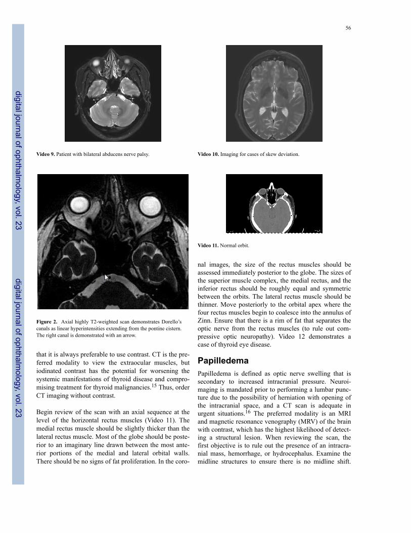
Abducens Nerve PalsyThe abducens nerve lies at the dorsal aspect of the cau-dal pons. It lies immediately ventral to the facial colli-culli, which are protrusions of the dorsal pons into thefourth ventricle. Each colliculus is formed by the motorfibers of the facial nerve wrapping around the nucleus ofthe abducens nerve. This area should be assessed for anyhyperintensity in the axial FLAIR, DWI, and postcon-trast T1 sequences (Video 8). The abducens nerve exitsthe brainstem ventrally at the junction of the pons andmedulla. It then ascends toward the cavernous sinusthrough the pontine cistern. Within the cisternal seg-ment, the nerve can be visualized with heavily T2-weighted images, appearing as a linear hypointensitynearly parallel to the sagittal plane. Assess for the pres-ence of any abnormal soft tissue in the heavily T2-weighted and T1 postcontrast images (Video 9). Theabducens nerve transitions from the pontine cistern intothe cavernous sinus via Dorello’s canal, a groove in thepetrous bone. It can be visualized on the axial T2 imagesas a linear hyperintensity extending from the pontinecistern into the hypointense petrous bone (Figure 2).Coronal postcontrast images throughout the cavernoussinus should then be reviewed. While the oculomotorand trochlear nerves are adjacent to the lateral wall ofthe cavernous sinus, the abducens nerve is more central,situated inferior-lateral to the internal carotid artery. Thecaliber is typically too small to be visualized, but theobserver should be alert for a T2 hyperintense orenhancing lesion inferolateral to the carotid artery,which would be likely to produce an abducens nervepalsy.
Skew DeviationA skew deviation is an acquired vertical strabismuscaused by a supranuclear lesion. There is defective inputto the oculomotor and trochlear nerves, which controlthe vertical position of the globe. Relevant lesions canoccur in the peripheral vestibular system, the brainstem,the cerebellum or the thalamus.14 These structures canbe assessed best on axial T2-weighted imaging (Video10).
Thyroid Eye DiseaseThe most common etiology of an orbitopathy causingdiplopia is Thyroid eye disease. It is perhaps the bestexception to the “rules” that MRI is superior to CT and
Kruger et al. 55
digital journal of ophthalmology, vol. 23
digital journal of ophthalmology, vol. 23
that it is always preferable to use contrast. CT is the pre-ferred modality to view the extraocular muscles, butiodinated contrast has the potential for worsening thesystemic manifestations of thyroid disease and compro-mising treatment for thyroid malignancies.15 Thus, orderCT imaging without contrast.
Begin review of the scan with an axial sequence at thelevel of the horizontal rectus muscles (Video 11). Themedial rectus muscle should be slightly thicker than thelateral rectus muscle. Most of the globe should be poste-rior to an imaginary line drawn between the most ante-rior portions of the medial and lateral orbital walls.There should be no signs of fat proliferation. In the coro-
Video 9. Patient with bilateral abducens nerve palsy.
Figure 2. Axial highly T2-weighted scan demonstrates Dorello’scanals as linear hyperintensities extending from the pontine cistern.The right canal is demonstrated with an arrow.
nal images, the size of the rectus muscles should beassessed immediately posterior to the globe. The sizes ofthe superior muscle complex, the medial rectus, and theinferior rectus should be roughly equal and symmetricbetween the orbits. The lateral rectus muscle should bethinner. Move posteriorly to the orbital apex where thefour rectus muscles begin to coalesce into the annulus ofZinn. Ensure that there is a rim of fat that separates theoptic nerve from the rectus muscles (to rule out com-pressive optic neuropathy). Video 12 demonstrates acase of thyroid eye disease.
PapilledemaPapilledema is defined as optic nerve swelling that issecondary to increased intracranial pressure. Neuroi-maging is mandated prior to performing a lumbar punc-ture due to the possibility of herniation with opening ofthe intracranial space, and a CT scan is adequate inurgent situations.16 The preferred modality is an MRIand magnetic resonance venography (MRV) of the brainwith contrast, which has the highest likelihood of detect-ing a structural lesion. When reviewing the scan, thefirst objective is to rule out the presence of an intracra-nial mass, hemorrhage, or hydrocephalus. Examine themidline structures to ensure there is no midline shift.
Video 10. Imaging for cases of skew deviation.
Video 11. Normal orbit.
56
digital journal of ophthalmology, vol. 23
digital journal of ophthalmology, vol. 23

Look at the ventricles to evaluate for ventricularenlargement. Ensure that the sulci extend all the way tothe inner calvarium and are not separated from it by asubdural hematoma. Look for displaced structures, swol-len regions of the brain, and mass effect. The clinicianshould then proceed to assess for signs of increasedintracranial pressure, which can be seen in idiopathicintracranial hypertension. Begin with the sagittal T1images and identify the pituitary gland. Increased intra-cranial pressure causes the CSF in the suprasellar cisternto compress the pituitary gland against the floor of thesella resulting in a “partially empty sella” (Video 13).On the axial T2-weighted images, the anterior aspect ofthe CSF cuff of the optic nerve should be inspected. Innormal individuals, mild flaring can occur (Video 14).Larger amounts of CSF fluid are concerning forincreased intracranial pressure (Video 13). The posterioraspect of the globe should have a spherical appearance.It typically becomes “flattened” in idiopathic intracra-nial hypertension (IIH).
MRV should also be performed in all patients before IIHis presumed, particularly in atypical cases, to rule out thepossibility of a sinus venous thrombosis.17 The courseof each sinus should be reviewed in both the raw dataand the MIP images. The presence of a filling defect inthe lumen of a sinus is known as the empty delta signand is specific for a thrombus.18 Stenosis of the trans-verse sinuses is a common finding in IIH (see Video15).19
Horner SyndromeTwo causes of anisocoria that require urgent neuroimag-ing are an oculomotor nerve palsy, discussed above, anda Horner syndrome. A Horner syndrome is caused bydecreased sympathetic stimulation to the eye, resulting
Video 12. Thyroid orbitopathy.
in miosis and ptosis due to disrupted innervation of thepupillary dilators and Muller’s muscle. The sympatheticpathway runs a circuitous course, descending from the
Video 13. Idiopathic intracranial hypertension – magnetic reso-nance imaging (MRI).
Video 14. Idiopathic intracranial hypertension ruled out – MRI.
Video 15. Idiopathic intracranial hypertension – MR venography.
Kruger et al. 57
digital journal of ophthalmology, vol. 23
digital journal of ophthalmology, vol. 23

hypothalamus to the level of the thoracic spine and thenascending to the orbit.
Until proved otherwise, any case of a Horner syndromemust be presumed to be due to a carotid dissection.Imaging of the carotid artery should be performed usinga CTA of the head and neck. The goal is to trace theentire course of each carotid artery, ensuring that theartery retains its full caliber throughout its path. Thescan is reviewed with axial sections. Begin at the levelof the aortic arch (Video 16). As one scrolls superiorly,the aortic arch transitions into the brachiocephalic (thatis, innominate), left common carotid, and left subclavianarteries. Starting on the right, the brachiocephalicdivides into the right subclavian artery (posteriorly) andcommon carotid artery (anteriorly). The common carotidwill then bifurcate into the external carotid artery (ante-riorly) and the internal carotid artery (posteriorly). Theinternal carotid artery then ascends through the neck andenters the skull base through the carotid canal, in thepetrous bone. It then runs transversely across the bonyspace within the petrous segment, which it should com-pletely fill. Moving further superiorly, the internal caro-tid artery passes through the cavernous sinus. The inter-nal carotid artery exits the cavernous sinus and entersthe dura roughly at the level of the anterior clinoid. Thesupraclinoid internal carotid artery then bifurcates intothe middle cerebral artery and the anterior cerebralartery. Volume rendering reconstruction technologies,such as MIP, allow for a sagittal view of the internal car-otid artery, which is often more readily followed (Video16). Any tapered narrowing of the internal carotid arteryis concerning for the possibility of a dissection. In somecases, MRI may be useful to evaluate for a high signalcrescent around the carotid on fat-saturated T1-weightedimages. This high signal crescent represents clot in thefalse lumen of the vessel.
Video 16. Horner syndrome.
While ruling out a carotid dissection is a high priority ina Horner syndrome, imaging must be performed to scanthe pathway of the three orders of neurons involved insupplying sympathetic innervation to the eye. The first-order neuron originates in the hypothalamus. Thus, anMRI of the brain is required to properly image this area.An apical lung tumor (Pancoast tumor) can injure thesecond-order neuron, and a CT of the chest should beperformed to rule out this possibility. Imaging of thethird-order neuron is achieved in the previously men-tioned imaging of the carotid artery. Therefore, Hornersyndrome requires a variety of imaging modalities torule out these structural causes in the differential diagno-sis.
References1. Kruger JM, Lessell S, Cestari DM. Neuro-imaging: a review for the
general ophthalmologist. Semin Ophthalmol 2012;27:192-6.2. Lee AG, Lin DJ, Kaufman M, Golnik KC, Vaphiades MS, Eggen-
berger E. Atypical features prompting neuroimaging in acute opticneuropathy in adults. Can J Ophthalmol 2000;35:325-30.
3. Polman CH, Reingold SC, Banwell B, et al. Diagnostic criteria formultiple sclerosis: 2010 revisions to the McDonald criteria. AnnNeurol 2011;69:292-302.
4. Optic Neuritis Study Group. Multiple sclerosis risk after optic neuri-tis:final optic neuritis treatment trial follow-up. Arch Neurol2008;65:727-32.
5. Abboud CF, Laws ER Jr. Diagnosis of pituitary tumors. EndocrinolMetab Clin North Am 1988;17:241-80.
6. Kasner SE, Liu GT, Galetta SL. Neuro-ophthalmologic aspects ofaneurysms. Neuroimaging Clin N Am 1997;7:679-92.
7. Nawar RN, AbdelMannan D, Selman WR, Arafah BM. Pituitarytumor apoplexy: a review. J Intensive Care Med 2008;23:75-90.
8. Kucharczyk W, Lenkinski R, Kucharczyk J, Henkelmann RM. Theeffect of phospholipid vesicles on the NMR relaxation of water: anexplanation for the appearance of the neurohypophysis? AJNR AmJ Neuroradiol 1990;11:693-700.
9. Tsunoda A, Okuda O, Sato K. MR height of the pituitary gland as afunction of age and sex: especially physiological hypertrophy inadolescence and in climacterium. AJNR Am J Neuroradiol1997;18:551-4.
10. Wolpert SM, Molitch ME, Goldman JA, Wood JB. Size, shape,and appearance of the normal female pituitary gland. AJR Am JRoentgenol 1984;143:377-81.
11. Huige WM, van Vliet AG, Bastiaensen LA. Early symptoms ofsubarachnoid haemorrhage due to aneurysms of the posterior com-municating artery. Doc Ophthalmol 1988;70:251-6.
12. Lee AG, Hayman LA, Brazis PW. The evaluation of isolated thirdnerve palsy revisited: an update on the evolving role of magneticresonance, computed tomography, and catheter angiography. SurvOphthalmol 2002;47:137-57.
13. Chi SL, Bhatti MT. The diagnostic dilemma of neuro-imaging inacute isolated sixth nerve palsy. Curr Opin Ophthalmol2009;20:423-9.
14. Wong AM. Understanding skew deviation and a new clinical testto differentiate it from trochlear nerve palsy. J AAPOS2010;14:61-7.
58
digital journal of ophthalmology, vol. 23
digital journal of ophthalmology, vol. 23

15. van der Molen AJ, Thomsen HS, Morcos SK, Contrast MediaSafety Committee, European Society of Urogenital Radiology(ESUR). Effect of iodinated contrastmedia on thyroid function inadults. Eur Radiol 2004;14:902-7.
16. Gower DJ, Baker AL, Bell WO, Ball MR. Contraindications tolumbar puncture as defined by computed cranial tomography. JNeurol Neurosurg Psychiatry 1987;50:1071-4.
17. Lin A, Foroozan R, Danesh-Meyer HV, De Salvo G, Savino PJ,Sergott RC. Occurrence of cerebral venous sinus thrombosis in
patients with presumed idiopathic intracranial hypertension. Oph-thalmology 2006;113:2281-4.
18. Virapongse C, Cazenave C, Quisling R, Sarwar M, Hunter S. Theempty delta sign: frequency and significance in 76 cases of duralsinus thrombosis. Radiology 1987;162:779-85.
19. Riggeal BD, Bruce BB, Saindane AM, et al. Clinical course of idi-opathic intracranial hypertension with transverse sinus stenosis.Neurology 2013;80:289-95.
Kruger et al. 59
digital journal of ophthalmology, vol. 23
digital journal of ophthalmology, vol. 23