Ophthalmology - mohp.gov.eg§مراض العيون .pdf · 2 Acknowledgments This two-year...
83
1 Ophthalmology Collected and organized by Ihab Saad Othman, MD, FRCS Professor of Ophthalmology, Cairo University Director, EyeWorld Hospital, Giza , Egypt Second Year 2018-2019
Transcript of Ophthalmology - mohp.gov.eg§مراض العيون .pdf · 2 Acknowledgments This two-year...

1
Ophthalmology
Collected and organized by
Ihab Saad Othman, MD, FRCS
Professor of Ophthalmology, Cairo University
Director, EyeWorld Hospital, Giza , Egypt
Second Year
2018-2019

2
Acknowledgments
This two-year curriculum was developed through a participatory and collaborative approach between the
Academic faculty staff affiliated to Egyptian Universities as Alexandria University, Ain Shams University,
Cairo University , Mansoura University, Al-Azhar University, Tanta University, Beni Souef University , Port
Said University, Suez Canal University and MTI University and the Ministry of Health and
Population(General Directorate of Technical Health Education (THE). The design of this course draws on
rich discussions through workshops. The outcome of the workshop was course specification with Indented
learning outcomes and the course contents, which served as a guide to the initial design.
We would like to thank Prof.Sabah Al- Sharkawi the General Coordinator of General Directorate of
Technical Health Education, Dr. Azza Dosoky the Head of Central Administration of HR Development, Dr.
Seada Farghly the General Director of THE and all share persons working at General Administration of the
THE for their time and critical feedback during the development of this course.
Special thanks to the Minister of Health and Population Dr. Hala Zayed and Former Minister of
Health Prof. Ahmed Emad Edin Rady for their decision to recognize and professionalize health education
by issuing a decree to develop and strengthen the technical health education curriculum for pre-service
training within the technical health institutes.

3
ف مقرر دراسىتوصي
بيانات المقرر -1
أمراض العيون : اسم المقرر :الرمز الكودى
Ophthalmology
الثانية: المستوى /الفرقة
البصريات: التخصص عملى 0نظرى 2: عدد الوحدات الدراسية
To provide training for the Ophthalmic Technician in general :هدف المقرر -2
concepts and main principles related to eye diseases and
systemic diseases related to the eye to be able to examine vision
and eye under the appropriate direction or supervision of an
ophthalmologist.
: المستهدف من تدريس المقرر -3
المعلومات . ا
:والمفاهيم
By the end of this course the trainee will be able to:
● State different parts of the eye.
● Describe frequently encountered ophthalmic conditions
affecting different parts of the eye.
● Define different ophthalmic terms.
● Describe the ocular manifestations of common systemic
diseases.
:المهارات الذهنية -ب
By the end of this course the trainee will be able to:
● Identify a normal eye from an abnormal pathological entity.
● Distinguish between different pathological entities of the eye.
● Explain the relation between common systemic disease and
the eye.
● Select the appropriate investigation procedure for different
ocular conditions.
المهارات المهنية -ج
:الخاصة بالمقرر
By the end of this course the trainee will be able to: ● Take medical history and identify high risk patients.
● Test and record visual acuity appropriately for patients with all
levels of acuity.
● Use proper technique for external eye examination, pupillary
examination and Slit-lamp Biomicroscopy.
● Provide instructions to preoperative and postoperative care
المهارات -د
:العامة
By the end of this course the trainee will be able to:
● Identify problems
● Communicate properly information to physician and patient.
:محتوى المقرر -4
1- Introduction to Ophthalmology:
● Basic Anatomy of the eye and adnexa.
2- Clinical examination of the eye:
● History taking
● Visual Acuity measurement and confrontation visual
field assessment.
● Clinical assessment of the eye and its adnexa: Penlight

4
examination, Slit-lamp examination.
3- Errors of refraction:
Myopia
Hypermetropia
Astigmatism
Presbyopia
Aphakia
Anisometropia
4- Common Ocular diseases involving:
● Lids
● Lacrimal system
● Conjunctiva
● Cornea
● Lens
● Orbit
● Vitreous
● Retina
● Optic nerve
● Uvea
● Trauma
أساليب التعليم -5
والتعلم
● Academic Lectures.
● Assignments.
● Practical hands–on training.
أساليب التعليم -6
والتعلم للطالب ذوى
.القدرات المحدودة
● Supportive feed-back.
● Involve in projects and helpful assignments.
● Forming peer support groups.
اختبارات نظرية تقويم الطالب -7
اختبارات عملية
األساليب المستخدمة -أ
(Essay & MCQ)اختبارات نظرية
(Slide show)اختبار عملي على صور وسائط متعددة
Mid-term assessment (MCQ) by 7 التوقيت -بth
week
Final assessment (Essay, MCQ & Slide show) by 15th
week
توزيع الدرجات -ج
درجة 60درجة والصغرى 100النهاية العظمى
درجة 20اعمال سنة
درجة 00اختبار تحريري
:قائمة الكتب الدراسية والمراجع -0● The Ophthalmic Assistant, 9th Edition, (H.A. Stein, R.M. Stein and M.I.
Freeman, Eighth Edition, 2013, Elsevier Inc.)
● Fundamentals for Ophthalmic Technical Personnel, (B. Cassin, 1995, W.B.
Sanders Company.
----- مذكرات -أ
----- كتب ملزمة -ب
.Certified Ophthalmic Assistant Exam Review Manual, (J ● كتب مقترحة -ج
K. Ledford, Slack.)
● Ophthalmic Medical Assisting, An Independent Study

5
Course, (E. Newmark & M.A. O’Hara, 6th Edition, 2012,
AAO).
دوريات علمية أو -د
الخ...... نشرات
▪ International Council of Ophthalmology website:
www.icoph.org/resources.html
▪ American Academy of Ophthalmology website:
Eyewiki.aao.org
▪ American Academy of Optometry website and journal:
www.aaopt.org

6
Course overview.............................................................................7
Chapter 1: Basic Anatomy of the eye...............................................8
Chapter 2: Ophthalmic History taking and clinical examination....13
Chapter 3: Errors of Refraction......................................................24
Chapter 4: Ocular diseases...............................................................42
لوزارة الصحة والسكان ويحذر بيعهحقوق النشر والتأليف
Contents

7
Course overview
Week Theory Practice
1st week Introduction 1:
- Anatomy of the eye.
-----------------
2nd
week Clinical examination of the eye 1: - History taking
- Vision assessment
-----------------
3rd
week Clinical examination of the eye 2: - Clinical examination of the eye.
-----------------
4th
week Errors of refraction 1:
- Refraction
- Myopia
-----------------
5th
week Errors of refraction 2:
- Hypermetropia
- Astigmatism
-----------------
6th
week Errors of refraction 3:
- Presbyopia
- Aphakia
- Anisometropia
-----------------
7th
week Mid-term Assessment -----------------
8th
week Ocular diseases 1: - Lids
- Conjunctiva
- Cornea
-----------------
9th
week Ocular diseases 2: - Cornea (continued)
- Uvea
- Lens
-----------------
10th
week Ocular diseases 3: - Vitreous
- Retina
-----------------
11th
week Ocular diseases 4: - Optic nerve
-----------------
12th
week Ocular diseases 5: - Orbit
-----------------
13th
week Ocular diseases 6:
- Trauma
-----------------
14th
week Revision -----------------
15th
week Final Examination ------------

8
The eyeball has three coats (Layers) (Figure 1).
A. Outer layer
• Sclera
• Cornea
B. Middle layer (uvea)
• Iris
• Ciliary body
• Choroid
C. Inner layer
• Retina
Chapter 1 Basic Anatomy of the eye

9
Figure 1
Sclera & Cornea
Sclera
Consists largely of collagen.
Provides support and protection, and maintains shape of eye.
Cornea
Transparent anterior part of eye, the most powerful optical component of the eye.
Lacks blood vessels, gets oxygen directly from the air and the aqueous humor.
Very sensitive nerve endings, responds rapidly to injury.
Uvea: Iris
Uvea
Highly vascularized, provides nutrition to various elements of the eye.
Iris
• The colored part of the eye.
• The iris muscles control pupil size which regulates the amount of light entering the eye.
• It influences sharpness of retinal image.

10
Uvea: Ciliary Body & Choroid
Ciliary body
Produces aqueous humor (function: nourishes the cornea and lens).
Ciliary muscle plays a major role in accommodation (change in lens shape to focus at distance or
near).
Choroid
Provides nourishment to the retina.
Other Ocular Structures
• Crystalline lens
• Vitreous humor
• Canal of Schlemm
• Fovea
• Optic Nerve
Crystalline Lens
• It provides ~1/3 of the power of the eye.
• Accommodation: this is the ability of the lens to change its shape to focus at different distances.
The lens loses this ability to change shape easily above the age of 40 years, decreasing the eye’s
ability to focus near objects i.e. presbyopia.
• Also with age, lens becomes less transparent and eventually develops opacification i.e. cataract.
Vitreous Humor
• It consists primarily of collagen and hyaluronic acid and has a gel-like structure.
• The vitreous gel provides structural support to the eye and helps nourishes the retina.
• With age, may liquefy causing floaters.

11
Canal of Schlemm
• It provides drainage for the aqueous humor.
• Canal of Schlemm is located at the angle of the eye (where the iris inserts into the ciliary body).
• Production and drainage of aqueous humor help maintain the intraocular pressure (IOP).
Optic Nerve
It is formed from the axons of the ganglion cells (leaving the eye).
Optic nerve head, or optic disc: the part of the fundus where the axons of ganglion cells exits the
eye.
No photoreceptors in optic disc, therefore no perception of light forming the physiological blind
spot.
Retina
A sheet of neural tissue, ~0.2 to 0.4 mm thick.
5 classes of neurons: photoreceptors, bipolar cells, ganglion cells, horizontal cells and amacrine
cells. Each of these classes has subtypes, as well.
Fovea
It is a Part of the macula (area centralis) (Figure 2).
Specialized area of the retina that provides sharpest vision.
Foveal pit: neural elements of inner retina piled up on the side of pit.
The Macula contains Xanthophyll pigment that absorbs blue light, thus giving the macula a
yellowish appearance (Macula lutea = Yellow spot)

12
Figure 2 - Fovea.
Photoreceptors
2 classes of photoreceptors: rods and cones.
Rods: night-time vision, very sensitive at dim light level, no rods in the fovea.
Cones: daytime vision, not as sensitive as rods but work well in bright light, most densely packed
in the fovea. Three cone types provide trichromatic (color) vision.

13
History taking:
1- Start by introducing yourself
2- Personal history: a. Name, age, marital status, address, occupation
b. Smoking habits: how many cigarettes/day, for how many years (toxic
amblyopia, atherosclerosis, smoking worsens diabetic retinopathy, age-related
macular degeneration, thyroid exophthalmos)
3- Complaint: a. Use patient’s own words, do not lead the patient
4- History of present illness:
a. Onset: sudden (minutes) , acute (hours- days), chronic
b. Course: stationary, progressive, regressive
c. Duration:
i. Use since when you have a fixed date (since July, 2000)
ii. Use ago when you have a vague date of onset (3 years ago)
5- Past medical history: a. Diabetes mellitus: how many years (duration related to complications), drug
therapy, is blood glucose controlled or not (symptoms, does the patient monitor
blood glucose at home ? do we have a reading on glycosylated hemoglobin?)
b. Hypertension: duration, medications, control
c. Trauma
d. Allergy to medications
6- Past surgical history a. Did the patient do any surgeries before (ocular, non-ocular)
b. Any complications of anesthesia
Clinical examination:
1- Visual acuity:
Definition: this is a measure of the capability of visual system to resolve a
target. It is measured by the smallest object which can be seen at certain
distance from the eye and depends on
i. Target illumination
ii. Background illumination (best if in Photopic conditions for cone
function)
iii. Visual angle the target subtends at the nodal point of the eye
Chapter 2
Ophthalmic History taking and clinical examination

14
Minimal angle of resolution (figure 1):
i. This is the smallest angle subtended by 2 points at the nodal point of the
eye
ii. Snellen’s letters or Landolt’s broken rings subtend an angle of 1 minute
of an arc at the specified distance
Figure 1: Minimal angle of resolution based on Snellen’s chart
How to measure VA (figure 2):
i. Patient sits at 6 m from the chart
ii. Lowest line that can be read is recorded (6/6 up to 6/60)
iii. If patient does not see biggest ring (6/60), patient is asked to come
closer to the chart to see the 6/60 target. The distance at which he sees
the target is recorded if he sees it at 5 m distance is recorded as 5/60
iv. When patient does not see target object at 1m (ie: 1/60) then we move
to counting fingers
v. CF: in good illumination, the patient is asked to count fingers till a
distance of 10 cm in front of the eye and this is recorded ( CF: 50 cm )
vi. Hand movement: patient does not see CF at 10 cm , do HM
vii. Light perception: If no HM do LP and write:
1. LP
2. NLP
viii. In HM , and LP vision, proceed to projection of light test:
1. Patient is in a dim room and looking straight forward
2. Ideally test is performed using the light of an indirect
ophthalmoscope (6V)
3. light comes from the 4 cardinal positions from the periphery
towards the pupil, an patient is asked to point directions from
which light is projected
4. This is noted as :
a. Good projection
b. Bad projection

15
Figure 2: How to test visual acuity
ix. Testing near vision:
1. By using targets for near (Allen cards)
x. Testing color vision (figure 3) :
1. Important in optic neuritis (red color desaturation)
2. Red-topped bottle is presented to the patient’s eyes sequentially
and patient is asked if top has same brightness or not
Figure 3: color vision testing
How to assess macular function in opaque media (presence of mature cataract):
:
i. Macular function tests:
1. VA:
a. HM , indicates good prognosis if correlates with the
degree of opacity
b. <HM indicates complicated cataract with worse
prognosis
2. Pupillary reaction: if sluggish indicates bad prognosis
3. Color vision
4. Form sense:
a. Black perforated disc is put in a trial frame
b. Light is thrown onto the disc
c. The patient is asked to count the number of holes in
the disc
d. Maddox rod test : tests for form and color
5. Potential acuity meter: a mini-Snellen’s chart is projected on
the fovea through the lens opacity

16
6. Laser interferometry: detects diffraction fringes projected
on the fovea
7. Visual evoked potential:
a. Light stimulation of the retina waves recorded
over occipital lobe
b. It measures VA in children
c. Macular function due to large area of representation
at occipital lobe
How to assess VA in babies:
1. Fixation and following: at 2-3 months. Useful colorful attractive
objects. Test each eye separately and look for avoidance
movements.
2. Red reflex
3. Fundus examination
4. Optokinetic nystagmus: jerky nystagmus in response to a
rotating drum
5. Preferential looking: to patterned stimulus
2- Field of vision:
Definiton: it is the part of the outer world that can be seen at one time by one or
both eyes.
Tests for Visual fields:
1. Condition: VA > 33 cm
2. Light projection:
a. VA HM
b. Patient is seated in a dark room, with the fellow eye
covered
c. Patient looks forward
d. Light of an indirect ophthalmoscope (6Volts) is thrown
onto the eye from the four cardinal positions.
e. Each time the patient is asked to point the direction
where the light is coming from
3. Confrontation method (figure 4):
a. Patient and examiner sit at 1 m distance
b. Left patient’s eye is covered and he looks into the
observer’s left eye (right observer’s eye is closed
c. The observer moves his finger between him and the
patient in a plane from periphery to the center and asks
the patient to report when he sees it
d. Normally, both patient and examiner see the finger
simultaneously

17
Figure 3: Confrontation method to detect gross visual field changes
Central VF changes:
1- These changes occur in the central 30 of the visual field
2- Evaluation:
* Bjerrrum’s screen: this is a 1m x 1m screen placed at 1 meter from the patient..
Target tests vary in size and color
* Automated visual field testing: target tests vary in intensity, and size
PS: Changes in central VF are early and diagnostic and include:
a- Baring of the blind spot: exclusion of the blind spot from the central field
b- Seidel scotoma: superior or inferior extension of the blind spot
c- Bjerrum (arcuate) scotoma: an arcuate sotoma that is continuous with the blind spot
and concentric with the point of fixation
d- Annular scotoma: fusion of 2 arcuate scotomas
Peripheral VF changes:
1- These changes occur in the peripheral field beyond the central 30. Normal visual field is
limited by (figure 4) :
- Inferiorly by the inferior orbital rim ( 70)
- Superiorly by orbital tim and brow (60)
- Nasally by the nose (60)
- Temporally : 90
2- Evaluation:
- Goldmann Perimeter
- Automated perimeter
3- Changes include:
a- Nasal contraction of VF
b- Roenne nasal step: nasal defect with sharp horizaontal border
c- Concentric contraction of VF (more on the nasal side)
d- Tubular field: the papillomacular bundle are the last to be lost
e- Total loss of VF (and vision) wjen the central and peripheral fields meet

18
Figure 4: normal limitation of visual field
3- External examination:
Used to examine the anterior segment of the eye and ocular adnexal
General inspection:
i. Use ambient light
ii. Head posture, ptosis, proptosis, squint
Oblique illumination: using a penlight
i. Observe with the naked eye for diffuse corneal opacities, conjunctival
lesions
ii. Use magnifiers as corneal loupes
iii. Use slit lamp biomicroscopy:
1. Strong illumination
2. Slit lamp allows to make sections in cornea, AC, lens, and
anterior vitreous
Gonioscopy:
i. This is a method of evaluation of anterior chamber angle using special
lenses : as Goldmann 3 mirrors lens or Zeis 4 mirror lenses
ii. Used in evaluation of glaucomas
4- IOP measurement:
Definition: it is the pressure that maintains normal ocular function
Measurement of IOP:
Digital method: finger fluctuation test:
a. Patient is asked to look down to the floor without closing
his eyes
b. Examiner’s index fingers are placed on the upper lid above
the tarsus
c. One finger is held still and it is the recipient finger
d. The other finger is gently applying pressure
e. Compare the two eyes

19
f. Tension is expressed as:
i. Tn = Normal
ii. T+= High
iii. T- = Low
Tonometry:
a. Indentation Tonometry (figure 5):
i. Principle:
The IOP is measured by finding the amount of
indentation of the cornea produced by placing a known
weight on it
ii. Method:
1. Patient lies supine
2. Local anesthetic drops are instilled in
conjunctival sac
3. The lids are gently separated
4. The Schiotz tonometer is applied on the
cornea with its known weight
5. Conversion tables are supplied with the
tonometer to get the IOP
iii. Advantages:
1. Cheap
2. Simple
3. Portable
iv. Disadvantages:
1. Affected by scleral rigidity:
2. Less reliable in myopes
3. Less reliable in hypermetropes
Figure 4: Schiotz indentation tonometer

20
b. Applanation tonometry (Figure 5):
i. Principle:
1. Measuring the IOP by finding the force needed to flatten an area of
the cornea measuring 3.06 mm in diameter
2. An applanation cone is used
ii. Method:
1. Surface anesthesia drops
2. A fluorescein drop is instilled in lacrimal sac (Why? To be able to
see the tear film as the RI of tears and cornea are the same)
3. The patient looks straight ahead with his eyes wide open
4. The applanation cone is gently applied to the cornea
5. The inner edges of the half circles (myres) should gently touch each
other
6. The reading obtained is multiplied by 10
iii. Advantages: more accurate (less affected by scleral rigidity)
iv. Types:
1. Goldmann’s applanation tonometry (SL)
2. Perkins applanation tonometry (portable)
Figure 5: Goldmann’s applanation tonometer
c. Air Puff tonometers: non-contact methods using an air
puff to applanate the cornea
5- Pupil examination: for light reflex (figure 6)
Light Reflex:
i. Definition:
When the light falls on the retina of one eye:
The ipsilateral pupil constricts “Direct reflex”
The contralateral pupil also constricts “ Consensual reflex”
Pathway:
Stimulus: light
Receptors: Rods and cones
Afferent: o Optic nerve

21
o Optic tract:
Temporal fibers uncrossed
Nasal fibers cross
o Midbrain:
Pretectal nucleus
Intercalated neurons Edinger-Westphal nucleus on both sides
Center: o Edinger Westphal nucleus (part of 3
rd CN) parasympathetic fibers travel in 3
rd
CN
Efferent: o 3
rd N inferior division nerve to inferior oblique ciliary ganglion
synapse short ciliary nerves constrictor pupilllae muscle
Figure 6: Light reflex pathway
6-Fundus examination:
Methods of detection of cataract:
1- Oblique illumination: using
a. Penlight
b. Slit lamp: offers a strong illumination with magnification
Principle: Iris shadow (Figure 7)
Definition: it is the shadow of the iris on the immature cataract.
How to elicit:
a- observer looks from the front and through the light on the pupil obliquely
(45)
b- A black crescent is seen at the pupillary border if a clear interval intervenes
between the iris and the opacity (immature cortical cataract, hypermature
cataract)

22
Figure7: Iris shadow
2- Red reflex method (figure 8):
a. A light source is located behind the left ear of the patient
b. The examiner holds a perforated plane mirror while sitting in fromt of the
patient and at the same level
c. The examiner looks through the small whole throwing the reflected light at the
patient’s pupil
d. Examiner sees the reflected light from the chroidal circulation as a red reflex in
clear ocular media
e. The lens opacity appears dark with clear areas inbetween
f. Used in retinoscopy
g. Diseases associated:
i. Grey RR: retinal detachment
ii. Whitish RR: Mature cataract, retinoblastoma
iii. Yellowish RR: endophthalmitis
iv. Black: vitreous hemorrhage, cataracta nigra
Figure 8: A: Red reflex. B: Gray reflex due to cataract
Fundus examination:
Ophthalmoscopy:
Direct Indirect
Image Erect Inverted
Magnification X15 X5

23
Field Smaller Larger
Binocularity Uniocular Binocular , good stereopsis
Uses: Optic nerve and macula
evaluation in non-high
myopes
Retinal periphery
examination
Slit lamp biomicroscopy:
o In conjunction with a contact lens (Goldmann 3 mirrors, or non-contactlenses:
Volk 90 )
Methods of evaluation of ON:
1- Slit lamp biomicroscopy using:
1. Contact or non-contact lens (+78D)
2. Red free filter (evaluates nerve fiber layer)
2- Serial ON drawings
3- Visual field defects
4- Nerve fiber layer analyzer
5- Confocal laser ophthalmoscopy
6- Optical coherence tomography (OCT)
Other tests in opaque media :
Ultrasongraphy:
a. Sound waves of high frequency are transmitted through
a probe in contact with the eye
b. Sound waves are reflected from different tissues at
different speeds
c. The reflected sound waves are amplified and reflected as
:
i. Waves: A-scan: to detect axial length of the eye,
and the spike indicates tissue density
ii. Two dimensional picture: B-scan:
1. detects RD
2. IOFB
3. Vitreous hemorrhage
4. Intraocular tumors
2. VEP:
a. Light stimulation of the retina waves recorded at
occipital lobe
b. Uses:
i. Measure VA in children
ii. Macular function tests (the macula has a large
area of cortical representation)

24
Definitions:
Emmetropia (Figure 1): This is a refractive condition in which incident parallel rays come to a focus on the retina with
accommodation fully relaxed. Rays coming out of the retina leave the eye parallel and meet at
infinity. The retina and infinity are conjugate foci.
Figure 1: Emmetropia status
Ametropia:
This is a refractive condition in which parallel rays do not come to a focus on the retina with
accommodation fully relaxed. This includes:
Myopia, hypermetropia
Astigmatism
Aphakia and anisometropia
Factors affecting the eye as an optical system:
1- Axial length:
This represents the anteroposterior axis, which is usually 24 mm
2- Refractive power:
Which depends on the curvature of the cornea and lens as well as the refractive indices
of different ocular structures
Chapter 3
Errors of Refraction

25
Accommodation
Definition:
It is the ability of the lens to increase its power to see near objects clearly.
Mechanism:
Contraction of ciliary muscles decreases the diameter of the ciliary ring resulting in relaxation
of zonular fibers and increased convexity of the lens and its refractive power allowing the eye
to focus on the near object.
Near point (Punctum proximum, Figure 2):
It is the nearest point on the line of sight which is seen clear when accommodation is fully
active
Far point (Punctum remotum, Figure 2):
It is the furthest point on the line of sight which is seen clear when accommodation is fully
relaxed. This point varies with the refractive condition of the eye:
Emmetrope: it is located at infinity
Myope: nearer than infinity
Hypermetrope: point behind the eye
Figure 2: PP: Punctum remotum, PR: punctum proximum and relation to refractive
status of the eye. PP is when accommodation is fully active.
Range of Accommodation:
It is the distance between the near point and the far point
Amplitude of Accommodation:
It is the total number of diopters which an eye can accommodate. It is the difference between
accommodation for near (maximum) and accommodation for far (at rest).
Amplitude of accommodation = Refractive power for near – refractive power for far
Accommodation is strongest in early childhood and decreases with age. It may be weak or lost
in the following conditions:
Presbyopia
Cycloplegic drugs: parasympatholytics: atropine, cyclopentolate, tropicamide

26
Third nerve palsy
Lens subluxation
Aphakia
Presbyopia
Definition:
It is a physiological recession of the near point (punctum proximum, figure 2, 3) due to a
decrease in accommodative power of the lens making near work uncomfortable
Aetiology:
Increase in lens sclerosis with age results in decrease in lens elasticity and decrease
in accommodative power
Decrease power of ciliary muscles
Pathological presbyopia: in chronic simple glaucoma due to ischemia of ciliary
muscle
NB: Presbyopia is not an error of refraction, it is an ageing process
CP:
1- Difficulty in near work
2- The patient holds the near object away to be able to read
3- Accommodative asthenopia
4- Age of onset: varies with refractive state of the eye:
Emmetrope: around age of 45 yrs
Hypermetrope: < 45 yrs
Myope: > 45 yrs
Figure 3: Presbyopia and correction
Treatment:
Reading glasses:
1- Correct the error for far if present (eg: +1 D hypermetrope)
2- Measure near point (punctum proximum) by asking the patient to put the near object where
he sees it (50 cm)

27
3- Estimate power of accommodation = 100/ punctum proximum in cm (100/50 = 2 D)
4- Keep 1/3 of accommodation in reserve (2 x 1/3 = 2/3 D reserve)
5- Allow patient to use 2/3 of accommodation (= + 1 1/3 D)
6- Add plus lenses to make the total sum (+3D) ie: the near distance at 33 cm
3- 1 1/3 = 1 2/3 D
7- Add this plus lens to patient far glasses = +1 + 1 2/3 D = 2 2/3 D near add
The near add can be given as:
Separate pairs of glasses for distance and near vision
A pair of bifocal lenses, where the near correction is added to the lower segment of the
distance lens
Varifocal lenses where the power of the lens gradually changes from the distance
correction (in the upper part) to near correction (in the lower part). This provides
sharper middle-distance vision.
Multifocal contact lenses
Multifocal intraocular lenses
Surgical correction of presbyopia: under investigation
Diopter:
It is the unit of lens power. It is defined as the power of a lens which brings parallel rays
falling on it to a focus at a distance of 1 meter. (figure 4)
Figure 4: one diopter
Types of lenses:
1- Spherical lenses:
They represent segments of a sphere. They bring light rays to a point of focus. They my be
convex or concave.
Convex lenses Concave lenses
Thickest at Center Periphery
Objects appears Larger Smaller
On moving the lens Objects move in opposite
direction of lens mvt
Objects move in same
direction of lens movement
2- Cylindrical lenses:
They represent segments of a cylinder
Light rays passing in the plane of axis of the cylinder undergo no refraction
Light rays passing in a plain perpendicular to the cylinder undergo refraction

28
How to estimate lens power ??
By neutralizing with lenses of opposite kind and of known power
Geneva lens meter
Phacometer = lens meter = focimeter

29
Refraction Determination of refractive correction can be done using objective or subjective methods.
1- Objective Refraction (Retinoscopy):
a. Manual Retinoscopy:
i. Patient is sitting in dim illumination and is asked to look at a far point
ii. Physician is sitting in front of the patient at a working distance of 75 cm
iii. Light is reflected on the pupil by either:
1. Using a plain mirror with reflecting light from a source of light
behind the patient
2. Using a retinoscope
iv. Light is swept across the patient's pupil and the status of the reflex is
noted:
1. Against movement: myopia > -1 : use concave lenses to
neutralize the reflex (No movments is noted)
2. With movement:
a. Emmetropia
b. Hypermetropia
b. Automated retinoscopy:
2- Subjective refraction:
a. Depends on the patients response to different lenses in front of the eye placed
in the trial frame
b. As a rule: the lowest minus and highest plus that achieve the best corrected
vision are prescribed
3- Cycloplegic refraction:
a. Necessary in children and young adults with active accommodation
b. Aims at abolishing accommodation to determine full hyperopic refractive error
using cycloplegic drugs:
i. Cyclopentolate 1%
ii. Atropine 1%

30
Myopia
Definition (Figure 5):
It is a refractive condition in which the optical power of the eye is too high so that with
accommodation fully relaxed:
1- Incident parallel rays come to a focus in a point in front of the retina
2- Rays emerging from a point on the retina leave the eye converging to a point in front of the
eye (Punctum remotum)
3- The distance of the punctum remotum from the eye will depend on the degree of myopia.
The relation is inverse.
Figure 5: Refractive status in emmetropia, myopia and hypermetropia. Note the relation of
punctum remotum.
Aetiology:
1- Axial Myopia:
It is due to increased axial length of the eye (commonest type). Classified into:
a- Simple
b- Degenerative: progressive
c- Congenital
Simple Myopia Degenerative
myopia
Congenital myopia
Onset Teenagers (14 yrs) Children (7 yrs) Since birth
Progression Until age 25 yrs Beyond 25 yrs Stationary
Degree < -6D 15-25 D About –10D
Degenerative
changes
Absent Present Less severe
Heredity - + -

31
2- Refractive myopia:
It is due to refractive power of the eye. This includes:
a- Curvature myopia:
curvature of the cornea (keratoconus)
curvature of the lens (lenticonus)
Anterior displacement of the lens
b- Index myopia:
refractive index of the nucleus (nuclear cataract)
refractive index of the cortex (uncontrolled DM)
Treatment:
1- Optical:
Glasses: (Concave or minus lenses, Figure 6)
o Simple myopia: give full correction= the least power leading to 6/6 vision
o High myopia:
Children : full correction to allow for normal mental development
Adults: undercorrect. Two pairs of glasses for far and for near. As
the patient is not accustomed to the small, sharp, and bright retinal
image given by the high minus lens, leading to coular discomfort.
For a myope of –20D, a –18 D correction is given for far and a –
16D for near (weak ciliary muscle weak accommodation)
Contact lenses:
o Better cosmetic appearance
o Bigger field
o No in retinal image size
Figure 6: Concave lens correction of myopia
2- Surgical:
Laser procedures:
o Excimer laser photorefractive surgery: this flattens the corneal center by
ablating the central part using excimer laser
o LASIK: laser in situ keratomileusis: this involves removal of a corneal flap
(150µ) using a microkeratome and ablating a central part of the cornea,
then replacing the flaps

32
o LASEK: laser in situ epithelial keretmileusis: the flap consists only of
epithelium with basement membrane (50µ) using alcohol 20% or
femtosecond laser (Epilasik procedure).
o Indications:
Low to moderate errors of refraction (up to -8D sphere, and -4D
astigmatism)
Age more than 21 years (stable refraction)
Surgical procedures:
o High errors:
Anterior chamber phakic IOL implantation: in high errors
Implantable posterior chamber phakic lenses (ICL)
Clear lens extraction with implantation of an IOL in high minus
errors: contraindicated in patients below 40 years of age as the
incidence of rhegmatogenous retinal detachment is high in
younger population
Complications of surgery:
Surgery carries a significant risk of rhegmatogenous
retinal detachment specially in young ge ( up to 20%)
Hypermetropia
Definition
It is a refractive condition in which the optical power of the eye is too low, so that with
accommodation fully relaxed (Figure 6):
1- Incident parallel rays come to a focus in a point behind the retina
2- Rays emerging from a point on the retina leave the eye diverging to a virtual point coming
from behind the retina (Punctum remotum)
Accommodative effort will bring distant objects into focus by increasing the lens power. This
will use up accommodative reserve for near objects leading to early fatigue (asthenopia)
Aetiology:
1- Axial Hypermetropia:
Due to a small antero-posterior axis (small eyes). Commonly seen in children below
age 7 as their eyes are not fully developed yet
Acquired: if the retina is pushed forward as in central serous retinopathy or orbital
tumors flattening the globe
2- Refractive Hypermetropia:
Due to refractive power of the eye:
Curvature Hypermetropia: cornea plana: flat cornea
Index Hypermetropia: refractive index of the lens (immature cortical cataract)

33
3- Aphakia and posterior lens dislocation
Components of Hypermetropia:
Total Hypermetropia
Latent Manifest
Facultative Absolute
Total Hypermetropia: The amount of hypermetropia measured under the effect of atropine.
This is equal to the power of convex lens allowing 6/6 vision with the eye fully atropinized
Latent Hypermetropia: The amount of hypermetropia corrected by the tone of ciliary
muscles (about 1D)
Latent hypermetropia = Total - manifest
Manifest hypermetropia: The amount of hypermetropia not corrected by the tone of ciliary
muscle. Highest power convex lens that allows 6/6 vision without atropine
Facultative Hypermetropia: part of hypermetropia corrected by accommodation
Facultative hypermetropia = Manifest – Absolute
Absolute: remaining of manifest hypermetropia not corrected by accommodation. It is the
least power convex lens giving 6/6 vision without atropine
Treatment:
Mild forms: no symptoms = no treatment
High degrees:
1- Optical correction:
Children: give full correction
Adults: o Highest tolerated convex lens (the patient cannot tolerate full correction due
to spasm of the ciliary muscle) (figure 7)
o After 6 months , the full correction is given
Elderly: o Far correction: full correction
o Near correction: add a +3D for near correction
2- Refractive surgery:
Less successful than myopia

34
Figure 7: Optical correction of hypermetropia
Astigmatism
Definition:
It is a condition of refraction in which the eye does not have the same power of refraction in
all meridia. Incident parallel rays do not come to a point focus on the retina and come to form
a line (or a geometric figure called the conoid of Sturm) at varying distances from the retina
(Figure 8).
Figure 8: Astigmatism
Aetiology:
Astigmatism is due to irregularities in curvature of the cornea or the lens
1- Corneal astigmatism:
Degenerative: Keratoconus
Acquired: corneal opacity, keratectasia, following cataract surgery, keratoplasty
2- Lenticular astigmatism:
Congenital: lenticonus
Acquired:
o Subluxation
o Pressure on the lens by a ciliary body tumor
Types:
1- Regular:
Meridia of highest and lowest power are perpendicular to each other
Transition from the highest power to the lowest power is gradual.

35
When the two-principle meridia are not the vertical and horizontal, this is known as oblique
astigmatism
It includes:
Simple Astigmatism: One meridian is emmetrope and the other ametrope:
o Simple myopic astigmatism
o Simple hyperopic astigmatism
Compound astigmatism: both meridia are ametrope and of the same type:
o Compound myope astigmatism
o Compound hypermetrope astigmatism
Mixed astigmatism (figure 9):
o One meridian is myope and the other hypermetrope
o Figure 9: Mixed astigmatism
2- Irregular Astigmatism (Figure 10)
Meridia of highest and lowest power are not perpendicular to each other
Transition from the highest to the least is not regular
Figure 10: No, regular and irregular astigmatism
Rule of Astigmatism:
Physiological astigmatism (Figure11): in emmetropia: the vertical meridian is
more curved than the horizontal meridian due to pressure of the lids on the cornea,
resulting in a more myopic (more powerful) vertical meridian. The diameter of the
horizontal meridian is 12 mm while the vertical is 11 mm. The eye doesn’t suffer

36
from physiological astigmatism as the retina is stretched on the sclera, which is also
oval.
Astigmatism with the rule: vertical meridian is more myopic
Astigmatism against the rule: vertical meridian is less myopic (horizontal meridian
is more myopic)
Figure 11: With and against the rule astigmatism
CP:
1- Symptoms:
Accommodative asthenopia (small errors)
Indistinct vision for near and far (high errors)
Distorted objects
2- Signs:
Signs of aetiology: Keratoconus, corneal opacity, subluxation
Retinoscopy
Signs of pathology:
o Landolt’s chart: some of the openings of the Cs are not seen
o Astigmatic fan: some lines are sharp and black, others are blurred and grey
o Placido’s disc: shows irregular circles
o Keratometry: measures the curvature and power of different corneal
meridia
o Retinoscopy: measures the diopteric power of each meridian
o Corneal topography
o Fundus picture:

37
Oval optic disc
Retinal blood vessels running in different planes are not focused
simultaneously
Treatment:
1- Regular astigmatism:
Corrected by glasses (figure 12) or hard type contact lenses
Simple:
o A cylindrical lens is given with axis perpendicular to the ametropic
meridian
Compound or mixed: sphero-cylindrical lenses
Figure 12: Cylindrical and sphero-cylindrical lenses in treating astigmatism
Irregular astigmatism:
o Hard CL: the hard CL cancels the irregular corneal surface and the
refraction exerted by the cornea is transmitted to the anterior lens
surface
o Keratoplasty.
Anisometropia
Definition:
It is a condition of refraction in which the difference in refractive power between the two eyes
is > 4 Diopters
Aneisokonia: it is the difference in size of retinal images. On correcting the error, minus
lenses will minify the size of the retinal image and plus lenses will magnify it. When the
difference between the sizes of the retinal image is >4 D, the brain will not fuse the images of
marked size difference

38
Aetiology:
Congenital
Acquired: unilateral aphakia
CP:
1- Small degrees of anisometropia are very common and causing no problems
2- Accommodative asthenopia
3- Binocular diplopia : due to inability of the brain to fuse the images
4- Amblyopia: the image of the more ametropic eye will be suppressed by the brain leading to
amblyopia of the eye (amblyopia ex-anopsia)
5- Divergent squint
Treatment:
Contact lenses: reduce the difference in retinal image size to about 6%, allowing the
brain to fuse both images to obtain a single binocular vision
IOLs: reduce the difference to less than 1%
Refractive corneal surgery: epikeratophakia
Asthenopia:
Definition: Eyestain noticed specially after close work including eye ache, frequent blinking, lacrimation
and periorbital headache.
Aetiology:
Accommodative asthenopia:
o Hypermetropia
o Astigmatism
o Presbyopia
o Anisometropia
Muscular asthenopia:
o Disproportion between convergence and accommodation (hypermetropia)
Aphakia:
Definition:
Absence of the crystalline lens
Aetiology:
Cataract extraction
Trauma:
o Lens dislocation

39
o Ruptured lens capsule and lens matter absorption
Congenital: rare
CP:
Symptoms:
Loss of accommodation
Blurred vision: an emmetropic patient transforms into hyperope of about +10D when
aphakic
Signs:
Signs of aetiology: limbal wound, trauma
Signs of aphakia:
o Deep anterior chamber
o Iridodonesis
o Jet black papillary reflex
o Purkinje-Sanson image (Figure 12):
When reflecting a penlight on the anterior surface of the eye, the
following images are formed:
First image: on anterior corneal surface and is virtual and erect
(moves in same direction of penlight)
Second image: on anterior lens surface and is also virtual and
erect (moves in same direction of penlight)
Third image: on posterior lens surface and is true and inverted
(moves in opposite direction of penlight)
Figure 12: Purkinje-Sanson image
In aphakia, there is loss of the 2 images belonging to the lens.
Correction of aphakia:
Strong plus glasses are to be given to correct the aphakia
Strong plus glasses magnify the retinal image x1.6 times the size of the image on the
fellow normal eye (Aneisokonia)
The brain cannot fuse a normal and a magnified image due to aneisokonia
binocular diplopia
Management includes correction of unilateral aphakia using contact lenses or IOL
implantation to decrease the size of the magnified image on the retina

40
Contact lenses
Definition:
They are special types of plastic lenses applied directly to the cornea
Principle:
Abolishes the cornea as a refractive surface and replaces it by “contact lens-fluid lens
interface”
Indications:
1- Optical :
a- High errors: high myopia
b- Unilateral aphakia
c- Irregular astigmatism
d- Anisometropia
2- Therapeutic:
a- Neuroparalytic keratitis, resistant ulcers
b- Keratitis metaherpetica
c- Prophylaxis against symbepharon (Simmond’s hard lenses)
d- Aniridia: guards against excessive
3- Cosmetic: colored CL hide dense corneal opacities
4- Occupational
5- Diagnostic:
a- Gonioscopy lenses
b-LASER delivery CL
Types:
1- Soft contact lenses:
Formed of plastic material with pores in it allowing oxygenation through the lens itself. The
duration of use depends on water contents of the lens and its thickness. It is recommended that
any type of lenses should be removed before sleep.
2- Hard CL
Made of PMMA.Used for high degrees of astigmatism
3- Gas permeable CL:
It is a type of hard CL, but allow more corneal oxygenation, so that the ocular tolerance and
time of use is greater
Advantages:
1-Larger field: no frame, move with the eye
2- No anisokonia: absent binocular diplopia
3- Cosmetically better
4- Avoid spherical, and chromatic aberrations of spectactles

41
Disadvantages:
1- Intolerance
2- Corneal abrasions, keratitis: from overwear
4- Lens infection and deposits: if not properly cleaned

42
The Eyelid
Diseases of the Eyelid
I. Congenital (defects present at birth)
Coloboma
Epicanthus
Blepharophimosis
II. Anomalies of position of lashes and lid margin
Entropion
Ectropion
Ptosis
Lagophthalmos
III. Inflammatory disorders
Blepharitis
Chalazion
Hordeolum internum
Hordeolum externum
IV. Lid Tumors
Benign
Malignant
V. Lid Injuries
Lacerations
Penetrating injuries
Chemical burns
Chapter 4
Ocular diseases
43
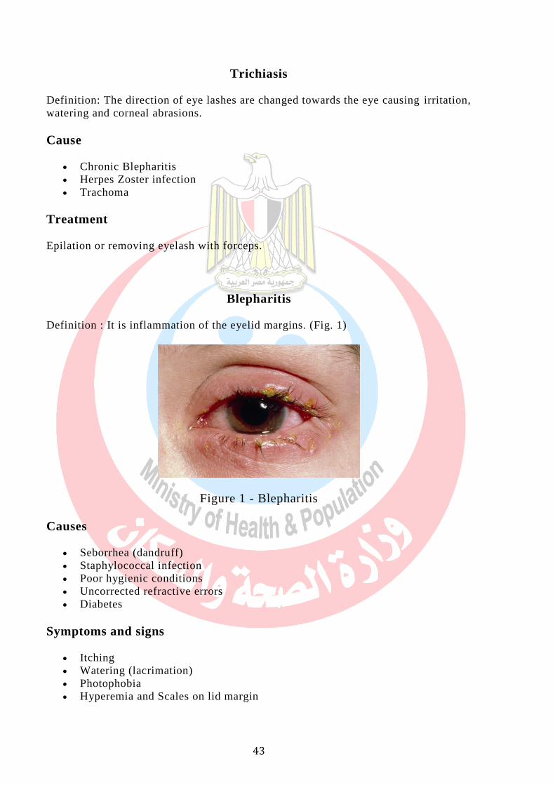
Trichiasis
Definition: The direction of eye lashes are changed towards the eye causing irritation,
watering and corneal abrasions.
Cause
Chronic Blepharitis
Herpes Zoster infection
Trachoma
Treatment
Epilation or removing eyelash with forceps.
Blepharitis
Definition : It is inflammation of the eyelid margins. (Fig. 1)
Figure 1 - Blepharitis
Causes
Seborrhea (dandruff)
Staphylococcal infection
Poor hygienic conditions
Uncorrected refractive errors
Diabetes
Symptoms and signs
Itching
Watering (lacrimation)
Photophobia
Hyperemia and Scales on lid margin

44
Treatment
Improvement of general health
Treatment of dandruff
Lid hygiene
Antibiotic eye ointment
Correction of refractive error
Screening for diabetes in adults and treating if present .
Stye (Hordeolum Externum)
Definition
Inflammation of follicle of the eyelash including the gland of Zeis.
Causative organism
Staphylococcus (a bacterial infection)
Risk factors
Children and young adults
Diabetes
Very sick patients
Uncorrected refractive errors
Symptoms and signs
Pain and swelling of the lid margin
Edema of lids
Tenderness
Treatment
Hot fomentation
Antibiotic eye ointment
Analgesic
Treatment of refractive errors, blepharitis, diabetes.
Acute Chalazion (Hordeolum Intermum
Definition
Acute inflammation of the meibomian gland.

45
Causative organism
Staphylococcus
Risk factors
Same as hordeolum externum
Symptoms
Same as hordeolum externum but more severe
Signs
Point of maximum tenderness is away from lid margin
Pus points on the tarsal conjunctiva
Treatment
Same as hordeolum externum
Chronic Chalazion
Definition
Chronic inflammation of the meibomian gland (Figure 2)
Figure 2 - Chalazion

46
Causes
Blepharitis
Chronic conjunctivitis
Diabetes in adults
Errors of refraction
Symptoms
Painless nodular swelling in the eye lid
Signs
Well defined swelling
Firm and not tender
Eversion of the lid shows purplish discoloration of conjunctiva
Treatment
1. Hot fomentation
2. Antibiotic eye ointment
3. Incision and curettage
Ectropion
Definition
Rolling in out of the margin of thelower eye lid (Figure 3).
Figure 3– Ectropion

47
Types
Senile - Old age
Paralytic - Paralysis of orbicularis seen in facial palsy
Congenital - Present since birth
Mechanical - Due to weight of swelling in the lower lid
Cicatrical - Chemical burns
Symptoms
- Epiphora : Because punctum is not apposed to the globe.
- Excoriation of skin around the lid
Treatment
Temporary
Antibiotic ointment
Adhesive tape to close the lid at night
Permanent
Surgical corrections
Entropion
Definition
Rolling in of the lid margin with its lashes (Figure 4)
Figure 4 – Entropion

48
Types
Senile - old age
Spastic - excessive contraction of orbicularis oculi
Cicatrical - due to scarring (trachoma, chemical burns)
Congenital - present since birth
Mechanical - due to weight of the swellings in the lid
Symptoms
Foreign body sensation
Pain
Lacrimation
Complications
Corneal ulceration
Corneal opacities
Treatment
Temporary procedures
- Adhesive tape : pulling the skin out with strip of plaster
- Cautery : over skin below lashes
- Soft contact lenses
Permanent procedures: Surgical
Lagophthalmos
Definition
Inadequate closure of eyelids when an attempt is made to close them.
Causes
Facial nerve palsy
Proptosis
Patient in coma
Deformity of upper lid
Complications
Dryness of conjunctiva and cornea
Exposure keratitis

49
Treatment
Temporary
Artificial tear drops at day time
Antibiotic ointment at night time
Closure of lids with adhesive tape
Soft contact lenses to prevent corneal damage
Permanent
Surgery: Tarsorrhaphy
Ptosis
Definition
Drooping of the upper eye lid (Fig. 5)
Figure 5 – Ptosis of the Right upper eyelid.
Causes
Old age
Abnormalities in the development of muscles
Malfunctioning of the nerves
Injury or trauma in the eye
Inflammation, diabetic, stroke, tumors, cancers, zaneurysms
Mechanical
Congenital
Present from birth

50
Acquired
Due to nerve damage - 3rd nerve palsy
Muscle disease - Myasthenia gravis
Mechanical - Tumors, chalazion etc.
pulling the lid down
Traumatic - Following injury
Treatment
Neostigmine tablets are given for myasthenia disease
Surgical correction is done in most cases
Tumors of the Eyelids
Benign
Papilloma
Molluscum contagiosum
Naevus
Xanthelesma
Warts
Hemangioma
Neurofibroma
Malignant
Rhodent ulcer (Basal cell carcinoma)
Squamous cell carcinoma
Meibomian gland carcinoma

51
Diseases of the Lacrimal System
Lacrimal apparatus diseases are divided into 2 groups
Diseases of Lacrimal Gland
Infection: Acute dacryoadenitis
Tumors : Adenoma carcinoma, adenoma
Diseases of Lacrimal sac
Infection - Dacryocystitis
Tumors - Very rare e.g.: papilloma
Dacryocystitis
Definition
Acute or chronic inflammation of lacrimal sac.
Causative organisms
Pneumococcus (most common)
Streptococcus
Staphylococcus
Mycobacterium etc.
Causes
Congenital deformation of the lacrimal drainage system
Diseases of nose : deviated nasal septum
Nasal polyps
Infection spreading from naso-pharynx
Trauma causing distortion of anatomy and stagnation of tears
Sequence of events
Obstruction at the junction of the lower end of lacrimal sac and the upper end of
naso-lacrimal duct Stagnation of tears in the sac Predisposing to infection
↓
Stagnation of tears in the sac
Predisposing to infection

52
Dacryocystitis
Types
Acute dacryocystitis
Chronic dacryocystitis
Congenital dacryocystitis
Symptoms
Acute
Sudden onset of pain, redness and swelling in the sac area
Watering of the eyes
Sometimes redness of the eyes
Congenital - watering of one or both eyes since birth
Chronic
Watering of the eyes
Sometimes discharge
Pus like material coming from lacrimal puncta when pressing over the sac
area.
Diagnosis
Acute : Syringing contraindicated
Chronic : On syringing, fluid will regurgitate through the same punctum or
upper punctum, Mucus or pus may also be seen in the regurgitant fluid.
Congenital: 1-2 drops of 2% fluorescent dye instilled in the eye and after
some time, a cotton swab is introduced in the nostril to see whether the dye
has drained into the nose.
Complications
Chronic conjunctivitis
Can lead to infection within the eye after surgery in the eye
Promote bacterial corneal ulcers
Acute dacryocystitis may progress to orbital cellulitis, lacrimal fistula [skin
opening in sac area].
Treatment
I. Acute Dacryocystitis
Hot compresses: 3-4 times / day
Broad spectrum systemic antibiotics
Topical antibiotics: 4 - 6 times / day

53
DCT (dacryocystectomy) or dacryocysto -rhinostomy (DCR) done after
inflammation subsides.
II. Chronic dacryocystitis
In younger age DCR is done
In old age DCT is done
III. Congenital dacryocystitis
Massaging and antibiotic drop instillation
Probing : probe is passed down naso-lacrimal duct
DCR if the above procedures fail to open lacrimal passages after 3 years
Dry Eyes
It is one of the most common problems treated by ophthalmologist.
Definition
There is decreased secretion of tears or deficiency in the composition of tears.
Causes
Aging
Hot, dry or windy climates
High altitude
Air conditioning
Cigarette smoking
Working on computer for long time
Contact lens wearers
Symptoms
Itching
Burning
Irritation
Redness
Blurred vision that improves with blinking
Excessive tearing
Increased discomfort after reading, watching TV, or working on a computer
F.B sensation
Diagnosis
Schirmer test is done

54
Treatment
No permanent cure, can only relieve symptoms
I. Preservation of existing tears
Reduction of room temperature
Humidifiers
Punctual occlusion
II. Supplementation of tears
1. Drops - Methyl Cellulose
- Hydroxy ethyl cellulose
- Hyper mellose
2. Ointments - HPMC gel at bed time

55
Diseases of the Conjunctiva
The most common conjunctival disease is conjunctivitis which is infective. Others
being pterygium, pinguecula and subconjunctival haemorrhage.
Conjunctivitis
Definition
Infection of the conjunctiva
Classification
Infections
Bacterial
Staphylococus aureus
Haemophillus
Gonococcal (Ophthalmic Neonatorum)
Mycobacterium
Viral
Adenovirus
Varicella
Herpes Zoster
Mumps
Influenza
Chalamydial (Trachoma)
- Causative organism is Chlamydia trachomatis
Table : 1
Conjunctival congestion Ciliary congestion
i. Site Fornices Limbus
ii. Color Bright red Violet
iii. Depth Vessels superficial Vessels deep
iv. Branching Dichotomously Radially
v. 1.1000 epinephrine test Whitens conjunctiva No effect
vi. Disease Conjunctivits Iridocystitis, Glaucoma

56
Allergic
Simple allergic conjunctivitis
Vernal catarrh
Trauma
Infective conjunctivitis
Symptoms
Discomfort and foreign body sensations due to engorgement of blood vessels
Sticking together of eye lashes due to discharge
Photophobia and watering of the eye
Defective vision due to thin layer of discharge on the cornea
Haloes around light due to thin layer of discharge on the corneal surface.
Signs
Discharge : causes sticking together of eyelashes especially when waking up
in the morning
Congestion : should be differentiated from other causes of congestion
(redness) (Table:1).
Subconjunctival hemorrhage: more common in viral conjunctivitis. Rupture
of tiny conjunctival vessel.
Follicles: Round swellings (0.5 - 2 mm) surrounded by tiny blood vessel in the
tarsal conjunctiva, more commonly seen in viral conjunctivitis and allergic
conjunctivitis.
Papillae : Round swelling with blood vessels in the centre over the tarsal
conjunctiva more commonly seen in vernal conjunctivitis.
Pre Auricular Lymphadenopathy: Seen in viral, chlamydial infection
Treatment
Frequent wash with luke warm saline solution to clear crusting and discharge
Use dark glasses to prevent photophobia
Broad spectrum antibiotic drops are used
o E.g.,: ciprofloxacin dosage hourly to 4 times depending on severity.
o Ointment: Tetracycline / gentamycin/ Chloramphenicol at bed time
Prophylaxis (Prevention)
Patient must keep his hands clean and avoid touching around the eye.
Personal belongings of the patient like towel, handkerchief, and pillow should
be kept separately.
Other family members if infected should be treated simultaneously.

57
Simple allergic conjunctivitis
Definition
Allergic reaction due to large amount of allergens reaching the conjunctiva
Causes
Pollen grains
Certain topical drugs e.g.. Neomycin
Contact with pet animals
Dust, cosmetics, chemicals
Treatment
Removal of allergen
Antihistamine tablets & drops
Topical 2% Sodium chromoglycolate to prevent recurrence
Corticosteroid drops in severe cases
Vernal conjunctivitis
Definition
Hypersensitivity reaction of conjunctiva to exogenous allergens
Causes
Age - 6-20 yrs, usually males
Seasonal variation - Prevalent in summer
Exciting factors - Dust, dry heat, pollens
Symptoms
Intense itching
Discharge
Photophobia, burning and foreign body sensation
Signs
Cobble stone appearance due to papillary hypertrophy in the palpaberal
conjunctiva
Multiple small nodules around the limbus
Trantas spots superficial white spots scattered around the limbus.

58
Treatment
Cold compresses
Disodium chromoglycolate 4 times / day reduces itching
Topical steroids like dexamethasone 4 times daily and tapering dose depending
on the severity. Long term use of steroids can cause cataract and glaucoma.
Ophthalmia neonatorum
Definition
Bilateral purulent conjunctivitis occurring in new born within first 3 weeks of life.
Causative organisms
Gonococcus (Most common)
Chlamydia
Mode of Infection
Before birth - very rare
During birth - face presentation is the most common
After birth - from soiled linen
Clinical picture
Watering, redness & discharge.
Treatment
1) Prophylaxis
Proper antenatal care of mother. Any vaginal discharge should be treated
meticulously.
Crede's prophylaxis: 1% silver nitrate is instilled into the baby's eyes
immediately after birth.
2) Curative
Swab taken for culture and sensitivity
Gonococcal - Ciprofloxacin hourly for 3-5 days
Chlamydial - 1% tetracycline 2 times/day

59
Pterygium
Definition
It is a triangular growth on the conjunctiva encroaching on the cornea in the
horizontal meridian, in the palpaberal fissure, either from the nasal or temporal side
of bulbar conjunctiva or from both sides.
Cause
Prolonged exposure to the sun (UV radiation)
Exposure to hot, sandy and dusty weather
Symptoms
Appearance of lesion on nasal or temporal side
Dimness of vision due to obstruction of visual axis
Redness and burning sensation.
Signs
1. Decreased visual acuity
2. Triangular fold of conjunctival fleshy growth encroaching upon the cornea
Treatment
Stationary pterygium - no treatment (If inflamed - NSAIDS or steroids eye
drops)
Progressive pterygium - excision
Recurrent pterygium - mitomycin C, Beta radiation
Pterygium in the pupillary area - Excision of pterygium and keratoplasty
Subconjunctival Haemorrhage
Definition
Spontaneous rupture of conjunctival blood vessels
Clinical picture
Sectoral red patch under the conjunctiva where blood collects.
Causes
Trauma

60
Diabetic and hypertensives
Sneezing
Coughing
Straining
Lifting heavy weight
Vomiting
Treatment:
None
Reassurance to the patient

61
Glaucoma
It is a condition characterized by an abnormal increase in the intraocular pressure
(IOP) with subsequent changes in the optic nerve head and the visual field . The
pressure is normally between 11mm to 20mm of Hg. If the pressure increases it
affects the optic nerve and gradually causes loss of vision.
Types of glaucoma
Open angle glaucoma (POAG)or chronic simple glaucoma:
o The most common type.
o The angle between the iris and the cornea is normal but the trabecular
meshwork get blocked from inside.
o In this type the patient is asymptomatic until late (called silent thief of
sight).
o First, the peripheral field of vision is lost.
o In course of time central vision is also affected..
Angle closure glaucoma. o This disease causes headache, severe pain, watering and redness of the
eye and loss of sight suddenly.
o The patients will see rainbow like circles, halos, around lamps.
o The patient is usually seen in the emergency department.
Other types of glaucoma:
o Congenital type:
The infant at birth will have big eyes. This is called
Buphthalmos.
The cornea will be large. Photophobia, redness and watering of
the eye will be seen.
The child usually needs to be examined under anesthesia to
measure the IOP.
o Other causes may be secondary to trauma, inflammation and Cataract.
Diagnosis
Visual Acuity
Slit lamp examination
Tonometry
Fundus examination
Gonioscopy
Testing visual fields by computerized field analysis (Perimetry).
Risk factors for POAG
Generally above 40
Family history
Diabetic patients
African heritage

62
Myopia
Treatment
Medical treatment: o Some patients suffering from glaucoma may be treated with eye drops
and tablets.
o Depending on the severity of the intraocular pressure, one or two types
of eye drops are used. Some may require tablets also.
o It is important that the treatment is continued according to the
physician's directions.
Laser treatment:
o They are called laser trabeculoplasty and Laser Iridotomy.
o Patients do need not stay long in the hospital.
Surgery: If the medical and laser treatments are not effective, surgery is the
only alternative.
Glaucoma patients should be advised to
Follow instructions of the ophthalmologist.
Eye drops must be used without fail
Eye drops must be applied to the eye
Must not stop treatment without doctor's advice
People in the family and relatives must get themselves examined
Must inform about it to other doctors when consulted
It is not possible to cure glaucoma completely. But it is possible to control it
and keep the vision by continuous treatment

63
Diseases of the Cornea
A) Corneal ulcers
Bacterial ulcer
Fungal ulcer
Viral ulcer
Parasitic ulcer
B) Degenerative Conditions
Arcus Senelis
Band Shaped Keratoplasty
C) Dystrophies
Granular
Macular
Lattice
D) Corneal opacities
Nebula
Leucoma (adherent/ non-adherent)
Anterior Staphyloma.
A) Corneal ulcers
Definition
It is defined as a break in the corneal epithelium with added infection
Source of infection
Exogenous route - From outside source
Secondary route - From adjacent structures like conjunctiva, sclera, uvea
Endogenous route - Systemic sources

64
Causative organisms
Bacteria Fungal Parasitic Virus
1.Staphylococcus aureus Candida Albicans Acanthamoeba Herpes simplex
2.Pseudomonas Aspergillus Herpes zoster
3. Streptococcus Fusarium
4. E.Coli
5. Klebsciella
Symptoms
Lacrimation (watering)
Photophobia
Pain
Defective vision
Signs
Edema of lids and blepharospasm
Circum ciliary congestion
+ve floourescein staining (Figure)
Localised area of necrosis (Figure)
Vascularisation of cornea
Hypopyon may be present (Figure)
Figure – Corneal ulcer stained with fluorescein stain. A- with normal illumination, B-
with cobalt blue filter.

65
Figure – infective corneal ulcer with hypopyon.
Investigations: Corneal Scraping
Done under local anesthesia by the ophthalnmologist. The material obtained is used
for the following investigations.
Gram and Giemsa stains for bacteria
10% KOH wet preparation for fungus
Culture on blood agar for aerobic organisms
Culture on Sabouraud's dextrose agar for fungus
Features Bacterial ulcer Fungal ulcer
i. History of Injury Non specific With Vegetable matter
ii. Predisposing factors Non specific Immunocompromised, steroids
iii. Course Rapid Slow
iv. Symptoms and signs Proportionate Sign out of proportionto symptoms
v. Satellite lesions Absent Present
vi. Margins Well defined Feathery
vii. Hypopyon Usually not seen Usually seen
Complications
Perforation - (Hole in cornea)
Corneal opacities
Endophthalmitis or panophthalmitis
Secondary glaucoma

66
Treatment
a. Control of infections
Antibiotics are used until the ulcer heals - e.g., fortified gentamycin,
Cefazolin, Ciprofloxacin
Antifungal - e.g.,: Natamycin, Nystatin etc.
b. Rest to the eye
Cycloplegics: atropine 1% eye ointment 2 times / day gives rest to eye by
paralysing the ciliary muscle
Dark glasses: protect the eye from irritating effect of strong light
c. Relief of pain
Hot compresses
Oral pain killers
d. Removal of any septic focus in the neighbourhood
If sac is infected, it is to be removed without delay
e. Improve general health condition
By control of diabetes and improving nutrition.
Don'ts in a case of corneal ulcer
No Bandage
No Steroid drop
No Schiotz Tonometer.
Viral ulcer (Dendritic ulcer)
Definition
Ulcer with a branched appearance caused by herpes simplex virus.
Symptoms
Pain, watering and photophobia
Clinical signs
The ulcer appears as star shaped or branched pattern
Absent corneal sensation
Enlarged and tender pre auricular lymph nodes

67
Treatment
Antiviral drops or ointment (e.g.,: acyclovir eye ointment 3%)
Steroids to suppress the host response
Antibiotic eye drops to prevent secondary infection
B) Degenerative conditions
Keratoconus
Definition
Bilateral conical protrusion of central part of the cornea due to thinning
Symptoms
Defective vision due to irregular myopic astigmatism and watering due to rupture of
hydrops
Signs
1. Munson 's sign: bulging of the lower lid when patient looks down.
2. Retinoscopy: scissoring reflex
3. Ophthalmoscopy: oil drop sign
4. Slit lamp:
o Thinning of cornea at the centre
o Prominent corneal nerves
o Fleischer ' s ring - iron deposits at the base of cone
5. Keratometry ('K' reading) showing high astigmatism
Complications
Acute hydrops: rupture of Descemet 's membrane and seepage of aqueous in the
corneal stroma and epithelium
Treatment
Hard contact lens
Penetrating keratoplasty.
Arcus senilis
Corneal degeneration normally seen at peripheral cornea.
Lesion: starts as crescent grey line around 12o clock and 6 o clock and finally
it becomes circular.

68
Site: periphery of cornea parallel to limbus. There is clear space seen between
the lesion and the limbus
Does not affect vision or vitality of cornea.
C) Corneal dystrophies
Definition
Condition in which cornea loses its clarity due to deposition of materials in various
layers of the cornea
Features
1. Usually inherited
2. Not caused by outside factors like injury or diet
3. Progresses gradually
4. Occurs in otherwise healthy people
Symptoms
Some detected on routine examination
Some cause visual impairment
Some cause repeated episodes of pain without visual loss
Types
1. Epithelial dystrophy - Map-dot finger print dystrophy
2. Stromal dystrophy - granular dystrophy, macular dystrophy and lattice
dystrophy
3. Endothelial dystrophy - Fuch's dystrophy
D) Corneal opacities Nebula:
o Peripheral: correct irregular astigmatism by
Glasses
Hard contact lens
o Central:
Lamellar keratoplasty
Leucoma non-adherent:
o Non-seeing eye:
Tinted contact lens
Tattoing
o Seeing eye:
Peripheral: use glasses or hard CL to correct irregular astigmatism

69
Central opacity:
Postmydriatic test: Measure vision after dilating drops:
o Marked improvement: do a visual iridectomy
o No or little improvement: do penetrating keratoplasty
(PKP)
Leucoma adherent:
o Non-seeing eye:
Blind:
Tinted contact lens
Tattoing
Blind and painful:
Enucleation
o Seeing eye: Measure IOP:
High IOP:
Do glaucoma surgery initially then manage as leucoma non-
adherent
Simultaneous glaucoma surgery and penetrating keratoplasty
can be combined in one surgery
Normal IOP:
Manage as leucoma non-adherent
Anterior staphyloma:
o Partial:
Combined glaucoma surgery and PKP
o Total:
Enucleation for a blind, painful, cosmetically disfiguring eye
70
Diseases of the Lens
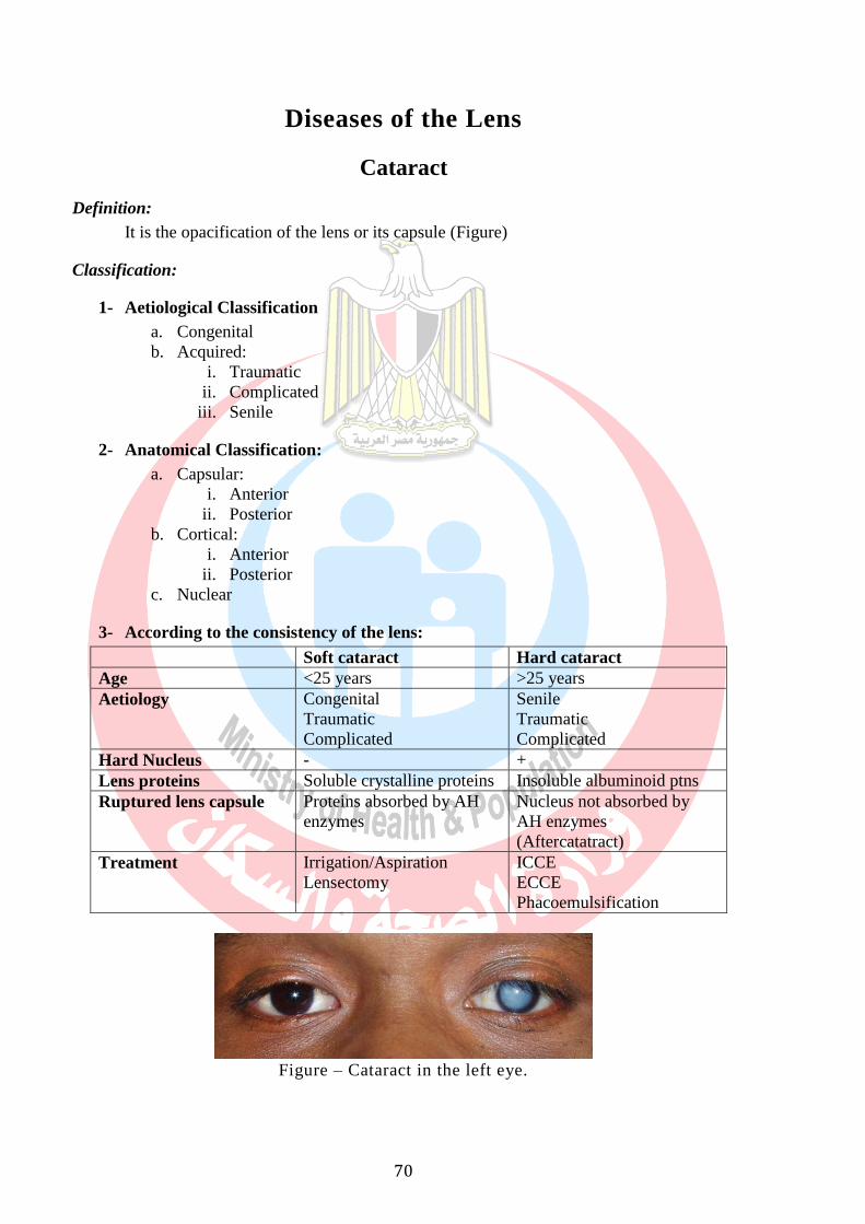
Cataract
Definition:
It is the opacification of the lens or its capsule (Figure)
Classification:
1- Aetiological Classification
a. Congenital
b. Acquired:
i. Traumatic
ii. Complicated
iii. Senile
2- Anatomical Classification:
a. Capsular:
i. Anterior
ii. Posterior
b. Cortical:
i. Anterior
ii. Posterior
c. Nuclear
3- According to the consistency of the lens:
Soft cataract Hard cataract
Age <25 years >25 years
Aetiology Congenital
Traumatic
Complicated
Senile
Traumatic
Complicated
Hard Nucleus - +
Lens proteins Soluble crystalline proteins Insoluble albuminoid ptns
Ruptured lens capsule Proteins absorbed by AH
enzymes
Nucleus not absorbed by
AH enzymes
(Aftercatatract)
Treatment Irrigation/Aspiration
Lensectomy
ICCE
ECCE
Phacoemulsification
Figure – Cataract in the left eye.

71
Subluxation / dislocation of the lens
Definition
Subluxation: partial displacement of the lens from its normal position but
remains behind the pupil
Dislocation: total displacement of the lens either
o Forward - into the anterior chamber (can block the pupil and cause
glaucoma)
o Backward - into the vitreous
Ectopia lentis - congenital bilateral, subluxation or dislocation of the lens
Causes
Congenital (ectopia lentis)
Acquired
Excessive stretching of zonules - trauma
Degeneration of zonules - pseudo exfoliation
Clinical features
Mono ocular diplopia
Unequal depth of AC
Iridodonesis
Phacodonesis
Figure – Subluxated lens

72
Lens induced glaucoma
Phacolytic glaucoma
Hypermature cataract
Leakage of lens material
Macrophages engulf lens material
Obstruct trabecular meshwork
Secondary open angle glaucoma
Phacomorphic glaucoma
Intumescent cataract (swollen white lens)
Shallow anterior chamber
Secondary angle closure glaucoma
Fixed dilated pupil
Note: (Pupil should never be dilated)

73
Diseases of the Retina and Vitreous
Diseases of vitreous Some common problems of the vitreous are
1. Floaters
2. Vitreous hemorrhage
1. Floaters
They are various kinds of opacities moving in front of the eye. They are due to presence of
opacities in the vitreous which cast a shadow on the retina. They are a common complaint and
are usually harmless.
Causes
- Blood in vitreous
Diabetic retinopathy
Vein occlusion
Trauma
- Degeneration of the vitreous
High myopia
Aging
- Inflammatory exudates
Retinitis
Uveitis
Optic neuritis
Synchysis scintillans- cholesterol crystals in the vitreous
Management
Treatment is that of the cause in most cases.
Vitreous Hemorrhage Definition: bleeding into the vitreous
Source of hemorrhage: blood vessels in the retina
Causes
Trauma to the eye
Diseases of the blood vessels
Diabetic retinopathy
Retinal vein occlusions
Inflammation of the retinal veins
Diseases of retina
Retinal tears
Retinal detachment
Management
Blood usually gets absorbed over a few months, hence observation initially
Vitrectomy: surgical method of removing vitreous if blood does not get absorbed

74
Diseases of the retina Some of the common and important diseases are;
1. Diabetic retinopathy
2. Hypertensive retinopathy
3. Retinal vein occlusions
4. Retinitis pigmentosa
5. Retinal detachment
6. Central serous retinopathy (CSR)
7. Retinoblastoma
Diabetic retinopathy It is one of the most important ocular manifestations of diabetes. It is now a major cause of
blindness in developed countries and is rapidly becoming an important cause of preventable
blindness in developing countries.
Definition
It is a change seen in the retina of patients suffering from diabetes mellitus. Control of blood
sugar level decreases the risk of developing severe diabetic retinopathy.
Classification
1. Non proliferative diabetic retinopathy Ophthalmoscopic features
Micro aneurysms
Hemorrhages
Hard exudates
Retinal oedema
2. Proliferative diabetic retinopathy
Ophthalmoscopic features
New vessels at the disc (NVD)
Fibrovascular bands
Vitreous detachment
Vitreous hemorrhage
Investigation
Urine and blood sugar examination
FFA (fundus flourescein angiography)
Management
Medical treatment: good diabetic control,
Laser treatment: photocoagulation- to stop leaking from retinal vessels and bleeding
from new vessels
Surgical treatment: vitrectomy is done in case of vitreous hemorrhage, traction
retinal detachment
2. Hypertensive retinopathy
vascular changes in the retina associated with systemic hypertension
Clinical features
Grade I - mild generalised narrowing of arterioles, particularly of small branches
Grade II - marked generalized narrowing associated with focal narrowing of arterioles
Grade III - grade II changes and also hemorrhages, cotton-wool spots and hard exudates
Grade IV - all changes of grade III plus papilloedema
Management

75
No special management is required for the retinopathy as most of the changes are reversible
with adequate control of blood pressure.
Retinal vein occlusion More common than the artery occlusions, it affects elderly patients in sixth or seventh decades
of life.
Risk factors
Hypertension
Diabetes
Arteriosclerosis
Clinical features
Sudden loss of vision
Oedema and hemorrhages all over the fundus including the macula
Complications
Neovascular glaucoma: occurs in 50% of cases within three months (so called 90 days
glaucoma)
Vitreous hemorrhage
Proliferative retinopathy
Treatment
Proper control of diseases like hypertension, diabetes etc.
Laser photocoagulation: for macular oedema and new vessels
Retinitis Pigmentosa Definition: It is a hereditary condition of the retina affecting the rods
Clinical features
Night blindness (nyctalopia)
Tubular vision: advanced cases
Fundus changes
Waxy pallor of disc
Narrowed vessels
Bony spicule pigmentation
Treatment
No permanent cure at present
Supportive treatment
Vitamin A
Low vision aids - night vision devices, field expanders etc.
Visual rehabilitation
Genetic counselling- no consanguineous marriage (marriage among relatives)and
affected individuals educated about possibility of affected kids.
Retinal Detachment Definition: Separation of the retina from the retinal pigment epithelial layer
Risk factors
Myopia
Retinal degeneration
Trauma
Symptoms
Floaters

76
Flashes of light (due to excitement of retina by vitreous movements)
Early stages - field defects
Sudden painless loss of vision - (in large and central detachments)
Signs
Direct ophthalmoscopy: shows grey reflex instead of normal pink reflex
Indirect ophthalmoscopy: detached retina is grey, wavy and moves with eye
movements -retinal break is seen
Treatment
Surgery: scleral buckling procedure
Central Serous Retinopathy (CSR) Definition: It is due to detachment of retina in the macular region due to accumulation of fluid
resulting in defective vision
Causes: not known
Symptoms
Sudden onset of painless loss of vision
Central scotoma (dark area)
Micropsia (objects appears small)
Metamorphopsia (irregularity of the objects)
Signs
Ophthalmoscopy
Mild elevation of macular area
Foveal reflex is absent
Treatment
Reassurance to the patient (usually resolves spontaneously without any treatment)
Long standing cases (more than 4 months) : laser photocoagulation
Retinoblastoma
Definition: it is a malignant tumour of the retina occurring in children under 5 years. It is the
most common intraocular malignant tumour of childhood.
Symptoms
White reflex over the pupil (leukocoria)
Squint
Signs
Indirect ophthalmoscopy: elevated fungating mass with satellite lesions seen in the retina.
Treatment
Radiation therapy, chemotherapy
Photocoagulation
Cryotherapy
Enucleation / excentration

77
Diseases of Orbit The eye along with its surrounding structures like connective tissue, nerves, blood vessels, fat,
muscles and glands have the potential to give rise to virtually any known neoplasm anywhere
in the body. Certain types of tumors are more common and specific to this organ. A brief
discussion of the more important tumors follows.
Common orbital diseases of children
Orbital cellulitis
Rhabdomyosarcoma
Capillary hemangioma
Dermoid and epidermoid cysts
Leukemia
Lymphangioma
Psueudotumor
Optic nerve glioma
Metastatic neuroblastoma
Neuro fibroma
Common orbital diseases of adults
Thyroid related ophthalmopathy
Pseudo tumour
Cavernous haemangioma
Lacrimal gland tumour
Lymphoma
Lymphangioma
Meningioma
Proptosis
Definition: forward protrusion of the eyeball beyond the orbital margin.
Causes
Inflammatory
Acute - orbital cellulitis
Chronic - pseudotumor
Neoplastic
Benign - dermoid
Malignant - rhabdomyosarcoma
Parasitic
Cysticercosis
Hydatid cyst
Vascular- retrobulbar haemorrhage
Orbital varices
Aneurysm
Systemic diseases
Thyroid ophthalmopathy
Leukaemic deposits
Measurement of proptosis: Exophthalmometry
Investigations
Thyroid function test
X-ray orbit

78
USG
CT and MRI
Biopsy
Treatment
Medical - for orbital cellulitis, thyroid disease.
Radiotherapy- for secondary deposits and malignant tumours
Surgical a. tarsorrhaphy: to protect the exposed cornea, b. orbitotomy to remove the
tumour
Thyroid ophthalmopathy It is the most common cause of both unilateral and bilateral proptosis in adults.
Ocular manifestations
Retraction of eyelids
Lid lag
Lagophthalmos - incomplete closure of eye lids
Infrequent blinking
Increased lid pigmentation
Extra ocular muscle palsies
Investigations
Thyroid function test: T3 and T4 levels
USG: shows enlargement of extra ocular muscles
CT scan: enlargement of muscles
Optic nerve compression can be demonstrated.
Orbital cellulitis Definition: infection of the fat and cellular tissues of the orbit.
Causes
Sinus infection
Penetrating orbital injury
Thrombophlebitis
Post operative: following enucleation of the globe
Dental or naso-pharyngeal infection. Causative organism: streptococcus,
staphylococcus, haemophilus
Types
Preseptal
Orbital
Symptoms
Pain and swelling of the lids
Lacrimation and photophobia
Diplopia due to limitation of movements
Impairment of vision at later stages
Frequently associated with fever
Signs
Lid oedema and tenderness
Conjunctival congestion
Proptosis
Limitation of ocular movements
Ophthalmoscopy: features of optic neuritis in severe cases

79
Complications: cavernous sinus thrombosis
Treatment
Admit the patient
Broad spectrum antibiotics systemically
Neuro ophthalmology Some of the common and important diseases in neuro ophthalmology are
1. Optic neuritis
2. Papilloedema
3. Optic atrophy
4. Cranial nerve palsies
Optic neuritis Definition: Inflammation of the optic nerve
Causes
Idiopathic
Childhood infection (e.g.,: measles, mumps)
Viral infection (e.g.,: encephalitis, herpes zoster)
Systemic diseases (e.g.,: syphilis, tuberculosis)
Local inflammations (e.g,: sinusitis, meningitis, orbit)
Intraocular inflammation
Symptoms
Uniocular sudden loss of vision
Pain behind the eye ball particularly while moving
Signs
Visual acuity may be 6/60 or less
Pupil: relative afferent pupillary defect
Colour vision: defective
Field defect: central, centrocaecal scotoma
Fundus
Hyperemia of the disc
Oedema of the disc
Small hemorrhages on the disc
Treatment: systemic and oral steroids
Papilloedema Definition: Non-inflammatory swelling of the optic disc produced by raised intracranial
pressure.
Causes
Congenital: e.g.,: aqueductal stenosis
Space occupying intracranial lesions
Head injury
Infection: meningitis and encephalitis
Malignant hypertension
Symptoms
a. General symptoms:
1. Headache
2. Vomiting
b. Ocular symptoms
1. Visual acuity normal

80
2. Transient blackout of vision for few seconds
3. Fields: enlargement of blind spot
Signs
1. Blurring of disc margin
2. Disc hyperemia
3. Dilated vessels
4. Hemorrhages on disc
Treatment: Given according to the cause of the disease.
Optic atrophy Definition: death of the optic nerve fibres characterized by pallor of the disc.
Causes
Glaucoma
Trauma
Retinitis pigmentosa
Central retinal artery occlusion
Papilloedema
Optic neuritis
Symptoms
Sudden or gradual loss of vision
Defective color vision
Signs
Visual acuity impaired
Relative afferent pupillary defect (RAPD)
Fundus
Pallor of disc
Narrowing of blood vessels
Treatment: The specific cause should be treated before the onset of optic atrophy. The
damaged part of optic nerve cannot be recovered.
Cranial Nerve Lesions Third Nerve Palsy
Ptosis (falling down of upper eyelid) - levator muscle paralysis
Restriction of adduction- medial rectus paralysis
Restriction of elevation- superior rectus paralysis
Restriction of depression - inferior rectus paralysis
Fourth Nerve Palsy
Double vision on looking down
Paralysis of superior oblique muscle
Sixth Nerve Palsy
Double vision on looking to the side of lesion
Paralysis of lateral rectus muscle

81
Diseases of the uvea
Uveitis
Definition: inflammation of the uvea (iris, ciliary body and choroid)
Inflammation of iris - Iritis
Inflammation of ciliary body - Cyclitis
Inflammation of choroid - Choroiditis
Classification
Anterior uveitis (irido-cyclitis) : inflammation of the iris and the ciliary body.
Intermediate uveitits - inflammation of ciliary body.
Posterior uveitis: inflammation of the choroid with or without cyclitis.
Pan uveitis : iritis + cyclitis + choroiditis
Anterior uveitis
Causes
Bacteria - tuberculosis, leprosy
Virus - herpes simplex, herpes zoster
Fungus - histoplasmosis
Parasite - toxoplasmosis, toxocariasis
Systemic diseases - joint diseases
Trauma
Symptoms
Pain
Redness
Defective vision
Photophobia
Watering
Signs
Vision diminished
Conjunctiva - circumciliary congestion
Cornea - oedema keratitic
o Keratitic precipitates (KP 's): cells that stick to the back surface of cornea
(endothelium)
Anterior chamber
o Cells : inflammatory cells
o Flare : due to increased protein content of the aqueous
o Hypopyon: large accumulation of inflammatory cells in the lower part of
anterior chamber.
Pupil - small and irregular (sluggish pupillary reaction)
Synechiae
o Sticking of iris to the lens capsule (posterior synechiae)
Treatment
Mydriatics: e.g., Atropine eye drop or ointment (1%)
Steroids
Topical : dexamethosone - 1 drop hourly to 4 times per day - eye ointment at bed time
Subconjunctival - 0.5 ml per day dexamethasone
Oral - 1mg prednisolone per kg per day and tapered as condition improves, Painkillers
- paracetamol or ibuprofen

82
Complications
Band keratopathy
Complicated cataract
Retinal detachment
Optic neuritis
Posterior Uveitis (Choroiditis)
Symptoms
Defective vision
Floaters - seeing black dots
If central area is involved
o Metamorphopsia (distortion)
o Micropsia (objects appear smaller)
o Macropsia (objects appear larger)
Photopsia (flashes of light )
Signs
Requires indirect ophthalmoscopy to detect the various abnormalities in the retina and
choroid.
Treatment
It depends upon the cause. It is better to refer the case for investigation and treatment in a
specialized eye center

83
Ocular Trauma
Normal Protective ocular mechanisms:
Eyelids and eyelashes
Tear fluid (washes debris and bactericidal)
Corneal sensation
Bony orbit
Retrobulbar fat: cushion-like effect
Bell’s phenomenon
Miosis: pupillary consrtriction secondary to light exposure
Neck withdrawal reflex
Types of ocular injuries:
1- Blunt trauma
2- Sharp trauma:
Penetrating
Perforating
3- Physical injuries
4- Chemical injuries
Book Coordinator ; Mostafa Fathallah
General Directorate of Technical Education for Health
والسكان ويحذر بيعهلوزارة الصحة حقوق النشر والتأليف