Ophthalmology Grand Rounds - SUNY Downstate Medical Center · 2019-02-28 · 60 yo F c/o...
40
Ophthalmology Grand Rounds Matthew Gorski, MD SUNY Downstate Medical Center December 15, 2011
Transcript of Ophthalmology Grand Rounds - SUNY Downstate Medical Center · 2019-02-28 · 60 yo F c/o...

Ophthalmology Grand Rounds
Matthew Gorski, MD
SUNY Downstate Medical Center
December 15, 2011

History
• 60 year old Caucasian woman presents with blurry
vision x 20 years OU. No acute change. Happy with
near vision. States that she has worn glasses since
she was a young child.
• Denies pain, photophobia, HA, diplopia
• Denies metamorphopsia, photopsia, blockages in
vision
Patient Care

History
•PMHx: CVA with residual left hemiparesis 2004, DM,
HTN, HL
•POHx: “Glasses since I was a child”
•Gtts: none
•FHx: denies glaucoma/blindness
•Social: denies EtOH, smoking, drugs
•All: NKDA
Patient Care

Examination
• dVAcc: 20/30 PH NI, 20/40-1 PH NI OS
• EOM: Full OU, no diplopia or pain in any gaze
• P: 53 OU, no APD OU
• cVF: Full OU
• Tapp: 15 OD, 16 OS @ 10:00 AM
•Amsler Grid: WNL OU
Patient Care

Examination
SLE
LLA: WNL OU
C/S: white and quiet OU
K: Tr SPK OU
AC: Deep and Quiet OU
P/I: round/reactive OU, no NVI OU
L: 1+ NS OU, Tr PSC OS
Patient Care

Dilated Examination

Fluorescein Angiogram
(Kim, YM et al. Eye, 2011)

Dilated Examination
• Vitreous: clear OU; no vitritis OU
• C/D:
OD: 0.3, sharp, tilted, peripapillary atrophy
OS: 0.3, sharp, tilted, myopic crescent
•Macular: flat OD; flat OS, Hyperpigmented spot foveal
center OD
•Vessels:
• OU: WNL • Periphery: Tigroid fundus OU
Patient Care

Differential Diagnosis
•Degenerative Myopia
• Presumed Ocular Histoplasmosis Syndrome (POHS)
• Age-Related Macular Degeneration
• Staphyloma
• Angioid streaks
• Gyrate Atrophy
• Choroideremia
• Polypoidal choroidal vasculopathy
Medical Knowledge
60 yo F c/o progressive, chronic blurry vision with
Tigroid fundus, PPA OU, and macular choroidal
neovascularization OD

Presumed Ocular Histoplasmosis Syndrome
Right: Macular grey-green CNV, PPA
Left: Classic POHS, punched out histo-spots, pigmented PPA
Medical Knowledge

Presumed Ocular Histoplasmosis Syndrome Fungi, Histoplasma capsulatum,
endemic in Ohio and Mississippi
River Valley
asymptomatic, unless + CNVget
metamorphopsia, central scotoma,
60% bilateral, immunocompetent
Signs:
Punched out chorioretinal “histo
spots”
PPA with pigment separating disc
No vitritis
CNVTreatment
Antifungal not indicated
Management of CNV: PDT vs Anti-VEGF vs Laser

Choroideremia
Medical Knowledge

Choroideremia•X-linked recessive, Rod-cone
dystrophy
• M>>F, presents in 1st-2nd decade of
life, slow progression
• Presents with
Nyctalopia, and progressive VF deficit
Fundus: Dispersed pigment granules, peripapillary RPE
atrophytotal RPE and choriocapillaris loss
• Normal color vision, abnormal ERG
• FA: scalloped hypofluorescence adjacent to bright
hyperfluorescence
• No TreatmentMedical Knowledge

Angioid Streaks
Medical Knowledgehttp://dro.hs.columbia.edu/angstreaks.htm

Angioid Streaks Breaks in a thickened or calcified
Bruch’s membrane, reddish-
brown curvilinear, radiations from
ON sub-retinal
50% associated with systemic
disease, most commonly:
Pseudoxanthoma elasticum,
Ehlers-Danlos, Paget’s and SS
Disease, (mnemonic: PEPSI)
Asymptomatic, unless CNV
develop
Signs
- Peau d’orange, ON drusen,
histo-like spots
Atrophic RPE overlying the streak
FA: granular early phase
hyperfluorescence
Treatment
- NONE, unless CMV develop
-Polycarbonate lenses
Medical Knowledge

http://disorders.eyes.arizona.edu/disorders/pseudoxanthoma-elasticum

Gyrate Atrophy Well demarcated,
lobulated areas
of chorioretinal
atrophy
http://disorders.eyes.arizona.edu/disorders/gyr
ate-atrophy-0 MEDICAL KNOWLEDGE

Gyrate Atrophy Autosomal Recessive
Mutation in gene for
ornithine
aminotransferase
(OAT) 10x [plasma]
ornithine, which is toxic
to retina
Presents 1st-2nd decade
with night blindness and
VF deficit
Hyperpigmented fundus
with lobulated RPE
atrophy in midperiphery
Measure ornithine levelshttp://disorders.eyes.arizona.edu/disorders/gyr
ate-atrophy-0 MEDICAL KNOWLEDGE
Treatment
- Vitamin B6, restrict
Arginine

Back to our patient…
Mrx:
OD: : -10.25 x -3.25 x 160 (20/40)
OS: - 11.50 x -1.75 x 155 (20/60)
Patient Care, Systems-Based Practice:

Degenerative Myopia Definition:
High Myopia: spherical equivalence greater than -6.00 D,
axial length > 26-27 mm
Degenerative Myopia: usually > - 8.00 D, axial length
>32.5mm
Epidemiology:
7th leading cause of blindness in USA, 2% of general
population
Of myopic population, 6 to 18% progress to high myopia
Higher incidence in Asians, Mediterranean, less likely in
African Americans
Medical Knowledge

Pathophysiology Progressive elongation of axial diameter leads to
thinning of RPE, choroid, sclera and deviation of the
optic nerve
Biomechanical thinning stromal and vascular
obliteration metabolic disturbance
retinal/RPE degeneration and neovascularization
Genetics: nine Autosomal Dominant loci have been
identified
Associated with Ehler Danlos, Noonans, Downs,
Marfans Syndromes
Medical Knowledge

Clinical Presentation
Symptoms
Asymptomatic
Blurry vision…Myopia
Metamorphopsias
Photopsias
Scotoma
Medical Knowledge

Fundus Manifestations
Tilted optic disc
Peripapillary atrophy
Lacquer Cracks
Subretinal, macular heme
Forster-Fuchs spots
Medical Knowledge
Posterior Staphyloma
Lobular RPE Atrophy
Lattice Degeneration
Cobblestone Degeneration
Choroidal
Neovascularization

Lacquer Cracks
rupture of Elastic
lamina of Bruch’s
membrane
Malagola et al, 2006
Medical Knowledge
Foreshadows
subretinal heme and
CNV

Lacquer Cracks vs Angioid Streak
vs Choroidal Rupture
All three diseased statesof Bruch’s membrane
Angioid streaks emanate radially from disc, are
straighter,and are reddish in color.
Choroidal ruptures, similar distribution, color,
and fluoresceinangiographic appearance to LC,
but are caused by a traumaticevent.

Fuch’s Spot
Hyperpigmented
spot due to
subretinal or
intraretinal RPE
hyperplasia in
response to a small
CNV that does not
regress, or from
resolved micro-
hemorrhage
Medical Knowledge

Posterior Staphyloma

Lattice vs Paving Stone Degeneration
Paving Stone: protective Risk Factor for RD

Diagnosis
Clinical
FA—if CNV suspected
Classification of myopic CNV– 90% ―classic‖
Type I: early hyperfluorescence without late leakage
Type II: early hyperfluorescence with late leakage
ICG (less sensitive for CNV Identification)
+/- OCT
Medical Knowledge

Complications
Retinal Detachment--Rhegmatogenous
Choroidal Neovascularization
5-10% develop with axial length > 26.5 mm
89% subfoveal ( Secretan et al. 1997)
Majority progress to <20/200 within 5-10 years
Retinal Tears
Retinal or Choroidal Hemorrhage
Chorioretinal Atrophy
Medical Knowledge

Treatment
Not as studied as CNV related to wet ARMD
Laser-thermal photocoagulation (extrafoveal)
Photodynamic Therapy with verteporfin
(sub/juxtafoveal)
Anti-VEGF (sub/juxtafoveal ? )
Polycarbonate Lens given increased risk of
rupture from minor trauma
Medical Knowledge

Objective: To determine if PDT with Verteporfin
improves or stabilizes VA in patients with subfoveal
CNV from pathologic myopia
Methods: Prospective, multi-center, placebo controlled,
randomized study of 120 patients with VA > 20/100,
and CNV < 5.4mm in diameter
Medical Knowledge
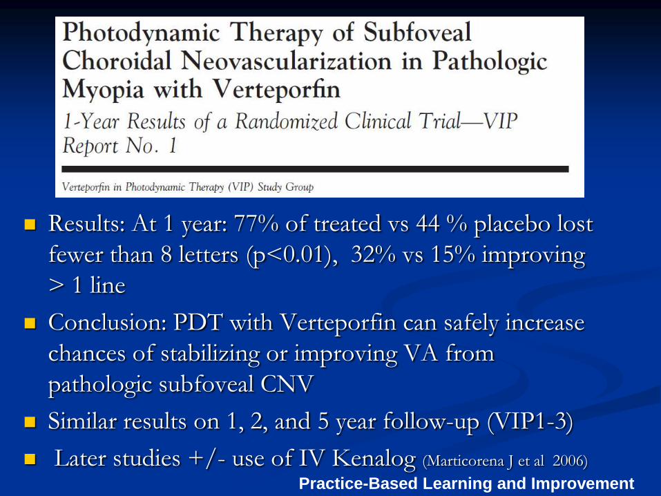
Results: At 1 year: 77% of treated vs 44 % placebo lost
fewer than 8 letters (p<0.01), 32% vs 15% improving
> 1 line
Conclusion: PDT with Verteporfin can safely increase
chances of stabilizing or improving VA from
pathologic subfoveal CNV
Similar results on 1, 2, and 5 year follow-up (VIP1-3)
Later studies +/- use of IV Kenalog (Marticorena J et al 2006)
Practice-Based Learning and Improvement

Purpose: To Assess effect of IV bevacizumab on CMV in
pathological myopia using FA and VA
Methods: Prospective, non-controlled, non-randomized 63 eyes,
received 1mg of IVB, with avg 2.4 injections during first year.
Subfoveal (43%), juxtafoveal (49%), extrafoveal (8%)
Results: BCVA improved 3 ETDRS lines in 40%, worsened > 3
lines in 5%, unchanged in 56% (P<0.01). FA leakage ceased in 48%,
diminished in 44%, unchanged in 8%. No chorioretinal atrophy
Conclusion: IVB is effective Tx for myopic CNV
Medical Knowledge

The Futureof Treatment
Anti-VEGF vs PDT
More prospective, double blind, placebo controlled
studies needed
VEGF-Trap
For now… Laser has a questionable role for
extrafoveal CNV associated with pathologic
myopia given propensity for chorioretinal
atrophy
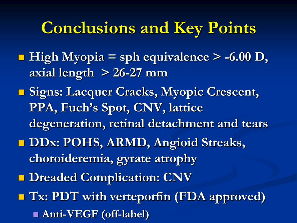
Conclusions and Key Points
High Myopia = sph equivalence > -6.00 D,
axial length > 26-27 mm
Signs: Lacquer Cracks, Myopic Crescent,
PPA, Fuch’s Spot, CNV, lattice
degeneration, retinal detachment and tears
DDx: POHS, ARMD, Angioid Streaks,
choroideremia, gyrate atrophy
Dreaded Complication: CNV
Tx: PDT with verteporfin (FDA approved)
Anti-VEGF (off-label)

Core CompetenciesPatient Care: The patient received compassionate care, based on the appropriate and most
effective management techniques that addressed her physical, emotional, and mental health
issues
Medical Knowledge: The literature was reviewed, a differential was formed. Diagnostic and
therapeutic modalities were discussed using evidence-based medicine and general practice
guidelines. The basic and clinical science of the disease was reviewed to better understand
this condition
Practice-Based Learning and Improvement: The literature was reviewed, as was the full
The clinical evidence was assimilated to better treat the patient as well as learn from her
clinical course in order to manage patients in the future.
Interpersonal and Communication Skills: We communicated extensively with the patient
regarding the process of diagnosing and treating her disease. All of her questions were
answered in a compassionate manner. We worked as a team to limit her fears of vision loss.
Professionalism: Our responsibility as a physician to do no harm was adhered to at all times.
Necessary tests were suggested and the ethical principles of informed consent were utilized.
The patients clinical information remained confidential at all times.
Systems-Based Practice: We showed awareness of the healthcare system, using cost-
effective mechanisms of diagnosis and management. We worked with the optometrists to
better to best correct the patient’s visual acuity

Reflective PracticeThis case demonstrated a classic presentation of an
uncommon disease process. After considering a wide
differential diagnosis and examining the literature, the
appropriate diagnostic modalities were chosen to narrow our
differential and formulate a diagnosis. The patient was
appropriately and compassionately managed. Understandably,
she was quite concerned regarding her visual prognosis. She
was educated about her disease process and its natural
course. We worked closely with the optometrists to improve
the patient’s vision as best as we could.

ReferencesAvila MP, Weiter JJ, Jalkh AE, et al. Natural history of choroidal neovascularization in degenerative myopia.
Ophthalmology 1984;91:1573–1581.
Cohen AW, et al. Presumed Ocular Histoplasmosis Syndrome. [online] . Accessed 12/12/11.
http://webeye.ophth.uiowa.edu/eyeforum/cases/83-presumed-ocular-histoplasmosis-pohs.htm
University of Iowa. Dept. of Ophthalmolgy and Visual Sciences. 3/9/08.
Hayashi K., et al. Comparison of visual outcome and regression pattern of myopic choroidal neovascularization
after Intravitreal Bevacizumab or after photodynamic therapy. Am J Ophthalmol 2009; 148: 396-408
Ikuno Y et al. Intravitreal Bevacizumab for Choroidal Neovascularization Attributable to Pathologic Myopia: One
Year Results. Am J Ophthal 2009.147: 94-100.
Malagola R et al. Peripheral Lacquer Cracks as an Early Finding in Pathological Myopia
Arch Ophthalmol. 2006;124:1783-1784.
Marticorena J, Gomez-Ulla F, Fernandez M, Pazos B, Rodriguez- Cid MJ, Sanchez-Salorio M. Combined
photodynamic therapy and intravitreal triamcinolone acetonide for the treatment of myopic
subfoveal choroidal neovascularization. Am J Ophthalmol 2006; 142: 335-7.
Multiple Chapters in “Retina and Vitreous.” Basic Clinic Science Course Book. American Academy of Ophthalmology, 2011.
Secretan M, Kuhn D, Soubrane G, Coscas G. Long-term visual outcome of choroidal neovascularization in
pathologic myopia: natural history and laser treatment. Eur J Ophthalmol 1997; 7: 307- 16.
Verteporfin in Photodynamic Theory Study Group. Photodynamic therapy of subfoveal choroidal
neovascularization in pathologic myopia with verteporfin. 1-year results of a randomized clinical
trial--VIP report no. 1. Ophthalmology 2001; 108: 841-52.
Wakabayashi T et al. Different Dosing of Intravitreal Bevacizumab for CNC Because of Pathologic Myopia Retina.
31:880–886, 2011.
Yoshida T, Ohno-Matsui K, Yasuzumi K, et al. Myopic choroidal neovascularization: a 10-year follow-up.
Ophthalmology 2003;110:1297–1305.

Thank You!
- Dr. Scott
- Dr. Shrier
- KCHC Faculty, Staff,
and Residents