OPERATION TECHNIC OF DENTOALVEOLAR SURGERY...
71
OPERATION TECHNIQUES OF DENTOALVEOLAR SURGERY Dr. György Komlós Semmelweis University - Oral Surgery Department
Transcript of OPERATION TECHNIC OF DENTOALVEOLAR SURGERY...

OPERATION TECHNIQUES OF DENTOALVEOLAR SURGERY
Dr. György Komlós Semmelweis University - Oral Surgery Department

MINOR ORAL SURGERY (DENTOALVEOLAR SURGERY)
MAXILLOFACIAL SURGERY
(Head and Neck Surgery)

SURGICAL THERAPY
SURGICAL THEORY
=
THE KNOWLEDGE OF THE METHODS & TECNIQUES (& BACKGROUND) OF SURGICAL
INTERVENTIONS

SPECIFIC PROPERTIES OF DENTOALVEOLAR SURGERY
• Frequent
• Bad reputation
• Outpatient
• Flap preparation surgery
• Bone surgery
• Unfavorable environment

TOPICS OF THE LECTURE
INSTRUMENTS
INTRAORAL FLAPS
SUTURES
INSTRUMENTS

SURGICAL MOTOR
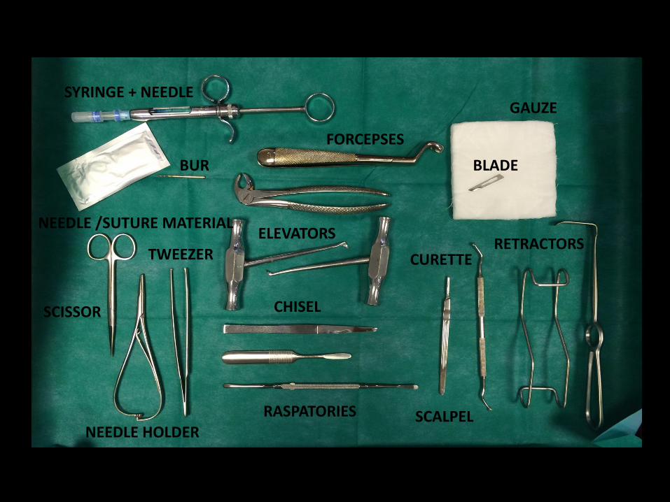
SYRINGE + NEEDLE
SCALPEL
BLADE
RETRACTORS
CHISEL
RASPATORIES
FORCEPSES
ELEVATORS
CURETTE
BUR
NEEDLE /SUTURE MATERIAL
NEEDLE HOLDER
TWEEZER
SCISSOR
GAUZE

Example: Surgical tooth removal
1. Incision (scalpel)
2. Preparating the flap (tweezers, raspatories,chisels)
3. Drilling the bone (sugical motor, burs)
4. Removing the tooth (forcepses, elevators)
5. Cleaning the wound (curettes)
6. Closing the wound (needle/suture material, needleholder, scissor)

Scalpel


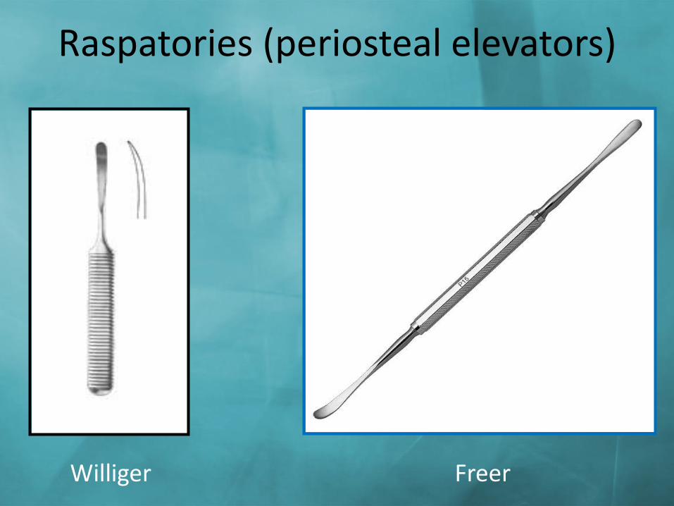
Raspatories (periosteal elevators)
Williger Freer

CASE by Dr. Katalin Csurgay

Chisels

Chisels
MÓCZÁR PARTSCH
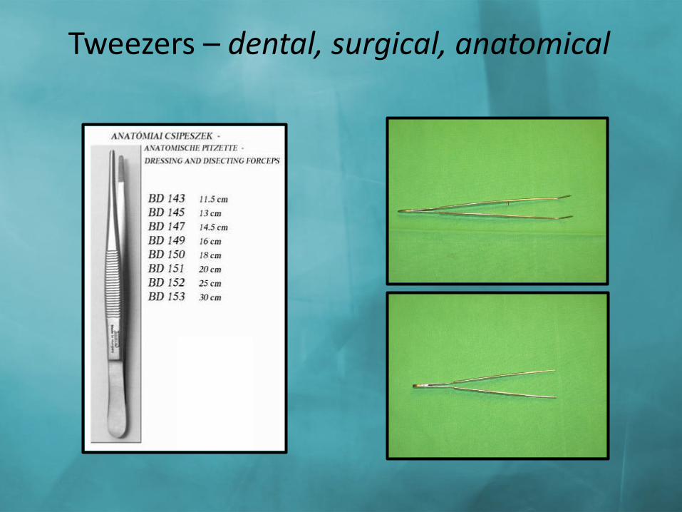
Tweezers – dental, surgical, anatomical

CASE by Dr. Fanni Sára Kálmán

Surgical burs
ROUND and LINDEMANN
Elevators and forcepses

Curettes
Kerpel Volkmann

Curettes
Lucas

Needle holders
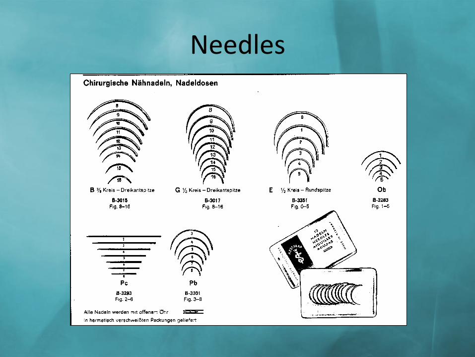
Needles

Needles
INTRAORAL FLAPS
FLAPS - REQUIREMENTS
1. Avoid main anatomical structures
– mentalis neurovascularis plexus
– palatal art.
– ducts
– nasopalatine art.
– infraorbital n.
– pterygoid plexus
FLAPS - REQUIREMENTS
2. Mucoperiosteal flap preparation

FLAPS - REQUIREMENTS
3. Sufficient approaching

FLAPS - REQUIREMENTS
4. Enlargebility

FLAPS - REQUIREMENTS
5. Good blood supply

FLAPS - REQUIREMENTS
6. Tensionless closure

FLAPS - REQUIREMENTS
7. Sutures on intact bone surface

Types of the intraoral flaps

1. Marginal / gingival flap
Mesio-distally performed incision on the gingiva of the edentulous alveolar crest
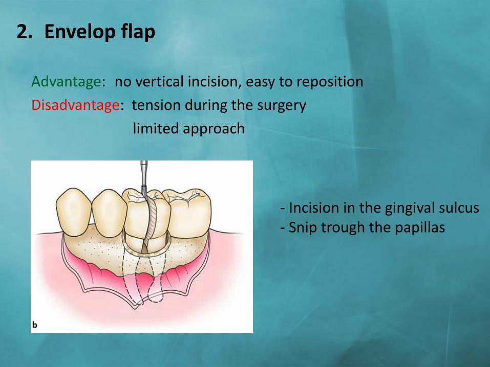
2. Envelop flap
Advantage: no vertical incision, easy to reposition
Disadvantage: tension during the surgery
limited approach
- Incision in the gingival sulcus - Snip trough the papillas

3. Envelop flap on the palatal side
– Incision in the gingival sulcus
Advantage: easy to reposition
good approach
Disadvantage: difficult to prepare
SZABÓ GY: Szájsebészet, maxillofacialis sebészet. Semmelweis Kiadó, Budapest, 2004; 33-47, 49-58, 59-68, 69-84. VÁMOS I, BERÉNYI B, INOVAY J: Szájsebészet. Medicina Könyvkiadó, Budapest, 1980; 120-122, 134-135, 156-157.

CASE by Dr. Attila Szűcs
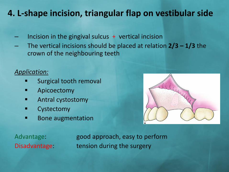
4. L-shape incision, triangular flap on vestibular side
– Incision in the gingival sulcus + vertical incision
– The vertical incisions should be placed at relation 2/3 – 1/3 the crown of the neighbouring teeth
Application:
Surgical tooth removal
Apicoectomy
Antral cystostomy
Cystectomy
Bone augmentation
Advantage: good approach, easy to perform
Disadvantage: tension during the surgery

CASE by Dr. Fanni Sára Kálmán
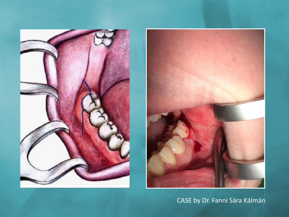
CASE by Dr. Fanni Sára Kálmán

CASE by Dr. Fanni Sára Kálmán

5. L-shape incision, triangular flap on palatal side
– Incision in the gingival sulcus + vertical incision
Application: in case of retained canine
Advantage: good approach
Disadvantage: difficullt to prepare

CASE by Dr. Attila Szűcs

6. Wassmund- flap (trapezoidal flap, four cornered) – Incision in the gingival sulcus (horizontal) + 2 vertical incisions
• Divergating (in direction of the vestibular fornix) vertical incisions • The vertical incisions should be placed at relation 2/3 – 1/3 the crown of
the neighbouring teeth
Advantage: better approach Disadvantage: gingival recession, narrowing of the
vestibule
Application
Apicoectomy Antral cystostomy Sinus closure
SZABÓ GY: Szájsebészet, maxillofacialis sebészet. Semmelweis Kiadó, Budapest, 2004; 33-47, 49-58, 59-68, 69-84. VÁMOS I, BERÉNYI B, INOVAY J: Szájsebészet. Medicina Könyvkiadó, Budapest, 1980; 120-122, 134-135, 156-157. GOPIKRISHNA, KANDASWAMY D, NANDINI S: Newer Classification of Endodontic Flaps. Endodontology 2005; 17:14-19.

CASE by Dr. Attila Szűcs

CASE by Dr. Attila Szűcs

CASE by Dr. Attila Szűcs

CASE by Dr. Attila Szűcs
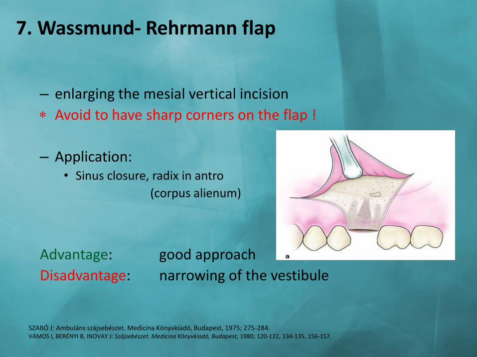
7. Wassmund- Rehrmann flap
– enlarging the mesial vertical incision
Avoid to have sharp corners on the flap !
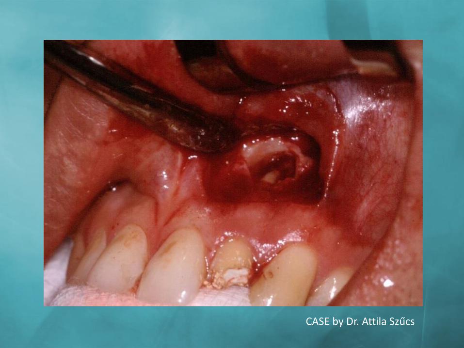
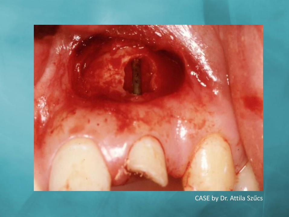
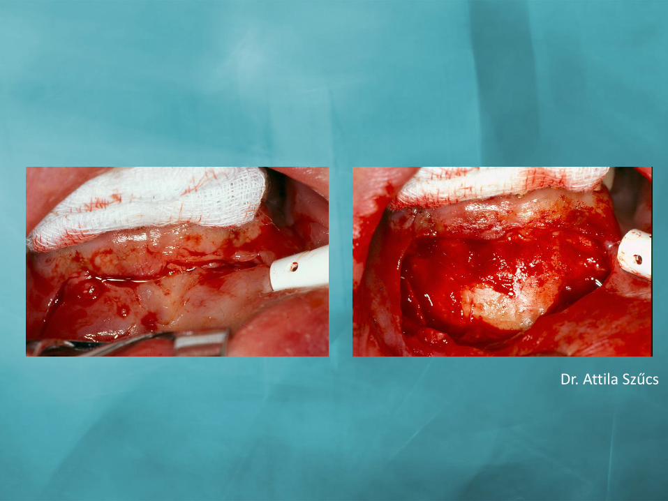
– Application: • Sinus closure, radix in antro
(corpus alienum)
Advantage: good approach
Disadvantage: narrowing of the vestibule
SZABÓ J: Ambuláns szájsebészet. Medicina Könyvkiadó, Budapest, 1975; 275-284. VÁMOS I, BERÉNYI B, INOVAY J: Szájsebészet. Medicina Könyvkiadó, Budapest, 1980; 120-122, 134-135, 156-157.

8. Reinmöller (submarginal) flap
– L-shape – vertical incision parallel with frenulum – horisontal incison min. 4-5 mm from the marginal gingiva
(parallel with it)
Application : apicoectomy, cystectomy Advantage: - enlargebility in distal direction - no gingiva recession
Disadvantage: - poor flap design = not adequate blood supply
Blunt edges are suggested!
CASE by Dr. Attila Szűcs

9. Partsch flap – CONVEX in the direction of the gingival margin
• horizontal incision – Semilunar
– Lower flap margin sholud be 4-5 mms from the marginal gingiva
Application: apicoectomy, cystostomy and cystectomy
Advantage: enlargeable easy to perform, no gingival recession Disadvantage: visible scar limited approach
SZABÓ GY: Szájsebészet, maxillofacialis sebészet. Semmelweis Kiadó, Budapest, 2004; 33-47, 49-58, 59-68, 69-84. VÁMOS I, BERÉNYI B, INOVAY J: Szájsebészet. Medicina Könyvkiadó, Budapest, 1980; 120-122, 134-135, 156-157. GOPIKRISHNA, KANDASWAMY D, NANDINI S: Newer Classification of Endodontic Flaps. Endodontology 2005; 17:14-19. CHINDIA ML, VALDERHAUG J: Periodontal status following trapezoidal and semilunar flaps in apicectomy. East Afr Med J 1995; 72:564-567.
CASE by Dr. Attila Szűcs

10. Pichler flap
INVERSE PARTSCH

11. Ochsenbein-Luebke (submarginal) flap
– Base sholud be 4-5 mms from the marginal gingiva – Follows the contour of the gingiva Application : in esthetic zone in case of fix prosthesises Advantage: the gingival margin is not involved no gingiva recession safe closure Disadvantage: visible scar tissue corners on the flap can necrotize
AHMED MV, RASTOGI S, BAAD RK, GUPTA AK, NISHAD SG, BANSAL M ÉS MTSAI: Comparative Study Between Two Flaps-Trapezoidal flap (TZF) and Ocshenbein- Leubke Flap (OLF) in Periapical Surgeries. J Maxillofac Oral Surg 2013; 12:440- 446. MUSHTAQ I, MALIK A. Evaluation of the Ochsenbein-Luebke flap technique in periapical surgery at Punjab Dental Hospital Lahore Pakistan. J Ayub Med Coll Abbottabad 2003;15:50-53.
Dr. Attila Szűcs

12. Palatal Pichler flap
– Contains the palatal art.
– Angle it in 45 ⁰
per secundam healing on the palate
Advantage: Good blood supply
Disadvantage: Rigid
Painful healing

SUTURES

Principles of suturing • Use suture needle of suitable shape and size • Use suture material that is of suitable type and
size for the tissue being sutured • Good bite (2-3 mm from the free edge of the soft
tissue) • Sutures should NOT be placed under tension • Knots should be tied 2-3 mm away from the
incision line • Suture material is cut 4-5 mm away from the knot • Superficial sutures should be removed 5-7 days
after (exception: sinus closure) surgery to prevent infection / foreign body reaction

Advantages of suturing (closure)
• Promotes healing
• Prevents complications
– INFECTION
– HAEMORRHAGE
– TISSUE NECROSIS
• Preserve the normal contur and shape of tissues

Suture material / needle

Grasping the needle

HOW TO MAKE A KNOT?
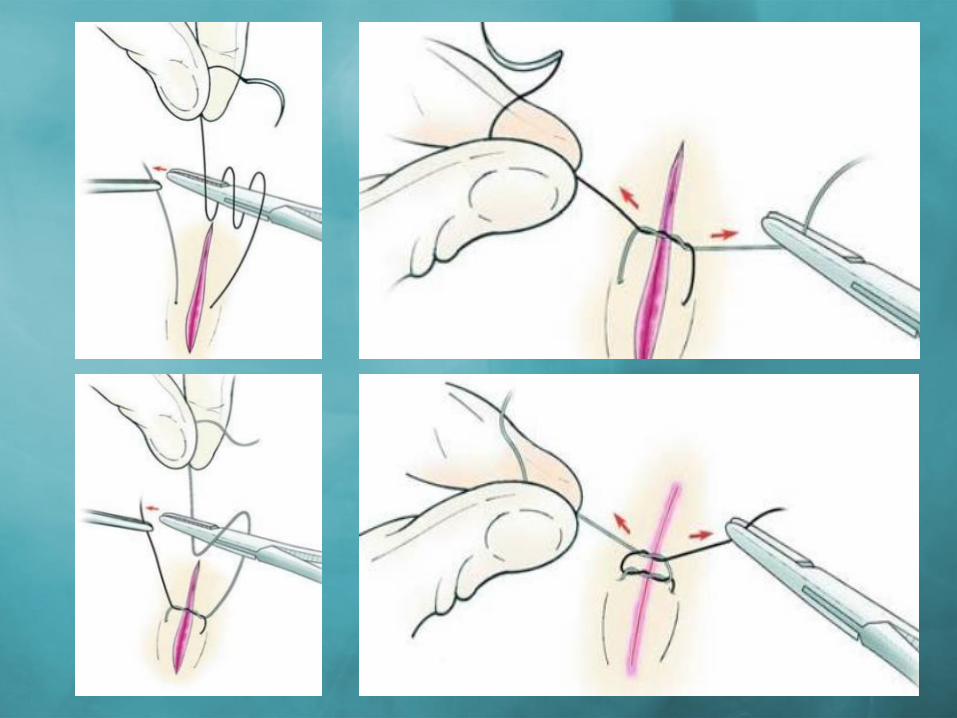

SUTURING TECHNIQUES

SINGLE INTERRUPTED SUTURE
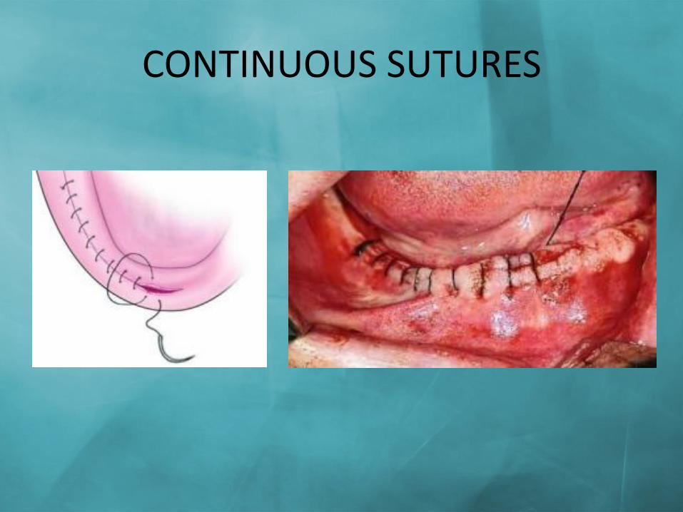
CONTINUOUS SUTURES
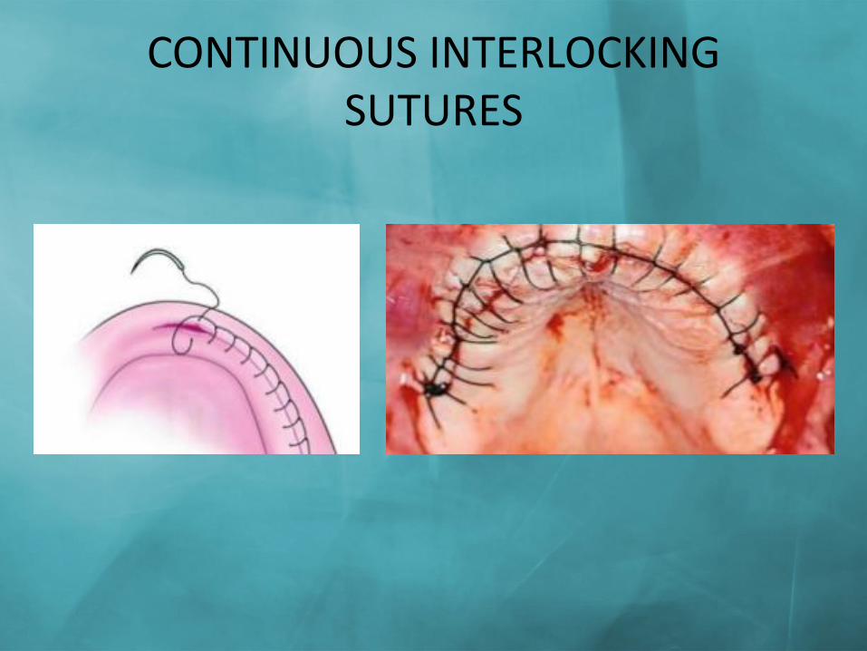
CONTINUOUS INTERLOCKING SUTURES

HORIZONTAL MATRESS SUTURE

VERTICAL MATRESS SUTURE (Donáti)