OPEN Structure of the mouse TRPC4 ion...
10
ARTICLE Structure of the mouse TRPC4 ion channel Jingjing Duan 1,2 , Jian Li 1,3 , Bo Zeng 4 , Gui-Lan Chen 4 , Xiaogang Peng 5 , Yixing Zhang 1 , Jianbin Wang 1 , David E. Clapham 2 , Zongli Li 6 & Jin Zhang 1 Members of the transient receptor potential (TRP) ion channels conduct cations into cells. They mediate functions ranging from neuronally mediated hot and cold sensation to intra- cellular organellar and primary ciliary signaling. Here we report a cryo-electron microscopy (cryo-EM) structure of TRPC4 in its unliganded (apo) state to an overall resolution of 3.3 Å. The structure reveals a unique architecture with a long pore loop stabilized by a disulfide bond. Beyond the shared tetrameric six-transmembrane fold, the TRPC4 structure deviates from other TRP channels with a unique cytosolic domain. This unique cytosolic N-terminal domain forms extensive aromatic contacts with the TRP and the C-terminal domains. The comparison of our structure with other known TRP structures provides molecular insights into TRPC4 ion selectivity and extends our knowledge of the diversity and evolution of the TRP channels. DOI: 10.1038/s41467-018-05247-9 OPEN 1 School of Basic Medical Sciences, Nanchang University, 330031 Nanchang, Jiangxi, China. 2 Howard Hughes Medical Institute, Janelia Research Campus, Ashburn, VA 20147, USA. 3 Department of Molecular and Cellular Biochemistry, University of Kentucky, Lexington, KY 40536, USA. 4 Key Laboratory of Medical Electrophysiology, Ministry of Education, and Institute of Cardiovascular Research, Southwest Medical University, 646000 Luzhou, Sichuan, China. 5 The Key Laboratory of Molecular Medicine, The Second Affiliated Hospital of Nanchang University, 330006 Nanchang, China. 6 Department of Biological Chemistry and Molecular Pharmacology, Howard Hughes Medical Institute, Harvard Medical School, Boston, MA 02115, USA. These authors contributed equally: Jingjing Duan, Jian Li, Bo Zeng. Correspondence and requests for materials should be addressed to Z.L. (email: [email protected]) or to J.Z. (email: [email protected]) NATURE COMMUNICATIONS | (2018)9:3102 | DOI: 10.1038/s41467-018-05247-9 | www.nature.com/naturecommunications 1 1234567890():,;
Transcript of OPEN Structure of the mouse TRPC4 ion...

ARTICLE
Structure of the mouse TRPC4 ion channelJingjing Duan1,2, Jian Li1,3, Bo Zeng 4, Gui-Lan Chen4, Xiaogang Peng5, Yixing Zhang1, Jianbin Wang1,
David E. Clapham 2, Zongli Li6 & Jin Zhang1
Members of the transient receptor potential (TRP) ion channels conduct cations into cells.
They mediate functions ranging from neuronally mediated hot and cold sensation to intra-
cellular organellar and primary ciliary signaling. Here we report a cryo-electron microscopy
(cryo-EM) structure of TRPC4 in its unliganded (apo) state to an overall resolution of 3.3 Å.
The structure reveals a unique architecture with a long pore loop stabilized by a disulfide
bond. Beyond the shared tetrameric six-transmembrane fold, the TRPC4 structure deviates
from other TRP channels with a unique cytosolic domain. This unique cytosolic N-terminal
domain forms extensive aromatic contacts with the TRP and the C-terminal domains. The
comparison of our structure with other known TRP structures provides molecular insights
into TRPC4 ion selectivity and extends our knowledge of the diversity and evolution of the
TRP channels.
DOI: 10.1038/s41467-018-05247-9 OPEN
1 School of Basic Medical Sciences, Nanchang University, 330031 Nanchang, Jiangxi, China. 2 Howard Hughes Medical Institute, Janelia Research Campus,Ashburn, VA 20147, USA. 3 Department of Molecular and Cellular Biochemistry, University of Kentucky, Lexington, KY 40536, USA. 4 Key Laboratory ofMedical Electrophysiology, Ministry of Education, and Institute of Cardiovascular Research, Southwest Medical University, 646000 Luzhou, Sichuan, China.5 The Key Laboratory of Molecular Medicine, The Second Affiliated Hospital of Nanchang University, 330006 Nanchang, China. 6 Department of BiologicalChemistry and Molecular Pharmacology, Howard Hughes Medical Institute, Harvard Medical School, Boston, MA 02115, USA. These authors contributedequally: Jingjing Duan, Jian Li, Bo Zeng. Correspondence and requests for materials should be addressed to Z.L. (email: [email protected])or to J.Z. (email: [email protected])
NATURE COMMUNICATIONS | (2018) 9:3102 | DOI: 10.1038/s41467-018-05247-9 | www.nature.com/naturecommunications 1
1234
5678
90():,;

Mammalian transient receptor potential (TRP) channelsare activated by a wide spectrum of ligands, tempera-ture, lipids, pH, and as yet unknown stimuli. They
are classified into six subfamilies based on sequence similarity:TRPC (canonical), TRPM (melastatin), TRPV (vanilloid),TRPA (ankyrin), TRPML (mucolipin), and TRPP (or PKD)(polycystin)1. The TRPC subfamily are non-selective cationchannels (Na+, K+, Ca2+) that alter proliferation, vascular tone,and synaptic plasticity2,3. This family can be further subdividedinto two subgroups: TRPC2/3/6/7 and TRPC1/4/5. TRPC4 isbroadly expressed in human tissues and can assemble as homo-meric channels or form heteromeric channels with TRPC1 andTRPC54–7. Studies of Trpc4-deficient mice have shown thatTRPC4 affects endothelial-dependent regulation of vascular tone,endothelial permeability, and neurotransmitter release from tha-lamic interneurons8. Stimulation of Gq and Gi/oG-protein-coupled receptors (GPCRs) as well as tyrosine kinase receptorspotentiate channel activity9,10. Activation of TRPC4 is regulatedby intracellular Ca2+, phospholipase C, and membrane lipids byunclear mechanisms. In addition, Storch et al.11 have proposed apotential mechanism related to phosphatidylinositol 4,5-bispho-sphate and Na+/H+ exchanger regulatory factor proteins11.
Along with the revolution in cryo-electron microscopy (cryo-EM), improved sample preparation, data acquisition, and imageprocessing strategies, the structures of TRPVs12–17, TRPA118,TRPP119, TRPML120, TRPM421–24, and TRPM825 have beensolved. Here we present the structure of mouse TRPC4 in its apostate at pH 7.5 at an overall resolution of 3.3 Å. The structureprovides detailed information on the ion permeation, selectivity,and gating mechanism of TRPC subfamily.
ResultsOverall structure of the mouse TRPC4 tetrameric ion channel.The mouse TRPC4 (residues amino acids (a.a.) 1–758, excludinga.a. 759–974) was expressed using the BacMam expression system(Methods) and purified protein (pH 7.5) was used for single-particle cryo-EM analysis (Supplementary Fig. 1). The overallresolution of TRPC4 reconstruction was 3.3 Å (SupplementaryFig. 2 and Table 1), which enabled us to construct a near-atomicmodel (Supplementary Fig. 3). Disordered regions led to poordensities for 4 residues in the S1–S2 loop, 2 residues in the S3–S4loop, 27 residues in the distal N terminus, and 28 residues in thetruncated distal C terminus. In total, the TRPC4 structure is afour-fold symmetric homotetramer (Fig. 1a) with dimensions of100 Å × 100 Å × 120 Å (Fig. 1b). Each monomer consists of atransmembrane domain (TMD) and a compact cytosolic domain.The cytosolic domain is composed of two subdomains: the N-terminal subdomain consisting of four ankyrin repeats(AR1–AR4) and seven α-helices (H1–H7), and the C-terminalsubdomain containing a connecting helix and a coiled-coildomain (Fig. 1c, d).
Ca2+ measurements and electrophysiological studies wereperformed to verify that the truncated construct used forstructural investigation (residues a.a. 1–758, excluding a.a.759–974) was permeable to cations and retained sensitivity tochannel activator englerin A, blocker ML204, and blocker 2-APB(Fig. 2). Englerin A induced a robust rise of intracellular Ca2+ inboth full-length and truncated TRPC4-transfected 293T cells, butnot in the empty vector-transfected control cells (Fig. 2a). Inpatch clamp experiments, englerin A potentiated whole-cellcurrents in a dose-dependent manner for 293T cells expressingfull-length and truncated TRPC4. In addition, currents wereinhibited by the TRPC4 blocker ML204 and the non-selectiveblocker 2-APB (Fig. 2b). The current–voltage (I–V) relationshipsfrom both constructs were typical of TRPC4/5, with flattening of
the curve between 10 and 40 mV due to outward Mg2+ block26.These results suggest that, in the context of chemical modulation,the biophysical properties of the truncated construct are similarto that of the full-length TRPC4.
To futher test the functional properties of the truncatedconstruct, we examined receptor-operated activation of TRPC4by coexpression with GPCRs. In cells transfected with TRPC4constructs and P2Y1 and P2Y2 receptors (both coupled to Gq
proteins), extracellular application of ADP (P2Y1 agonist) orATP (P2Y2 agonist) induced large currents for full-lengthTRPC4. In contrast, the currents for truncated TRPC4 weremuch smaller but still exhibited the characteristic I–V relation-ship of the TRPC4 channel (Supplementary Fig. 4a). Thetruncated TRPC4 did not respond to Gi/o receptor agonistsDAMGO (μOR) or carbachol (M2R) (Supplementary Fig. 4b).
Major structural differences with other TRP subfamilies. InFig. 3, we compare the TRPC4 structure with previously reportedTRP structures. Not surprisingly, the organization of six helices ineach TMD is similar to that of other TRP channels, while theintracellular architecture is distinct. By superimposing a TRPC4monomer with representative TRP monomers from each sub-family, we found that the overall fold of TRPC4 is closest to thatof TRPM4 (Fig. 3a). TRPC4 has marked similarities to TRPM4 inthe TMDs despite their different tissue functions and lack ofsequence conservation (<20% identical residues) (SupplementaryFig. 5a, b). Distinctive features of TRPC4 include: (1) the
Table 1 Data collection and refinement statistics
Cryo-EM of TRPC4 [EMD:6901][PDB:5Z96]
Data collection and processing
Microscope FEI Tecnai PolaraDetector Gatan K2Calibrated magnification 40,607Voltage (kV) 300Electron exposure (e–/Å2) 52.8Defocus range (μm) 1.5–3.0Pixel size (Å) 1.23Symmetry imposed C4Initial particle images (no.) 381,165Final particle images (no.) 232,858FSC threshold 0.143Map resolution (Å) 3.3Refinement
Refinement software phenix.real_space_refineInitial model used (PDB code) De novoModel composition
Non-hydrogen atoms 21,506Protein residues 2608Ligands 12
B factors (Å2)
Average 65.46r.m.s. deviations
Bond lengths (Å) 0.01Bond angles (°) 1.11
Validation
MolProbity score 1.83Clashscore 7.25
Ramachandran plot
Favored (%) 97.39Allowed (%) 2.08Disallowed (%) 0.53
ARTICLE NATURE COMMUNICATIONS | DOI: 10.1038/s41467-018-05247-9
2 NATURE COMMUNICATIONS | (2018) 9:3102 | DOI: 10.1038/s41467-018-05247-9 | www.nature.com/naturecommunications

arrangement of S2–S3 linker, S5, S6, and the pore loop. InTRPC4, the S2–S3 linker has two-helical turns, shorter than thatof TRPM4 (Supplementary Fig. 5a), which limits the interactionsof S2 and S3 with their cytoplasmic regions; (2) the disulfide bondbetween TRPC4’s Cys549 and Cys554 lies in the loop linking S5and the pore helix (Fig. 3b, c), while TRPM family’s disulfidebond is located in the loop between the pore helix and S624
(Supplementary Fig. 5c). Note that these two cysteines are con-served in TPRC1/4/5, but not in other TRPC members (Supple-mentary Fig. 5b); (3) a pre-S1 elbow helix connects the Nterminus and TMD in TRPC4 (Fig. 3d), as in TRPM4 andNOMPC (no mechanoreceptor potential C)21,27; however,TRPC4 and TRPM4’s pre-S1 helix is not found in NOMPC27. InTRPC4 the pre-S1 elbow helix is longer, bending andconnecting to the pre-S1 helix directly, while in TRPM4 a
characteristic bridge loop (approximately 60 residues) connectsthe pre-S1 helix with the pre-S1 elbow (Fig. 3d and Supplemen-tary Fig. 5c).
To understand the role of the disulfide bond in the pore loop ofTRPC4, we performed patch clamp experiments on wild-type(wt) and cysteine mutants of TRPC4. Reducing agents dithio-threitol (DTT; membrane permeable) and tris(2-carboxyethyl)phosphine hydrochloride (TCEP; membrane impermeable)potentiated wt TRPC4 whole-cell currents (Fig. 3e). The I–Vrelationship of the DTT-induced current was characteristic ofTRPC4, while that induced by TCEP was similar to the I–Vcommonly seen for other TRP channels such as TRPV1/M7/M8.This may be due to its stronger reducing effect andexclusive extracellular action. The double cysteine mutant(C549A+C554A) could be activated by englerin A but not DTT
a
b
c
Coiled-coildomain
S1
Porehelix
S5S6
S4
S2 S3
Disulfidebond
d
N-term
inal domain
TRP domain
S5
S6
S1
S2
S3 S4
Disulfide bond
Top viewSide view
AR1 AR1
AR2 AR2
AR3 AR3
AR4 AR4
H1
H2H3
H4 H5 H6 H7
ARD
HLH
Pore
helix
Pre-S1helix
Elbow
Connecting helix
TRP domainS2-S
3 linke
r
Connecting helix
120 Å
100 Å
100 Å
100 Å
90°
90°
Pre-S1elbow
N-terminaldomain
Coiled-coil dom
ain
S2-S3linkerPre-S1
Fig. 1 Overall structure of mTRPC4. a Side and top views of the cryo-EM density map of mouse TRPC4 at 3.3 Å overall resolution. Each monomer isrepresented in different colors. b Ribbon diagrams of the mouse TRPC4 model with channel dimensions indicated. c Ribbon diagrams depicting structuraldetails of a single subunit. d Linear diagram depicting the major structural domains of the TRPC4 monomer, color-coded to match the ribbon diagram in (c)
NATURE COMMUNICATIONS | DOI: 10.1038/s41467-018-05247-9 ARTICLE
NATURE COMMUNICATIONS | (2018) 9:3102 | DOI: 10.1038/s41467-018-05247-9 | www.nature.com/naturecommunications 3
(Fig. 3f), suggesting that the TRPC4 channel without the poreloop disulfide bond is still functional but lacks redox sensitivity.Surprisingly, mutation of a single cysteine (C549A or C554A)resulted in insensitivity to englerin A and DTT (Fig. 3g). If thismutated TRPC4 trafficked to the plasma membrane, the loss ofenglerin A and DTT sensitivities suggests that the channel’s poreloop architecture has been severely disrupted, resulting in achannel that cannot be exogenously activated.
Cytosolic domain features and interactions. The cytosolicdomains of TRP channels include regulatory components anddomain interactions that may tune channel gating. The cytosolicdomain of TRPC4 adopts a pedestal-like architecture (Fig. 4a andSupplementary Fig. 6a). The large and unique N-terminal domain
of TRPC4 contains a long loop followed by an ankyrin repeatdomain and helix–loop–helix (HLH) motifs. These HLH motifsconsist of seven helices and several connecting loops (Fig. 1c, dand Supplementary Fig. 6a). Similar to TRPM structures, the C-terminal domain of TRPC4 is composed of two helices, a con-necting helix and a coiled-coil domain helix (Fig. 1c, d). Theconnecting and coiled-coil domain helices bend ~120° to form aninverted “L” (Supplementary Fig. 6b). The coiled-coil domaincontains three heptad repeats that exhibit the characteristic per-iodicity (a-b-c-d-e-f-g)n (Fig. 4b, c and Supplementary Fig. 7),with hydrophobic residues at positions “a” and “d”. The presenceof Val and Ile at the “a” position, and Leu and Phe at the “d”position in the core of the coiled-coil domain supports the for-mation of a tetramer (Fig. 4c).
0
–12
–8
–4
0
4
8Englerin A 100 nM
Cur
rent
(nA
)
Time (min)
2-APB 100 μM
–100 100
–16
–12
–8
–4
4
8
2-APBBasal
nA Englerin A
mV
0
–8
–4
0
4
8
12 Englerin A 100 nM
Cur
rent
(nA
)
2-APB 100 μM
–100 100
–18
–12
–6
6
12
2-APBBasal
nA Englerin A
mV
0–8
–4
0
4
8
Cur
rent
(nA
)
–100 100
–8
–4
4
8
BasalEnglerin A
nA
mV
0
–0.2
0.0
0.2
0.4
0.6 Englerin A 1 nM
Cur
rent
(nA
)
Time (min)
ML204 10 μM
100
0.5
1.0
1.5
ML204Basal
nA Englerin A
mV
TRPC4 full-length TRPC4 truncated
TRPC4 full-length TRPC4 truncated
Empty vector
0.1 1 10 100
0
20
40
60
80
100
Act
ivat
ion
(%)
Englerin A (nM)
–0.3
0.0
0.3
0.6
0.9 Englerin A 1 nM
Cur
rent
(nA
)ML204 10 μM
0 2
0.0
0.1
0.2
0.3
0.4
TRPC4 full-lengthTRPC4 truncated Empty vector
2-APB 100 μM
Time (min)
Englerin A 100 nM
b
a
–100 100
ML204Basal
Englerin AnA
mV
Englerin A 100 nM
Full-length (+80 mV)Full-length (–80 mV)Truncated (+80 mV)Truncated (–80 mV)
ΔRat
io (F
340/F
380)
1
2 4 6 0Time (min)2 4 6
1.5
1.0
0.5
1 2 3
–12
Time (min)1 2 3 1 2 3
Time (min)
–100
Fig. 2 The truncated TRPC4 construct used in cryo-EM encodes a functional channel. a Intracellular Ca2+ measurements of truncated mTRPC4, full-lengthmTRPC4, and empty vector-transfected 293T cells loaded with Fura-2 AM. Data are expressed as mean ± s.e.m.; n= 10 cells in each group. bRepresentative whole-cell patch clamp recordings and I–V relationships of truncated mTRPC4, full-length mTRPC4, and empty vector expressed in293T cells. The time course of currents measured at +80 and −80mV and I–V relationships of the peak currents from different conditions are shown. Theupper right panel shows dose–response curves of englerin A in activating TRPC4 currents at +80 and −80mV (values with error bars are expressed asmean ± s.e.m.; n= 8 for each concentration). Current induced by 100 nM englerin A in each cell was considered as maximum current (100%); currentsinduced by other concentrations of englerin A in the same cell were normalized. Englerin A is a TRPC4/5 activator and ML204 is an inhibitor for TRPC4/5channels. 2-APB is a non-selective blocker of TRP channels
ARTICLE NATURE COMMUNICATIONS | DOI: 10.1038/s41467-018-05247-9
4 NATURE COMMUNICATIONS | (2018) 9:3102 | DOI: 10.1038/s41467-018-05247-9 | www.nature.com/naturecommunications

180°
b
hTRPM4mTRPC4 mTRPV1 hTRPA1 hTRPP1 mTRPML1
a
c
d
C993
C1011
C549
C554
Intracellular
Extracellular
S5S6
S5
S6
0 2 4–2
–1
0
1
2C
urre
nt (
nA)
Time (min)
DTT 10 mM
Englerin A100 nM
0 1 2 3–2
–1
0
1
Englerin A 100 nM
Cur
rent
(nA
)
Time (min)
0 1 2
0
1
2TCEP 10 mM
Cur
rent
(nA
)
Time (min)
–100 100
1
2
3
Basal
TCEPnA
mV
0 1 2 3–2
–1
0
1
2
Cur
rent
(nA
)
Time (min)
DTT 10 mM
–100 100
–1.0
–0.5
0.5
1.0
BasalDTT
nA
mV
0 2 4 6
–2
0
2
4DTT 10 mM
Cur
rent
(nA
)
Time (min)
–100 100
–4
–2
2
4
6
Basal
DTTnA
mV
TRPC4 C549A+C554ATRPC4 wild type
TRPC4 C549A+C554ATRPC4 wild type TRPC4 C554A
e f g
–100 100
–2
–1
1
2
Basal
Englerin AnA
mV
0 1 2 3–2
–1
0
1
2
Cur
rent
(nA
)
Time (min)
DTT 10 mM
Englerin A100 nM
–100 100
–1.0
–0.5
0.5
1.0
DTT
Basal
Englerin AnA
mV
TRPC4 C549A
–100 100
–1.0
–0.5
0.5
1.0
DTT
Basal
Englerin A
nA
mV
Pre-S1 elbow
Pre-S1 helix
Bridge loopPre-S1 elbow
Fig. 3 Comparison of the TRPC4 structure with previously solved TRP channel structures (apo state). a Side views of an mTRPC4 subunit compared withother TRP family members including human TRPM4 [PDB:6BWI]24, mouse TRPV1 [PDB:3J5P]14, human TRPA1 [PDB:3J9P]18, human PKD2/TRPP1[PDB:5T4D]19, and mouse TRPML1 [PDB:5WPV]20. b Superimposition of TRPC4 and TRPM4. c Key pore loop disulfide bond between Cys549 and Cys554in TRPC4 and the corresponding pore loop disulfide bond between Cys993 and Cys1011 in TRPM4 (black arrows). d Differences in the organizations of thelinker (pre-S1 elbow and pre-S1 helix) between the N terminus and transmembrane domains in TRPC4 and TRPM4. e Activation of whole-cell currents byreducing agents DTT and TCEP in 293T cells overexpressing wild-type TRPC4. f The double cysteine TRPC4 mutant (C549A+C554A) is activated byenglerin A but not DTT. g Single cysteine mutants, C549A or C554A, are insensitive to 100 nM englerin A and 10mM DTT. The time course of currentsmeasured at +80 and −80mV and I–V relationships of the peak currents in different conditions are shown
NATURE COMMUNICATIONS | DOI: 10.1038/s41467-018-05247-9 ARTICLE
NATURE COMMUNICATIONS | (2018) 9:3102 | DOI: 10.1038/s41467-018-05247-9 | www.nature.com/naturecommunications 5
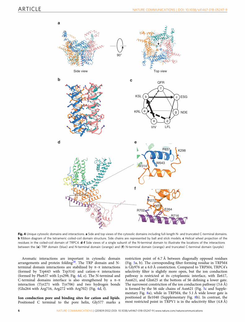
Aromatic interactions are important in cytosolic domainarrangements and protein folding28. The TRP domain and N-terminal domain interactions are stabilized by π–π interactions(formed by Trp643 with Trp314) and cation–π interactions(formed by Phe637 with Lys298; Fig. 4d, e). The N-terminal andC-terminal domains interface is also strengthened by a π–πinteraction (Tyr271 with Tyr706) and two hydrogen bonds(Glu264 with Arg716, Arg272 with Arg702) (Fig. 4d, f).
Ion conduction pore and binding sites for cation and lipids.Positioned C terminal to the pore helix, Gly577 marks a
restriction point of 6.7 Å between diagonally opposed residues(Fig. 5a, b). The corresponding filter-forming residue in TRPM4is Gly976 at a 6.0 Å constriction. Compared to TRPM4, TRPC4’sselectivity filter is slightly more open, but the ion conductionpathway is restricted at its cytoplasmic interface, with Ile617,Asn621, and Gln625 at the bottom of S6 defining a lower gate.The narrowest constriction of the ion conduction pathway (3.6 Å)is formed by the S6 side chains of Asn621 (Fig. 5c and Supple-mentary Fig. 8a), while in TRPM4, the 5.1 Å wide lower gate ispositioned at Ile1040 (Supplementary Fig. 8b). In contrast, themost restricted point in TRPV1 is in the selectivity filter (4.8 Å)
R716
E264
R272
R702
90°
a d
g
c
f
b
e
VIV LFL
KRL NDE
ESGKSL
QFRc
e
W314W643
F637 K298
f
Side view Top view
Y706
Y271
a
b
d
Fig. 4 Unique cytosolic domains and interactions. a Side and top views of the cytosolic domains including full-length N- and truncated C-terminal domains.b Ribbon diagram of the tetrameric coiled-coil domain structure. Side chains are represented by ball and stick models. c Helical wheel projection of theresidues in the coiled-coil domain of TRPC4. d–f Side views of a single subunit of the N-terminal domain to illustrate the locations of the interactionsbetween the (e) TRP domain (blue) and N-terminal domain (orange) and (f) N-terminal domain (orange) and truncated C-terminal domain (purple)
ARTICLE NATURE COMMUNICATIONS | DOI: 10.1038/s41467-018-05247-9
6 NATURE COMMUNICATIONS | (2018) 9:3102 | DOI: 10.1038/s41467-018-05247-9 | www.nature.com/naturecommunications

between opposing Gly643 residues (Supplementary Fig. 8c)29. InTRPA1, the narrowest point (6.1 Å) is Val961 at its lower gate18
(Supplementary Fig. 8d). These ~0.5–2.5 Å differences in thenarrowest point of TRPs structures may give some clue as to ionselectivity, but we also are aware that current resolution optimi-zation in cryo-EM is still being improved by methods such asmodel-based local density sharpening30, and resolution varieswith location within the particle, conditions such as vitrification,and electron density map fitting.
The simplest hypothesis, with these caveats in mind, is thatTRPC4 is in a closed or inactivated state since the lower gate istoo narrow to allow the passage of a fully or partially hydratedion. In support of this idea is the fact that Gln625 (located in theion conduction exit pathway) is conserved in all the TRPCchannels (Supplementary Fig. 9), suggesting it plays an importantrole in ion permeation.
TRPC4 is non-selective and thus permeable to monovalents(Na+, K+) and some divalents, such as Ca2+. A strong non-protein density peak in our TRPC4 structure is present in ahydrophilic pocket on the cytoplasmic side of the S1–S4 fold,
consistent with the corresponding location of a presumed Ca2+ inTRPM421 (Fig. 5d, e and Supplementary Fig. 10). We tentativelymodeled this non-protein density as Na+ since sodium was themost abundant cation in our purification buffer. The assumedNa+ located at the cytoplasmic face is apparently coordinated byside chains of Glu417 and Gln420 from S2 and the Asp438 andAsn435 from S3 (Fig. 5e). The negatively charged Glu417 andAsp438 are conserved within the TRPC subfamily (exceptTRPC1) (Supplementary Fig. 9). S1’s Tyr373 and the positivelycharged S4 Arg491 are located above the cation binding site,forming a lid that may prevent the outward movement of cations(Fig. 5e).
Eight densities corresponding to lipid molecules were clearlyresolved and identified as cholesteryl hemisuccinates (CHS) andphospholipids (the density fitting ceramide-1-phosphate orphosphatidic acid) (Supplementary Figs. 3 and 11). Four CHSlocated at the interface of the N-terminal domain and the S4/S5linker are bound to each protomer, stabilizing the domaininteraction (Supplementary Fig. 11). The phospholipid isembedded in the gap between the four monomeric subunits with
8.2 Å
6.7 Å
13.2 Å
3.6 Å
4.7 Å
N580
G577
F576
I575
N621
Q625
I6175.4 Å
dc
G577
N621
Q625
I617
N580
TRPC4
TRPM4
G577N580
0
20
40
60
0
Pore radius (Å)
Dis
tanc
e al
ong
pore
axi
s (Å
)
I617
N621
Q625
Selectivity filter
Lower gate
Central cavity
ba
e
Y373S1
S4
R491
E417 D438
Q420 N435
2 4 6 8
Fig. 5 TRPC4 ion conduction pathway. a Ion conduction pathway shown as dots and mapped using HOLE. b Pore radius along the central axis. The sidechains of G577 form a narrow constriction at the selectivity filter. N621 is the most restricted site of the lower gate. The dashed line represents TRPM4 forcomparison. c Side view of TRPC4’s pore region with chains A and C. The distances between diagonal residues in the selectivity filter and lower gate arelabeled. d A putative Na+ binding site is found on the cytosolic side in the hydrophilic pocket of the S1–S4 domain. e Enlarged view of putative Na+ (orangesphere) ion interacting with E417, Q420, N435, and D438
NATURE COMMUNICATIONS | DOI: 10.1038/s41467-018-05247-9 ARTICLE
NATURE COMMUNICATIONS | (2018) 9:3102 | DOI: 10.1038/s41467-018-05247-9 | www.nature.com/naturecommunications 7

its polar head interacting with the pore helix and neighboring S6helix (Supplementary Fig. 11). In vivo phosphorylation ordephosphorylation of membrane lipids could thus alter thetopology of the ion conduction pathway.
DiscussionTRPC4 has been characterized as a non-selective cation channelwith moderate selectivity for Ca2+ over monovalent cations.Comparison of this TRPC structure with other TRP channelstructures highlights some commonalities and differences. AllTRP channels are tetramers with domain swapping interactions,pore loops, selectivity filters, and extracellular and intracellular-facing constriction sites, as first shown for six TM K+ channels31.However, the lower gate in the TRPC4 appears to have an unu-sual set of three constriction sites not found in other TRP channelstructures. Another interesting feature that bears functionalinvestigation is the extracellular pore loop disulfide bond (e.g.,TRPC4 and TRPM4).
Our structure of TRPC4 provides insights into the architectureof the selectivity filter and lower gate. Mutations within this poreregion leads to changes in ion permeability; however, mutagenesisof the pore-localized LFW motif (a.a. 571–573 in TRPC4), whichis conserved in all TRPC members, results in a dominant-negativechannel4. This phenomenon can be explained in our structure, bythe pore helix located at the LFW motif (which has a π–πinteraction; Supplementary Fig. 12a, b). This mutation likelyperturbs the stability of the key pore region, leading to proteinmisfolding and subsequent degradation. Notably, mutation ofGly503 to serine resulted in spontaneously active channelswhich could not be further activated by receptor and intracellularCa2+32. In our TRPC4 structure, Gly503, which is located on theS4/S5 linker, forms a hydrogen bond with Trp635 on the TRPdomain, suggesting that the S4/S5 linker and TRP domaininteract to regulate channel activity32 (Supplementary Fig. 12c).
The role of the disulfide bond in TRPC4’s pore loop has notbeen previously investigated, but electrophysiological studies onthe closely related TRPC5 have shown that reduction of thedisulfide bond with DTT potentiated TRPC5 currents33,34.However, the results of TRPC5 cysteine mutations are disparate.Xu et al.33 reported that mutation of either a single cysteine(C553A, C558A, or C553S) or both cysteines (C553A+C558A)resulted in constitutively active TRPC5 channels. Moreover, thedisulfide bond was formed between C553 and C558 in the samemonomer and not crosslinked between different monomers.These observations were challenged by a later study stating thatboth C553S and C558S mutants completely lost channel activity,and dimerization of channel proteins was significantly impairedfor cysteine mutants of both TRPC4 and TRPC534.
Our structural results demonstrate the existence of a disulfidebond in the pore region. If one of the two cysteines is mutated, aninter-monomer disulfide bond may be formed which wouldsubstantially change the architecture of the pore loop and affectchannel activity. This is supported by our electrophysiologicalobservations from the C549A and C554A TRPC4 mutants. Spe-cifically, results of the double cysteine mutant (C549A+C554A)suggest that the pore loop disulfide bond is dispensible forchannel activation by the activator, but is essential for redoxregulation of channel activity.
TRPCs have multiple activation mechanisms. TRPC1/4, 1/5heterotetramers and TRPC4 or 5 homotetramers can be activatedby Gq protein-coupled receptors and increasing intracellularCa2+ as well as by PIP2 hydrolysis, while TRPC3, 6, and 7 areadditionally sensitive to diacylglycerol35. From our observations,we propose that our TRPC4 structure represents a closed statebecause of the narrow distance between the residues in the lower
gate. In the present study, we truncated a.a. 759–974 fromTRPC4, resulting in the absence of the PDZ-binding motif, a Gα-binding sites, the second camodulin-binding domain and apotential PIP2-binding region. We found that this truncationgreatly impaired or abolished channel activity in response to Gq-mediated and Gi/o-mediated signaling, which confirmed theimportance of the C terminus in receptor-operated activation ofTRPC4. However, the truncated TRPC4 currents stimulated byP2Y1 and P2Y2 were small and thus there are other structuralconstraints related to Gq-mediated TRPC4 activation. The trun-cated TRPC4 retained the first camodulin-binding domain andthus could still be sensitive to intracellular Ca2+ changes, suchCa2+ release from the ER stores induced by IP3. Our currentstudy should help guide future mutagenesis, functional, andstructural studies.
MethodsProtein expression and purification. The mouse TRPC4 construct (a.a. 1–758 of974) was cloned into the pEG BacMam vector36 and a maltose-binding protein(MBP) tag was added to its N terminus (all primer sequences used in this study arein a Supplementary Table 1). P3 baculovirus were produced in the Bac-to-BacBaculovirus Expression System (Invitrogen). HEK293S GnTI− cells (from ATCC)were infected with 10% (v/v) P3 baculovirus at a density of 2.0–3.0 × 10⁶ cells/mlfor protein expression at 37 °C. After 12–24 h, 10 mM sodium butyrate was addedand the temperature reduced to 30 °C. Cells were harvested at 72 h after trans-duction, and resuspended in a buffer containing 30 mM HEPES, 150 mM NaCl, 1mM DTT, pH 7.5, with EDTA-free protease inhibitor cocktail (Roche). After 30min, cells were solubilized for 2–3 h in a buffer containing 1.0% (w/v) N-dodecyl-β-D-maltopyranoside (Affymetrix), 0.1% (w/v) CHS (Sigma), 30 mM HEPES, 150mM NaCl, 1 mM DTT, pH 7.5, with EDTA-free protease inhibitor cocktail(Roche). The supernatant was isolated by 100,000 × g centrifugation for 60 min,followed by incubation in amylose resin (New England BioLabs) at 4 °C overnight.The resin was washed with 20 column volumes of “wash buffer” containing 30 mMHEPES, 150 mM NaCl, 0.1% (w/v) digitonin, 0.01% (w/v) CHS, 1 mM DTT, pH7.5, with EDTA-free protease inhibitor cocktail (Roche). The protein was elutedwith four column volumes of wash buffer with 40 mM maltose. The protein wasthen concentrated to 0.5 ml with a 100 kDa molecular weight cut-off concentrator(Millipore). PreScission protease was added to the samples and incubated overnightat 4 °C to remove the MBP tag. After incubation at 4 °C overnight, the protein wasthen purified on a Superose 6 column in a buffer composed of 30 mM HEPES, 150mM NaCl, 0.1% (w/v) digitonin, 1 mM DTT, pH 7.5. The peak corresponding totetrameric TRPC4 was collected and concentrated to 4.5 mg/ml for cryo-EMstudies.
EM data collection. Purified TRPC4 protein (3.5 µl) in digitonin at 4.5 mg/ml wasapplied onto a glow-discharged, 400 mesh copper Quantifoil R1.2/1.3 holey carbongrid (Quantifoil). Grids were blotted for 7 s at 100% humidity and flash frozen byliquid nitrogen-cooled liquid ethane using a FEI Vitrobot Mark I (FEI). The gridwas then loaded onto an FEI TF30 Polara electron microscope operated at 300 kVaccelerating voltage. Image stacks were recorded on a Gatan K2 Summit (Gatan)direct detector set in super-resolution counting mode using SerialEM37, with adefocus range between 1.5 and 3.0 μm. The electron dose was set to 8 e−/physicalpixel/s and the sub-frame time to 200 ms. A total exposure time of 10 s resulted in50 sub-frames per image stack. The total electron dose was 52.8 e−/Å2 (~1.1 e−/Å2
per sub-frame).
Image processing and 3D reconstruction. Image stacks were gain normalizedand binned by 2× to a pixel size of 1.23 Å prior to drift and local movementcorrection using motionCor2 38. The images from the sum of all frames with doseweighting were subjected to visual inspection and poor images were removedbefore particle picking. Particle picking and subsequent bad particle eliminationthrough 2D classification was performed using Python scripts/programs39 withminor modifications in the 8× binned images. The selected 2D class averages wereused to build an initial model using the common lines approach implemented inSPIDER40 through Maofu Liao’s Python scripts39, which was applied to later 3Dclassification using RELION41. Contrast transfer function parameters were esti-mated using CTFFIND442 using the sum of all frames without dose weighting.Quality particle images were then boxed out from the dose-weighted sum of all 50frames and subjected to RELION 3D classification. RELION 3D refinements werethen performed on selected classes for the final map43. The resolution of this mapwas further improved by using the sum of sub-frames 1–14.
Model building, refinement, and validation. For the TRPC4 structure, a poly-alanine model was first built in COOT44. Taking advantage of the defined geometryof helices and clear bumps for Cα atoms in the TMD, amino acid assignment wassubsequently achieved based primarily on the clearly defined side chain densities of
ARTICLE NATURE COMMUNICATIONS | DOI: 10.1038/s41467-018-05247-9
8 NATURE COMMUNICATIONS | (2018) 9:3102 | DOI: 10.1038/s41467-018-05247-9 | www.nature.com/naturecommunications

bulky residues. The refined atomic model was further visualized in COOT. A fewresidues with side chains moving out of the density during the refinement werefixed manually, followed by further refinement. The TRPC4 model was thensubjected to global refinement and minimization in real space using the PHENIX45
module “phenix.real_space_refine”46 and geometry of the model was assessedusing MolProbity47 in the comprehensive model validation section of PHENIX.The final model exhibited good geometry as indicated by the Ramachandran plot(preferred region, 97.39%; allowed region, 2.08%; outliers, 0.53%). The pore radiuswas calculated using HOLE48.
Electrophysiology and Ca2+ measurements. Mouse TRPC4α, TRPC4β, and μORwere cloned from mouse brain complementary DNA (cDNA) (primer sequencesused in this study are in Supplementary Table 1). Human P2Y1 and P2Y2 werecloned from cDNA of HEK293T/17 cells (from ATCC). The human M2R con-struct was purchased from Cyagen (Guangzhou, China). C549A, C554A, andC549A+C554A mutants were generated by mutagenesis on mTRPC4α. mTRPC4βwas used as the full-length control to assess the functionality of the truncatedconstruct used for cryo-EM analysis. The TRPC4/GPCR constructs or emptyvector were transfected into 293T cells together with an mCherry plasmid. Cellswith red fluorescence were selected for whole-cell patch recordings (HEKA EPC10USB amplifier, Patchmaster 2.90 software). A 1-s ramp protocol from –100 to+100 mV was applied at a frequency of 0.2 Hz. Signals were sampled at 10 kHz andfiltered at 3 kHz. The pipette solution contained (mM): 140 CsCl, 1 MgCl2, 0.03CaCl2, 0.05 EGTA, 10 HEPES, and the pH was titrated to 7.2 using CsOH. Thestandard bath solution contained (mM): 140 NaCl, 5 KCl, 1 MgCl2, 2 CaCl2, 10HEPES, 10 D-glucose, and the pH was adjusted to 7.4 with NaOH. The recordingchamber had a volume of 150 µl and was perfused at a rate of ~2 ml/min. For Ca2+
imaging experiments, transfected 293T cells were seeded on coverslips and incu-bated with Fura-2 AM (2 μM) for 30 min at 37 °C in standard bath solution. Theratio (F340/F380) of Ca2+ dye fluorescence was measured by a Nikon Ti-E systemwith the NIS-Elements software. All the experiments were performed at roomtemperature.
Data availability. Data supporting the findings of this manuscript are availablefrom the corresponding authors upon reasonable request. Data deposition: Cryo-EM electron density map of the mouse TRPC4 has been deposited in the ElectronMicroscopy Data Bank, [EMD:6901] https://www.ebi.ac.uk/pdbe/emdb/, and thefitted coordinate has been deposited in the Protein Data Bank, [PDB:5Z96] www.pdb.org.
Received: 21 March 2018 Accepted: 19 June 2018
References1. Clapham, D. E. TRP channels as cellular sensors. Nature 426, 517–524 (2003).2. Selvaraj, S., Sun, Y. & Singh, B. B. TRPC channels and their implication in
neurological diseases. CNS Neurol. Disord. Drug Targets 9, 94–104 (2010).3. Gees, M., Colsoul, B. & Nilius, B. The role of transient receptor potential
cation channels in Ca2+ signaling. Cold Spring Harb. Perspect. Biol. 2, a003962(2010).
4. Strubing, C., Krapivinsky, G., Krapivinsky, L. & Clapham, D. E. Formation ofnovel TRPC channels by complex subunit interactions in embryonic brain. J.Biol. Chem. 278, 39014–39019 (2003).
5. Strubing, C., Krapivinsky, G., Krapivinsky, L. & Clapham, D. E. TRPC1 andTRPC5 form a novel cation channel in mammalian brain. Neuron 29, 645–655(2001).
6. Zhu, X. et al. Trp, a novel mammalian gene family essential for agonist-activated capacitative Ca2+ entry. Cell 85, 661–671 (1996).
7. Clapham, D. E., Julius, D., Montell, C. & Schultz, G. International Union ofPharmacology. XLIX. Nomenclature and structure–function relationships oftransient receptor potential channels. Pharmacol. Rev. 57, 427–450 (2005).
8. Freichel, M. et al. Functional role of TRPC proteins in vivo: lessons fromTRPC-deficient mouse models. Biochem. Biophys. Res. Commun. 322,1352–1358 (2004).
9. Schaefer, M. et al. Receptor-mediated regulation of the nonselective cationchannels TRPC4 and TRPC5. J. Biol. Chem. 275, 17517–17526 (2000).
10. Thakur, D. P. et al. Critical roles of Gi/o proteins and phospholipase C-delta1in the activation of receptor-operated TRPC4 channels. Proc. Natl Acad. Sci.USA 113, 1092–1097 (2016).
11. Storch, U. et al. Dynamic NHERF interaction with TRPC4/5 proteins isrequired for channel gating by diacylglycerol. Proc. Natl Acad. Sci. USA 114,E37–E46 (2017).
12. Hughes, T. E. T. et al. Structural basis of TRPV5 channel inhibition byeconazole revealed by cryo-EM. Nat. Struct. Mol. Biol. 25, 53–60 (2018).
13. Gao, Y., Cao, E., Julius, D. & Cheng, Y. TRPV1 structures in nanodiscs revealmechanisms of ligand and lipid action. Nature 534, 347–351 (2016).
14. Liao, M., Cao, E., Julius, D. & Cheng, Y. Structure of the TRPV1 ion channeldetermined by electron cryo-microscopy. Nature 504, 107–112 (2013).
15. Huynh, K. W. et al. Structure of the full-length TRPV2 channel by cryo-EM.Nat. Commun. 7, 11130 (2016).
16. McGoldrick, L. L. et al. Opening of the human epithelial calcium channelTRPV6. Nature 553, 233–237 (2018).
17. Deng, Z. Q. et al. Cryo-EM and X-ray structures of TRPV4 reveal insight intoion permeation and gating mechanisms. Nat. Struct. Mol. Biol. 25, 252–260(2018).
18. Paulsen, C. E., Armache, J. P., Gao, Y., Cheng, Y. & Julius, D. Structure of theTRPA1 ion channel suggests regulatory mechanisms. Nature 520, 511–517(2015).
19. Shen, P. S. et al. The structure of the polycystic kidney disease channel PKD2in lipid nanodiscs. Cell 167, 763–773 (2016). e711.
20. Chen, Q. et al. Structure of mammalian endolysosomal TRPML1 channel innanodiscs. Nature 550, 415–418 (2017).
21. Autzen, H. E. et al. Structure of the human TRPM4 ion channel in a lipidnanodisc. Science 359, 228–232 (2018).
22. Winkler, P. A., Huang, Y., Sun, W., Du, J. & Lu, W. Electron cryo-microscopystructure of a human TRPM4 channel. Nature 552, 200–204 (2017).
23. Guo, J. et al. Structures of the calcium-activated, non-selective cation channelTRPM4. Nature 552, 205–209 (2017).
24. Duan, J. et al. Structure of full-length human TRPM4. Proc. Natl Acad. Sci.USA 115, 2377–2382 (2018).
25. Yin, Y. et al. Structure of the cold- and menthol-sensing ion channel TRPM8.Science 359, 237–241 (2018).
26. Obukhov, A. G. & Nowycky, M. C. A cytosolic residue mediates Mg2+ blockand regulates inward current amplitude of a transient receptor potentialchannel. J. Neurosci. 25, 1234–1239 (2005).
27. Jin, P. et al. Electron cryo-microscopy structure of the mechanotransductionchannel NOMPC. Nature 547, 118–122 (2017).
28. Meyer, E. A., Castellano, R. K. & Diederich, F. Interactions with aromaticrings in chemical and biological recognition. Angew. Chem. Int. Ed. Engl. 42,1210–1250 (2003).
29. Cao, E., Liao, M., Cheng, Y. & Julius, D. TRPV1 structures in distinctconformations reveal activation mechanisms. Nature 504, 113–118 (2013).
30. Jakobi, A. J., Wilmanns, M. & Sachse, C. Model-based local densitysharpening of cryo-EM maps. Elife 6, https://doi.org/10.7554/eLife.27131(2017).
31. Long, S. B., Campbell, E. B. & Mackinnon, R. Crystal structure of amammalian voltage-dependent Shaker family K+ channel. Science 309,897–903 (2005).
32. Beck, A. et al. Conserved gating elements in TRPC4 and TRPC5 channels. J.Biol. Chem. 288, 19471–19483 (2013).
33. Xu, S. Z. et al. TRPC channel activation by extracellular thioredoxin. Nature451, 69–72 (2008).
34. Hong, C. et al. Extracellular disulfide bridges stabilize TRPC5 dimerization,trafficking, and activity. Pflug. Arch. 467, 703–712 (2015).
35. Venkatachalam, K., Zheng, F. & Gill, D. L. Regulation of canonical transientreceptor potential (TRPC) channel function by diacylglycerol and proteinkinase C. J. Biol. Chem. 278, 29031–29040 (2003).
36. Goehring, A. et al. Screening and large-scale expression of membrane proteinsin mammalian cells for structural studies. Nat. Protoc. 9, 2574–2585 (2014).
37. Mastronarde, D. N. Automated electron microscope tomography using robustprediction of specimen movements. J. Struct. Biol. 152, 36–51 (2005).
38. Zheng, S. Q. et al. MotionCor2: anisotropic correction of beam-inducedmotion for improved cryo-electron microscopy. Nat. Methods 14, 331–332(2017).
39. Ru, H. et al. Molecular mechanism of V(D)J recombination from synapticRAG1–RAG2 complex structures. Cell 163, 1138–1152 (2015).
40. Frank, J. et al. SPIDER and WEB: processing and visualization of images in 3Delectron microscopy and related fields. J. Struct. Biol. 116, 190–199 (1996).
41. Scheres, S. H. RELION: implementation of a Bayesian approach to cryo-EMstructure determination. J. Struct. Biol. 180, 519–530 (2012).
42. Rohou, A. & Grigorieff, N. CTFFIND4: fast and accurate defocus estimationfrom electron micrographs. J. Struct. Biol. 192, 216–221 (2015).
43. Kucukelbir, A., Sigworth, F. J. & Tagare, H. D. Quantifying the local resolutionof cryo-EM density maps. Nat. Methods 11, 63–65 (2014).
44. Emsley, P., Lohkamp, B., Scott, W. G. & Cowtan, K. Features and developmentof Coot. Acta Crystallogr. 66, 486–501 (2010).
45. Adams, P. D. et al. PHENIX: a comprehensive Python-based system formacromolecular structure solution. Acta Crystallogr. D 66, 213–221 (2010).
46. Afonine, P. V., Headd, J. J., Terwilliger, T. C. & Adams, P. D. New tool:phenix.real_space_refine. Comput. Crystallogr. Newsl. 4, 43–44 (2013).
47. Chen, V. B. et al. MolProbity: all-atom structure validation formacromolecular crystallography. Acta Crystallogr. D 66, 12–21 (2010).
NATURE COMMUNICATIONS | DOI: 10.1038/s41467-018-05247-9 ARTICLE
NATURE COMMUNICATIONS | (2018) 9:3102 | DOI: 10.1038/s41467-018-05247-9 | www.nature.com/naturecommunications 9

48. Smart, O. S., Neduvelil, J. G., Wang, X., Wallace, B. A. & Sansom, M. S. HOLE:a program for the analysis of the pore dimensions of ion channel structuralmodels. J. Mol. Graph. 14, 354–360, 376 (1996).
AcknowledgementsWe thank Steve Harrison and the Cryo-EM Facility (Harvard Medical School) for use oftheir microscopes. We also thank Maofu Liao for providing the Python scripts and helpin image processing, and Corey Valinsky’s for help on manuscript revision. J.Z. wassupported by the Thousand Young Talents Program of China and National NaturalScience Foundation of China (Grant No. 31770795). J.L. was supported by the NationalNatural Science Foundation of China (Grant No. 81402850). Functional studies in thisproject were supported by the National Natural Science Foundation of China (31300949to B.Z. and 31300965 to G.-L.C.)
Author contributionsJ.Z. and J.D. designed and made constructs for BacMam expression and determinded thecondition to enhance protein stability. J.Z. purified the protein. Z.L. carried out detailedcryo-EM experiments, including data acquisition and processing. J.L. and J.Z. built theatomic model on the basis of cryo-EM maps. B.Z. and G.-L.C. performed functionalstudies. X.P., Y.Z., and J.W. assisted with protein purification and the mutation of TRPC4constructs for functional studies. J.D., D.E.C. and J.Z. drafted the initial manuscript. Allauthors contributed to structure analysis/interpretation and manuscript revision. J.Z.and Z.L. initiated the project, planned and analyzed experiments, and supervised theresearch.
Additional informationSupplementary Information accompanies this paper at https://doi.org/10.1038/s41467-018-05247-9.
Competing interests: The authors declare no competing interests.
Reprints and permission information is available online at http://npg.nature.com/reprintsandpermissions/
Publisher's note: Springer Nature remains neutral with regard to jurisdictional claims inpublished maps and institutional affiliations.
Open Access This article is licensed under a Creative CommonsAttribution 4.0 International License, which permits use, sharing,
adaptation, distribution and reproduction in any medium or format, as long as you giveappropriate credit to the original author(s) and the source, provide a link to the CreativeCommons license, and indicate if changes were made. The images or other third partymaterial in this article are included in the article’s Creative Commons license, unlessindicated otherwise in a credit line to the material. If material is not included in thearticle’s Creative Commons license and your intended use is not permitted by statutoryregulation or exceeds the permitted use, you will need to obtain permission directly fromthe copyright holder. To view a copy of this license, visit http://creativecommons.org/licenses/by/4.0/.
© The Author(s) 2018
ARTICLE NATURE COMMUNICATIONS | DOI: 10.1038/s41467-018-05247-9
10 NATURE COMMUNICATIONS | (2018) 9:3102 | DOI: 10.1038/s41467-018-05247-9 | www.nature.com/naturecommunications