OPEN ACCESS Review Article Gingival Plastic Surgery · ing the rhomboid or diamond flap, ......
14
Cronicon OPEN ACCESS EC DENTAL SCIENCE Review Article Gingival Plastic Surgery Marcos Di Pascua D’Angelo* Faculty of Dentistry, University of the Republic, Montevideo, Uruguay Citation: Marcos Di Pascua D’Angelo. “Gingival Plastic Surgery”. EC Dental Science 17.5 (2018): 561-574. *Corresponding Author: Marcos Di Pascua D’Angelo, Faculty of Dentistry, University of the Republic, Montevideo, Uruguay. Received: January 29, 2018; Published: April 26, 2018 Abstract At present, dental specialties such as periodontics and oral surgery are responsible for the surgical treatment of defects in the gum and oral mucosa to correct and harmonize the mouth and smile of our patients aesthetically and functionally. Keywords: Gingival Plastic Surgery; Gingival Recessions; Frenectomy; Gum Smile Introduction Periodontal tissue alterations (gingiva, alveolar bone and root cement), do not exclusively represent an aesthetic problem for the patient’s smile, but on different occasions they interpose and hinder other rehabilitation treatments of dentistry such as orthodontics or prostheses, also favoring the accumulation of plaque and, therefore, the inflammation of the gingival tissues. This article summarizes all surgical techniques and tools for the resolution of these mucogingival defects. Periodontal or mucogingival surgical therapy consists of surgical treatments aimed at the correction of morphological defects, posi- tion or number of soft tissues and alveolar bone underlying the teeth and osseointegrated implants. In 1993 Dr. Miller defined them as “surgical procedures performed to prevent or correct anatomical, developmental, traumatic defects or due to other diseases of the gingiva, alveolar mucosa and bone tissue” [1]. These surgical procedures include a variety of treatments such as gingival augmentation, root plating, coronary lengthening, gingival preservation in ectopic eruptions, elimination of short lingual braces, elimination of high insertion labial braces, correction of mucosal defects adjacent to dental implants, prevention of alveolar collapse associated with tooth extraction, increase of tissue in an edentulous section, treatment of the gingival smile, exposure of an unerupted tooth, corrections in gingival coloration, reconstruction of the interden- tal papilla and superior lip repositioning. General concepts of gingival aesthetics When referring to the Aesthetic in Periodontics, we mean that through clinical and surgical procedures, the gum is as healthy and harmonious as possible, since it is the “framework” of dental aesthetics, essential to achieve a “ perfect smile”. We know that the three basic components of a smile are the dental pieces, the lips and the periodontium, where the three structures must share and respect different aesthetic patterns for a harmonious and pleasant result. As aesthetic parameters we have the line of the smile that we can define as a hypothetical line drawn along the incisal edges of the up- per antero teeth, which must coincide or run parallel to the curvature of the inner edge of the lower lip [2].
-
Upload
truongcong -
Category
Documents
-
view
218 -
download
0
Transcript of OPEN ACCESS Review Article Gingival Plastic Surgery · ing the rhomboid or diamond flap, ......

CroniconO P E N A C C E S S EC DENTAL SCIENCE
Review Article
Gingival Plastic Surgery
Marcos Di Pascua D’Angelo*
Faculty of Dentistry, University of the Republic, Montevideo, Uruguay
Citation: Marcos Di Pascua D’Angelo. “Gingival Plastic Surgery”. EC Dental Science 17.5 (2018): 561-574.
*Corresponding Author: Marcos Di Pascua D’Angelo, Faculty of Dentistry, University of the Republic, Montevideo, Uruguay.
Received: January 29, 2018; Published: April 26, 2018
AbstractAt present, dental specialties such as periodontics and oral surgery are responsible for the surgical treatment of defects in the
gum and oral mucosa to correct and harmonize the mouth and smile of our patients aesthetically and functionally.
Keywords: Gingival Plastic Surgery; Gingival Recessions; Frenectomy; Gum Smile
IntroductionPeriodontal tissue alterations (gingiva, alveolar bone and root cement), do not exclusively represent an aesthetic problem for the
patient’s smile, but on different occasions they interpose and hinder other rehabilitation treatments of dentistry such as orthodontics or prostheses, also favoring the accumulation of plaque and, therefore, the inflammation of the gingival tissues.
This article summarizes all surgical techniques and tools for the resolution of these mucogingival defects.
Periodontal or mucogingival surgical therapy consists of surgical treatments aimed at the correction of morphological defects, posi-tion or number of soft tissues and alveolar bone underlying the teeth and osseointegrated implants.
In 1993 Dr. Miller defined them as “surgical procedures performed to prevent or correct anatomical, developmental, traumatic defects or due to other diseases of the gingiva, alveolar mucosa and bone tissue” [1].
These surgical procedures include a variety of treatments such as gingival augmentation, root plating, coronary lengthening, gingival preservation in ectopic eruptions, elimination of short lingual braces, elimination of high insertion labial braces, correction of mucosal defects adjacent to dental implants, prevention of alveolar collapse associated with tooth extraction, increase of tissue in an edentulous section, treatment of the gingival smile, exposure of an unerupted tooth, corrections in gingival coloration, reconstruction of the interden-tal papilla and superior lip repositioning.
General concepts of gingival aestheticsWhen referring to the Aesthetic in Periodontics, we mean that through clinical and surgical procedures, the gum is as healthy and
harmonious as possible, since it is the “framework” of dental aesthetics, essential to achieve a “ perfect smile”.
We know that the three basic components of a smile are the dental pieces, the lips and the periodontium, where the three structures must share and respect different aesthetic patterns for a harmonious and pleasant result.
As aesthetic parameters we have the line of the smile that we can define as a hypothetical line drawn along the incisal edges of the up-per antero teeth, which must coincide or run parallel to the curvature of the inner edge of the lower lip [2].
562
Gingival Plastic Surgery
Citation: Marcos Di Pascua D’Angelo. “Gingival Plastic Surgery”. EC Dental Science 17.5 (2018): 561-574.
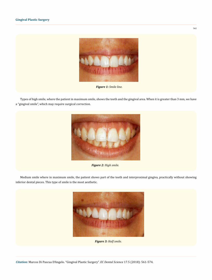
Figure 1: Smile line.
Types of high smile, where the patient in maximum smile, shows the teeth and the gingival area. When it is greater than 3 mm, we have a “gingival smile”, which may require surgical correction.
Figure 2: High smile.
Medium smile where in maximum smile, the patient shows part of the teeth and interproximal gingiva, practically without showing inferior dental pieces. This type of smile is the most aesthetic.
Figure 3: Half smile.

563
Gingival Plastic Surgery
Citation: Marcos Di Pascua D’Angelo. “Gingival Plastic Surgery”. EC Dental Science 17.5 (2018): 561-574.
Low smile is where the patient in maximum smile only shows the incisal third of the upper anterior pieces [3].
Figure 4: Low smile.
Another esthetic gingival parameter to study in each patient is the zenith that we can define as the most apical point of the free mar-ginal gingiva in relation to the vertical axis of the tooth, where in the central incisors and the upper canines it is more apical than the teeth. upper lateral incisors.
Figure 5: Gingival center.
The zenith may be compromised by restorations and iatrogenic prostheses, but it can and must be restored by means of excellent restorative dentistry and mainly by periodontal plastic surgery [4].
Gingival enlargements
The increase of gingival tissue volume is due to the increase of stroma and not to the number of cells.

564
Gingival Plastic Surgery
Citation: Marcos Di Pascua D’Angelo. “Gingival Plastic Surgery”. EC Dental Science 17.5 (2018): 561-574.
Figure 6: Generalized gingival hyperplasia.
The same can be generalized, localized, marginal or papillary and is caused by different factors such as sustained gingivitis for a long time, taking certain medications such as immunosuppressants, antihypertensives and anticonvulsants, systemic factors such as preg-nancy and mouth breather patients.
Figure 7 and 8: Generalized gingival hyperplasia and its treatment.
The treatment consists of a resective surgery that removes excess tissue [5].
Figure 9 and 10: Localized gingival hyperplasia.
Elimination of bridles and flanges with high insertion
The existence of anomalous braces can cause diastema (spaces) between the anterior teeth and limit the mobility of the lip or tongue.
For these situations, the release of the frenulum is indicated by a frenectomy.

565
Gingival Plastic Surgery
Citation: Marcos Di Pascua D’Angelo. “Gingival Plastic Surgery”. EC Dental Science 17.5 (2018): 561-574.
Figure 11: Upper insertion lip brace.
The surgical technique can be in different ways depending on the particular case and the possibility of applying one or the other, exist-ing the rhomboid or diamond flap, technique of apical reposition of the flap, zheplasty of the frenum and the use as a cutting element and hemostasis a laser.
Figure 12 to 16: Surgical technique of elimination of the upper labial frenum.

566
Gingival Plastic Surgery
Citation: Marcos Di Pascua D’Angelo. “Gingival Plastic Surgery”. EC Dental Science 17.5 (2018): 561-574.
The ankyloglossia is an abnormality present from birth in which the membrane that joins the tongue to the floor of the mouth (lingual frenulum) is too short, which prevents the tongue protruding and have normal mobility.
Figure 17: Short lingual frenulum in child.
The most common symptom in babies is the difficulty with breastfeeding due to problems of latching onto the nipple.
Figure 18: Short lingual frenulum in infant.
It can develop difficulties in speech, specifically articulation problems of sounds that require the elevation of the tongue.
Figure 19 to 24: Surgical technique of short lingual frenulum removal.

567
Gingival Plastic Surgery
Citation: Marcos Di Pascua D’Angelo. “Gingival Plastic Surgery”. EC Dental Science 17.5 (2018): 561-574.
It can interfere with a normal swallowing, preventing the tongue from coming into contact with the roof of the mouth [6].
Increase of the edentulous ridge
The alveolar ridge may be deformed as a result of dental extractions, advanced periodontal disease or abscess and cysts formation.
The defects may involve loss of the vestibulolingual dimension, apicocoronary or both. The procedures that correct these deformities include soft tissue grafts (free or pedicled) and bone tissue.
Figure 25: Deficiency of tissue in the lingual vestibule direction.
Figure 26: Compensation of the missing tissue with pink acrylic in the prosthesis.

568
Gingival Plastic Surgery
Citation: Marcos Di Pascua D’Angelo. “Gingival Plastic Surgery”. EC Dental Science 17.5 (2018): 561-574.
Figure 27 to 35: Palate conjunctival graft to improve vestibular defect in osseointegrated implant.
Coronary lengthening, gingivectomy and gingivoplasty
The increase of the clinical crown in periodontal plastic surgery is fundamentally indicated to correct aesthetic anomalies in the dental pieces of the anterior sector that have not completely erupted and are short or have irregular margins.

569
Gingival Plastic Surgery
Citation: Marcos Di Pascua D’Angelo. “Gingival Plastic Surgery”. EC Dental Science 17.5 (2018): 561-574.
We also resort to plastic surgery to expose healthy dental tissue in teeth that are badly damaged and must be reconstructed.
Figure 36 and 37: Gingivoplasty before and after treatment.
Reduces the amount of exposed gum by altering the configuration and shape of the anterior teeth in a favorable way.
For the correct planning of coronary lengthening surgery, we must carry out a detailed individualized study of the relationships be-tween the clinical crown, the dental crown, the root, the alveolar bone and the periodontal biotype.
Gingivectomy is the removal of gingival tissue for the purpose of removing or reducing the periodontal pocket. Gingivoplasty is the remodeling of the gum to achieve a more physiological contour and eliminate gingival defects [7].
Figure 38 and 39: Surgical treatment of gingivectomy.
Covering of exposed roots and implants with free grafts
Oral exposure of the radicular surface caused by the dislocation of the gingival margin in the apical direction to the cementoenamel junction, frequently related to a detriment of the dental and gingival aesthetics, as well as dentin hypersensitivity.
Figure 40: Generalized gingival recessions.

570
Gingival Plastic Surgery
Citation: Marcos Di Pascua D’Angelo. “Gingival Plastic Surgery”. EC Dental Science 17.5 (2018): 561-574.
The tooth becomes more sensitive to cold and is at risk of having root caries.
It is common that over time they increase in size and compromise neighboring teeth.
Among the most frequent causes we have traumatic brushing, sustained gingival inflammation, alteration of the occlusion, bruxism and iatrogenic orthodontic movements.
All this associated with a very thin type of gum.
Classification of recessions
Class I: would be equivalent to a recession of marginal tissue that does not extend to the mucogingival line, and in which there is no bone loss or interdental soft tissue.
Figure 41: Miller’s Class I.
Class II: corresponds to recessions of marginal tissue that do extend or exceed the mucogingival line, and in which there is no bone loss or interdental soft tissue.
Figure 42: Miller’s Class II.

571
Gingival Plastic Surgery
Citation: Marcos Di Pascua D’Angelo. “Gingival Plastic Surgery”. EC Dental Science 17.5 (2018): 561-574.
Class III: Recession of marginal tissue that reaches or exceeds the mucogingival line, affecting the interproximal bone or the papilla.
Figure 43: Miller’s Class III.
Class IV: Recession of marginal tissue that reaches or exceeds the mucogingival line, affecting the interproximal bone and the papilla [8].
Figure 44: Miller’s Class IV.
The treatment consists of eliminating the causal factor and performing a plastic surgery that consists of taking a gum graft (usually from the palate of the patient) and its placement on the root surface. With this, the lost tissue is recovered in a high percentage and the thickness of the gum increases [9].

572
Gingival Plastic Surgery
Citation: Marcos Di Pascua D’Angelo. “Gingival Plastic Surgery”. EC Dental Science 17.5 (2018): 561-574.
Figure 45 to 50: Palate conjunctival graft for root coating in sensitive area.10.
Upper lip repositioning
Excessive exposure of the gum can be addressed by several therapeutic modalities, depending on the specific diagnosis.
The superior labial repositioning is carried out by removing a mucosal band from the upper vestibule, and performing the posterior suture of the labial mucosa to the mucogingival line [11].

573
Gingival Plastic Surgery
Citation: Marcos Di Pascua D’Angelo. “Gingival Plastic Surgery”. EC Dental Science 17.5 (2018): 561-574.
Figure 51 and 52: Extremely high gingival smiles.
This results in a narrower vestibule and a restriction of muscle traction, which reduces the gingival exposure when smiling.
Figure 53 and 54: Treatment of gingivectomy, upper lip repositioning and hyaluronic acid in lips.
ConclusionsIn 1993, Miller stated that periodontal plastic surgery was the most appropriate term to define mucogingival surgery described by
Friedman in 1957, because it is a surgical procedure that goes beyond the traditional treatment of problems related to the amount of gingiva and gingival recessions, since it also includes the correction of alveolar ridge shape and soft tissue aesthetics.
Some of the consequences of gingival recessions are radicular hypersensitivity, caries in the cervical area, and the aesthetic problem due to root exposure. The techniques used in root coverage are intended not only to cover the root surface, but also to modify the peri-odontal biotype of the area, generating an increase in the thickness and height of the soft tissues.
Many of these techniques are conditioned by the skill of the operator and postoperative care by the patient. These two aspects, as well as a good diagnosis of the etiology, surgical possibilities and prognosis are essential to obtain a good functional and aesthetic result, satisfying the patient’s long-term demand.
Bibliography
1. Miller PD. “Root coverage grafting for regeneration and aesthetics”. Periodontology 2000 1 (1993): 118-127.
574
Gingival Plastic Surgery
Citation: Marcos Di Pascua D’Angelo. “Gingival Plastic Surgery”. EC Dental Science 17.5 (2018): 561-574.
2. Ker AJ., et al. “Esthetics and smile characteristics from the lay person’s perspective: a computer-based survey study”. Journal of the American Dental Association 139.10 (2008): 1318-1327.
3. Blanco O and Solorzano A. “Estética en Odontología, parte II Papel de los principios estéticos de la Odontología”. Acta Odontológica Venezolana 37.3 (1999).
4. Hatjó J. “A Beleza Natural dos Dentes Anteriores”. São Paulo: Santos (2008).
5. Atlas de las enfermedades de la mucosa oral (4th edition). Barcelona: Salvat (1986).
6. Cuestas Giselle., et al. “Tratamiento quirúrgico del frenillo lingual corto en niños”. Archivos Argentinos de Pediatria 112.6 (2013).
7. Carranza F., et al. ‘Principios generales de la cirugía periodontal”. En: Carranza FA. Periodontología clínica. 9th edición. México D.F.: McGraw Hill (2003).
8. Lindhe J., et al. “Terapia mucogingival”. En: Periodontología clínica e implantología odontológica, 3rd edition. Editorial Panamericana.
9. The American Academy of Periodontology (US). “Parameter on Mucogingival Conditions”. Journal of Periodontology 71.5 (2000): 861-862.
10. Zuchelli G and De Sanctus M. “Treatment of multiple recession-type defects in patients with esthetic demands”. Journal of Periodontol-ogy 71.9 (2000): 1506-1514.
11. Chacón Martínez H., et al. “Simplificando el tratamiento quirúrgico de la sonrisa gingival”. Cirugía Plástica Iberolatinoamericana 37.1 (2011).
Volume 17 Issue 5 May 2018©All rights reserved by Marcos Di Pascua D’Angelo.