Open Access Journal Indian J Medical Research and ...ijmrps.com/Issues...
12
Open Access Journal Indian Journal of Medical Research and Pharmaceutical Sciences September 2016; 3(9) ISSN: ISSN: 2349-5340 DOI: 10.5281/zenodo.61775 sImpact Factor: 3.052 © Indian Journal of Medical Research and Pharmaceutical Sciences http://www.ijmprs.com/ [16] RESISTANCE TRAINING FOR MUSCLE STRENGTH AND LEAN MASS IN ADULTS OLDER THAN 60 YEARS – A SYSTEMATIC REVIEW Maria da Lapa Rosado * , Maria Teresa Tomás ** , Sílvia Collaço Correia * , Cristina Ribeiro Gonçalves * , Mónica Henriques de Abreu * , Susana Ferreira Cardoso * * Physiotherapy Department, Alcoitão School of Health Sciences, Alcabideche, Portugal ** Interdisciplinary Center for the Study of Human Movement at Faculty of Human Kinetics at Lisbon University, Cruz-Quebrada, Portugal Abstract Keywords: Resistance training; Body Composition; Sarcopenia; Older adults. Objectives: Verify the effect of resistance training (RT) in muscle mass and muscle strength in older adults. Methods: Randomized Controlled Trials (RCT) published between 2005 and 2015, with a study population aged 65 and up that went through an RT based intervention were analysed. Body composition should be assessed by Dual Energy X-ray Absorptiometry or Computed Tomography Scan. Internal validity of each article was assessed using the PEDro scale. Results: Five RCTs whit score of 5/10 met the inclusion criteria and globally 162 participants were assessed. Each study was based on a RT program of 6-16 weeks of 2-3times/week. Discussion Main results show that high intensity and even low intensity RT, increased muscle mass, cross sectional area, strength of the quadriceps and functionality. RT has shown great outcomes in preventing sarcopenia. Results magnitude is proportional to RT characteristics. Introduction Sarcopenia, first coined by Irwin Rosenberg in 1989, is now accepted to describe the involuntary loss of skeletal muscle mass and muscle strength and function during aging 1 . Is a complex medical condition that leads to loss of independence, high risk of falls, decreased quality of life, increased expenses in health and possibly increased mortality 2 . Rate of loss is estimated to be 1%–2% per year after the age of 50, especially in the lower limbs, in conjunction with strength declines of 1.5% per year that accelerates to 3% annually after the age of 60. These losses result in a decreased total muscle cross sectional area (CSA) of approximately 40% between 20 and 60 years of age 1 . While this decline occurs gradually in men, it is quite rapid in women, especially after menopause 2 . It has been estimated that up to 5%-15% of people older than 65 years and 11% - 50% of people older than 80 years have sarcopenia 1,3 . Some data reports that 53% males and 43% females older than the age of 80 were sarcopenic 1 . Sarcopenia can be considered ‘primary’ (or age-related) when no other cause is evident but aging itself, or ‘secondary’ when one or more causes are evident. In general, etiology of sarcopenia is multi -factorial becoming difficult to characterize each individual as having a primary or secondary condition. This situation is consistent with recognizing sarcopenia as a multi-faceted geriatric syndrome 4 . Multiple factors appear to be involved in the development of this condition including loss of muscle fibers (mainly type II), changes in muscle fiber quantity and quality, protein synthesis rates, inflammation and altered hormonal levels 1,5 . Two other major risk factors are under-nutrition and obesity 6 . Female gender or some organ diseases, such as cancer, hypoxia-related diseases, diabetes mellitus II, kidney disease and/or kidney failure and HIV may be predisposing factors to sarcopenia 2,6 . Several authors agree that the most prominent cause of sarcopenia is physical inactivity 3,6 . At present there are no standardized diagnostic criteria for sarcopenia, although the following criteria could be used: walking speed below 0.8 m/s in the 4 meter walking test, decreased handgrip strength below 26kg in males or 16kg
Transcript of Open Access Journal Indian J Medical Research and ...ijmrps.com/Issues...

Open Access Journal
Indian Journal of Medical Research and Pharmaceutical Sciences September 2016; 3(9) ISSN: ISSN: 2349-5340 DOI: 10.5281/zenodo.61775 sImpact Factor: 3.052
© Indian Journal of Medical Research and Pharmaceutical Sciences http://www.ijmprs.com/
[16]
RESISTANCE TRAINING FOR MUSCLE STRENGTH AND LEAN MASS
IN ADULTS OLDER THAN 60 YEARS – A SYSTEMATIC REVIEW
Maria da Lapa Rosado*, Maria Teresa Tomás**, Sílvia Collaço Correia*, Cristina Ribeiro
Gonçalves*, Mónica Henriques de Abreu*, Susana Ferreira Cardoso* * Physiotherapy Department, Alcoitão School of Health Sciences, Alcabideche, Portugal ** Interdisciplinary Center for the Study of Human Movement at Faculty of Human Kinetics at Lisbon University,
Cruz-Quebrada, Portugal
Abstract
Keywords:
Resistance training; Body
Composition; Sarcopenia;
Older adults.
Objectives: Verify the effect of resistance training (RT) in muscle mass and muscle
strength in older adults. Methods: Randomized Controlled Trials (RCT) published
between 2005 and 2015, with a study population aged 65 and up that went through an
RT based intervention were analysed. Body composition should be assessed by Dual
Energy X-ray Absorptiometry or Computed Tomography Scan. Internal validity of
each article was assessed using the PEDro scale. Results: Five RCTs whit score of
5/10 met the inclusion criteria and globally 162 participants were assessed. Each
study was based on a RT program of 6-16 weeks of 2-3times/week. Discussion Main
results show that high intensity and even low intensity RT, increased muscle mass,
cross sectional area, strength of the quadriceps and functionality. RT has shown great
outcomes in preventing sarcopenia. Results magnitude is proportional to RT
characteristics.
Introduction Sarcopenia, first coined by Irwin Rosenberg in 1989, is now accepted to describe the involuntary loss of skeletal
muscle mass and muscle strength and function during aging1. Is a complex medical condition that leads to loss of
independence, high risk of falls, decreased quality of life, increased expenses in health and possibly increased
mortality2.
Rate of loss is estimated to be 1%–2% per year after the age of 50, especially in the lower limbs, in conjunction with
strength declines of 1.5% per year that accelerates to 3% annually after the age of 60. These losses result in a
decreased total muscle cross sectional area (CSA) of approximately 40% between 20 and 60 years of age 1. While
this decline occurs gradually in men, it is quite rapid in women, especially after menopause 2. It has been estimated
that up to 5%-15% of people older than 65 years and 11% - 50% of people older than 80 years have sarcopenia 1,3.
Some data reports that 53% males and 43% females older than the age of 80 were sarcopenic 1.
Sarcopenia can be considered ‘primary’ (or age-related) when no other cause is evident but aging itself, or
‘secondary’ when one or more causes are evident. In general, etiology of sarcopenia is multi-factorial becoming
difficult to characterize each individual as having a primary or secondary condition. This situation is consistent with
recognizing sarcopenia as a multi-faceted geriatric syndrome 4.
Multiple factors appear to be involved in the development of this condition including loss of muscle fibers (mainly
type II), changes in muscle fiber quantity and quality, protein synthesis rates, inflammation and altered hormonal
levels 1,5.
Two other major risk factors are under-nutrition and obesity 6. Female gender or some organ diseases, such as
cancer, hypoxia-related diseases, diabetes mellitus II, kidney disease and/or kidney failure and HIV may be
predisposing factors to sarcopenia 2,6. Several authors agree that the most prominent cause of sarcopenia is physical
inactivity 3,6.
At present there are no standardized diagnostic criteria for sarcopenia, although the following criteria could be used:
walking speed below 0.8 m/s in the 4 meter walking test, decreased handgrip strength below 26kg in males or 16kg
Open Access Journal
Indian Journal of Medical Research and Pharmaceutical Sciences September 2016; 3(9) ISSN: ISSN: 2349-5340 DOI: 10.5281/zenodo.61775 sImpact Factor: 3.052
© Indian Journal of Medical Research and Pharmaceutical Sciences http://www.ijmprs.com/
[17]
in females, a distance in six minutes’ walk test lower than 400 m and specially low appendicular lean mass 7,8.
Reference values depends on population and assessment methodologies, especially for muscle mass 9.
A growing body of evidence indicates that physical activity can slow sarcopenia and recent evidence on RT seems
to support earlier research showing that RT may be the most effective strategy to prevent or/and treat sarcopenia
through muscle hypertrophy and increased muscular strength and power 1.
The purposes of this systematic review were to verify the effect of RT, in adults older than 60 in muscle strength and
lean mass.
Materials and methods This systematic review followed Preferred Reporting Items for Systematic Reviews and Meta-Analyses (PRISMA) 10 to conduct this review. A bibliographic database search of PubMed, PEDro and Cochrane Library was performed
to identify RCTs of Resistance Training (RT) or Strength Training (ST) in older adults (age 60 and up) with no prior
exercise training and with assessment of body composition performed by DEXA, CT scan, ultrasonography or
magnetic resonance image (MRI), published between 2005 and 2015 in English, French, Portuguese or Spanish.
RCTs where study population have diagnosis of Diabetes Mellitus type II, Cancer, Obesity or Osteoporosis and
studies with included diet, nutrition, medication or supplements in the intervention or interventions not based only in
RT were not eligible for analysis (exclusion criteria).
Study Selection
Selection of studies was performed by two of the reviewers, to avoid the exclusion of relevant articles. When the
reviewers did not reach a consensus, other independent reviewer was contacted.
PEDro scale for assessing RCTs has sufficient reliability to be used in systematic reviews. It should be applied by
more than one reviewer individually, followed by a discussion in order to analyse the results and see the level of
agreement between the assessors 11.
This scale includes criteria of internal validation assessment and statistical analysis. Each satisfied item adds 1 point
to the total score; with the exception of item one, as it assesses external validity. If the criterion is not met, then no
points are added.
In this systematic review, four independent reviewers (SFC, CG, MA, and SMC) read and evaluated the 5 studies in
accordance with the PEDro scale. When a consensus was not reached, consultation by an independent reviewer
(MLR) was required.
Results A total of 340 manuscripts were found (figure 1). After applying all the inclusion and exclusion criteria and
excluding all the duplicates a total of 5 RCTs were analysed.

Open Access Journal
Indian Journal of Medical Research and Pharmaceutical Sciences September 2016; 3(9) ISSN: ISSN: 2349-5340 DOI: 10.5281/zenodo.61775 sImpact Factor: 3.052
© Indian Journal of Medical Research and Pharmaceutical Sciences http://www.ijmprs.com/
[18]
Figure (1): Flowchart for selection of papers
All five studies selected for the review were RCTs published in English and released between 2009 and 2014.
A final PEDro score of 5/10 for all studies was obtained (table 1), which according Maher et al 11 is of “moderate
reliability”, which indicates that the results tend to show greater effects of intervention than it really is.
Table 1 – The PEDro scale summary of included studies
Criteria
(Bickel et al.,
2011)
(Fragala et al.,
2014)
(Scanlon et al.,
2014)
(Mueller et al.,
2009)
(Watanabe et al.,
2014)
1 Yes Yes Yes Yes Yes
2 Yes Yes Yes Yes Yes
3 No No No No No
4 Yes Yes Yes Yes Yes
5 No No No No No
6 No No No No No
7 Yes No No No No
8 No Yes Yes Yes Yes
9 No No No No No

Open Access Journal
Indian Journal of Medical Research and Pharmaceutical Sciences September 2016; 3(9) ISSN: ISSN: 2349-5340 DOI: 10.5281/zenodo.61775 sImpact Factor: 3.052
© Indian Journal of Medical Research and Pharmaceutical Sciences http://www.ijmprs.com/
[19]
A total of 201 participants were analysed (Table 2) but only 162 participants were included, as one trial 12 included
young and older participants (39 of the 70 participants in this study were outside the age criterion). All trials except
one 12 analysed subjects of both genders, with ages over 60 years 12. Although our PICO question (Population,
Intervention, Comparator and Outcome) included participants with age over 65 years and some studies presented
some participants with 60 years, it was not exclusion criterion, as the rest of the sample in the study covered the
inclusion criteria (RCTs median age was superior to 65 years).
There was a considerable diversity in the frequency, intensity, and duration of interventions (Table 3). Some studies
recommend RT frequency of twice a week 13–16 and only one study suggested a frequency of three times per week 12.
Duration of different exercise training programs varied between 6 weeks 13,15, 12 weeks 14,16 and 48 weeks 12. Three
of the studies 12–14 followed the recommended guidelines for older adults by the American College of Sports
Medicine 17.
Two studies 13,14 reported supervision in exercise training program by a certified strength and conditioning specialist.
Others exercise training programs were supervised by one coach per two participants 16, or a direct supervision from
a clinical exercise physiologist 12. In one study the level of supervision was unclear 15.
10 Yes Yes Yes Yes Yes
11 Yes Yes Yes Yes Yes
Total score 5/10 5/10 5/10 5/10 5/10

Open Access Journal
Indian Journal of Medical Research and Pharmaceutical Sciences September 2016; 3(9) ISSN: ISSN: 2349-5340
DOI: 10.5281/zenodo.61775 sImpact Factor: 3.052
© Indian Journal of Medical Research and Pharmaceutical Sciences http://www.ijmprs.com/
[20]
Table 2 – Characteristics of different samples of analysed studies.
Reference Sample Characteristics
Strength Characteristics
Body Composition Characteristics Population; Number studied (M/F);
Age (y) (Mean ± SD) [Range]
Mueller et al.
2009
Men & women with stable medication and health
conditions;
Group RT: n = 23 (10/13); (80.1 ± 0.8 y)
Group EET: n = 23 (10/13); (80.3 ± 0.7 y)
Group CT: n = 16 (6/10); (81.8 ± 0.8 y)
Maximal isometric extension of the legs
(Nkg-1)
Group RT:
Baseline – 15.3
12 weeks – 15.8
Group EET:
Baseline – 14.8
12 weeks – 16.1
Group CT:
Baseline – 15.7
12 weeks – 15.1
Body fat mass (kg)
Group RT:
Baseline – 19.1
12 weeks – 19.0
Group EET:
Baseline – 17.6
12 weeks – 16.7
Group CT:
Baseline – 18.9
12 weeks – 19.1
Bickel et al.
2011
Community-dwelling;
Group 1: n = 31; (64.1 ± 0.6 y) [60-75]
Group 2: n = 39; (27.5 ± 0.6 y) [20-35]
Knee extension 1RM (kg)
Phase 1:
Group old: Baseline – 36.9 ± 2.1
16 weeks – 51.5 ± 2.9
Group young: Baseline – 54.2 ± 2.54
16 weeks – 76.2 ± 2.9
Tigh lean mass (kg) Phase 1:
Group old: Baseline – 10.72 ± 0.53
16 weeks – 11.16 ± 0.57
Group young: Baseline – 12.43 ± 0.49
16 weeks – 13.13 ± 0.51
Fragala et al.
2014
Community-dwelling;
Group 1: n = 12 (8/4); (70,8 ± 6.8 y)
Group 2: n = 11 (5/6); (69.6 ± 5.5 y)
Hand grip strength (kg)
Group 1:
Baseline – 36.25 ± 10.94
Phase 1 – 39.17 ± 13.58
Phase 2 – 40.08 ± 15.72
Group 2:
Baseline – 32.82 ± 14.17
Phase 1 – 34.18 ± 16.48
Phase 2 – 35.55 ± 15.71
Lean body mass (kg)
Group 1:
Baseline – 47.70 ± 10.67
Phase 1 – 47.92 ± 10.51
Phase 2 – 49.19 ± 10.88
Group 2:
Baseline – 49.58 ± 13.48
Phase 1 – 49.51 ± 13.23
Phase 2 – 48.97 ± 12.84
Scanlon et al.
2014
Healthy men & women;
Group RT: n = 13; (71.1 ± 6.7 y)
Group control: n = 12; (70.1 ± 5.5 y)
Knee extensor strength (kg)
Group RT:
Baseline – 39.20 ± 15.90
6 weeks – 51.70 ± 17.60
Group control:
Tigh lean mass
(kg)
Group RT:
Baseline – 5.4 ± 1.3
6 weeks – 5.4 ± 1.3
Lean body mass (kg)
Group RT:
Baseline – 47.6 ± 10.6
6 weeks – 47.9 ± 10.5
Group control:
Open Access Journal
Indian Journal of Medical Research and Pharmaceutical Sciences September 2016; 3(9) ISSN: ISSN: 2349-5340
DOI: 10.5281/zenodo.61775 sImpact Factor: 3.052
© Indian Journal of Medical Research and Pharmaceutical Sciences http://www.ijmprs.com/
[21]
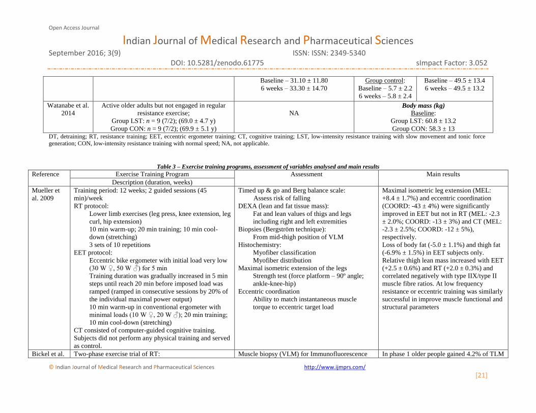
Baseline – 31.10 ± 11.80
6 weeks – 33.30 ± 14.70
Group control:
Baseline – 5.7 ± 2.2
6 weeks – 5.8 ± 2.4
Baseline – 49.5 ± 13.4
6 weeks – 49.5 ± 13.2
Watanabe et al.
2014
Active older adults but not engaged in regular
resistance exercise;
Group LST: n = 9 (7/2); (69.0 ± 4.7 y)
Group CON: n = 9 (7/2); (69.9 ± 5.1 y)
NA Body mass (kg)
Baseline:
Group LST: 60.8 ± 13.2
Group CON: 58.3 ± 13 DT, detraining; RT, resistance training; EET, eccentric ergometer training; CT, cognitive training; LST, low-intensity resistance training with slow movement and tonic force
generation; CON, low-intensity resistance training with normal speed; NA, not applicable.
Table 3 – Exercise training programs, assessment of variables analysed and main results
Reference Exercise Training Program Assessment Main results
Description (duration, weeks)
Mueller et
al. 2009
Training period: 12 weeks; 2 guided sessions (45
min)/week
RT protocol:
Lower limb exercises (leg press, knee extension, leg
curl, hip extension)
10 min warm-up; 20 min training; 10 min cool-
down (stretching)
3 sets of 10 repetitions
EET protocol:
Eccentric bike ergometer with initial load very low
(30 W ♀, 50 W ♂) for 5 min
Training duration was gradually increased in 5 min
steps until reach 20 min before imposed load was
ramped (ramped in consecutive sessions by 20% of
the individual maximal power output)
10 min warm-up in conventional ergometer with
minimal loads (10 W ♀, 20 W ♂); 20 min training;
10 min cool-down (stretching)
CT consisted of computer-guided cognitive training.
Subjects did not perform any physical training and served
as control.
Timed up & go and Berg balance scale:
Assess risk of falling
DEXA (lean and fat tissue mass):
Fat and lean values of thigs and legs
including right and left extremities
Biopsies (Bergström technique):
From mid-thigh position of VLM
Histochemistry:
Myofiber classification
Myofiber distribution
Maximal isometric extension of the legs
Strength test (force platform – 90º angle;
ankle-knee-hip)
Eccentric coordination
Ability to match instantaneous muscle
torque to eccentric target load
Maximal isometric leg extension (MEL:
+8.4 ± 1.7%) and eccentric coordination
(COORD: -43 ± 4%) were significantly
improved in EET but not in RT (MEL: -2.3
± 2.0%; COORD: -13 ± 3%) and CT (MEL:
-2.3 ± 2.5%; COORD: -12 ± 5%),
respectively.
Loss of body fat (-5.0 ± 1.1%) and thigh fat
(-6.9% ± 1.5%) in EET subjects only.
Relative thigh lean mass increased with EET
(+2.5 ± 0.6%) and RT (+2.0 ± 0.3%) and
correlated negatively with type IIX/type II
muscle fibre ratios. At low frequency
resistance or eccentric training was similarly
successful in improve muscle functional and
structural parameters
Bickel et al. Two-phase exercise trial of RT: Muscle biopsy (VLM) for Immunofluorescence In phase 1 older people gained 4.2% of TLM
Open Access Journal
Indian Journal of Medical Research and Pharmaceutical Sciences September 2016; 3(9) ISSN: ISSN: 2349-5340
DOI: 10.5281/zenodo.61775 sImpact Factor: 3.052
© Indian Journal of Medical Research and Pharmaceutical Sciences http://www.ijmprs.com/
[22]
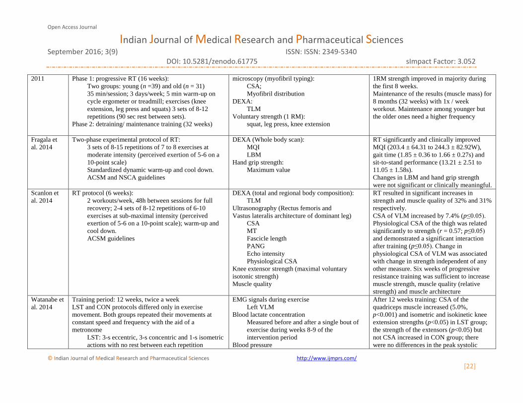
2011 Phase 1: progressive RT (16 weeks):
Two groups: young (n =39) and old (n = 31)
35 min/session; 3 days/week; 5 min warm-up on
cycle ergometer or treadmill; exercises (knee
extension, leg press and squats) 3 sets of 8-12
repetitions (90 sec rest between sets).
Phase 2: detraining/ maintenance training (32 weeks)
microscopy (myofibril typing):
CSA;
Myofibril distribution
DEXA:
TLM
Voluntary strength (1 RM):
squat, leg press, knee extension
1RM strength improved in majority during
the first 8 weeks.
Maintenance of the results (muscle mass) for
8 months (32 weeks) with 1x / week
workout. Maintenance among younger but
the older ones need a higher frequency
Fragala et
al. 2014
Two-phase experimental protocol of RT:
3 sets of 8-15 repetitions of 7 to 8 exercises at
moderate intensity (perceived exertion of 5-6 on a
10-point scale)
Standardized dynamic warm-up and cool down.
ACSM and NSCA guidelines
DEXA (Whole body scan):
MQI
LBM
Hand grip strength:
Maximum value
RT significantly and clinically improved
MQI (203.4 ± 64.31 to 244.3 ± 82.92W),
gait time (1.85 ± 0.36 to 1.66 ± 0.27s) and
sit-to-stand performance (13.21 ± 2.51 to
11.05 ± 1.58s).
Changes in LBM and hand grip strength
were not significant or clinically meaningful.
Scanlon et
al. 2014
RT protocol (6 weeks):
2 workouts/week, 48h between sessions for full
recovery; 2-4 sets of 8-12 repetitions of 6-10
exercises at sub-maximal intensity (perceived
exertion of 5-6 on a 10-point scale); warm-up and
cool down.
ACSM guidelines
DEXA (total and regional body composition):
TLM
Ultrasonography (Rectus femoris and
Vastus lateralis architecture of dominant leg)
CSA
MT
Fascicle length
PANG
Echo intensity
Physiological CSA
Knee extensor strength (maximal voluntary
isotonic strength)
Muscle quality
RT resulted in significant increases in
strength and muscle quality of 32% and 31%
respectively.
CSA of VLM increased by 7.4% (p≤0.05).
Physiological CSA of the thigh was related
significantly to strength (r = 0.57; p≤0.05)
and demonstrated a significant interaction
after training (p≤0.05). Change in
physiological CSA of VLM was associated
with change in strength independent of any
other measure. Six weeks of progressive
resistance training was sufficient to increase
muscle strength, muscle quality (relative
strength) and muscle architecture
Watanabe et
al. 2014
Training period: 12 weeks, twice a week
LST and CON protocols differed only in exercise
movement. Both groups repeated their movements at
constant speed and frequency with the aid of a
metronome
LST: 3-s eccentric, 3-s concentric and 1-s isometric
actions with no rest between each repetition
EMG signals during exercise
Left VLM
Blood lactate concentration
Measured before and after a single bout of
exercise during weeks 8-9 of the
intervention period
Blood pressure
After 12 weeks training: CSA of the
quadriceps muscle increased (5.0%,
p<0.001) and isometric and isokinetic knee
extension strengths (p<0.05) in LST group;
the strength of the extensors (p<0.05) but
not CSA increased in CON group; there
were no differences in the peak systolic
Open Access Journal
Indian Journal of Medical Research and Pharmaceutical Sciences September 2016; 3(9) ISSN: ISSN: 2349-5340
DOI: 10.5281/zenodo.61775 sImpact Factor: 3.052
© Indian Journal of Medical Research and Pharmaceutical Sciences http://www.ijmprs.com/
[23]
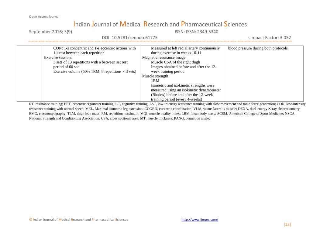
CON: 1-s concentric and 1-s eccentric actions with
1-s rest between each repetition
Exercise session:
3 sets of 13 repetitions with a between set rest
period of 60 sec
Exercise volume (50% 1RM, 8 repetitions × 3 sets)
Measured at left radial artery continuously
during exercise in weeks 10-11
Magnetic resonance image
Muscle CSA of the right thigh
Images obtained before and after the 12-
week training period
Muscle strength
1RM
Isometric and isokinetic strengths were
measured using an isokinetic dynamometer
(Biodex) before and after the 12-week
training period (every 4-weeks)
blood pressure during both protocols.
RT, resistance training; EET, eccentric ergometer training; CT, cognitive training; LST, low-intensity resistance training with slow movement and tonic force generation; CON, low-intensity
resistance training with normal speed; MEL, Maximal isometric leg extension; COORD, eccentric coordination; VLM, vastus lateralis muscle; DEXA, dual-energy X-ray absorptiometry;
EMG, electromyography; TLM, thigh lean mass; RM, repetition maximum; MQI; muscle quality index; LBM, Lean body mass; ACSM, American College of Sport Medicine; NSCA,
National Strength and Conditioning Association; CSA, cross sectional area; MT, muscle thickness; PANG, pennation angle;
Open Access Journal
Indian Journal of Medical Research and Pharmaceutical Sciences September 2016; 3(9) ISSN: ISSN: 2349-5340 DOI: 10.5281/zenodo.61775 sImpact Factor: 3.052
© Indian Journal of Medical Research and Pharmaceutical Sciences http://www.ijmprs.com/
[24]
Discussion Sarcopenia is a growing societal healthcare problem due to rapid expansion of the elderly population and the limited
number of therapeutic approaches to this problem 6. Evidence has shown that older adults, who are less physically
active, are more likely to have lower skeletal muscle mass and strength and are at an increased risk of developing
sarcopenia 5.
Several studies show that RT increases the functionality of the elderly and that participation earlier in life may
provide superior effectiveness in the prevention / treatment of sarcopenia 18. According ACSM 19, effectiveness and
outcomes of such exercise depends on several factors: intensity, training volume, frequency and type of exercises
recommended, recovery time between workouts and frequency of training.
In a systematic review in 2013 authors referred that RT should be done two days or more per week, using one set of
8-10 exercises for the whole body and at moderate to vigorous level of effort enabling 8-12 repetitions 2. The type of
exercises recommended includes strengthening of the entire body with progressive strength or RT and other
activities involving large muscle groups. The RT in these five studies meets the standard criteria of two or more
days per week and more than one set, but some studies referred to perform more than 12 repetitions, because
intensity was lower 14,15.
In two studies 13,14, the RT duration was 6 weeks, but the number of repetitions was different ranging between 3 sets
of 7-8 exercises with 8-15 repetitions at moderate intensity 13 and 2–4 sets of 6-10 exercises with 8–12 repetitions
with an intensity at 85% of 1RM. Despite the difference between them, both RTs showed an increase in the MQI of
18% in group 1 and 31% in group 2. The different results in these may be due to the different intensities of training.
Those results are in accordance with ACSM guidelines 19, where high-intensity RT showed better results than low
intensity RT training. The study of Mueller et al. 16 used a low intensity for the RT. The same low intensity used by
Watanabe et al. (30% 1RM) was maintained during the entire RT program. Both authors referred an increment in
maximal isometric extension strength but in the study of Mueller et al. (2009) the increment in strength was
significant only in the group performing eccentric exercise. This show the importance of intensity of RT for
improvements in strength and confirms also the importance of intensities higher than 40% of 1RM for a significant
improvement in muscle strength 20. While low intensity resistance training is not as effective as higher intensity
training, it still shows some benefits, which may be of significant importance to older adults who are not able to
perform exercise at higher intensities. In general, all the five studies showed increases in strength between 2% and
25% and in muscle quality between 18% and 31.5%. Thus, it seems that 6 weeks of RT is sufficient to increase
muscle strength in elderly and the majority of the strength improvement occurs during the first 8 weeks. This is in
accordance with physiological changes since in the first phase of an exercise training program directed for muscle
strength, the improvements in muscle strength seems to be primarily due to neuromuscular adaptations 21,22 and only
after 6-8 weeks the increments in muscle strength are due to increments in muscle mass.
One systematic review has found that an average of 20.5 weeks of RT has produced a significant main effect equal
to a 1.1 kilogram increase in LBM in aging men and women 18 particularly in programs with higher volume of
training.
From the analyses of these 5 studies it seems that the duration of 6 weeks of RT was not enough to increase LBM.
Only three studies show increments in lean mass and they used duration of 12 weeks of training 15,16 or 16 weeks 12.
This confirms the importance and contribution of duration of RT programs for the increments in strength, showing
that only after larger periods of time in training the strength will be due to increments of muscle mass (more
contractile mass) and not only to neuromuscular adaptations.
As a limitation in this systematic review we do not exclude the possibility of not identifying all of the relevant
studies since the language of all the included studies were only English, Spanish, French and Portuguese due to
unfamiliarity of reviewers with other languages. Also we must not forget that a general limitation is that analysis is
that do not infer a causal-effect since RT increases in fact increase strength and lean mass which in turn enables an
increase in functional capacity of participants also enabling many other behavioural interventions (for example
nutritional interventions) that will promote increments in strength and mass and functionality. This should be
planned in very old populations or in those whit difficulties in engaging RT programmes.

Open Access Journal
Indian Journal of Medical Research and Pharmaceutical Sciences September 2016; 3(9) ISSN: ISSN: 2349-5340 DOI: 10.5281/zenodo.61775 sImpact Factor: 3.052
© Indian Journal of Medical Research and Pharmaceutical Sciences http://www.ijmprs.com/
[25]
Conclusion Results of current systematic review suggest that RT improves strength and lean mass in older adults which
attenuates the development of sarcopenia. However, they should have duration equal or higher than 12 weeks in
order to achieve an improvement in muscle mass which could contribute even more for the increments in strength.
The increments in strength seems to be achievable in programs of 6 weeks of duration but the intensity should be
higher than 30% of 1RM and should be progressively incremented in order to achieve a progressive adaptation.
References 1. Waters DL, Baumgartner RN, Garry PJ, Vellas B. Advantages of dietary, exercise-related, and therapeutic
interventions to prevent and treat sarcopenia in adult patients: an update. Clinical interventions in aging.
2010;5:259-270. doi:10.2147/CIA.S6920.
2. Montero-Fernández N, Serra-Rexach JA. Role of exercise on sarcopenia in the elderly. European Journal of
Physical and Rehabilitation Medicine. 2013;49:131-143. doi:10.1093/fampra/cmr063.
3. Morley JE. Undernutrition in older adults. Family Practice. 2012;29(SUPPL. 1).
doi:10.1093/fampra/cmr054.
4. Woo J. 2013: That was the year that was. Age and Ageing. 2014;43(2):152-156. doi:10.1093/ageing/aft215.
5. Burton LA, Sumukadas D. Optimal management of sarcopenia. Clinical interventions in aging. 2010;5:217-
228. doi:10.2147/CIA.S11473.
6. Buford TW, Anton SD, Judge AR, et al. Models of accelerated sarcopenia: Critical pieces for solving the
puzzle of age-related muscle atrophy. Ageing Research Reviews. 2010;9(4):369-383.
doi:10.1016/j.arr.2010.04.004.
7. Cooper C, Fielding R, Visser M, et al. Tools in the assessment of sarcopenia. Calcified Tissue International.
2013;93(3):201-210. doi:10.1007/s00223-013-9757-z.
8. Cruz-Jentoft AJ, Baeyens JP, Bauer JM, et al. Sarcopenia: European consensus on definition and diagnosis:
Report of the European Working Group on Sarcopenia in Older People. Age and ageing. 2010;39(4):412-
423. doi:10.1093/ageing/afq034.
9. Bijlsma AY, Meskers CGM, Ling CHY, et al. Defining sarcopenia: the impact of different diagnostic
criteria on the prevalence of sarcopenia in a large middle aged cohort. AGE. 2013;35(3):871-881.
doi:10.1007/s11357-012-9384-z.
10. Moher D, Liberati A, Tetzlaff J, Altman DG, PRISMA Group. Preferred reporting items for systematic
reviews and meta-analyses: the PRISMA statement. PLoS medicine. 2009;6(7):e1000097.
doi:10.1371/journal.pmed.1000097.
11. Maher CG, Sherrington C, Herbert RD, Moseley AM, Elkins M. Reliability of the PEDro scale for rating
quality of randomized controlled trials. Physical therapy. 2003;83(8):713-721. doi:10.1016/s0895-
4356(00)00360-7.
12. Bickel CS, Cross JM, Bamman MM. Exercise dosing to retain resistance training adaptations in young and
older adults. Medicine and Science in Sports and Exercise. 2011;43(7):1177-1187.
doi:10.1249/MSS.0b013e318207c15d.
13. Scanlon TC, Fragala MS, Stout JR, et al. Muscle architecture and strength: Adaptations to short-term
resistance training in older adults. Muscle and Nerve. 2014;49(4):584-592. doi:10.1002/mus.23969.
14. Fragala MS, Fukuda DH, Stout JR, et al. Muscle quality index improves with resistance exercise training in
older adults. Experimental Gerontology. 2014;53:1-6. doi:10.1016/j.exger.2014.01.027.
15. Watanabe Y, Madarame H, Ogasawara R, Nakazato K, Ishii N. Effect of very low-intensity resistance
training with slow movement on muscle size and strength in healthy older adults. Clinical Physiology and
Functional Imaging. 2014;34(6):463-470. doi:10.1111/cpf.12117.
16. Mueller M, Breil FA, Vogt M, et al. Different response to eccentric and concentric training in older men and
women. European Journal of Applied Physiology. 2009;107(2):145-153. doi:10.1007/s00421-009-1108-4.
17. ACSM. American College of Sports Medicine Position Stand. Exercise and physical activity for older
adults. Medicine and science in sports and exercise. 1998;30(6):992-1008.
http://www.ncbi.nlm.nih.gov/pubmed/9624662.
18. Peterson MD, Sen A, Gordon PM. Influence of resistance exercise on lean body mass in aging adults: A
meta-analysis. Medicine and Science in Sports and Exercise. 2011;43(2):249-258.
Open Access Journal
Indian Journal of Medical Research and Pharmaceutical Sciences September 2016; 3(9) ISSN: ISSN: 2349-5340 DOI: 10.5281/zenodo.61775 sImpact Factor: 3.052
© Indian Journal of Medical Research and Pharmaceutical Sciences http://www.ijmprs.com/
[26]
doi:10.1249/MSS.0b013e3181eb6265.
19. ACSM. American College of Sports Medicine position stand. Progression models in resistance training for
healthy adults. Medicine and science in sports and exercise. 2009;41(3):687-708.
doi:10.1249/MSS.0b013e3181915670.
20. Peterson MD, Rhea MR, Sen A, Gordon PM. Resistance exercise for muscular strength in older adults: A
meta-analysis. Ageing Research Reviews. 2010;9(3):226-237. doi:10.1016/j.arr.2010.03.004.
21. Arnold P, Bautmans I. The influence of strength training on muscle activation in elderly persons: A
systematic review and meta-analysis. Experimental Gerontology. 2014;58:58-68.
doi:10.1016/j.exger.2014.07.012.
22. Aagaard P, Suetta C, Caserotti P, Magnusson SP, Kjær M. Role of the nervous system in sarcopenia and
muscle atrophy with aging: Strength training as a countermeasure. Scandinavian Journal of Medicine and
Science in Sports. 2010;20(1):49-64. doi:10.1111/j.1600-0838.2009.01084.x.
Author Biblography
Maria da Lapa Rosado
Physiotherapist
PhD in Physical Activity and Health - Faculty of Human
Kinetics at Lisbon University
Adjunct Professor at Alcoitão School of Health Sciences,
Alcabideche, Portugal
Maria Teresa Tomás
Physiotherapist
MSc in Exercise and Health – Faculty of Human Kinetics at
Lisbon University
PhD in Physical Activity and Health - Faculty of Human
Kinetics at Lisbon University
Member of CIPER (Interdisciplinary Center for the Study of
Human Performance) at Faculty of Human Kinetics at Lisbon
University, Portugal.
Sílvia Collaço Correia
Physiotherapist graduated in 2015 by Alcoitão School of Health
Sciences, Alcabideche, Portugal
Private practice
Cristina Ribeiro Gonçalves
Physiotherapist graduated in 2015 by Alcoitão School of Health
Sciences, Alcabideche, Portugal
Private practice

Open Access Journal
Indian Journal of Medical Research and Pharmaceutical Sciences September 2016; 3(9) ISSN: ISSN: 2349-5340 DOI: 10.5281/zenodo.61775 sImpact Factor: 3.052
© Indian Journal of Medical Research and Pharmaceutical Sciences http://www.ijmprs.com/
[27]
Mónica Henriques de Abreu
Physiotherapist graduated in 2015 by Alcoitão School of Health
Sciences, Alcabideche, Portugal
Private practice
Susana Ferreira Cardoso
Physiotherapist graduated in 2015 by Alcoitão School of Health
Sciences, Alcabideche, Portugal
Private practice