Open Access Full Text Article Two cases of intraocular
4
© 2013 Matsuoka et al, publisher and licensee Dove Medical Press Ltd. This is an Open Access article which permits unrestricted noncommercial use, provided the original work is properly cited. Clinical Ophthalmology 2013:7 691–694 Clinical Ophthalmology Two cases of intraocular lymphoma diagnosed by analyses of vitreous and infusion fluid Masato Matsuoka 1 Hideyuki Yoshida 1 Yuichi Kinoshita 2 Tetsuya Nishimura 1 1 Department of Ophthalmology, 2 Department of Histopathology, Kansai Medical University, Takii Hospital, Osaka, Japan Correspondence: Masato Matsuoka Department of Ophthalmology, Kansai Medical University, Fumizono-cho 10-15, Moriguchi, Osaka 570-8507, Japan Tel +81 6 6992 1001 ext 3324 Fax +81 6 6993 2222 Email [email protected] Purpose: Intraocular lymphomas are rare, and they have poor prognosis. Thus, early diagnosis and treatment are needed. A definitive diagnosis of a lymphoma is based on cytological analysis of the intraocular fluids or tissues. We report two cases of intraocular lymphoma diagnosed by the analyses of vitreous and infusion fluid. Patients: Case 1 was a 66-year-old woman who complained of eye floaters and was found to have diffuse vitreous opacification bilaterally. She received corticosteroid therapy, however the vitreous opacification was not resolved, and her visual acuity (VA) remained reduced. She underwent pars plana vitrectomy (PPV), and vitreous and infusion fluid were collected to determine the cause of the reduced VA. The undiluted vitreous obtained from core PPV was submitted for cytokine analysis, and infusion fluid was obtained from the machine cassette after full PPV and used for cytological analysis. Case 2 was a 62-year-old man referred with low vision and was found to have diffuse vitreous opacification in the right eye and dot hemorrhages in both eyes. Four years earlier, he had been diagnosed with diffuse large B-cell lymphoma of the paranasal sinuses and was in remission after chemotherapy. Because metastasis of the lymphoma was suspected, he underwent PPV, and intraocular samples were collected as in Case 1. Results: Atypical lymphoid cells were detected from the infusion fluid in both cases. The ratio of interleukin (IL)-10 to IL-6 was greater than 1.0 in both cases. These results allowed us to make a diagnosis of intraocular lymphoma: primary intraocular lymphoma in Case 1 and metastatic intraocular lymphoma in Case 2. Conclusion: Vitreous and infusion fluid collected during PPV can be used for diagnosing an intraocular lymphoma. Keywords: intraocular lymphoma, cytological analysis, vitreous, pars plana vitrectomy, infusion fluid Introduction Intraocular lymphomas are rare, and they make up less than 1% of intraocular tumors 1 and 1% of intraocular inflammations. 2 Blurred vision and vitreous opacifications are the most common ocular symptoms and signs of an intraocular lymphoma. 3 The life- time prognosis of this disease is poor: 4 the 5-year survival rate is 61.1% in Japan. 3 Thus, early diagnosis and treatment are needed. A definitive diagnosis of an intraocular lymphoma is based on cytological findings of the intraocular fluid or tissues obtained from a biopsy or pars plana vitrectomy (PPV). 5,6 However, cytological examination can be difficult because only a small amount of sample can be obtained from a biopsy or during PPV. In addition, the mechanical trauma associated with PPV may limit the use of the sample. 7 In addition, Dovepress submit your manuscript | www.dovepress.com Dovepress 691 CASE REPORT open access to scientific and medical research Open Access Full Text Article http://dx.doi.org/10.2147/OPTH.S44353 Clinical Ophthalmology downloaded from https://www.dovepress.com/ by 95.216.75.56 on 18-Nov-2018 For personal use only. 1 / 1
Transcript of Open Access Full Text Article Two cases of intraocular
© 2013 Matsuoka et al, publisher and licensee Dove Medical Press Ltd. This is an Open Access article which permits unrestricted noncommercial use, provided the original work is properly cited.
Clinical Ophthalmology 2013:7 691–694
Clinical Ophthalmology
Two cases of intraocular lymphoma diagnosed by analyses of vitreous and infusion fluid
Masato Matsuoka1
Hideyuki Yoshida1
Yuichi Kinoshita2
Tetsuya Nishimura1
1Department of Ophthalmology, 2Department of Histopathology, Kansai Medical University, Takii Hospital, Osaka, Japan
Correspondence: Masato Matsuoka Department of Ophthalmology, Kansai Medical University, Fumizono-cho 10-15, Moriguchi, Osaka 570-8507, Japan Tel +81 6 6992 1001 ext 3324 Fax +81 6 6993 2222 Email [email protected]
Purpose: Intraocular lymphomas are rare, and they have poor prognosis. Thus, early diagnosis
and treatment are needed. A definitive diagnosis of a lymphoma is based on cytological analysis
of the intraocular fluids or tissues. We report two cases of intraocular lymphoma diagnosed by
the analyses of vitreous and infusion fluid.
Patients: Case 1 was a 66-year-old woman who complained of eye floaters and was found to
have diffuse vitreous opacification bilaterally. She received corticosteroid therapy, however
the vitreous opacification was not resolved, and her visual acuity (VA) remained reduced.
She underwent pars plana vitrectomy (PPV), and vitreous and infusion fluid were collected to
determine the cause of the reduced VA. The undiluted vitreous obtained from core PPV was
submitted for cytokine analysis, and infusion fluid was obtained from the machine cassette after
full PPV and used for cytological analysis. Case 2 was a 62-year-old man referred with low vision
and was found to have diffuse vitreous opacification in the right eye and dot hemorrhages in
both eyes. Four years earlier, he had been diagnosed with diffuse large B-cell lymphoma of the
paranasal sinuses and was in remission after chemotherapy. Because metastasis of the lymphoma
was suspected, he underwent PPV, and intraocular samples were collected as in Case 1.
Results: Atypical lymphoid cells were detected from the infusion fluid in both cases. The
ratio of interleukin (IL)-10 to IL-6 was greater than 1.0 in both cases. These results allowed
us to make a diagnosis of intraocular lymphoma: primary intraocular lymphoma in Case 1 and
metastatic intraocular lymphoma in Case 2.
Conclusion: Vitreous and infusion fluid collected during PPV can be used for diagnosing an
intraocular lymphoma.
Keywords: intraocular lymphoma, cytological analysis, vitreous, pars plana vitrectomy,
infusion fluid
IntroductionIntraocular lymphomas are rare, and they make up less than 1% of intraocular tumors1
and 1% of intraocular inflammations.2 Blurred vision and vitreous opacifications are
the most common ocular symptoms and signs of an intraocular lymphoma.3 The life-
time prognosis of this disease is poor:4 the 5-year survival rate is 61.1% in Japan.3
Thus, early diagnosis and treatment are needed.
A definitive diagnosis of an intraocular lymphoma is based on cytological findings
of the intraocular fluid or tissues obtained from a biopsy or pars plana vitrectomy
(PPV).5,6 However, cytological examination can be difficult because only a small
amount of sample can be obtained from a biopsy or during PPV. In addition, the
mechanical trauma associated with PPV may limit the use of the sample.7 In addition,
Dovepress
submit your manuscript | www.dovepress.com
Dovepress 691
C A s E R E P O RT
open access to scientific and medical research
Open Access Full Text Article
http://dx.doi.org/10.2147/OPTH.S44353
C
linic
al O
phth
alm
olog
y do
wnl
oade
d fr
om h
ttps:
//ww
w.d
ovep
ress
.com
/ by
95.2
16.7
5.56
on
18-N
ov-2
018
For
per
sona
l use
onl
y.
Powered by TCPDF (www.tcpdf.org)
1 / 1
Clinical Ophthalmology 2013:7
if steroid therapy had been performed, the cellular necrosis
induced will prevent any useful cytological analysis of fluids
collected during PPV.8
We have reported that infusion fluid can be used for
the cytological diagnosis of sarcoidosis.9 The purpose of
this paper is to report two cases of intraocular lymphoma
diagnosed with the results obtained from cytokine and
cytological analyses of the vitreous and infusion fluid
collected during PPV.
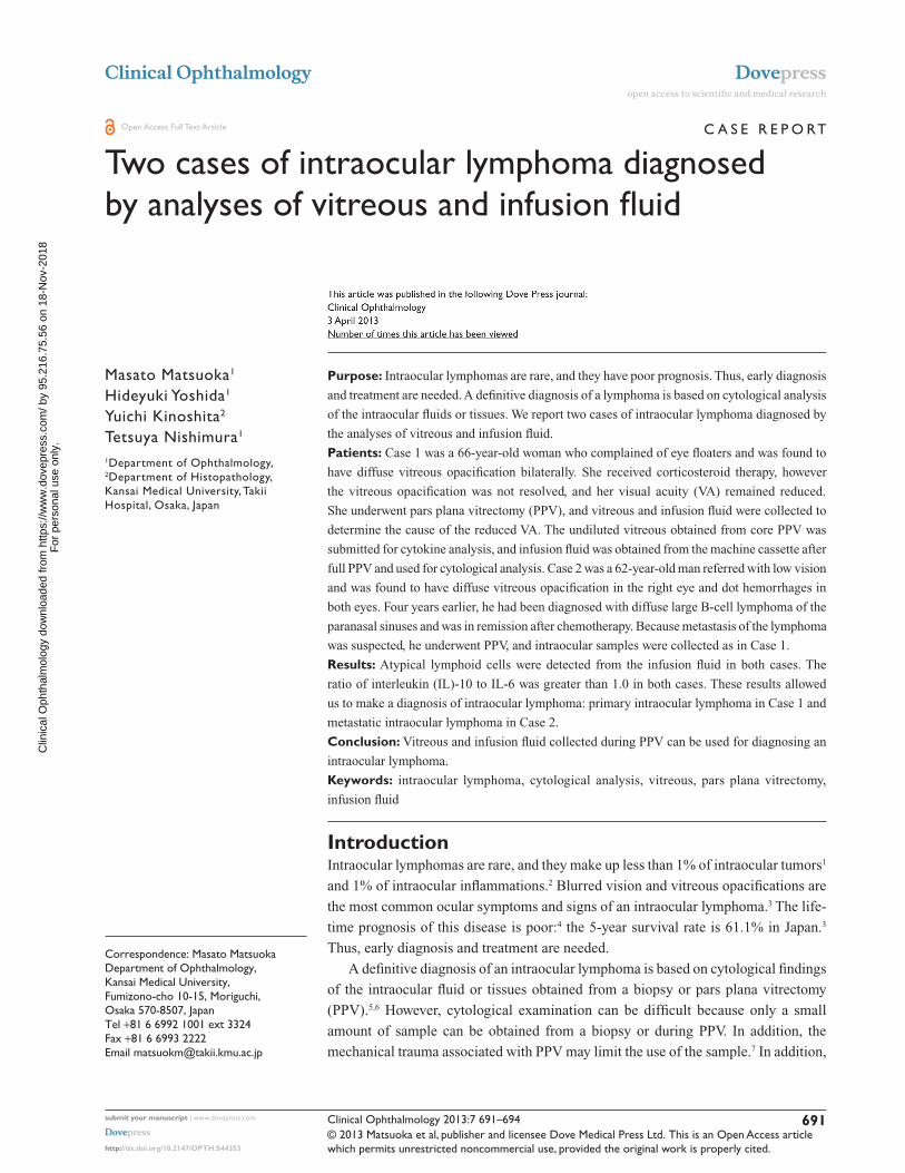
Case 1A 66-year-old woman was referred to our hospital
complaining of floaters in both eyes in February 2010. At the
initial examination, her best-corrected visual acuity (BCVA)
was 20/25 in both eyes, and diffuse vitreous opacifications
with aggregation of large cells were present in both eyes
(Figure 1A and B). Fluorescein angiography showed no
abnormalities. The results of the laboratory examinations
were within normal limits. Cerebral magnetic resonance
imaging (MRI) detected no abnormalities.
Although she was treated with corticosteroid therapy for
3 months, her VA had decreased to 20/50 in both eyes with
increased vitreous opacification. She then underwent PPV
by the standard three-port approach (20-gauge). The core
vitreous was cut with a vitreous cutter, and about 1 mL of
undiluted vitreous was collected by simultaneous manual
aspiration. After the infusion line was opened, a full PPV
was done, and the cortical vitreous was removed as much
as possible. The undiluted vitreous was immediately frozen
at −20°C, and submitted for cytokine analysis. Infusion fluid
(BSS PLUS®; Alcon Japan Ltd., Tokyo, Japan) used during
full PPV was obtained from the machine cassette (Fortas™
CV-30000; Nidek Co, LTD, Aichi, Japan) after the surgery
(about 300 mL), and immediately submitted for cytological
analysis with use of cytospin.
Atypical lymphocytes with anisonucleosis, aberrant
chromatin, and dyskaryosis were detected by cytological
analysis (Figure 1C). The lymphoma cells were positive
for leukocyte common antigen (LCA: Figure 1D). The
ratio of interleukin (IL)-10 to IL-6 (IL-10/IL-6 ratio) of the
undiluted vitreous was greater than 1.0 (IL-10 = 7650 pg/mL:
IL-6 = 139 pg/mL). She was diagnosed with primary
intraocular lymphoma in both eyes and received intravitreal
and intraspinal methotrexate therapy. At 3 years after the
surgery, the vitreous opacification had disappeared and her
corrected VA had improved to 20/20.
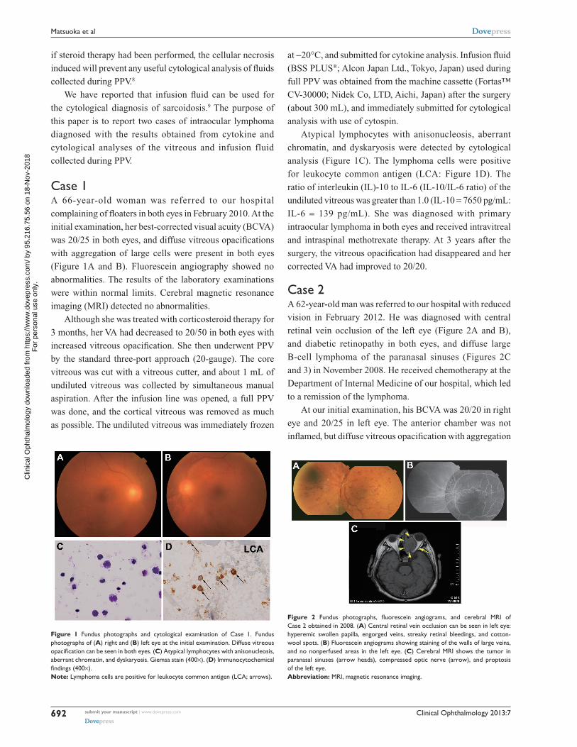
Case 2A 62-year-old man was referred to our hospital with reduced
vision in February 2012. He was diagnosed with central
retinal vein occlusion of the left eye (Figure 2A and B),
and diabetic retinopathy in both eyes, and diffuse large
B-cell lymphoma of the paranasal sinuses (Figures 2C
and 3) in November 2008. He received chemotherapy at the
Department of Internal Medicine of our hospital, which led
to a remission of the lymphoma.
At our initial examination, his BCVA was 20/20 in right
eye and 20/25 in left eye. The anterior chamber was not
inflamed, but diffuse vitreous opacification with aggregation
Figure 1 Fundus photographs and cytological examination of Case 1. Fundus photographs of (A) right and (B) left eye at the initial examination. Diffuse vitreous opacification can be seen in both eyes. (C) Atypical lymphocytes with anisonucleosis, aberrant chromatin, and dyskaryosis. Giemsa stain (400×). (D) Immunocytochemical findings (400×). Note: Lymphoma cells are positive for leukocyte common antigen (LCA; arrows).
Figure 2 Fundus photographs, fluorescein angiograms, and cerebral MRI of Case 2 obtained in 2008. (A) Central retinal vein occlusion can be seen in left eye: hyperemic swollen papilla, engorged veins, streaky retinal bleedings, and cotton-wool spots. (B) Fluorescein angiograms showing staining of the walls of large veins, and no nonperfused areas in the left eye. (C) Cerebral MRI shows the tumor in paranasal sinuses (arrow heads), compressed optic nerve (arrow), and proptosis of the left eye.Abbreviation: MRI, magnetic resonance imaging.
submit your manuscript | www.dovepress.com
Dovepress
Dovepress
692
Matsuoka et al
Clin
ical
Oph
thal
mol
ogy
dow
nloa
ded
from
http
s://w
ww
.dov
epre
ss.c
om/ b
y 95
.216
.75.
56 o
n 18
-Nov
-201
8F
or p
erso
nal u
se o
nly.
Powered by TCPDF (www.tcpdf.org)
1 / 1

Clinical Ophthalmology 2013:7
of large cells was observed in right eye, and dot hemorrhages
were observed in both eyes. Fluorescein angiography
showed several small nonperfused areas, but no retinal
neovascularization. The results of laboratory examinations
were essentially normal except high blood glucose level.
Cerebral MRI and positron emission tomography (PET) did
not detect any abnormalities. The lymphoma in his right
eye was suspected to be a metastasis because of the history
of paranasal lymphoma and the ocular findings. Therefore,
he underwent PPV on the right eye. Vitreous samples and
infusion fluid were collected as in Case 1.
From the infusion fluid, atypical lymphoid cells with
large and irregular nuclei and prominent nucleoli were
detected by cytological analysis (Figure 4A and B).
Immunohistochemistry showed that the cells were positive
for B-lymphocyte surface antigen (CD20) and negative for
T-lymphocyte surface antigen (CD3) (Figure 4C and D). The
IL-10/IL-6 ratio of the undiluted vitreous was greater than
1.0 (IL-10 = 48 pg/mL; IL-6 = 9.0 pg/mL). Immunoglobulin
heavy chain (IgH) gene rearrangements were detected in
the undiluted vitreous. He was diagnosed with metastatic
intraocular lymphoma in the right eye and received irradiation
therapy. At 1 year after the surgery, his BCVA had improved
to 20/20 in both eyes.
DiscussionWe report two cases in which an intraocular lymphoma was
diagnosed from cytokine and cytological analyses of vitreous
and infusion fluid collected during PPV. The undiluted
vitreous was used for cytokine analysis, and the infusion
fluid for cytological analysis.
Generally, the undiluted vitreous obtained from core
PPV or vitreous biopsy is submitted for cytological analysis
in cases suspected of having an intraocular lymphoma.
Submitting infusion fluid obtained from full PPV for
cytological examination has four advantages. First, a larger
number of cells can be collected. Mudhar et al reported that
the lymphoma cells were concentrated more in the cortical
vitreous than core vitreous.10 Additionally, Conlon et al
reported that vitrectomy did not cause any more cellular
degradation than simple aspiration.11 In our cases, we were
able to obtain a sufficient volume of lymphoma cells from
the infusion fluid.
The second advantage is that undiluted vitreous can be
submitted for other examination, such as cytokine analysis,
IgH gene arrangements, and flow cytometry. Several
studies have reported that cytokine analysis of vitreous
had the highest detection rate for intraocular lymphomas.3,5
However, the amount of undiluted vitreous obtained from
PPV or vitreous biopsy is usually too small to be submitted
for several types of analyses. Therefore, undiluted vitreous
can be submitted for cytokine analysis, if the infusion fluid
is submitted for cytological analysis.12
The third advantage is that the cell collection rate can be
increased by cytospin of the infusion fluid, and the collected
cells can be used for the cytological analysis. Undiluted
vitreous is not as effective for collecting cells by cytospin
because of its high viscosity. In contrast, the infusion fluid
Figure 3 Histopathological examination of paranasal-sinuses tumor of Case 2. (400×) (A) Atypical cells with anisonucleosis, aberrant chromatin, and dyskaryosis. Giemsa stain. Immunocytochemical findings. The cells are negative for T-lymphocyte surface antigen (CD3). (B) Immunocytochemical findings. The cells are negative for cytokeratin (AE1/AE3), a marker of epithelial cells. (C) Immunocytochemical findings. The cells are positive for B-lymphocyte surface antigen (CD20). (D) Immunocytochemical findings. The cells are negative for T-lymphocyte surface antigen (CD3).
Figure 4 Cytological examination of infusion fluid of Case 2 (400×). (A and B) Atypical lymphocytes with anisonucleosis, aberrant chromatin, and dyskaryosis. (A) Giemsa stain; (B) Papanicolaou stain. (C) Immunocytochemical findings. Lymphoma cells are positive for B-lymphocyte surface antigen (CD20). (D) Immunocytochemical findings. Lymphoma cells are negative for T-lymphocyte antigen (CD3).
submit your manuscript | www.dovepress.com
Dovepress
Dovepress
693
Vitreous cytology and intraocular lymphoma
Clin
ical
Oph
thal
mol
ogy
dow
nloa
ded
from
http
s://w
ww
.dov
epre
ss.c
om/ b
y 95
.216
.75.
56 o
n 18
-Nov
-201
8F
or p
erso
nal u
se o
nly.
Powered by TCPDF (www.tcpdf.org)
1 / 1

Clinical Ophthalmology
Publish your work in this journal
Submit your manuscript here: http://www.dovepress.com/clinical-ophthalmology-journal
Clinical Ophthalmology is an international, peer-reviewed journal covering all subspecialties within ophthalmology. Key topics include: Optometry; Visual science; Pharmacology and drug therapy in eye diseases; Basic Sciences; Primary and Secondary eye care; Patient Safety and Quality of Care Improvements. This journal is indexed on
PubMed Central and CAS, and is the official journal of The Society of Clinical Ophthalmology (SCO). The manuscript management system is completely online and includes a very quick and fair peer-review system, which is all easy to use. Visit http://www.dovepress.com/ testimonials.php to read real quotes from published authors.
Clinical Ophthalmology 2013:7
is not as viscous and does contain diluted cortical vitreous,
which allowed us to collect more cells.
Finally, infusion fluid contains cellular protectants such
as oxidized glutathione and glucose.13 The lymphoma cells
are fragile because of mechanical trauma associated with
PPV and cellular necrosis induced by steroid therapy. These
changes can be reduced by the cellular protectants.
There are limitations to this report. Lymphoma cells
are known to infiltrate the vitreous, subretinal pigment
epithelium, retina, choroid, iris, and optic nerve.3,5,8 In
some cases of intraocular lymphomas, tumor masses in the
subretinal pigment epithelium position can only be observed
without vitreous infiltration. In those cases, only analysis
of vitreous and infusion fluid may be difficult to detect
lymphoma cells without chorioretinal biopsy.5
In conclusion, we found that undiluted vitreous and
infusion fluid collected during full PPV could be used for
cytokine and cytological analyses for diagnosing intraocular
lymphomas.
Acknowledgments/disclosureNone of the authors report any conflicts of interest associated
with this work.
References1. Bardenstein DS. Intraocular lymphoma. Cancer Control. 1998;5:
317–325.2. Goto H, Mochizuki M, Yamaki K, et al. Epidemiological survey of
intraocular inflammation in Japan. Jpn J Ophthalmol. 2007;51:41–44.
3. Kimura K, Usui Y, Goto H. Clinical features and diagnostic significance of the intraocular fluid of 217 patients with intraocular lymphoma. Jpn J Ophthalmol. 2012;56:383–389.
4. Jahnke K, Korfel A, Komm J, et al. Intraocular lymphoma 2000–2005: results of a retrospective multicentre trial. Graefes Arch Clin Exp Ophthalmol. 2006;244:663–669.
5. Gonzales JA, Chan CC. Biopsy techniques and yields in diagnosing primary intraocular lymphoma. Int Ophthalmol. 2007;27:241–250.
6. Michels RG, Knox DL, Erozan YS, et al. Intraocular reticulum cell sarcoma. Diagnosis by pars plana vitrectomy. Arch Ophthalmol. 1975;93:1331–1335.
7. Char DH, Ljung BM, Miller T, et al. Primary intraocular lymphoma (ocular reticulum cell sarcoma) diagnosis and management. Ophthalmology. 1988;95:625–630.
8. Whitcup SM, de Smet MD, Rubin BI, et al. Intraocular lymphoma. Clinical and histopathologic diagnosis. Ophthalmology. 1993;100: 1399–1406.
9. Matsuoka M, Ogata N, Takahashi K, et al. Two cases of ocular sarcoidosis in which vitreous cytology was useful for supporting the diagnosis. Clin Ophthalmol. 2012;6:1207–1209.
10. Mudhar HS, Sheard R. Diagnostic cellular yield is superior with full pars plana vitrectomy compared with core vitreous biopsy. Eye (Lond). 2013;27:50–55.
11. Conlon MR, Craig I, Harris JF, et al. Effect of vitrectomy and cytopreparatory techniques on cell survival and preservation. Can J Ophthalmol. 1992;27:168–171.
12. Kinoshita Y, Takasu K, Adachi Y, et al. Retrospective cytological study of intraocular lymphoma using vitreous and intraocular perfusion fluid. Diagn Cytopathol. 2010;40:604–607.
13. Araie M, Shirasawa E, Hikita M. Effect of oxidized glutathione on the barrier function of the corneal endothelium. Invest Ophthalmol Vis Sci. 1988;29:1884–1887.
submit your manuscript | www.dovepress.com
Dovepress
Dovepress
Dovepress
694
Matsuoka et al
Clin
ical
Oph
thal
mol
ogy
dow
nloa
ded
from
http
s://w
ww
.dov
epre
ss.c
om/ b
y 95
.216
.75.
56 o
n 18
-Nov
-201
8F
or p
erso
nal u
se o
nly.
Powered by TCPDF (www.tcpdf.org)
1 / 1