· 2019-12-19 · op Wireless access and fixed access technology, 5G en advanced multiple antenna

Nechin et al. Epigenetics & Chromatin (2019) 12:64 https://doi.org/10.1186/s13072-019-0309-2
RESEARCH
Hemimethylation of CpG dyads is characteristic of secondary DMRs associated with imprinted loci and correlates with 5-hydroxymethylcytosine at paternally methylated sequencesJulianna Nechin, Emma Tunstall, Naideline Raymond, Nicole Hamagami, Chris Pathmanabhan, Samantha Forestier and Tamara L. Davis*
Abstract
Background: In mammals, the regulation of imprinted genes is controlled by differential methylation at imprinting control regions which acquire parent of origin-specific methylation patterns during gametogenesis and retain differ-ences in allelic methylation status throughout fertilization and subsequent somatic cell divisions. In addition, many imprinted genes acquire differential methylation during post-implantation development; these secondary differen-tially methylated regions appear necessary to maintain the imprinted expression state of individual genes. Despite the requirement for both types of differentially methylated sequence elements to achieve proper expression across imprinting clusters, methylation patterns are more labile at secondary differentially methylated regions. To understand the nature of this variability, we analyzed CpG dyad methylation patterns at both paternally and maternally methyl-ated imprinted loci within multiple imprinting clusters.
Results: We determined that both paternally and maternally methylated secondary differentially methylated regions associated with imprinted genes display high levels of hemimethylation, 29–49%, in comparison to imprinting control regions which exhibited 8–12% hemimethylation. To explore how hemimethylation could arise, we assessed the differentially methylated regions for the presence of 5-hydroxymethylcytosine which could cause methylation to be lost via either passive and/or active demethylation mechanisms. We found enrichment of 5-hydroxymethylcytosine at paternally methylated secondary differentially methylated regions, but not at the maternally methylated sites we analyzed in this study.
Conclusions: We found high levels of hemimethylation to be a generalizable characteristic of secondary differentially methylated regions associated with imprinted genes. We propose that 5-hydroxymethylcytosine enrichment may be responsible for the variability in methylation status at paternally methylated secondary differentially methylated regions associated with imprinted genes. We further suggest that the high incidence of hemimethylation at second-ary differentially methylated regions must be counteracted by continuous methylation acquisition at these loci.
Keywords: Genomic imprinting, DNA methylation, H19, Cdkn1c, Snrpn, Ndn, Peg12, Secondary DMR, Epigenetics
© The Author(s) 2019. This article is distributed under the terms of the Creative Commons Attribution 4.0 International License (http://creat iveco mmons .org/licen ses/by/4.0/), which permits unrestricted use, distribution, and reproduction in any medium, provided you give appropriate credit to the original author(s) and the source, provide a link to the Creative Commons license, and indicate if changes were made. The Creative Commons Public Domain Dedication waiver (http://creativecommons.org/publicdomain/zero/1.0/) applies to the data made available in this article, unless otherwise stated.
Open Access
Epigenetics & Chromatin
*Correspondence: [email protected] of Biology, Bryn Mawr College, 101 N. Merion Avenue, Bryn Mawr, PA 19010-2899, USA

Page 2 of 13Nechin et al. Epigenetics & Chromatin (2019) 12:64
BackgroundGenomic imprinting refers to the parent of origin-spe-cific expression of one parental allele over another. To date, approximately 150 mammalian genes have been found to exhibit this unusual form of regulation [1, 2]. Parent of origin-specific expression of imprinted genes is achieved via multiple mechanisms, including differential DNA methylation, differential distribution of modified histones and differential expression of long non-coding RNAs from the maternal vs. paternal alleles [3, 4]. Impor-tantly, all imprinted genes are associated with an imprint-ing control region, which is differentially methylated on the parental alleles and is responsible for the regulation of the genes located through the associated imprinting cluster [3].
Differential DNA methylation associated with imprinted genes can be categorized into two classes. One class is comprised of primary or gametic differentially methylated regions (DMRs), at which the differentially methylated state associated with the parental alleles is acquired during gametogenesis, inherited at fertilization, and maintained throughout development, including dur-ing the genome-wide demethylation that occurs prior to implantation [3]. These primary DMRs typically correlate with the imprinting control region and play a critical role in the establishment and maintenance of imprinted gene expression by affecting the activity of insulators or the expression of long non-coding RNAs that regulate adja-cent imprinted genes [5–8]. In addition, some primary DMRs directly affect the expression of protein-coding imprinted genes via differential methylation of their pro-moters [9]. In contrast, secondary DMRs acquire their differentially methylated state during post-implantation development [10–14]. The role of secondary DMRs is less clear, though evidence suggests they may be important for maintaining imprinted expression as they are typically located at promoters and failure to establish or maintain methylation at these loci results in the dysregulation of the associated imprinted gene [12, 15–18].
Previous work has illustrated that there is variation in the stability of DNA methylation at primary vs. second-ary DMRs associated with imprinted genes. Primary DMRs typically display very high levels of DNA meth-ylation on the methylated allele (90–100%) and very low levels of DNA methylation on the unmethylated allele (0–10%) [11, 14, 19–22]. In contrast, significantly more variations in DNA methylation patterns are observed at secondary DMRs. The methylated allele typically has less consistent DNA methylation than is observed at pri-mary DMRs [10, 11, 13, 23]. In addition, some secondary DMRs display DNA methylation on both parental alleles, although one allele contains significantly more meth-ylation than the other [24]. Investigation into the more
variably methylated secondary DMRs has shown that substantial levels of asymmetric DNA methylation are observed at CpG dyads. For example, the variably meth-ylated secondary DMRs associated with the imprinted Dlk1 and Gtl2 genes contain 29% and 32% hemimeth-ylation, as compared to 8% hemimethylation at the pri-mary IG-DMR associated with the Dlk1/Gtl2 imprinting cluster on mouse chromosome 12 [13, 24]. The high level of methylation asymmetry observed at these second-ary DMRs explains the variability in the DNA methyla-tion patterns and could be a consequence of TET activity at these loci, which could lead to the active demethyla-tion of cytosines in these regions and could also result in passive DNA demethylation via the reduced activity of Dnmt1 at oxidized methylcytosine [25–29].
To determine if high levels of hemimethylation are characteristic of secondary DMRs associated with imprinted loci, we investigated CpG dyad methylation patterns at well-characterized paternally and maternally methylated primary and secondary DMRs located in the central and distal imprinting clusters on mouse chromo-some 7; we did not analyze loci in the proximal imprint-ing cluster (Fig. 1). We further examined the correlation between hemimethylation levels and the presence of 5-hydroxymethylcytosine (5hmC) to test the hypothesis that oxidation and removal of methylcytosine is respon-sible for the variable DNA methylation patterns at these loci. Finally, we examined the sequence composition at primary vs. secondary paternal and maternal DMRs to test the hypothesis that sequence context may play a role in the difference in DNA methylation stability asso-ciated with primary vs. secondary DMRs. Briefly, we consistently observed high levels of hemimethylation at secondary DMRs, regardless of which parental allele was methylated, and high levels of hemimethylation were cor-related with the presence of 5hmC at paternally methyl-ated sequences.
ResultsCpG dyads within paternally and maternally methylated secondary DMRs associated with imprinted genes display high levels of hemimethylationPrevious work in our lab illustrated a high level of hemi-methylation at two paternally methylated secondary DMRs located in the Dlk1/Gtl2 imprinting cluster on mouse chromosome 12. To assess whether hemimethyla-tion is generally a feature of secondary DMRs or whether this phenomenon is unique to loci in the Dlk1/Gtl2 imprinting cluster, we examined DNA methylation at CpG dyads located within the paternally methylated sec-ondary DMRs associated with H19 and Cdkn1c; these DMRs are located in two different imprinting clusters located on mouse chromosome 7 (Fig. 1, [11, 12]). We

Page 3 of 13Nechin et al. Epigenetics & Chromatin (2019) 12:64
assessed methylation at various stages of development to assess both the establishment of differential methylation and its maintenance; in general, levels of methylation did not vary significantly during development indicating that overall levels of methylation remain similar over time (Additional file 1). All analyses were conducted using F1 hybrid mice to utilize C57BL/6J vs. Mus musculus cas-taneus SNPs to distinguish parental origin of each allele (see “Methods”).
At H19, we analyzed 8 CpG dyads that had previously been shown to be part of the promoter-proximal H19 secondary DMR (H19-ppDMR) [11]. Of note, in all the tissues we analyzed, we detected significantly more DNA methylation on both the paternal and the maternal alleles than had been observed previously ([11]; Fig. 2a). This difference may be attributed to differences in genetic background. Despite the higher levels of methylation we observed overall in this region, we determined that the amount of methylation on the paternal alleles was sig-nificantly higher than on the maternal alleles throughout development using a Mann–Whitney U test (P = 0.0012, 7.5 dpc embryo; 0.0477, 14.5 dpc embryo; 0.0001, 5 dpp liver; 0.0008, adult liver), indicating that this region was differentially methylated in the F1 hybrid mice used in our study. In addition, Mann–Whitney U tests indi-cated that methylation levels remained constant on the parental alleles across development (data not shown).
Hemimethylation at the H19 secondary DMR aver-aged 31.9% for both parental alleles across development (Fig. 2a, Additional file 2). Using a Chi square test of inde-pendence, we found that the level of hemimethylation at the H19 secondary DMR was not significantly different than the levels we had previously observed at the Dlk1- and Gtl2-DMRs (P = 0.2231 and 0.8370, respectively).
We also analyzed 29 CpG dyads located at the 5′ end of the Cdkn1c DMR analyzed by Bhogal et al. [12]. Cdkn1c displayed even higher levels of hemimethylation than we detected at H19. Methylation levels on each parental allele remained constant across development, averag-ing 48.5% for both parental alleles (Fig. 2b, Additional file 2). The level of hemimethylation at the Cdkn1c DMR was significantly higher than the level observed at the Gtl2, Dlk1 or H19 secondary DMRs (P = 2.45 × 10⁻⁷, 1.39 × 10⁻¹⁴ and 1.31 × 10⁻¹⁵, respectively).
Similar to paternally methylated secondary DMRs, maternally methylated secondary DMRs exhibit more variable DNA methylation patterns than primary DMRs associated with imprinted loci [10, 30–32]. Based on this similarity, we hypothesized that maternally methylated secondary DMRs would also exhibit high levels of hemi-methylation. We therefore examined DNA methylation at CpG dyads located within the maternally methylated sec-ondary DMRs associated with Ndn and Peg12 on mouse chromosome 7 [10, 30].
chr7cent7 dist7
10 Mb
Atp1
0a
Ube
3a
Ipw
Snor
d116
Snur
f/Snp
rn
Ndn
Mag
el2
Mkr
n3Pe
g12
200 kb
cent7*
*
dist7
5 kb
H19 Igf2
Ins2
Ascl
2
Kcnq
1ot1
Cdk
n1c
Nap
1I4
Phld
a2
Kcnq
1
Tssc
4
Cd8
1
Slc2
2a18
**
prox7a
c
b
#
#
Fig. 1 Imprinting clusters on mouse chromosome 7. a Location of proximal, central and distal imprinting clusters. Detail of central (b) and distal (c) imprinting clusters. Red and blue rectangles correspond to maternally and paternally expressed genes, respectively. Genes located above and below the line have + and − strand orientation, respectively. Pound signs and asterisks indicate the location, respectively, of primary and secondary DMRs analyzed in this study. Snrpn, maternally methylated primary DMR; Ndn and Peg12, maternally methylated secondary DMRs; H19 ICR, paternally methylated primary DMR; H19-pp and Cdkn1c, paternally methylated secondary DMRs

Page 4 of 13Nechin et al. Epigenetics & Chromatin (2019) 12:64
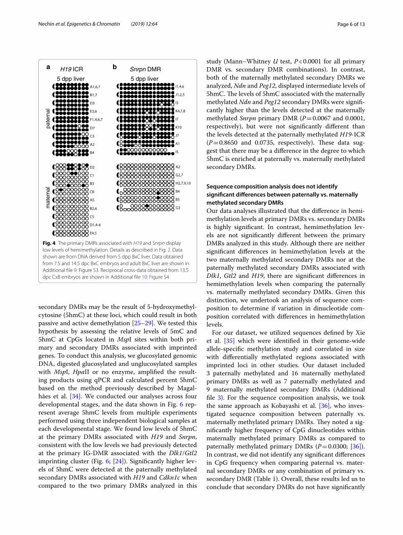
At Ndn, we analyzed the methylation status of 17 CpG dyads located 5′ relative to the start codon; these 17 CpGs had previously been shown to be differentially methylated in multiple adult tissues, including brain, where Ndn is expressed, as well as in tissues without Ndn expression such as liver and heart [10]. We there-fore assessed CpG dyad methylation in tissues derived from embryos, neonatal liver and brain, and adult brain. In every tissue analyzed, the level of methylation on the maternal vs. paternal alleles was significantly different, confirming that this region is differentially methylated throughout development and in multiple tissue types (Fig. 3a). The highest level of methylation we detected was on the maternal allele in 5 dpp liver; methylation of maternal alleles in 5 dpp liver was signif-icantly higher than on maternal alleles derived from 5 dpp or adult brain (Mann–Whitney U test, P = 0.0108, 0.0271), although this could be attributed to the small sample size. Overall, 39.0% of the CpG dyads at the Ndn DMR were hemimethylated on both parental alleles (Additional file 2).
We additionally assessed methylation at 29 CpG dyads located at the 3′ end of the CpG island associated with the maternally methylated Peg12 gene [30, 32]. Our analysis showed that the maternal allele has significantly more methylation than the paternal allele in embryonic, neona-tal and adult tissues (Fig. 3b). In addition, embryonic tis-sues have significantly less methylation on the maternal allele than neonatal and adult tissues. Overall, 35.4% of the CpG dyads at the Peg12 DMR were hemimethylated on both parental alleles (Additional file 2). Hemimethyla-tion levels at the maternally methylated Ndn and Peg12 DMRs were not significantly different from each other (P = 0.1745), although hemimethylation at both of these maternally methylated secondary DMRs was significantly higher than most of the paternally methylated secondary DMRs we analyzed, with the exception of Cdkn1c which contained the highest levels of hemimethylation amongst the loci examined in our study. Combined, these data support the hypothesis that high levels of hemimethyla-tion are characteristic of both maternally and paternally methylated secondary DMRs located throughout the
pate
rnal
mat
erna
l
7.5 dpc embryo 5 dpp liver 7.5 dpc embryo 5 dpp liver
A1
A4
A6
B3
C1
E1
E2
E4
E5
E6
E3
B2
A5
C4
D1,3,5
C2
B1,4
D2,6,7
C5
C1
A10
D3
A11
A12
G8
G2
G9
G12
A4
H2
H3
H6
H1
G13
A9
D4
D7
D1,9
G10
D2
H5
H4
H19-ppDMR Cdkn1c DMRa b
C6
F2
D4
C4
C2
D5
F6
F5
C1
B4
E4
A3
B1
B2
A5
A1,2,6,7
C5,6
D3,6
E6
F2,6
G6
G2
A8
C7
D5
G1
C4
F3
B3
E2
A2
B6
B5
E1
A1
C3
G4
F5
G3
E2
A4
D1
E5
D4
G5
F1
E8
F4
E7
D2
E4
Fig. 2 The paternally methylated secondary DMRs associated with H19 and Cdkn1c display high levels of hemimethylation. Bisulfite mutagenesis and sequencing of F1 hybrid DNA derived from 7.5 dpc BxC embryos and 5 dpp BxC liver. Individual circles in each row represent one of the potentially methylated CpG dinucleotides analyzed at the H19-ppDMR (a) or the Cdkn1c DMR (b), and each paired row of circles represents the complementary strands of an individual subclone; semi-circles to the right or left indicate the location of the linker connecting the complementary strands. Filled circles represent methylated cytosines, open circles represent unmethylated cytosines, absent circles represent ambiguous data. Alphanumeric labels identify subclones analyzed; letters represent independent amplification reactions, while numbers represent individual subclones. Subclones derived from the same amplification that have identical sequence and methylation patterns are grouped together, as it was not possible to determine if these amplicons were derived from the same or different template molecules. Data obtained from 14.5 dpc BxC embryos and adult BxC liver are shown in Additional file 7: Figure S1. Reciprocal cross-data obtained from 13.5 dpc CxB embryos are shown in Additional file 10: Figure S4

Page 5 of 13Nechin et al. Epigenetics & Chromatin (2019) 12:64
mouse genome and may be a unique epigenetic feature that further distinguishes secondary DMRs from primary DMRs.
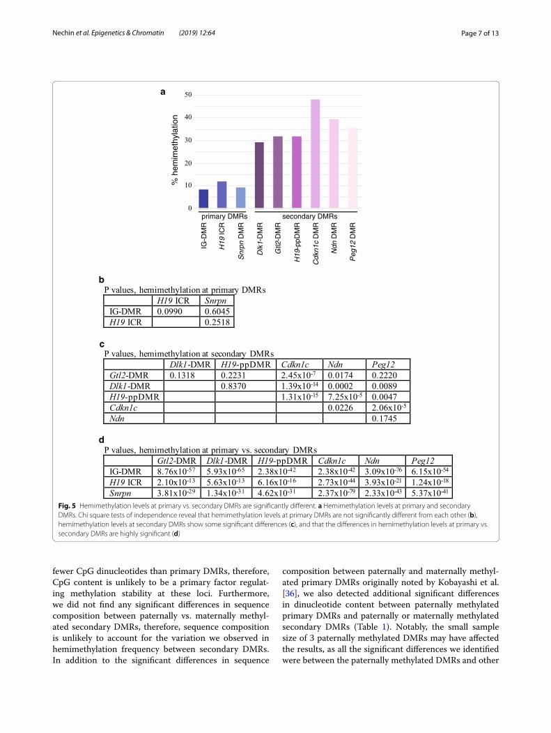
Hemimethylation levels are low at both paternally and maternally methylated primary DMRs associated with imprinted lociTo determine if the high levels of hemimethylation are a unique feature of secondary DMRs associated with imprinted genes, thereby distinguishing them further from primary DMRs, we examined the methylation sta-tus of CpG dyads associated with primary DMRs. We assessed DNA methylation at 9 CpG dyads located at the 5′ end of the H19 imprinting control region (ICR) and 15 CpG dyads located within the Snrpn DMR [11, 33]. We observed relatively low levels of hemimethyla-tion at both loci: 11.9% and 9.3% of the CpG dyads were hemimethylated within the paternally methylated H19 ICR and the maternally methylated Snrpn DMR, respec-tively (Fig. 4; Additional file 2). No significant differences were detected when comparing hemimethylation levels at primary DMRs associated with Dlk1/Gtl2 IG-DMR,
H19 or Snrpn (Fig. 5b; [24]). In contrast, the difference in hemimethylation levels at either paternally or maternally methylated primary DMRs when compared to any of the secondary DMRs we analyzed was highly significant (Fig. 5d) and the differences were notably more signifi-cant between primary vs. secondary DMRs than between secondary DMRs (Fig. 5c). These results are consistent with the hypothesis that high levels of hemimethylation are characteristic of the variably methylated secondary DMRs but are not associated with primary DMRs. In further support of our theory that high levels of hemi-methylation are a unique feature of secondary DMRs associated with imprinted genes, preliminary data show that the level of hemimethylation at tissue-specific DMRs is similar to hemimethylation levels at primary DMRs and is significantly lower than those observed at second-ary DMRs (T. Davis, data not shown).
5‑hydroxymethylcytosine is enriched within paternally methylated secondary DMRsWe hypothesized that the variability in DNA methyla-tion patterns and the high levels of hemimethylation at
B3
E2
B6
G8
G5
D3
A2,4
B5
H2
F1,4
D1
B4
E3,4
G6,7
F2,3
H3
H1
E3
A4
C2
I6
I4
I1
C4,7
E1,5,6
D5
I5
D1,3,4,6,8
A2,5,6
D5
E5
E3
D4
E1
F4,5
B6
C3
C4
C1
B2,3,4
C2,5
F1
D1
A2,5,7
B1
7.5 dpc embryo 5 dpp brain5 dpp liver
mat
erna
lpa
tern
al
5 dpp liver
Ndn DMR Peg12 DMRa b
C2
C9
E3
E5
E1
C8
D5
F4
D3,10
C5
C6
E7
F8
F5
C10
C1
D7
B1
F6
F3
E4
E6
E2
A3
D8
F7
F2
B2
Fig. 3 The maternally methylated secondary DMRs associated with Ndn and Peg12 display high levels of hemimethylation. a Methylation status at the Ndn DMR; F1 hybrid DNA derived from 7.5 dpc BxC embryos and 5 dpp BxC liver and brain. b Methylation status at the Peg12 DMR; F1 hybrid DNA derived from 5 dpp liver. Other details as described in Fig. 2. Data obtained from 7.5 and 14.5 dpc BxC embryos and adult BxC liver and brain are shown in Additional file 8: Figure S2. Reciprocal cross-data obtained from 13.5 dpc CxB embryos are shown in Additional file 10: Figure S4
Page 6 of 13Nechin et al. Epigenetics & Chromatin (2019) 12:64
secondary DMRs may be the result of 5-hydroxymethyl-cytosine (5hmC) at these loci, which could result in both passive and active demethylation [25–29]. We tested this hypothesis by assessing the relative levels of 5mC and 5hmC at CpGs located in MspI sites within both pri-mary and secondary DMRs associated with imprinted genes. To conduct this analysis, we glucosylated genomic DNA, digested glucosylated and unglucosylated samples with MspI, HpaII or no enzyme, amplified the result-ing products using qPCR and calculated percent 5hmC based on the method previously described by Magal-hães et al. [34]. We conducted our analyses across four developmental stages, and the data shown in Fig. 6 rep-resent average 5hmC levels from multiple experiments performed using three independent biological samples at each developmental stage. We found low levels of 5hmC at the primary DMRs associated with H19 and Snrpn, consistent with the low levels we had previously detected at the primary IG-DMR associated with the Dlk1/Gtl2 imprinting cluster (Fig. 6; [24]). Significantly higher lev-els of 5hmC were detected at the paternally methylated secondary DMRs associated with H19 and Cdkn1c when compared to the two primary DMRs analyzed in this
study (Mann–Whitney U test, P < 0.0001 for all primary DMR vs. secondary DMR combinations). In contrast, both of the maternally methylated secondary DMRs we analyzed, Ndn and Peg12, displayed intermediate levels of 5hmC. The levels of 5hmC associated with the maternally methylated Ndn and Peg12 secondary DMRs were signifi-cantly higher than the levels detected at the maternally methylated Snrpn primary DMR (P = 0.0067 and 0.0001, respectively), but were not significantly different than the levels detected at the paternally methylated H19-ICR (P = 0.8650 and 0.0735, respectively). These data sug-gest that there may be a difference in the degree to which 5hmC is enriched at paternally vs. maternally methylated secondary DMRs.
Sequence composition analysis does not identify significant differences between paternally vs. maternally methylated secondary DMRsOur data analyses illustrated that the difference in hemi-methylation levels at primary DMRs vs. secondary DMRs is highly significant. In contrast, hemimethylation lev-els are not significantly different between the primary DMRs analyzed in this study. Although there are neither significant differences in hemimethylation levels at the two maternally methylated secondary DMRs nor at the paternally methylated secondary DMRs associated with Dlk1, Gtl2 and H19, there are significant differences in hemimethylation levels when comparing the paternally vs. maternally methylated secondary DMRs. Given this distinction, we undertook an analysis of sequence com-position to determine if variation in dinucleotide com-position correlated with differences in hemimethylation levels.
For our dataset, we utilized sequences defined by Xie et al. [35] which were identified in their genome-wide allele-specific methylation study and correlated in size with differentially methylated regions associated with imprinted loci in other studies. Our dataset included 3 paternally methylated and 16 maternally methylated primary DMRs as well as 7 paternally methylated and 9 maternally methylated secondary DMRs (Additional file 3). For the sequence composition analysis, we took the same approach as Kobayashi et al. [36], who inves-tigated sequence composition between paternally vs. maternally methylated primary DMRs. They noted a sig-nificantly higher frequency of CpG dinucleotides within maternally methylated primary DMRs as compared to paternally methylated primary DMRs (P = 0.0300; [36]). In contrast, we did not identify any significant differences in CpG frequency when comparing paternal vs. mater-nal secondary DMRs or any combination of primary vs. secondary DMR (Table 1). Overall, these results led us to conclude that secondary DMRs do not have significantly
pate
rnal
mat
erna
l
5 dpp liver
I5
5 dpp liver
H19 ICR Snrpn DMRa b
A1,6,7
B1,7
F1,4,6,7
E3,6
D3
D7
C3
B4
A2
D2
C1
A5
C8
B3
B2,6
C5
E4,5
D1,4-6
A2
G2,7
H2,7,9,10
B4
B5
G3
I1,4,6
J1,2,5
I3
K4,7,8
I7
K10
J7
A1
Fig. 4 The primary DMRs associated with H19 and Snrpn display low levels of hemimethylation. Details as described in Fig. 2. Data shown are from DNA derived from 5 dpp BxC liver. Data obtained from 7.5 and 14.5 dpc BxC embryos and adult BxC liver are shown in Additional file 9: Figure S3. Reciprocal cross-data obtained from 13.5 dpc CxB embryos are shown in Additional file 10: Figure S4
Page 7 of 13Nechin et al. Epigenetics & Chromatin (2019) 12:64
fewer CpG dinucleotides than primary DMRs, therefore, CpG content is unlikely to be a primary factor regulat-ing methylation stability at these loci. Furthermore, we did not find any significant differences in sequence composition between paternally vs. maternally methyl-ated secondary DMRs, therefore, sequence composition is unlikely to account for the variation we observed in hemimethylation frequency between secondary DMRs. In addition to the significant differences in sequence
composition between paternally and maternally methyl-ated primary DMRs originally noted by Kobayashi et al. [36], we also detected additional significant differences in dinucleotide content between paternally methylated primary DMRs and paternally or maternally methylated secondary DMRs (Table 1). Notably, the small sample size of 3 paternally methylated DMRs may have affected the results, as all the significant differences we identified were between the paternally methylated DMRs and other
primary DMRs secondary DMRs
IG-D
MR
Peg
12 D
MR
Ndn
DM
R
Cdk
n1c
DM
R
H19
-ppD
MR
Gtl2
-DM
R
Dlk
1-D
MR
Snr
pn D
MR
H19
ICR
0
40
30
20
10
50
% h
emim
ethy
latio
n
P values, hemimethylation at primary DMRsH19 ICR Snrpn
IG-DMR 0.0990 0.6045H19 ICR 0.2518
P values, hemimethylation at secondary DMRsDlk1-DMR H19-ppDMR Cdkn1c Ndn Peg12
Gtl2-DMR 0.1318 0.2231 2.45x10⁻⁷ 0.0174 0.2220Dlk1-DMR 0.8370 1.39x10⁻¹⁴ 0.0002 0.0089H19-ppDMR 1.31x10⁻¹⁵ 7.25x10⁻⁵ 0.0047Cdkn1c 0.0226 2.06x10⁻⁵
Ndn 0.1745
P values, hemimethylation at primary vs. secondary DMRsGtl2-DMR Dlk1-DMR H19-ppDMR Cdkn1c Ndn Peg12
IG-DMR 8.76x10-57 5.93x10-65 2.38x10-42 2.38x10-42 3.09x10-76 6.15x10-54
H19 ICR 2.10x10⁻¹³ 5.63x10⁻¹³ 6.16x10⁻¹⁶ 2.73x10-44 3.93x10⁻²¹ 1.24x10⁻¹⁸
Snrpn 3.81x10⁻²⁹ 1.34x10⁻³¹ 4.62x10⁻³¹ 2.37x10-79 2.33x10-43 5.37x10-41
a
d
c
b
Fig. 5 Hemimethylation levels at primary vs. secondary DMRs are significantly different. a Hemimethylation levels at primary and secondary DMRs. Chi square tests of independence reveal that hemimethylation levels at primary DMRs are not significantly different from each other (b), hemimethylation levels at secondary DMRs show some significant differences (c), and that the differences in hemimethylation levels at primary vs. secondary DMRs are highly significant (d)

Page 8 of 13Nechin et al. Epigenetics & Chromatin (2019) 12:64
DMR categories; further analysis would be necessary to determine if these non-CpG differences are biologically relevant.
DiscussionThe research described herein focuses on characterizing DNA methylation at secondary DMRs associated with imprinted genes. Secondary DMRs, which acquire their differentially methylated status post-fertilization, display significantly more variation in their methylation patterns than do primary DMRs [10–13, 24, 37]. Despite the vari-ability in methylation at these loci, they appear to play a critical role in maintaining imprinted expression at the individual genes with which they are associated as loss of
imprinting at these genes can result either from deletion of the secondary DMR or from its loss of methylation due to mutations in Dnmt1 or deletion of the corresponding primary DMR [18, 37–41]. Understanding how meth-ylation at secondary DMRs influences imprinted gene expression despite the absence of highly stable methyla-tion patterns at these sites is, therefore, important. Our investigation into the variable nature of DNA methyla-tion at imprinted loci has identified high levels of hemi-methylation specifically at paternally and maternally methylated secondary DMRs, which we hypothesize is connected to the presence of 5-hydroxymethylcytosine leading to demethylation and hence the observed meth-ylation asymmetries. In this study, we obtained data consistent with this hypothesis: all three of the primary DMRs analyzed in this study and our previous work had low levels of 5hmC, while more 5hmC was prevalent at the paternally methylated secondary DMRs associated with H19, Cdkn1c and Dlk1 ([24] and data herein). How-ever, we did not detect similarly high levels of 5hmC at the Gtl2 secondary DMR, nor at the maternally methyl-ated Ndn or Peg12 secondary DMRs. These data may highlight a difference in the methylation state at pater-nally vs. maternally methylated secondary DMRs. It is also possible that these data are not representative of the overall level or distribution of 5hmC across these loci, as the scope of our 5hmC analysis was limited to CpGs located within MspI restriction sites. In addition, our assay was not allele-specific, preventing us from assessing the distribution of 5hmC on the methylated vs. unmeth-ylated allele. To resolve these questions, we are currently undertaking an oxidative bisulfite sequencing approach to interrogate these loci more broadly and determine if 5hmC is enriched at secondary DMRs and what its dis-tribution is on the parental alleles to better determine if the presence of 5hmC could be driving passive and/or active demethylation at secondary DMRs. In support of this hypothesis, we detected a significant enrichment of
primary DMRs secondary DMRs
Peg12Ndn
Cdkn1c
H19-ppSnrpn
H19 ICR
0
20
15
10
5
25
% 5
-hyd
roxy
met
hylc
ytos
ine
309.5 dpc embryo14.5 dpc embryo5 dpp liveradult liver
Fig. 6 5-hydroxymethylcytosine is enriched at paternally methylated secondary DMRs. Average 5hmC levels and standard deviations for primary DMRs associated with the paternally methylated H19-ICR, the maternally methylated Snrpn DMR, the paternally methylated H19-pp and Cdkn1c DMRs and the maternally methylated Ndn and Peg12 DMRs in DNA derived from 9.5 and 14.5 d.p.c. embryos and from 5 d.p.p. and adult liver
Table 1 Comparison of dinucleotide content within primary vs. secondary DMRs (P values)
(Pat1° paternally methylated primary DMR, Mat1° maternally methylated primary DMR, Pat2° paternally methylated secondary DMR, Mat2° maternally methylated secondary DMR. Frequency of each dinucleotide was determined as described in “Methods”. P values were calculated using a two-tailed t test for independent samples as described in “Methods”. Raw data are found in Additional file 3
CpG GpC ApT TpA ApA+TpT
ApC+GpT
ApG+CpT
CpA+TpG
CpC+GpG
GpA+TpC
Pat1° vs. Mat1° 0.0240 0.0139 0.0004 0.0444 0.4542 0.2515 0.9681 0.0074 0.0461 0.7065
Pat2° vs. Mat2° 0.4963 0.9369 0.9178 0.9987 0.3756 0.1281 0.2590 0.9287 0.7013 0.6743
Pat1° vs. Pat2° 0.0861 0.0835 0.0102 0.0387 0.2490 0.3482 0.6532 0.0768 0.0486 0.5086
Mat1° vs. Mat2° 0.7422 0.6371 0.7383 0.4821 0.6925 0.1175 0.2031 0.8800 0.9646 0.9638
Pat1° vs. Mat2° 0.1057 0.0472 0.0160 0.0218 0.7908 0.0570 0.2808 0.0187 0.1886 0.7569
Pat2° vs. Mat1° 0.5735 0.7343 0.8067 0.5679 0.4508 0.6926 0.6236 0.9804 0.6192 0.6434

Page 9 of 13Nechin et al. Epigenetics & Chromatin (2019) 12:64
5hmC on both parental alleles at two secondary DMRs but not at the primary DMR examined in our pilot study (Raymond and Davis, unpublished data).
Regardless of the causative mechanism, the high inci-dence of hemimethylation at secondary DMRs indi-cates that methylation is not well maintained at these loci. Previous research has shown that UHRF1 binds with high affinity to hemimethylated CpGs, recruit-ing DNMT1 specifically during S-phase and ensuring the maintenance of methylation [42–44]. The activity of UHRF1, therefore, helps to promote epigenetic stabil-ity which is necessary for the maintenance of imprint-ing marks. However, because recruitment of DNMT1 to hemimethylated DNA is S-phase dependent, UHRF1 would not be able to stimulate maintenance methylation when loss of methylation occurs outside of DNA rep-lication and would not be able to correct for the loss of DNA methylation in post-replicative DNA. Furthermore, although UHRF1 has been shown to bind 5hmC just as well as 5mC in vitro [45], UHRF2 has higher affinity for 5hmC and does not recruit DNMT1 to replication foci [44]. Therefore, it is possible that UHRF2 competes with UHRF1 at secondary DMRs containing 5hmC, prevent-ing maintenance methylation at these loci and contribut-ing to high frequency of hemimethylation at these loci. Additionally, the E3 ligase activity of UHRF2 is activated by its association with hemimethylated 5hmC [46], which may lead to increased activity of TET2 resulting in suc-cessive oxidation and eventual loss of methylation fol-lowing base excision repair [27, 47]. Enrichment of 5hmC at secondary DMRs could, therefore, contribute to the active demethylation of these loci via its association with UHRF2.
Given all the possible factors that could contribute to a loss of methylation in the presence of 5hmC, this raises the question as to how methylation is maintained in the absence of symmetrical DNA methylation patterns since epigenetic stability is dependent on consistent propaga-tion of DNA methylation profiles. Indeed, it has been shown that differentiated cells display a strong preference for concordant methylation [48]. Therefore, there must be a mechanism for maintaining methylation at CpG dyads within secondary DMRs despite the high level of hemimethylation, as the overall level of DNA methyla-tion at these loci is consistent throughout development once it is established ([12, 13, 16, 24] and data herein). We suggest that primary DMRs, which are responsible for the parent of origin-specific acquisition and/or main-tenance of DNA methylation at secondary DMRs during post-implantation [12, 49–51], must also act throughout development to consistently drive the remethylation of these sequences, countering the effects of demethylation and thereby maintaining the differentially methylated
state at these loci. This hypothesis could be tested by conditionally knocking out a primary DMR after meth-ylation is acquired at its corresponding secondary DMR(s) to determine if methylation levels continue to be maintained.
While Dnmt1 is responsible for methylation mainte-nance, mutations in Dnmt1 have uncoupled its ability to function in maintaining global DNA methylation vs. methylation at gametic DMRs, specifically those asso-ciated with imprinted genes [52, 53]. For example, the Dnmt1P allele has the ability to maintain methylation at gametic DMRs despite having greatly reduced levels of global DNA methylation, illustrating that the mouse-specific motif LESHTV within the intrinsically disor-dered domain is required for maintaining global DNA methylation [53]. These results suggest that Dnmt1 may be functioning differently at different genomic sequences. Indeed, Dnmt1 has been shown to interact with a large number of other proteins and its ability to function at global genomic loci vs. gametic DMRs may be influ-enced by its ability to interact with different partners via its intrinsically disordered domain [54]. This hypoth-esis is supported by the observation that the amino acid substitutions associated with the P allele result in a local increase in disorder [54], potentially affecting the proteins with which Dnmt1 can interact and therefore compromising its function globally without affecting its ability to act at gametic DMRs. Furthermore, the evi-dence that the maintenance of some DNA methylation may require both Dnmt1 and Dnmt3 [55, 56] is consist-ent with our hypothesis that secondary DMRs require remethylation throughout development.
ConclusionsOur analyses illustrate that the variable DNA methylation patterns observed at secondary DMRs associated with imprinted genes are a result of high levels of hemimeth-ylation which we show is a generalizable characteristic of both paternally and maternally methylated secondary DMRs. Hemimethylation could result from active dem-ethylation and/or from a failure of maintenance meth-ylation mechanisms and should, in theory, lead to loss of methylation over time. However, despite the high levels of hemimethylation we observed at secondary DMRs, overall methylation levels do not change significantly throughout development once methylation is acquired. We therefore conclude that parent of origin-specific methylation at secondary DMRs must be reacquired to counteract the mechanisms leading to hemimethylation at these loci, highlighting the complexities of DNA meth-ylation dynamics at imprinted genes. Further research is necessary to identify the components of the DNA methylation machinery that play a role in methylation

Page 10 of 13Nechin et al. Epigenetics & Chromatin (2019) 12:64
acquisition and maintenance at secondary DMRs as well as other factors involved.
MethodsMiceC57BL/6J (B) and Mus musculus castaneus (C) mice were purchased from the Jackson Laboratory. Natural matings between C57BL/6J and Mus musculus castaneus were used to generate BxC or CxB F1 hybrid tissues used for bisulfite analyses. For all F1 hybrid samples, the maternal allele is located on the left. Ethical approval for proce-dures involving animals was granted by the Bryn Mawr College Institutional Animal Care and Use Committee, PHS Welfare Assurance Number A3920-01.
DNA purification, template preparation and bisulfite analysisDNA was isolated from 7.5 dpc embryos using the DNeasy Blood & Tissue Kit (Qiagen. Germantown, MD, cat#69504). DNA was isolated from 9.5, 13.5 and 14.5 dpc embryos and from 5 dpp and adult liver and brain following proteinase K digestion and a series of phenol/chloroform extractions as described previously [57]. Prior to bisulfite mutagenesis, complementary strands of DNA were covalently attached as described by Laird et al. [58]; specific restriction enzymes and oligonucleo-tide sequences are listed in Additional file 4. For each sequence to be analyzed, 0.5 µg of genomic DNA was digested with the specified restriction enzyme and ligated to 1 µg of the appropriate phosphorylated hairpin linker. 0.5 µg of hairpin linked, ligated DNA was denatured by incubating in freshly prepared 3 M NaOH for 20 min at 42 °C, then subjected to bisulfite mutagenesis using an EZ DNA Methylation-Direct kit (Zymo Research, Irvine, CA, cat#D5020). All mutagenized DNAs were subjected to multiple independent PCR amplifications to ensure analysis of different strands of DNA, as sub-clones obtained from the same PCR reaction and display-ing the same sequence, including the same methylation pattern, cannot be definitively proven to derive from different template; subclones derived from independent PCR amplifications are distinguished by different letters of the alphabet. For Peg12, the hairpin linker included a random barcode that allowed for the identification of redundant sequences [59]. Data from multiple independ-ent tissue samples derived from the same developmen-tal stage were combined, as we did not detect variation between biological replicates when comparing meth-ylation and hemimethylation frequencies. Primer pairs used for nested amplification of mutagenized DNA were designed to incorporate at least one SNP as well as CpG dinucleotides within the previously analyzed DMRs [10–12, 32, 33]. Genomic coordinates, primers, PCR cycling
conditions and expected second round PCR product size for each DMR are detailed in Additional file 5. Sub-cloning of amplified products was achieved using a pGEM-T Easy vector (Promega Corporation, Madison, WI, cat#A1360). Sequencing reactions were conducted by Genewiz (South Plainfield, NJ) or using a Thermo Sequenase Cycle Sequencing Kit (Affymetrix, Cleveland, OH, cat#78500) and analyzed on a 4300 DNA Analyzer (LI-COR Biosciences, Lincoln, NE). Sequence polymor-phisms used to distinguish C57BL/6J vs. Mus muscu-lus castaneus DNA (B/C): H19-ppDMR, T/C at chr7: 142,578,903; H19 ICR, A/G at chr7:142,581,765, G/A at chr7:142,581,852; Cdkn1c, T/G at chr7:143,461,451; Ndn, C/G at chr7:62,348,216, A/G at 62,348,271; Peg12, T/C at chr7:62,463,607; Snrpn, G/T at chr7:60,005,215, C/T at chr7:60,005,265, C/T at chr7:60,005,282. Bisulfite conver-sion efficiency was determined for each locus analyzed; in total, 662 cytosines were detected at 74,735 non-CpG cytosine locations for a failed conversion rate of 0.89%, similar to error rates reported previously [24, 59]. Percent methylation was calculated based on data obtained from both complementary strands. Percent hemimethylation was calculated by dividing the number of hemimethyl-ated CpG dinucleotides by the number of hemimethyl-ated + homomethylated CpG dyads. Percent methylation for each strand was calculated and the raw data from each parental allele at each developmental stage was ranked and assessed for statistically significant differ-ences using a Mann–Whitney U test (http://vassa rstat s.net/utest .html). Chi square tests of independence were conducted in Microsoft Excel, utilizing the raw number of homo- and hemimethylated CpG dyads at different loci.
5‑hydroxymethylation analysisFor 5-hydroxymethylation analyses, DNA was iso-lated from 9.5 dpc embryos, 14.5 dpc embryos, 5 dpp liver and adult liver as described above. DNA derived from three different genetic backgrounds [C57BL/6J, B6x(CAST or CAST12) and (CAST or CAST12)xB] was used as the three biological replicates. 5-hydroxym-ethylation levels were assessed using an EpiMark 5hmC and 5mC Analysis Kit (NEB, Ipswich, MA, cat#E3317). Briefly, 2.5 µg genomic DNA was glucosylated using 30 units of T4 ß-glucosyltransferase at 37 °C overnight. Glucosylated and unglucosylated control DNA was treated with MspI, HpaII or no restriction endonucle-ase at 37 °C overnight. Following treatment with pro-teinase K, products were amplified via quantitative PCR (StepOnePlus, Applied Biosystems). Primers and PCR cycling conditions used are detailed in Additional file 6. qPCR was performed in triplicate for each of the three independent biological samples. Amount of 5mC and

Page 11 of 13Nechin et al. Epigenetics & Chromatin (2019) 12:64
5hmC was calculated according to Magalhães et al. [34]. 5hmC levels from each locus were calculated and pair-wise combinations of 5hmC levels were ranked and assessed for statistically significant differences between loci using a Mann–Whitney U test (http://vassa rstat s.net/utest .html). Genomic coordinates for MspI/HpaII sites: H19-ppDMR, chr7:142,578,770; H19 ICR, chr7:142,581,144; Cdkn1c DMR, chr7:143,461,739; Ndn DMR, chr7:62,348,492; Peg12 DMR, chr7:62,463,521; Snrpn DMR, chr7:60,005,094.
Sequence composition analysisSequences were obtained from GRCm38/mm10 based on data coordinates provided in Xie et al. [35]. Dinucleotide sequence composition was obtained using the Genom-atix Software Suite (http://www.genom atix.de/cgi-bin/tools /tools .pl). Raw data (Additional file 3) were analyzed using a two-tailed t test for independent samples to iden-tify significant differences between samples (http://vassa rstat s.net/tu.html).
Supplementary informationSupplementary information accompanies this paper at https ://doi.org/10.1186/s1307 2-019-0309-2.
Additional file 1. Methylation levels at imprinted DMRs generally do not vary across development. Mann–Whitney U tests were performed to compare methylation patterns on maternal and paternal alleles at each primary and secondary DMR across development. In general, methyla-tion levels on each parental allele did not significantly change across development, illustrating that overall levels of methylation are maintained developmentally once they are established.
Additional file 2. Hemimethylation data from each locus across develop-ment. Individual pages contain data for number of hemimethylated sites at the H19-ppDMR, Cdkn1c DMR, Ndn DMR, Peg12 DMR, H19 ICR and Snrpn DMR. Hemimethylation frequencies were determined from raw data illus-trated in Figs. 2, 3, 4 and Additional file 7: Figure S1; Additional file 8: Figure S2; Additional file 9: Figure S3; Additional file 10: Figure S4. Frequencies were calculated for maternal and paternal alleles at each developmental stage and combined to obtain total hemimethylation values.
Additional file 3. Dinucleotide frequencies at paternally- and maternally-methylated DMRs associated with imprinted genes. Frequencies were calculated as described in “Methods” using data coordinates provided in Xie et al. [35].
Additional file 4. Restriction enzymes and hairpin linker sequences for covalent attachment of complementary DNA strands for each DMR analyzed in this study.
Additional file 5. Primer and PCR cycling conditions for amplification of bisulfite-mutagenized DNA for each DMR analyzed in this study and rela-tive positions of amplicons to CpG islands/DMRs and transcription units.
Additional file 6. Primers and PCR cycling conditions for 5hmC analyses for each DMR analyzed in this study.
Additional file 7: Figure S1. The paternally methylated secondary DMRs associated with H19 and Cdkn1c display a high level of hemimethylation. Bisulfite mutagenesis and sequencing of F1 hybrid DNA derived from 14.5 dpc BxC embryos and adult BxC liver. Individual circles in each row repre-sent one of the potentially methylated CpG dinucleotides analyzed at the H19-ppDMR (A) or the Cdkn1c DMR (B), and each paired row of circles rep-resents the complementary strands of an individual subclone; semi-circles
to the right or left indicate the location of the linker connecting the complementary strands. Filled circles represent methylated cytosines, open circles represent unmethylated cytosines, absent circles represent ambiguous data. Alphanumeric labels identify subclones analyzed; letters represent independent amplification reactions, while numbers represent individual subclones. Subclones derived from the same amplification that have identical sequence and methylation patterns are grouped together, as it was not possible to determine if these amplicons were derived from the same or different template molecules.
Additional file 8: Figure S2. The maternally methylated secondary DMRs associated with Ndn and Peg12 display a high level of hemimethylation. (A) Methylation status at the Ndn DMR; F1 hybrid DNA derived from 14.5 dpc BxC embryos and adult brain. (B) Methylation status at the Peg12 DMR; F1 hybrid DNA derived from 7.5 and 14.5 dpc BxC embryos and adult BxC liver. Other details as described in Additional file 7: Figure S1.
Additional file 9: Figure S3. The primary DMRs associated with H19 and Snrpn display low levels of hemimethylation. Data shown are from DNA derived from 7.5 and 14.5 dpc BxC embryos and adult liver. Details as described in Additional file 7: Figure S1.
Additional file 10: Figure S4. Reciprocal crosses illustrate that allele-specific methylation is dependent on parental origin, not strain. Data shown are from DNA derived from a 13.5 dpc CxB embryo. (A) Paternally and maternally methylated secondary DMRs. (B) Paternally and maternally methylated primary DMRs. Details as described in Additional file 7: Figure S1.
AbbreviationsICR: imprinting control region; DMR: differentially methylated region; IG-DMR: intergenic DMR; dpc: days post-coitum; dpp: days post-partum; B: C57BL/6J; C: Mus musculus castaneus; PCR: polymerase chain reaction; 5mC: 5-methylcyto-sine; 5hmC: 5-hydroxymethylcytosine.
AcknowledgementsWe thank Jordan Ellis-Pugh for her contributions towards this work as well as Joshua Shapiro and Michelle Wien for advice on statistical analyses and thoughtful discussion.
Authors’ contributionsJN, ET, NR and NH contributed to experimental design and carried out molecular genetic studies and data analysis. CP and SF carried out molecular genetic studies and data analysis. TLD conceived of the study and experimen-tal design, carried out molecular genetic studies and wrote the manuscript. All the authors read and approved the final manuscript.
FundingThis work was supported by awards from the Bryn Mawr College Faculty Research Fund and National Science Foundation Grant 1514600 to TLD. In addition, JN, ET, NR, NH, CP and SF were supported in part by the Bryn Mawr College Summer Science Research program and the Francis Velay Fellowship.
Availability of data and materialsAll data generated or analyzed during this study are included in this published article and its Additional files.
Ethics approval and consent to participateEthical approval for procedures involving animals was granted by the Bryn Mawr College Institutional Animal Care and Use Committee, PHS Welfare Assurance Number A3920-01.
Consent for publicationNot applicable.
Competing interestsThe authors declare they have no competing interests.
Received: 12 July 2019 Accepted: 9 October 2019
Page 12 of 13Nechin et al. Epigenetics & Chromatin (2019) 12:64
References 1. Williamson CM, Blake A, Thomas S, Beechey CV, Hancock J, Cattanach
BM, Peters J. Mouse Imprinting Data and References. https ://www.mouse book.org/mouse book-catal ogs/impri nting -resou rce. Accessed 19 Sept 2019.
2. Morison IM, Ramsay JP, Spencer HG. A census of mammalian imprinting. Trends Genet. 2005;21(8):457–65.
3. Barlow D, Bartolomei MS. Genomic imprinting in mammals. Cold Spring Harbor Perspect Biol. 2014. https ://doi.org/10.1101/cshpe rspec t.a0183 82.
4. Adalsteinsson BT, Ferguson-Smith AC. Epigenetic control of the genome—lessons from genomic imprinting. Genes. 2014;5:635–55. https ://doi.org/10.3390/genes 50306 35.
5. Bell AC, Felsenfeld G. Methylation of a CTCF-dependent boundary con-trols imprinted expression of the Igf2 gene. Nature. 2000;405:482–5.
6. Hark AT, Schoenherr CJ, Katz DJ, Ingram RS, Levorse JM, Tilghman SM. CTCF mediates methylation-sensitive enhancer-blocking activity at the H19/Igf2 locus. Nature. 2000;405:486–9.
7. Sleutels F, Zwart R, Barlow DP. The non-coding Air RNA is required for silencing autosomal imprinted genes. Nature. 2002;415:810–3.
8. Mancini-DiNardo D, Steele SJS, Levorse JM, Ingram RS, Tilghman SM. Elongation of the Kcnq1ot1 transcript is required for genomic imprint-ing of neighboring genes. Genes Dev. 2006;20:1268–82. https ://doi.org/10.1101/gad.14169 06.
9. Arnaud P, Hata K, Kaneda M, Li E, Sasaki H, Feil R, Kelsey G. Stochastic imprinting in the progeny of Dnmt3L−/− females. Hum Mol Genet. 2006;15:589–98.
10. Hanel ML, Wevrick R. Establishment and maintenance of DNA methyla-tion patterns in mouse Ndn: implications for maintenance of imprinting in target genes of the imprinting center. Mol Cell Biol. 2001;21:2384–92.
11. Tremblay KD, Duran KL, Bartolomei MS. A 5′ 2-kilobase-pair region of the imprinted mouse H19 gene exhibits exclusive paternal methylation throughout development. Mol Cell Biol. 1997;17:4322–9.
12. Bhogal B, Arnaudo A, Dymkowski A, Best A, Davis TL. Methylation at mouse Cdkn1c is acquired during postimplantation development and functions to maintain imprinted expression. Genomics. 2004;84:961–70.
13. Gagne A, Hochman A, Qureshi M, Tong C, Arbon J, McDaniel K, Davis TL. Analysis of DNA methylation acquisition at the imprinted Dlk1 locus reveals asymmetry at CpG dyads. Epigenet Chromatin. 2014;7:9.
14. Takada S, Paulsen M, Tevendale M, Tsai C-E, Kelsey G, Cattanach BM, Ferguson-Smith AC. Epigenetic analysis of the Dlk1-Gtl2 imprinted domain on mouse chromosome 12: implications for imprinting control from comparison with Igf2-H19. Hum Mol Genet. 2002;11:77–86.
15. John RM, Ainscough JFX, Barton SC, Surani MA. Distant cis-elements regulate imprinted expression of the mouse p57Kip2 (Cdkn1c) gene: impli-cations for the human disorder, Beckwith-Wiedemann syndrome. Hum Mol Genet. 2001;10:1601–9.
16. Stöger R, Kubicka P, Liu C-G, Kafri T, Razin A, Cedar H, Barlow DP. Maternal-specific methylation of the imprinted mouse Igf2r locus identifies the expressed locus as carrying the imprinting signal. Cell. 1993;73:61–71.
17. Kobayashi H, Sakurai T, Sato S, Nakabayashi K, Hata K, Kono T. Imprinted DNA methylation reprogramming during early mouse embryogenesis at the Gpr1-Zdbf2 locus is linked to long cis-intergenic transcription. FEBS Lett. 2012;586:827–33.
18. Constância M, Dean W, Lopes S, Moore T, Kelsey G, Reik W. Deletion of a silencer element in Igf2 results in loss of imprinting independent of H19. Nat Genet. 2000;26:203–6.
19. Yatsuki H, Joh K, Higashimoto K, Soejima H, Arai Y, Wang Y, Hatada I, Obata Y, Morisaki H, Zhang Z, Nakagawachi T, Satoh Y, Mukai T. Domain regula-tion of imprinting cluster in Kip2/Lit1 subdomain on mouse chromo-some 7F4/F5: large-scale DNA methylation analysis reveals that DMR-Lit1 is a putative imprinting control region. Genome Res. 2002;12:1860–70.
20. Arnaud P, Monk D, Hichins M, Gordon E, Dean W, Beechey CV, Peters J, Craigen W, Preece M, Stanier P, Moore GE, Kelsey G. Conserved methyla-tion imprints in the human and mouse GRB10 genes with divergent allelic expression suggests differential reading of the same mark. Hum Mol Genet. 2003;12:1005–19.
21. Ono R, Shiura H, Aburatani H, Kohda T, Kaneko-Ishino T, Ishino F. Iden-tification of a large novel imprinted gene cluster on mouse proximal chromosome 6. Genome Res. 2003;13:1696–705.
22. Coombes C, Arnaud P, Gordon E, Dean W, Coar EA, Williamson CM, Feil R, Peters J, Kelsey G. Epigenetic properties and identification of an imprint
mark in the Nesp-Gnasx1 domain of the mouse Gnas imprinted locus. Mol Cell Biol. 2003;23:5475–88.
23. Nowak K, Stein G, Powell E, He LM, Naik S, Morris J, Marlow S, Davis TL. Establishment of paternal allele-specific DNA methylation at the imprinted mouse Gtl2 locus. Epigenetics. 2011;6:1012–20.
24. Guntrum M, Vlasova E, Davis TL. Asymmetric DNA methylation of CpG dyads is a feature of secondary DMRs associated with the Dlk1/Gtl2 imprinting cluster in mouse. Epigenet Chromatin. 2017;10:31. https ://doi.org/10.1186/s1307 2-017-0138-0.
25. Tahiliani M, Koh KP, Shen Y, Pastor WA, Bandukwala H, Brudno Y, Agarwal S, Iyer LM, Liu DR, Aravind L, Rao A. Conversion of 5-methylcytosine to 5-hydroxymethylcytosine in mammalian DNA by MLL partner TET1. Sci-ence. 2009;324:930–5.
26. Ito S, Shen L, Dai Q, Wu SC, Collins LB, Swenberg JA, He C, Zhang Y. Tet proteins can convert 5-methylcytosine to 5-formylcytosine and 5-car-boxylcytosine. Science. 2011;333:1300–3.
27. He YF, Li BZ, Li Z, Liu P, Wang Y, Tang Q, Ding J, Jia Y, Chen Z, Li L, Sun Y, Li X, Dai Q, Song CX, Zhang K, He C, Xu GL. Tet-mediated formation of 5-carboxylcytosine and its excision by TDG in mammalian DNA. Science. 2011;333:1303–7.
28. Kohli RM, Zhang Y. TET enzymes, TDG and the dynamics of DNA demeth-ylation. Nature. 2013;502:472–9. https ://doi.org/10.1038/natur e1275 0.
29. Valinluck V, Sowers LC. Endogenous cytosine damage products alter the site selectivity of human DNA maintenance methyltransferase DNMT1. Cancer Res. 2007;67:946–50.
30. Chai JH, Locke DP, Ohta T, Greally JM, Nicholls RD. Retrotransposed genes such as Frat3 in the mouse Chromosome 7C Prader-Willi syndrome region acquire the imprinted status of their insertion site. Mamm Genome. 2001;12:813–21.
31. Zeng T-B, He H-J, Han Z-B, Zhang F-W, Huang Z-J, Liu Q, Cui W, Wu Q. DNA methylation dynamics of a maternally methylated DMR in the mouse Dlk1-Dio3 domain. FEBS Lett. 2014;588:4665–71.
32. Brant JO, Riva A, Resnick JL, Yang TP. Influence of the Prader-Willi syndrome imprinting center on the DNA methylation landscape in the mouse brain. Epigenetics. 2014;9:1540–56.
33. Lucifero D, Mertineit C, Clarke H, Bestor T, Trasler J. Methylation dynamics of imprinted genes in mouse germ cells. Genomics. 2002;79:530–8.
34. Magalhães HR, Leite SBP, de Paz CCP, Duarte G, Ramos ES. Placental hydroxymethylation vs methylation at the imprinting control region 2 on chromosome 11p15.5. Braz J Med Biol Res. 2013;46:916–9. https ://doi.org/10.1590/1414-431x2 01330 35.
35. Xie W, Barr CL, Kim A, Yue F, Lee AY, Eubanks J, Dempster EL, Ren B. Base-resolution analyses of sequence and parent-of-origin dependent DNA methylation in the mouse genome. Cell. 2012;148:816–31.
36. Kobayashi H, Suda C, Abe T, Kohara Y, Ikemura T, Sasaki H. Bisulfite sequencing and dinucleotide content analysis of 15 imprinted mouse differentially methylated regions (DMRs): paternally methylated DMRs contain less CpGs than maternally methylated DMRs. Cytogenet Genome Res. 2006;113:130–7.
37. Nakagaki A, Osanai H, Kishino T. Imprinting analysis of the mouse chromosome 7C region in DNMT1-null embryos. Gene. 2014. https ://doi.org/10.1016/j.gene.2014.10.006.
38. Sutcliffe JS, Nakao M, Christian S, Orsatvik KH, Tommerup N, Ledbetter DH, Beaudet AL. Deletions of a differentially methylated CpG island at the SNPRN gene define a putative imprinting control region. Nat Genet. 1994;8:52–8.
39. Fitzpatrick GV, Soloway PD, Higgins MJ. Regional loss of imprinting and growth deficiency in mice with a targeted deletion of KvDMR1. Nat Genet. 2002;32:426–31.
40. Kagami M, O’Sullivan MJ, Green AJ, Watabe Y, Arisaka O, Masawa N, Mat-suoka K, Fukami M, Matsubara K, Kato F, Ferguson-Smith AC, Ogata T. The IG-DMR and the MEG3-DMR at human chromosome 14q32.2: hierarchical interaction and distinct functional properties as imprinting control cent-ers. PLOS Genet. 2010;6:1–13.
41. Thorvaldsen JL, Duran KL, Bartolomei MS. Deletion of the H19 differen-tially methylated domain results in loss of imprinted expression of H19 and Igf2. Genes Dev. 1998;12:3693–702.
42. Sharif J, Muto M, Takebayashi S-i, Suetake I, Iwamatsu A, Endo TA, Shinga J, Mizutani-Koseki Y, Toyoda T, Okamura K, Tajima S, Mitsuya K, Okano M, Koseki H. The SRA protein Np95 mediates epigenetic inheritance by recruiting Dnmt1 to methylated DNA. Nature. 2007;450:908–13.

Page 13 of 13Nechin et al. Epigenetics & Chromatin (2019) 12:64
• fast, convenient online submission
•
thorough peer review by experienced researchers in your field
• rapid publication on acceptance
• support for research data, including large and complex data types
•
gold Open Access which fosters wider collaboration and increased citations
maximum visibility for your research: over 100M website views per year •
At BMC, research is always in progress.
Learn more biomedcentral.com/submissions
Ready to submit your research ? Choose BMC and benefit from:
43. Bostick M, Kim JK, Estève P-O, Clark A, Pradhan S, Jacobsen SE. UHRF1 plays a role in maintaining DNA methylation in mammalian cells. Science. 2007;317:1760–4.
44. Zhang J, Gao Q, Li P, Liu X, Jia Y, Wu W, Li J, Dong S, Koseki H, Wong J. S phase-dependent interaction with DNMT1 dictates the role of UHRF1 but not UHRF2 in DNA methylation maintenance. Cell Res. 2011;21:1723–39.
45. Frauer C, Hoffmann T, Bultmann S, Cardoso MC, Antes I, Leonhardt H. Recognition of 5-hydroxymethylcytosine by the Uhrf1 SRA domain. PLoS ONE. 2011;6(6):e21306.
46. Vaughan RM, Dickson BM, Cornett EM, Harrison JS, Kuhlman B, Rothbard SB. Comparative biochemical analysis of UHRF proteins reveals molecular mechanisms that uncouple UHRF2 from DNA methylation maintenance. Nucleic Acids Res. 2018;46:4405–16.
47. Sprujit CG, Gnerlich F, Smits AH, Pfaffeneder T, Jansen PWTC, Bauer C, Münzel M, Wagner M, Müller M, Khan F, Eberl HC, Mensinga A, Brinkman AB, Lephikov K, Müller U, Walter J, Boelens R, van Ingen H, Leonhardt H, Carell T, Vermeulen M. Dynamic readers for 5-(hydroxy)methylcytosine and its oxidized derivatives. Cell. 2013;152:1146–59.
48. Choi M, Genereux DP, Goodson J, Al-Azzawi H, Allain SQ, Simon N, Palasek S, Ware CB, Cavanaugh C, Miller DG, Johnson WC, Sinclair KD, Stöger R, Laird CD. Epigenetic memory via concordant DNA methylation is inversely correlated to developmental potential of mammalian cells. PLoS Genet. 2017;13(11):e1007060.
49. Lin SP, Youngson N, Takada S, Seitz H, Reik W, Paulsen M, Cavaille J, Fer-guson-Smith AC. Asymmetric regulation of imprinting on the maternal and paternal chromosomes at the Dlk1-Gtl2 imprinted cluster on mouse chromosome 12. Nat Genet. 2003;35:97–102.
50. Thorvaldsen JL, Fedoriw AM, Nguyen S, Bartolomei MS. Developmen-tal profile of H19 differentially methylated domain (DMD) deletion alleles reveals multiple roles of the DMD in regulating allelic expression and DNA methylation at the imprinted H19/Igf2 locus. Mol Cell Biol. 2006;26:1245–58.
51. Williamson CM, Turner MD, Ball ST, Nottingham WT, Glenister P, Fray M, Tymowska-Lalanne Z, Plagge A, Powles-Glover N, Kelsey G, Maconochie
M, Peters J. Identification of an imprinting control region affecting the expression of all transcripts in the Gnas cluster. Nat Genet. 2006;38:350–5.
52. Borowczyk E, Mohan KN, D’Aiuto L, Cirio MC, Chaillet JR. Identification of a region of the DNMT1 methyltransferase that regulates the maintenance of genomic imprints. Proc Natl Acad Sci USA. 2009;106:20806–11. https ://doi.org/10.1073/pnas.09056 68106 .
53. Shaffer B, McGraw S, Xiao SC, Chan D, Trasler J, Chaillet JR. The Dnmt1 intrinsically disordered domain regulates genomic methylation during development. Genetics. 2015;199:533–41.
54. Liu H, Wei Q, Huang C, Zhang Y, Guo Z. Potential roles of intrinsic disorder in maternal-effect proteins involved in the maintenance of DNA methyla-tion. Int J Mol Sci. 2017;18:1898. https ://doi.org/10.3390/ijms1 80918 98.
55. Liang G, Chan MF, Tomigahara Y, Tsai YC, Gonzales FA, Li E, Laird PW, Jones PA. Cooperativity between DNA methyltransferases in the maintenance methylation of repetitive elements. Mol Cell Biol. 2002;22:480–91.
56. Jones PA, Liang G. Rethinking how DNA methylation patterns are main-tained. Nat Rev Genet. 2009;10:805–11. https ://doi.org/10.1038/nrg26 51.
57. Davis TL, Trasler JM, Moss SB, Yang GJ, Bartolomei MS. Acquisition of the H19 methylation imprint occurs differentially on the parental alleles dur-ing spermatogenesis. Genomics. 1999;58:18–28.
58. Laird CD, Pleasant ND, Clark AD, Sneeden JL, Hassan KMA, Manley NC, Vary JC, Morgan T, Hansen RS, Stöger R. Hairpin-bisulfite PCR: assessing epigenetic methylation patterns on complementary strands of individual DNA molecules. Proc Natl Acad Sci USA. 2004;101:204–9.
59. Genereux DP, Johnson WC, Burden AF, Stöger R, Laird CD. Errors in the bisulfite conversion of DNA: modulating inappropriate- and failed-conversion frequencies. Nucleic Acids Res. 2008;36(22):e150. https ://doi.org/10.1093/nar/gkn69 1.
Publisher’s NoteSpringer Nature remains neutral with regard to jurisdictional claims in pub-lished maps and institutional affiliations.


![Presentation SAFGRAD OP 2008.ppt [Mode de compatibilité]Issues.. • Land and water access and management relatedLand and water access and management related – Land degradation](https://static.fdocuments.in/doc/165x107/5f0ac0d07e708231d42d2c17/presentation-safgrad-op-2008ppt-mode-de-compatibilit-issues-a-land-and.jpg)