ONTOGENETIC NICHE SHIFT IN THE BRACHIOPOD … et al. 2008.pdf · ONTOGENETIC NICHE SHIFT IN THE...
26
ONTOGENETIC NICHE SHIFT IN THE BRACHIOPOD TEREBRATALIA TRANSVERSA : RELATIONSHIP BETWEEN THE LOSS OF ROTATION ABILITY AND ALLOMETRIC GROWTH by ADAM TOMAS ˇ OVY ´ CH* , SANDRA J. CARLSON à and MICHAEL LABARBERA§ *Department of Geophysical Sciences, University of Chicago, 5734 S. Ellis Ave., Chicago, Illinois 60637, USA Geological Institute, Slovak Academy of Sciences, Du ´ bravska ´ cesta 9, 84005 Bratislava, Slovakia, E-mail: [email protected] àDepartment of Geology, University of California, Davis, California 95616, USA, E-mail: [email protected] §University of Chicago, Department of Organismal Biology and Anatomy, 1027 East 57th Street, Chicago, Illinois 60637, USA, [email protected] Typescript received 5 April 2008; accepted in revised form 20 August 2008 Abstract: Many articulated brachiopods experience marked life habit variations during ontogeny because they experience their fluid environment at successively higher Reynolds num- bers, and they can change the configuration of their inhalant and exhalant flows as body size increases. We show that the extant brachiopod Terebratalia transversa undergoes a sub- stantial ontogenetic change in reorientation governed by rotation around the pedicle. T. transversa¢s reorientation angle (maximum ability to rotate on the pedicle) decreases during ontogeny, from 180 degrees in juveniles to 10–20 degrees in individuals exceeding 5 mm, to complete cessation of rotation in individuals larger than 10 mm. Rotation ability is substantially reduced after T. transversa achieves the adult lophophore configuration and preferred orientation with respect to ambient water currents at a length of 2.5–5 mm. We hypothesize that the rotation angle of T. transversa is determined mainly by the position of ventral and dorsal points of attachment of dorsal pedicle muscles relative to the pedicle. T. transversa shows a close correlation between the ontogenetic change in reorientation angle and ontogeny of morphological traits that are related to points of attachment of dorsal pedicle muscles, although other morphological fea- tures can also limit rotation in the adult stage. The major morphological change in cardinalia shape and the observed reduction of rotation affect individuals 2.5–10 mm in length. The position of ventral insertions of dorsal pedicle muscles remains constant, but contraction of dorsal pedicle muscles is functionally handicapped because dorsal insertions shift away from the valve midline, rise above the dorsal valve floor, and become limited by a wide cardinal process early in ontogeny (<5 mm). The rate of increase of cardinal process width and of distance between dorsal pedicle muscle scars substantially decreases in the subadult stage (5–10 mm), and most of the cardinalia shell traits grow nearly isometrically in the adult stage (>10 mm). T. transversa attains smaller shell length in crevices than on exposed substrates. The proportion of small-sized individuals and population density is lower on exposed substrates than in crevices, indicating higher juvenile mortality on substrates prone to grazing and physical distur- bance. The loss of reorientation ability can be a consequence of morphological changes that strengthen substrate attach- ment and maximize protection against biotic or physical dis- turbance (1) by minimizing torques around the pedicle axis and ⁄ or (2) by shifting energy investments into attachment strength at the expense of the cost involved in reorientation. Key words: Brachiopoda, ecomorphology, palaeoecology, functional morphology, ontogeny, allometry, pedicle. Articulated brachiopods are epifaunal suspension- feeders that dominated benthic communities in the Palae- ozoic and early Mesozoic (Rudwick 1970; Williams and Hurst 1977; Sepkoski and Miller 1985; Aberhan 1994), but have relatively restricted geographic ranges and low abun- dances in modern seas (Richardson 1997; Zezina 2001; Logan 2007). Variation in ecological traits among brachio- pod species (Rudwick 1970; Richardson 1981a; Valentine and Jablonski 1983; Logan et al. 2004, A ´ lvarez et al. 2005) and in genetics of extant brachiopod populations (Ayala et al. 1975; Cohen et al. 1991, 1993; Ostrow et al. 2001; Lu ¨ter and Cohen 2002) is relatively low in comparison to other benthic suspension-feeders such as bivalves (Stanley 1970; Bush et al. 2007; Novack-Gottshall 2007). The difference in ecological performance between articu- lated brachiopods and other successful suspension-feeders [Palaeontology, Vol. 51, Part 6, 2008, pp. 1471–1496] ª The Palaeontological Association doi: 10.1111/j.1475-4983.2008.00809.x 1471
Transcript of ONTOGENETIC NICHE SHIFT IN THE BRACHIOPOD … et al. 2008.pdf · ONTOGENETIC NICHE SHIFT IN THE...

ONTOGENETIC NICHE SHIFT IN THE BRACHIOPOD
TEREBRATALIA TRANSVERSA: RELATIONSHIP
BETWEEN THE LOSS OF ROTATION ABILITY AND
ALLOMETRIC GROWTH
by ADAM TOMASOVYCH*� , SANDRA J. CARLSON� and
MICHAEL LABARBERA§*Department of Geophysical Sciences, University of Chicago, 5734 S. Ellis Ave., Chicago, Illinois 60637, USA
�Geological Institute, Slovak Academy of Sciences, Dubravska cesta 9, 84005 Bratislava, Slovakia, E-mail: [email protected]
�Department of Geology, University of California, Davis, California 95616, USA, E-mail: [email protected]
§University of Chicago, Department of Organismal Biology and Anatomy, 1027 East 57th Street, Chicago, Illinois 60637, USA, [email protected]
Typescript received 5 April 2008; accepted in revised form 20 August 2008
Abstract: Many articulated brachiopods experience marked
life habit variations during ontogeny because they experience
their fluid environment at successively higher Reynolds num-
bers, and they can change the configuration of their inhalant
and exhalant flows as body size increases. We show that the
extant brachiopod Terebratalia transversa undergoes a sub-
stantial ontogenetic change in reorientation governed by
rotation around the pedicle. T. transversa¢s reorientation
angle (maximum ability to rotate on the pedicle) decreases
during ontogeny, from 180 degrees in juveniles to 10–20
degrees in individuals exceeding 5 mm, to complete cessation
of rotation in individuals larger than 10 mm. Rotation ability
is substantially reduced after T. transversa achieves the adult
lophophore configuration and preferred orientation with
respect to ambient water currents at a length of 2.5–5 mm.
We hypothesize that the rotation angle of T. transversa is
determined mainly by the position of ventral and dorsal
points of attachment of dorsal pedicle muscles relative to the
pedicle. T. transversa shows a close correlation between the
ontogenetic change in reorientation angle and ontogeny of
morphological traits that are related to points of attachment
of dorsal pedicle muscles, although other morphological fea-
tures can also limit rotation in the adult stage. The major
morphological change in cardinalia shape and the observed
reduction of rotation affect individuals 2.5–10 mm in length.
The position of ventral insertions of dorsal pedicle muscles
remains constant, but contraction of dorsal pedicle muscles
is functionally handicapped because dorsal insertions shift
away from the valve midline, rise above the dorsal valve
floor, and become limited by a wide cardinal process early in
ontogeny (<5 mm). The rate of increase of cardinal process
width and of distance between dorsal pedicle muscle scars
substantially decreases in the subadult stage (5–10 mm), and
most of the cardinalia shell traits grow nearly isometrically in
the adult stage (>10 mm). T. transversa attains smaller shell
length in crevices than on exposed substrates. The proportion
of small-sized individuals and population density is lower on
exposed substrates than in crevices, indicating higher juvenile
mortality on substrates prone to grazing and physical distur-
bance. The loss of reorientation ability can be a consequence
of morphological changes that strengthen substrate attach-
ment and maximize protection against biotic or physical dis-
turbance (1) by minimizing torques around the pedicle axis
and ⁄ or (2) by shifting energy investments into attachment
strength at the expense of the cost involved in reorientation.
Key words: Brachiopoda, ecomorphology, palaeoecology,
functional morphology, ontogeny, allometry, pedicle.
Articulated brachiopods are epifaunal suspension-
feeders that dominated benthic communities in the Palae-
ozoic and early Mesozoic (Rudwick 1970; Williams and
Hurst 1977; Sepkoski and Miller 1985; Aberhan 1994), but
have relatively restricted geographic ranges and low abun-
dances in modern seas (Richardson 1997; Zezina 2001;
Logan 2007). Variation in ecological traits among brachio-
pod species (Rudwick 1970; Richardson 1981a; Valentine
and Jablonski 1983; Logan et al. 2004, Alvarez et al. 2005)
and in genetics of extant brachiopod populations (Ayala
et al. 1975; Cohen et al. 1991, 1993; Ostrow et al. 2001;
Luter and Cohen 2002) is relatively low in comparison
to other benthic suspension-feeders such as bivalves
(Stanley 1970; Bush et al. 2007; Novack-Gottshall 2007).
The difference in ecological performance between articu-
lated brachiopods and other successful suspension-feeders
[Palaeontology, Vol. 51, Part 6, 2008, pp. 1471–1496]
ª The Palaeontological Association doi: 10.1111/j.1475-4983.2008.00809.x 1471

is generally attributed to morphological and physiological
traits associated with their low metabolic rates (Bambach
1993; Rhodes and Thompson 1993; Peck 1996), lack of
mobility (e.g. inability to actively escape from predation,
inability to re-attach after dislodgment, or reduced ability
to change orientation with respect to water currents) and
lack of infaunality (Vermeij 1977; LaBarbera 1981; Thayer
1983, 1985; Lescinsky 1995; Leighton 2000; Kowalewski
et al. 2005; Aberhan et al. 2006; Madin et al. 2006),
although other factors such as differences in dispersal may
also govern their differential ecological and evolutionary
success (Valentine and Jablonski 1983). The secular
decline in the diversity and abundance of brachiopods has
been hypothesized to be driven by increased consumer
(including predation and grazing) and competition pres-
sure initiated in the late Mesozoic (Vermeij 1977). Articu-
lated brachiopods typically fix themselves to hard
substrata by means of a pedicle after larval settlement.
Most of the extant brachiopods remain fixed with the
pedicle throughout life and use it as an anchor and ⁄ or as
an adjusting appendage (Surlyk 1972; Bromley and Surlyk
1973; LaBarbera 1977; Richardson 1981b; Curry 1981;
Bassett 1984; Emig 1989). Several species such as Magadina
cumingi and Neothyris lenticularis change from a peduncu-
late to a free-lying life mode during ontogeny because they
develop morphological traits (e.g. differential thickening in
posterior shell parts) that enable a more stable position
and restricted mobility on soft sediment (e.g. Mancenido
and Walley 1979; Richardson 1981a, 1987, 1997; Richard-
son and Watson 1975; Curry 1983; Alvarez 1990; Brunton
1996; Dattilo 2004; Simoes et al. 2007). On hard substrates,
types of pedicle attachment and development of pedicle
muscles affect reorientation ability with respect to water
currents (LaBarbera 1977; Richardson 1981a), resistance to
dislodgement (Thayer 1975), and avoidance of overgrowth
by other sessile benthos (LaBarbera 1985).
In this study, we evaluate an ontogenetic shift in
reorientation ability and a corresponding change in
morphology of cardinalia and pedicle muscle scars of a
hard-substrate-dwelling brachiopod. First, we address the-
oretical predictions for the rotation ability of brachiopods
that follow from the biomechanics of their pedicle and
pedicle muscles. Second, we document an ontogenetic
shift in rotation ability in a brachiopod, Terebratalia
transversa that inhabits hard-bottom habitats of the
Northeastern Pacific (Atkins 1959; Thayer 1975, 1977; Bu-
ening and Carlson 1992; Krause 2004; Logan 2007).
Although Terebratalia does not rotate in the adult stage
(LaBarbera 1977, 1978), the rate and timing of its rota-
tion loss remain unexplored. Third, to evaluate the
hypothesis that the change in rotation ability is deter-
mined by ontogenetic changes in the dorsal pedicle mus-
cles, we quantify ontogenetic variation in size and shape
of dorsal pedicle muscle scars in Terebratalia transversa
with traditional and landmark-based morphometric anal-
yses. Fourth, we infer functional and ecological conse-
quences of allometric growth of dorsal pedicle muscle
attachment sites (e.g. Ricklefs and Miles 1994; Wain-
wright 1994; Koehl 1996). We focus on dorsal pedicle
muscles because they were hypothesized to substantially
affect the rotation of the shell (LaBarbera 1978) and they
form clearly visible scars on internal shell structures in
the posterior parts of the dorsal valve (i.e. cardinalia).
The functional analysis of reorientation ability is based
on a simple geometric relationship that determines the
rotation angle of the brachiopod shell around the pedicle
(LaBarbera 1978). It can be used in autecological analyses
of fossil brachiopods because such biomechanical princi-
ples are distinct from comparative approaches based on
phylogenetic relationships and engineering approaches
that hypothesize an optimal structure (Plotnick and
Baumiller 2000). Comparative approaches may often
assume that structures of fossil organisms function simi-
larly to homologous structures in living relatives. How-
ever, this approach becomes less reliable with increasing
phylogenetic distance (Plotnick 1985). Functional analyses
that use engineering approaches in testing the function of
biological structures often assume that such structures
were optimized by natural selection (Gould and Lewontin
1979; Seger and Stubblefield 1996).
Although the role of pedicle muscles in determining
reorientation behaviour was evaluated in several extant
brachiopods in their adult stage (LaBarbera 1978; Rich-
ardson 1975, 1981a, 1981b, 1994, 1997; Richardson and
Mineur 1981), ontogenetic changes in pedicle and pedicle
muscles are poorly known. In addition, although varia-
tions in hinge-system geometry and in mantle cavity vol-
ume and lophophore complexity were explored in several
studies (e.g. Fursich and Hurst 1974; McGhee 1980; Carl-
son 1989, 1992; Aldridge 1998, 1999), temporal and spa-
tial changes in rotation ability also remain unexplored.
PEDICLE AND PEDICLE MUSCLES
Dorsal pedicle muscles govern a brachiopod’s ability to
rotate its shell around the pedicle because they are situ-
ated approximately perpendicular to the longitudinal axis
of the pedicle – they thus produce torques around the
pedicle axis (LaBarbera 1978). Ventral pedicle muscles
govern the ability to tilt and depress the shell with respect
to the substrate and also affect its attachment strength.
Ventral pedicle muscles mostly lie subparallel to the pedi-
cle axis and thus do not affect shell rotation around the
pedicle (LaBarbera 1978). The rotation of a brachiopod
shell around the pedicle depends on three parameters
(Text-fig. 1), including (1) the angle (a) determined by
the position of attachment sites of dorsal pedicle muscles
1472 P A L A E O N T O L O G Y , V O L U M E 5 1

on the pedicle and on the dorsal valve (LaBarbera 1978),
(2) the shape of the pedicle bulb at the insertions of the
dorsal pedicle muscles, and (3) the cross-sectional area of
the dorsal pedicle muscles (LaBarbera 1978). We note
that although changes in these parameters might result
directly from acclimatization or from selection pressure
on reorientation ability, changes (or lack of changes) in
morphology that constrain rotation can also be a second-
ary consequence of development of other traits that also
affect these parameters, such as shell convexity or hinge
geometry. The three parameters are described in detail
below.
(1) In an anterior view, parallel with the pedicle stalk
and approximately perpendicular to the dorsal pedicle
muscles (Text-fig. 1), the rotation of the shell around the
pedicle produced by contraction of the dorsal pedicle
muscles can be quantified by the angle a (Text-fig. 1)
(LaBarbera 1978). The expected net rotation of the shell
around the pedicle is 2a. The ventral side of angle a is
determined by the position of ventral attachment sites of
dorsal pedicle muscles to the pedicle. The dorsal side of
angle a is determined by the position of dorsal attach-
ment points of dorsal pedicle muscles relative to the pedi-
cle. This position depends on the vertical distance
between the pedicle and dorsal attachment sites of dorsal
pedicle muscles, and on the horizontal distance between
dorsal attachment sites of dorsal pedicle muscles and dor-
sal valve midline (Text-fig. 1). Dorsal attachment sites of
dorsal pedicle muscles are usually preserved as well-
marked depressions on the dorsal valve owing to shell
resorption (MacKinnon 1977; Williams and Hewitt 1977).
Although ventral attachment sites of pedicle muscles to
the pedicle are not preserved on brachiopod shells, evalu-
ating ontogenetic relationships among pedicle opening
diameter, distance between scars of dorsal pedicle mus-
cles, scar elevation, and shell length can indicate whether
and to what degree the rotation angle changes during
ontogeny in Terebratalia transversa.
If the geometry of the pedicle and attachment sites of
pedicle muscles change isometrically with an increase in
shell length, the angle a is expected to remain constant
(Text-fig. 1). In this case, ventral attachment sites of dor-
sal pedicle muscles have to grow in a ventral direction at
the same rate as the pedicle diameter grows. Although
between-scar distance is expected to increase under iso-
metric growth (i.e. dorsal attachment points shift laterally
from midline, thus decreasing the angle a), the depth of
dorsal attachment point is simultaneously increasing
under isometric growth, thus keeping the angle a con-
stant. However, dorsal attachment points of dorsal pedicle
muscles may change their position because cardinalia are
generally elevated above the dorsal valve floor in the adult
A B
C D
TEXT -F IG . 1 . Predicted changes in the angle a that determines rotation around the pedicle, based on the geometric relationship
between the pedicle (large circle) and ventral and dorsal attachment points of the dorsal pedicle muscles. Four possible changes can
reduce rotation angle during ontogeny: A, Dorsal attachment points shift in a horizontal direction. B, Dorsal attachment points shift
in a vertical direction. C, Ventral attachment points shift in a vertical and horizontal direction. D, Rotation angle is constrained by
wide cardinal process and diductor muscle.
T O M A S O V Y C H E T A L . : O N T O G E N E T I C N I C H E S H I F T I N T H E B R A C H I O P O D T E R E B R A T A L I A T R A N S V E R S A 1473

stage (Lee et al. 2006). In this case, dorsal attachment
points of dorsal pedicle muscles shift in a vertical direc-
tion, and the angle a can decrease. Several predictions
arise from the ontogenetic change of the attachment sites
of the dorsal pedicle muscles (Text-fig. 1). First, an allo-
metric shift of dorsal attachment points away from the
dorsal valve midline will decrease the angle a (Text-
fig. 1A). Second, any vertical shift of attachment points
above the dorsal valve floor during ontogeny will decrease
the angle a (Text-fig. 1B). Third, a shift of ventral attach-
ment points in the dorsal direction will decrease the angle
a (Text-fig. 1C). Fourth, an allometric growth of pedicle
diameter may lead to a shift of ventral muscle insertions.
The junction between pedicle epithelium and outer man-
tle epithelium forms a ring that is attached to the pos-
teromedian part of the brachiopod shell (Williams and
Hewitt 1977; Mackay and Hewitt 1978). During ontog-
eny, the junction associated with the pedicle and pedicle
capsule is enlarged and migrates in an anterior direction,
and proliferation of cells takes place within generative
zones of the pedicle epithelium. However, it is difficult to
predict whether a change in pedicle diameter will affect
insertion of the dorsal pedicle muscles on the pedicle
because it remains unknown whether enlargement of the
pedicle follows from localized or more general cell gener-
ation.
In addition to dorsal attachment points of dorsal
pedicle muscles, the dorsal side of the angle a is likely
to be constrained by the diductor muscle and its dorsal
attachment point, the cardinal process (Text-fig. 1D).
Although the cardinal process lies posterior to the dor-
sal attachment points of the dorsal pedicle muscles
(Brunton et al. 1996), the diductor muscles that attach
to the cardinal process run anteroventrally toward the
ventral valve. Therefore, a wide tendon of the diductor
muscle can constrain contraction of dorsal pedicle mus-
cles and restrict pedicle rotation because the ventral
insertion of the dorsal adjustor on the pedicle cannot
be rotated into line with a line connecting the pedicle
axis with the dorsal attachment point of the dorsal
adjustor muscle (Text-fig. 1D).
(2) The outline of the pedicle bulb (i.e. proximal,
slightly expanded part of the pedicle) in anterior, cross-
sectional view can partly determine the angle a and rota-
tion ability (LaBarbera 1978). Everything else being equal
and keeping the diameter of the pedicle bulb constant,
individuals with a circular outline of the pedicle bulb can
have a better ability to rotate than individuals with pedi-
cle bulbs elongated along the right-left axis.
(3) A change in size of the dorsal attachment area of
dorsal pedicle muscles, measured by a change in adjustor
scar size, can cause a change in rotation ability (e.g.
Chapman and Richardson 1981). This prediction follows
from the approximation that the cross-sectional area of a
muscle at resting length is proportional to the force that
the muscle is capable of generating.
METHODS
Reorientation ability
To evaluate the rate and timing of reorientation loss
during ontogeny of the juvenile and subadult Terebra-
talia, estimates by Carlson (1983, 1986, 1987a, 1987b)
and new estimates based on observations of nine juve-
nile and subadult individuals of Terebratalia transversa
(this study) are considered. Carlson (1983, 1986, 1987a,
1987b) placed brachiopods attached either to a scallop
or other brachiopods in a Plexiglas flow chamber under
current velocities of 1–2 cm ⁄ s. Reorientation of Terebra-
talia transversa was directly observed under a binocular
microscope placed above the flow chamber. In this
study, brachiopods attached to live individuals of the
mussel Modiolus modiolus cleaned of benthic settlers
were placed in a Plexiglas flow chamber and observed
under current velocities of 10 cm ⁄ s. In both studies,
the change in orientation of brachiopods was measured
after 24 hours. Live individuals of Modiolus modiolus
were oriented so that the anterior margin of brachio-
pods faced the water current either upstream or down-
stream. The initial orientation of brachiopods was
marked by a thin groove on the mussel valve that was
parallel to the anterior-posterior axis of the brachiopod
shell in the initial position because Modiolus individuals
commonly reoriented themselves during the observed
time period. After 24 hours of exposure to the current,
a new groove parallel to the anterior-posterior axis of a
brachiopod shell was marked on the mussel valve and
the resulting pattern was photographed under a micro-
scope (Text-fig. 2). Individuals were not filmed during
the whole time period and the observed angles thus
represent minimum estimates of reorientation ability.
Pedicle muscles
Six freshly dead individuals were dehydrated for several
days in alcohol and then dried. Their valves were
opened sufficiently to see the attachment of the dorsal
pedicle muscles to the dorsal valve and the pedicle in
anterior view. Three individuals with 2.2, 3.2, and
5.5 mm long dorsal valves were photographed using a
scanning electron microscope (SEM, Text-fig. 3A–C).
Three adult individuals reaching 20–21 mm in length
were photographed under a light microscope (Text-
fig. 3D–F). The angle a was directly measured on shells
with preserved muscles.
1474 P A L A E O N T O L O G Y , V O L U M E 5 1

Traditional morphometrics
We analyse size and shape changes of cardinalia with tra-
ditional and landmark-based morphometrics. On the one
hand, the hypotheses predicting rotation ability are
explicitly phrased in terms of the between-scar distance,
the size of attachment scars, and the size of the pedicle
bulb, and traditional morphometrics is thus suitable for
their testing. On the other hand, landmark-based mor-
phometrics allows rigorous comparison of complex
shapes by simultaneously detecting shape variation of
other characters of the cardinalia beside the adjustor
scars, including the diductor scars (cardinal process), the
socket ridges, and the crural bases.
All shells analysed were sampled by a dredge near the
western side of the San Juan Channel in rocky subtidal
habitats at depth of 60–80 km (Rock Point, Lopez Island,
Washington State). Digitized cardinalia of 189 dorsal
valves were based on camera lucida drawings because
light-microscope photographs of individuals over 3 cm in
length did not have sufficient contrast for detection of
morphological boundaries. 13 morphological traits were
measured on dorsal valves of 189 individuals (Text-
fig. 4A; Table 1). Dorsal valves range from 1 to 25 mm in
length and thus cover the ontogenetic size range of Tere-
bratalia transversa. Pedicle opening diameter was mea-
sured on the ventral valves of 165 individuals and shell
thickness on shells of 155 individuals. All individuals are
deposited in the Slovak National Museum (Bratislava).
The ontogenetic series was subdivided into five size
segments. Juvenile stages less than 2.5 mm-long are char-
acterized by trocholophe and schizolophe lophophores,
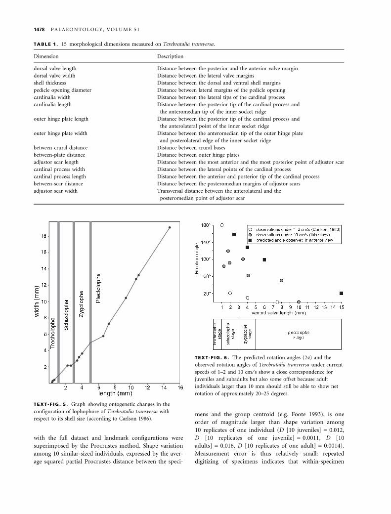
juvenile stages 2.5–5 mm-long are characterized by zygol-
ophe lophophores (Text-figs 5–6), and subadult stages 5–
10 mm-long are characterized by plectolophe lophophore
(Atkins 1959). Individuals smaller than 10 mm are gener-
ally less than two years old (Thayer 1977). Because Tere-
bratalia transversa can live for at least seven years (Paine
1969; Buening and Carlson 1992; Auclair et al. 2003), the
adult stage (10–25 mm in length) was subdivided into
two classes for descriptive purposes, with the arbitrary
breakpoint at 17.5 mm.
The relationship among size classes and shell traits was
explored using principal component analysis (PCA) of the
15 log-transformed shell traits, based on the variance-
covariance matrix. Bivariate and multivariate allometric
coefficients evaluate relationships among log-transformed
shell traits related to dorsal pedicle muscle attachment (i.e.
distance between adjustor scars, scar size, cardinal process
size, and pedicle opening diameter) and ontogenetic size
measures. Size is represented by centroid size (independent
variable in least-square regressions [LSR]) computed from
nine landmarks defined on cardinalia (see below), and dor-
sal valve length (dependent variable in reduced major axis
regressions [RMA]). The centroid size is defined as the
square root of the sum of squared distances between each
landmark and the centroid of the configuration, based on
landmark configuration in Text-fig. 4B (Bookstein 1991).
The multivariate allometric coefficient for a given shell trait
is estimated by dividing the PC1 loading for that variable
by the mean PC1 loading over all variables (Jolicoeur 1963;
Kowalewski et al. 1997). Regressed slopes of log-trans-
formed traditional measurements on log-transformed dor-
sal valve length, log-transformed centroid size, and PC 1
are expected to be significantly different from a value of
one under allometry.
Three methods explore non-linearity of ontogenetic
shape change, test whether complex allometry (i.e.
allometric trajectory is nonlinear and the allometric
coefficient is not constant during the ontogeny, e.g.
A B
TEXT -F IG . 2 . Examples of the rotation angle exhibited by Terebratalia transversa juveniles after 24 hours when attached to the
mussel Modiolus modiolus. In initial position, juveniles were oriented with their anterior margin upstream.
T O M A S O V Y C H E T A L . : O N T O G E N E T I C N I C H E S H I F T I N T H E B R A C H I O P O D T E R E B R A T A L I A T R A N S V E R S A 1475

Bookstein 1991; Zelditch et al. 1993) can better explain
ontogenetic trajectories than simple allometry, and evalu-
ate whether rates of shape change of juveniles differ from
those of adults. First, we evaluate residuals of shell traits
regressed on the centroid size and dorsal valve length.
Second, we compare differences in regression slopes
C D
E F
A B
TEXT -F IG . 3 . Ontogenetic change in position of dorsal (1) and ventral attachment points (2) of the dorsal pedicle muscles relative
to the pedicle, in elevation of outer hinge plates above the dorsal valve floor (3), in width of the cardinal process (4), and in shape of
the pedicle bulb (5) in Terebratalia transversa (anterior views). Outer hinge plates (3) are only slightly elevated above the dorsal valve
floor in the juvenile stage (A–B), and they are strongly elevated above the valve floor in the adult stage (D–F). The distance between
median attachment points of dorsal pedicle muscles increases from the juvenile to the adult stage. A, Ventral valve length is 2.9 mm.
B, Ventral valve length is 4 mm. C, Ventral valve length is 7.1 mm. D, Ventral valve length is 24.9 mm. E, Ventral valve length is
22.9 mm. F, Ventral valve length is 25.9 mm. Figures A–C were obtained using a scanning electron microscope and figures D–F were
photographed under a light microscope.
1476 P A L A E O N T O L O G Y , V O L U M E 5 1
between juveniles and adults. Third, an F-test evaluates
curvilinearity by testing the significance of reduction in
the sum of squares of residuals between linear and sec-
ond-order polynomial regression. This test is based on a
ratio of the difference in sum of squares of residuals
between the quadratic and linear model to the quadratic
residual mean square (McKinney 1984). Differentiating
the second order polynomial relationship (log trait
size = a(log centroid size)2 + b(log centroid size) + c),
one can derive an equation that approximates the slope
(k) at any given size:
k ¼ d log trait size=d log centroid size
¼ 2aðlog centroid sizeÞ þ b
The first derivative of the second order polynomial fit of
the growth trajectory thus estimates the scaling exponent,
corresponding to the growth ratio at a given centroid size
(McKinney 1984; Allmon 1994). The change in k along
the ontogenetic trajectory thus represents the magnitude
of the changes in growth ratio. Fourth, we use locally-
weighted polynomial regression (the lowess function, R
Development Core Team 2007) to estimate the centroid
size that corresponds to the breakpoint of the fitted line
curvature.
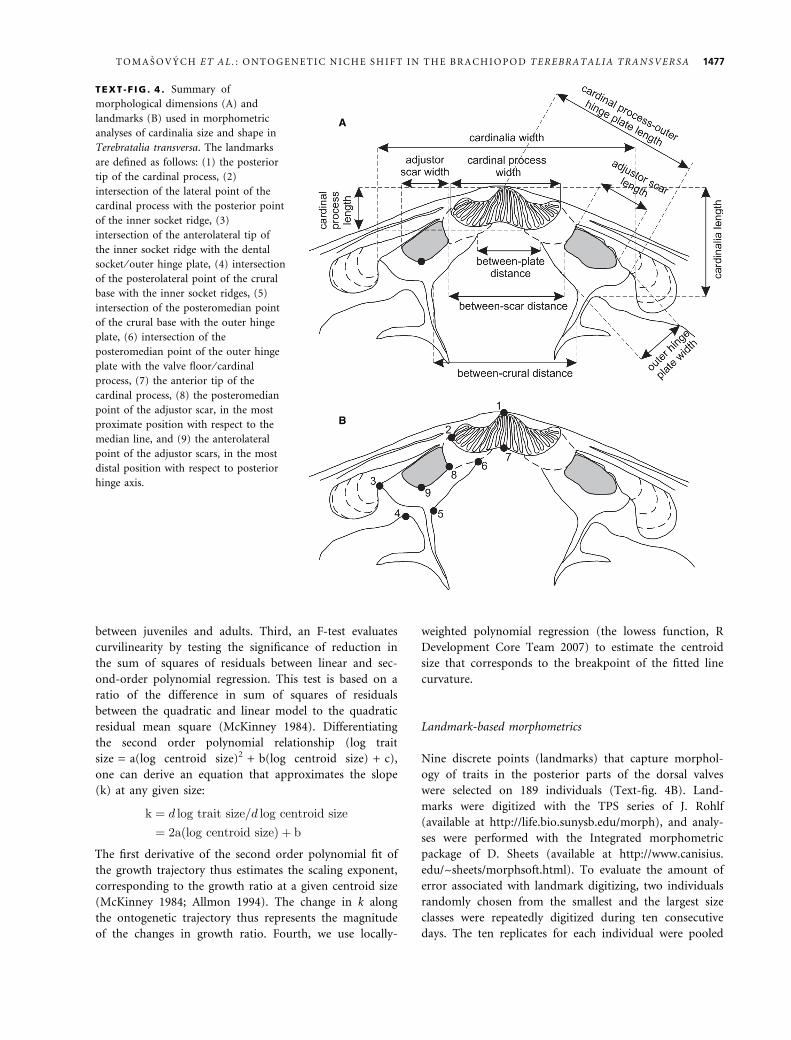
Landmark-based morphometrics
Nine discrete points (landmarks) that capture morphol-
ogy of traits in the posterior parts of the dorsal valves
were selected on 189 individuals (Text-fig. 4B). Land-
marks were digitized with the TPS series of J. Rohlf
(available at http://life.bio.sunysb.edu/morph), and analy-
ses were performed with the Integrated morphometric
package of D. Sheets (available at http://www.canisius.
edu/~sheets/morphsoft.html). To evaluate the amount of
error associated with landmark digitizing, two individuals
randomly chosen from the smallest and the largest size
classes were repeatedly digitized during ten consecutive
days. The ten replicates for each individual were pooled
A
B
TEXT -F IG . 4 . Summary of
morphological dimensions (A) and
landmarks (B) used in morphometric
analyses of cardinalia size and shape in
Terebratalia transversa. The landmarks
are defined as follows: (1) the posterior
tip of the cardinal process, (2)
intersection of the lateral point of the
cardinal process with the posterior point
of the inner socket ridge, (3)
intersection of the anterolateral tip of
the inner socket ridge with the dental
socket ⁄ outer hinge plate, (4) intersection
of the posterolateral point of the crural
base with the inner socket ridges, (5)
intersection of the posteromedian point
of the crural base with the outer hinge
plate, (6) intersection of the
posteromedian point of the outer hinge
plate with the valve floor ⁄ cardinal
process, (7) the anterior tip of the
cardinal process, (8) the posteromedian
point of the adjustor scar, in the most
proximate position with respect to the
median line, and (9) the anterolateral
point of the adjustor scars, in the most
distal position with respect to posterior
hinge axis.
T O M A S O V Y C H E T A L . : O N T O G E N E T I C N I C H E S H I F T I N T H E B R A C H I O P O D T E R E B R A T A L I A T R A N S V E R S A 1477
with the full dataset and landmark configurations were
superimposed by the Procrustes method. Shape variation
among 10 similar-sized individuals, expressed by the aver-
age squared partial Procrustes distance between the speci-
mens and the group centroid (e.g. Foote 1993), is one
order of magnitude larger than shape variation among
10 replicates of one individual (D [10 juveniles] = 0.012,
D [10 replicates of one juvenile] = 0.0011, D [10
adults] = 0.016, D [10 replicates of one adult] = 0.0014).
Measurement error is thus relatively small: repeated
digitizing of specimens indicates that within-specimen
TABLE 1 . 15 morphological dimensions measured on Terebratalia transversa.
Dimension Description
dorsal valve length Distance between the posterior and the anterior valve margin
dorsal valve width Distance between the lateral valve margins
shell thickness Distance between the dorsal and ventral shell margins
pedicle opening diameter Distance between lateral margins of the pedicle opening
cardinalia width Distance between the lateral tips of the cardinal process
cardinalia length Distance between the posterior tip of the cardinal process and
the anteromedian tip of the inner socket ridge
outer hinge plate length Distance between the posterior tip of the cardinal process and
the anterolateral point of the inner socket ridge
outer hinge plate width Distance between the anteromedian tip of the outer hinge plate
and posterolateral edge of the inner socket ridge
between-crural distance Distance between crural bases
between-plate distance Distance between outer hinge plates
adjustor scar length Distance between the most anterior and the most posterior point of adjustor scar
cardinal process width Distance between the lateral points of the cardinal process
cardinal process length Distance between the anterior and posterior tip of the cardinal process
between-scar distance Distance between the posteromedian margins of adjustor scars
adjustor scar width Transversal distance between the anterolateral and the
posteromedian point of adjustor scar
TEXT -F IG . 5 . Graph showing ontogenetic changes in the
configuration of lophophore of Terebratalia transversa with
respect to its shell size (according to Carlson 1986).
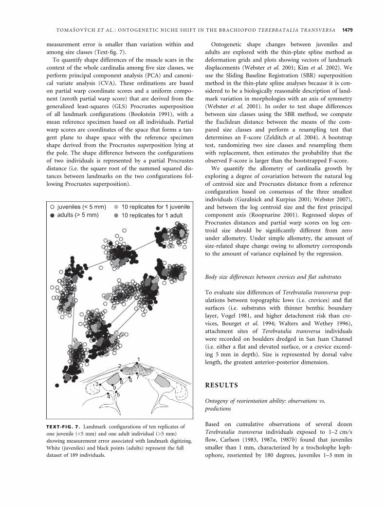
TEXT -F IG . 6 . The predicted rotation angles (2a) and the
observed rotation angles of Terebratalia transversa under current
speeds of 1–2 and 10 cm ⁄ s show a close correspondence for
juveniles and subadults but also some offset because adult
individuals larger than 10 mm should still be able to show net
rotation of approximately 20–25 degrees.
1478 P A L A E O N T O L O G Y , V O L U M E 5 1
measurement error is smaller than variation within and
among size classes (Text-fig. 7).
To quantify shape differences of the muscle scars in the
context of the whole cardinalia among five size classes, we
perform principal component analysis (PCA) and canoni-
cal variate analysis (CVA). These ordinations are based
on partial warp coordinate scores and a uniform compo-
nent (zeroth partial warp score) that are derived from the
generalized least-squares (GLS) Procrustes superposition
of all landmark configurations (Bookstein 1991), with a
mean reference specimen based on all individuals. Partial
warp scores are coordinates of the space that forms a tan-
gent plane to shape space with the reference specimen
shape derived from the Procrustes superposition lying at
the pole. The shape difference between the configurations
of two individuals is represented by a partial Procrustes
distance (i.e. the square root of the summed squared dis-
tances between landmarks on the two configurations fol-
lowing Procrustes superposition).
Ontogenetic shape changes between juveniles and
adults are explored with the thin-plate spline method as
deformation grids and plots showing vectors of landmark
displacements (Webster et al. 2001; Kim et al. 2002). We
use the Sliding Baseline Registration (SBR) superposition
method in the thin-plate spline analyses because it is con-
sidered to be a biologically reasonable description of land-
mark variation in morphologies with an axis of symmetry
(Webster et al. 2001). In order to test shape differences
between size classes using the SBR method, we compute
the Euclidean distance between the means of the com-
pared size classes and perform a resampling test that
determines an F-score (Zelditch et al. 2004). A bootstrap
test, randomizing two size classes and resampling them
with replacement, then estimates the probability that the
observed F-score is larger than the bootstrapped F-score.
We quantify the allometry of cardinalia growth by
exploring a degree of covariation between the natural log
of centroid size and Procrustes distance from a reference
configuration based on consensus of the three smallest
individuals (Guralnick and Kurpius 2001; Webster 2007),
and between the log centroid size and the first principal
component axis (Roopnarine 2001). Regressed slopes of
Procrustes distances and partial warp scores on log cen-
troid size should be significantly different from zero
under allometry. Under simple allometry, the amount of
size-related shape change owing to allometry corresponds
to the amount of variance explained by the regression.
Body size differences between crevices and flat substrates
To evaluate size differences of Terebratalia transversa pop-
ulations between topographic lows (i.e. crevices) and flat
surfaces (i.e. substrates with thinner benthic boundary
layer, Vogel 1981, and higher detachment risk than cre-
vices, Bourget et al. 1994; Walters and Wethey 1996),
attachment sites of Terebratalia transversa individuals
were recorded on boulders dredged in San Juan Channel
(i.e. either a flat and elevated surface, or a crevice exceed-
ing 5 mm in depth). Size is represented by dorsal valve
length, the greatest anterior-posterior dimension.
RESULTS
Ontogeny of reorientation ability: observations vs.
predictions
Based on cumulative observations of several dozen
Terebratalia transversa individuals exposed to 1–2 cm ⁄ sflow, Carlson (1983, 1987a, 1987b) found that juveniles
smaller than 1 mm, characterized by a trocholophe loph-
ophore, reoriented by 180 degrees, juveniles 1–3 mm in
TEXT -F IG . 7 . Landmark configurations of ten replicates of
one juvenile (<5 mm) and one adult individual (>5 mm)
showing measurement error associated with landmark digitizing.
White (juveniles) and black points (adults) represent the full
dataset of 189 individuals.
T O M A S O V Y C H E T A L . : O N T O G E N E T I C N I C H E S H I F T I N T H E B R A C H I O P O D T E R E B R A T A L I A T R A N S V E R S A 1479

length with a schizolophe lophophore showed a net rota-
tion of about 20 degrees, and juveniles 3–5 mm in length
with a zygolophe lophophore rotated very slightly by
about 10 degrees (Table 2). Individuals larger than 5 mm
in length did not show any reorientation.
Under a 10 cm ⁄ s flow, individuals oriented with the
anterior commissure downstream did not reorient after
24 hours. In contrast, several individuals with the anterior
commissure initially oriented upstream reoriented to
some degree. The initial orientation of attached juveniles
changed because Modiolus reoriented rapidly after their
placement in the flow chamber (Table 3). However,
observations indicate that Terebratalia transversa individ-
uals up to 3.5 mm in length can still reorient by 100
degrees and individuals 4 mm long can reorient by about
60 degrees. One individual reaching 8 mm in length
reoriented by 25 degrees. Juveniles smaller than 5 mm
show very high rotation angles, while subadult and adult
individuals invariably do not reorient at all in the adult
stage (lengths that exceed 10 mm). Repeated observations
of Thayer (1975) and LaBarbera (1977) also confirm that
adult Terebratalia transversa individuals do not rotate.
The observed cessation in reorientation of Terebratalia
transversa takes place at a ventral valve length of about 5–
8 mm. The rate of decrease in the potential rotation angle
is thus relatively rapid and coincides with the transition
from schizolophe to zygolophe ⁄ plectolophe stage (Text-
fig. 6). Direct measurements of the angle a on shells with
preserved soft tissues evaluate whether 2a is a good pre-
dictor of rotation ability of living brachiopods. The pre-
dicted 2a values show a good correspondence with the
observed rotation angles of living individuals for juveniles
and subadults (Text-fig. 6). The predicted net rotation
angle (2a) is about 160 degrees for juveniles 2 mm in
length (Text-fig. 3A), about 130–140 degrees for juveniles
reaching 4 mm in length (Text-fig. 3B), and 100–110
degrees for individuals 6 mm in length (Text-fig. 3C).
However, net rotation angle should still attain about
20–25 degrees for adults 15–25 mm in length (Text-
fig. 3D–F). This predicted rotation angle clearly overesti-
mates the observed rotation angle of zero exhibited by
living adults (Text-fig. 6).
Ontogeny of dorsal pedicle muscles and muscle scars
Position of dorsal shell attachment points relative to the
pedicle. The median attachment point of the dorsal pedi-
cle muscles lies on the anteromedian sides of outer hinge
plates that are slightly elevated above the floor of the dor-
sal valve in the juvenile stage (see arrows in Text-figs 3A–
B, 8A–B). In contrast, in the subadult and adult stage, the
TABLE 2 . The observed net rotation angles of Terebratalia transversa under current velocity of 1–2 cm ⁄ s (Carlson, 1983).
Attachment substrate Lophophore
stage
Current
velocity (cm ⁄ s)
Change after
24 hours (degrees)
Ventral
valve length
Terebratalia transversa Early trocholophe 1–2 180–210 0.3
Terebratalia transversa Late trocholophe 1–2 180 0.3–1
Terebratalia transversa Schizolophe 1–2 20 1–3
Terebratalia transversa Zygolophe 1–2 10 3–5
Terebratalia transversa Early plectolophe 1–2 0 5–10
TABLE 3 . Observed one-side rotation angles of Terebratalia transversa under current speed of 10 cm ⁄ s. One-side rotations should be
doubled to estimate net rotation angles (2a).
Attachment substrate Specimen ID Current
velocity
(cm ⁄ s)
Initial position –
exhalant current –
downstream
(degrees)
Change
after
24 hours
(degrees)
Initial position –
exhalant current –
upstream
(degree)
Change
after
24 hours
(degrees)
Ventral
valve
length
Modiolus modiolus #1 Terebratalia transversa #1 10 13 0 8 0 3.4
Modiolus modiolus #1 Terebratalia transversa #2 10 13 0 8 41 0.92
Modiolus modiolus #2 Terebratalia transversa #3 10 18 0 108 46 1.85
Modiolus modiolus #3 Terebratalia transversa #4 10 0 0 165 0 3.2
Modiolus modiolus #4 Terebratalia transversa #5 10 0 0 115 0 3.6
Modiolus modiolus #5 Terebratalia transversa #6 10 0 0 35 25 8.4
Modiolus modiolus #6 Terebratalia transversa #8 10 47 0 78 31 4.2
Modiolus modiolus #6 Terebratalia transversa #9 10 50 0 115 50 3.6
Modiolus modiolus #6 Terebratalia transversa #10 10 29 0 22 57 1.62
1480 P A L A E O N T O L O G Y , V O L U M E 5 1
outer hinge plates are not only substantially elevated
above the dorsal valve floor (Text-fig. 8C–D), but are also
elevated above the commissural plane (Text-fig. 3F).
A horizontal change in the position of the dorsal
attachment point with respect to the valve midline is
measured as an ontogenetic change in horizontal distance
between the dorsal pedicle muscle scars (i.e. between-scar
distance, Text-fig. 8). The between-scar distance increases
with a very strong positive allometry with increasing
centroid size (Text-fig. 9A, LSR slope = 1.93, 95%
CI = 1.87–1.99). The distance between adjustor muscles
becomes relatively larger than the length of the dorsal
valve with growth (RMA slope = 1.65; 95% CI = 1.6–
1.7). Dorsal attachment points of dorsal pedicle muscles
thus rise above the valve floor (Text-fig. 3A vs. Text-
fig. 3F) and allometrically shift away from the valve mid-
line (Text-fig. 9A).
However, the estimated slopes from linear regressions
are affected by curvilinearity. The locally-weighted poly-
nomial regression shows the break in the fitted line at
length of about 7.5 mm (Text-fig. 9A), and residuals of
the between-scar distance from linear regressions show
highly non-linear variations around zero, with negative
values at small and large sizes, and positive values at
intermediate stages (Text-fig. 9B). The rate of increase of
the between-scar distance with respect to centroid size in
juveniles (< 5 mm, LSR slope = 2.24, 95% CI = 1.98–
2.46) is significantly higher than the rate of increase of
the between-scar distance in adults (> 5 mm, LSR
slope = 1.57, 95% CI = 1.47–1.66). The F-value indicates
that the quadratic regression explains growth variations in
between-scar distance significantly better than the simple
linear regression (Table 4).
Position of ventral attachment points relative to the pedicle
bulb. The attachment sites to the pedicle are situated
approximately on the lateral sides of the pedicle in the
juvenile stage, and remain in this position or are slightly
A B
C D
TEXT -F IG . 8 . Ontogenetic increase in the distance between posteromedian parts of dorsal pedicle muscles scars (arrow 1), and
ontogenetic increase in the width of the cardinal process in Terebratalia transversa. Anterolateral parts of dorsal pedicle muscle scars
are marked with arrow 2. Lateral parts of the cardinal process are marked with arrow 3. A, Dorsal valve length is 1.06 mm. B, Dorsal
valve length is 1.2 mm. C, Dorsal valve length is 4.9 mm. D, Dorsal valve length is 19.1 mm. Figures A–C were obtained using a
scanning electron microscope and figure D was photographed under a light microscope.
T O M A S O V Y C H E T A L . : O N T O G E N E T I C N I C H E S H I F T I N T H E B R A C H I O P O D T E R E B R A T A L I A T R A N S V E R S A 1481

A B
DC
E F
1482 P A L A E O N T O L O G Y , V O L U M E 5 1

shifted in the ventral direction in the adult stage (Text-
fig. 3). The dorsal pedicle muscles are attached to the
pedicle epithelium in juveniles, but insertions extend
into the fibrous connective tissue in adults of Terebra-
talia transversa (Stricker and Reed 1985). Although the
connection between the attachment points and the pedi-
cle in the juvenile stage is not completely visible in
anterior views (Text-fig. 3), the ventral insertions proba-
bly also do not change markedly in size during ontog-
eny. The width of pedicle opening diameter increases
with a very slight but significantly positive allometry
with increasing centroid size (Text-fig. 10, LSR
slope = 1.14, 95% CI = 1.12–1.17). Its growth is almost
isometric relative to dorsal valve length (RMA
slope = 0.97, 95% CI = 0.95–0.997), and the locally-
weighted polynomial regression shows a relatively linear
fit (Text-fig. 10). Residuals of the pedicle opening dia-
meter width from linear regressions show a relatively
random variation around zero (Text-fig. 10). The rate
of increase of pedicle opening diameter also does not
differ between juveniles and adults, and the F-value is
very low (Table 4).
Cardinal process width. The cardinal process is formed
by a transversely oval depression in the juvenile stage
(Text-fig. 8A–B). In the adult stage, it is formed by an
elevation or raised ridge consisting of parallel, highly
irregular ridges, with lateral margins being curved in the
posterior direction (Text-fig. 8C). The width of the mus-
cle attachment site of the cardinal process changes with
a significantly positive allometry with respect to centroid
size (Text-fig. 9C, LSR = 1.3, 95%CI = 1.25–1.34), and
dorsal valve length (RMA slope = 1.11, 95%CI = 1.07–
1.14).
The locally-weighted polynomial regression shows a
break in the fitted line at length of about 5 mm (Text-
fig. 9C). Non-linear residuals of the cardinal process
width from linear regressions show a rather sharp break
that corresponds to the dorsal valve length of 3–5 mm
(Text-fig. 9D). Cardinal process width grows with a posi-
tive allometry in juveniles (Text-fig. 11, LSR slope = 1.75,
95% CI = 1.6–1.88), and grows isometrically in adults
(LSR slope = 0.99, 95% CI = 0.95–1.03). The complex
allometry is also demonstrated by the high and significant
F-value (Table 4).
TABLE 4 . Pearson correlations between principal components and 15 morphological traits, univariate (based on least-square regres-
sions of log-transformed traits on log centroid size) and multivariate allometric coefficients (the PC1 loading of each trait divided by
the mean PC1 loading over all traits), and results of the F-test for complex allometry in 15 morphological traits of Terebratalia
transversa. Allometric coefficents in boldface refer to means whose 95% bootstrapped confidence intervals did not overlap with 1. The
p-values in boldface are results that are significant after sequential Bonferroni correction at a = 0.05.
Shell trait Correlation
between PC1
and trait
Correlation
between PC2
and trait
Univariate
allometric
coefficient –
juvenile (<5 mm)
Univariate
allometric
coefficient –
adult (>5 mm)
Multivariate
allometric
coefficient
F-value p
Dorsal valve length 0.991 0.036 1.21 1.01 0.98 48.82 <0.0001
Dorsal valve width 0.992 )0.005 1.41 1.05 1.03 92.42 <0.0001
Shell thickness 0.991 0.055 1.34 1.28 1.16 4.24 0.041
Pedicle opening diameter 0.988 0.032 1.19 1.13 0.99 0.28 0.597
Cardinalia width 0.994 0.056 1.2 1.14 0.98 0.32 0.57
Cardinalia length 0.982 0.137 1.08 1.04 0.90 4.49 0.036
Outer hinge plate length 0.994 0.047 1.2 1.13 1.03 15.6 0.0001
Outer hinge plate width 0.969 0.150 0.98 0.99 0.80 10.4 0.0014
Between-crural distance 0.987 0.047 1.05 0.98 0.83 1.82 0.18
Between-plate distance 0.967 )0.225 2.29 1.13 1.30 136.1 <0.0001
Adjustor scar length 0.949 0.228 0.84 0.86 0.71 3.61 0.059
Cardinal process width 0.991 )0.033 1.75 0.99 0.98 163.8 <0.0001
Cardinal process length 0.983 0.088 1.28 1.17 1.03 5.44 0.021
Between-scar distance 0.986 )0.140 2.24 1.57 1.57 61.84 <0.0001
Adjustor scar width 0.959 0.142 0.98 0.79 0.71 11.48 0.0009
TEXT -F IG . 9 . Curvilinear bivariate relationships between centroid size (based on landmark configuration in Text-fig. 3B) and
between-scar distance (A), between centroid size and cardinal process width (C), and between centroid size and adjustor scar width
(E). The fitted lines are based on the locally-weighted polynomial regressions. The residuals from bivariate linear regressions show
curved patterns and demonstrate that complex allometric changes take place at centroid size corresponding to 3–7 mm long dorsal
valves (B, D, F).
T O M A S O V Y C H E T A L . : O N T O G E N E T I C N I C H E S H I F T I N T H E B R A C H I O P O D T E R E B R A T A L I A T R A N S V E R S A 1483
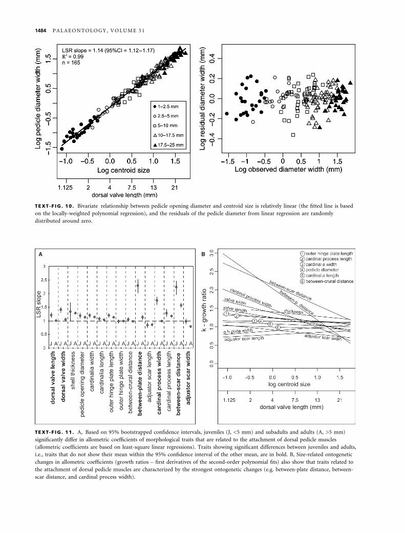
TEXT -F IG . 10 . Bivariate relationship between pedicle opening diameter and centroid size is relatively linear (the fitted line is based
on the locally-weighted polynomial regression), and the residuals of the pedicle diameter from linear regression are randomly
distributed around zero.
A B
TEXT -F IG . 11 . A, Based on 95% bootstrapped confidence intervals, juveniles (J, <5 mm) and subadults and adults (A, >5 mm)
significantly differ in allometric coefficients of morphological traits that are related to the attachment of dorsal pedicle muscles
(allometric coefficients are based on least-square linear regressions). Traits showing significant differences between juveniles and adults,
i.e., traits that do not show their mean within the 95% confidence interval of the other mean, are in bold. B, Size-related ontogenetic
changes in allometric coefficients (growth ratios – first derivatives of the second-order polynomial fits) also show that traits related to
the attachment of dorsal pedicle muscles are characterized by the strongest ontogenetic changes (e.g. between-plate distance, between-
scar distance, and cardinal process width).
1484 P A L A E O N T O L O G Y , V O L U M E 5 1

Outline of the pedicle bulb. In the smallest individuals,
ventral insertions of dorsal pedicle muscles and dorsal
insertions of ventral pedicle muscles penetrate or are
continuous with an epithelium that separates the pedicle
from the body cavity (Text-fig. 3A–B, see also Stricker
and Reed 1985). The pedicle bulb is transversely
elongated in the adult stage, mainly because the cardinal
process is highly elevated above the commissure (Text-
fig. 3E). This general change towards an elongate pedicle
bulb can also contribute to the decrease in the angle aduring ontogeny of T. transversa.
Size of dorsal pedicle muscle scars. The width of adjustor
scars shows a negative allometry with respect to centroid
size (Text-fig. 9E, LSR slope = 0.88, 95% CI = 0.85–0.9).
The scar width becomes relatively smaller than the length
of the dorsal valve with growth (RMA slope = 0.75; 95%
CI = 0.73–0.77). The locally-weighted polynomial regres-
sion shows a slight break in the fitted line at length
between 4–7 mm (Text-fig. 9E). Residuals of the adjustor
scar width from linear regressions are also non-linear,
with the breakpoint occurring at the length of dorsal
valve of about 5 mm (Text-fig. 9F). The rate of decrease
of the adjustor scar width is significantly lower and iso-
metric in juveniles (Text-fig. 11A, LSR slope = 0.98, 95%
CI = 0.88–1.08) than in adults (LSR slope = 0.79, 95%
CI = 0.74–0.83), and F-test confirm complex allometric
growth of the scar width (Table 4).
Ontogeny of cardinalia
Traditional morphometric analyses. Bivariate analyses with
the log-transformed centroid size as an independent vari-
able and morphological traits other than adjustor scars as
dependent variables show that shell length, width, and
thickness, cardinalia width, outer hinge plate length, and
cardinal process length and width change with a signifi-
cantly positive allometry. Cardinalia length show a slight
positive allometry (LSR slope = 1.07, 95% CI = 1.05–
1.08), and outer hinge plate width grows with a slight
negative allometry with centroid size (LSR slope = 0.93,
95% CI = 0.91–0.95). The between-crural distance
increases isometrically (LSR slope = 0.99, 95% CI = 0.97–
1). The between-plate distance shows a similarly high
negative allometry as the between-scar distance (LSR
slope = 1.72). Significant differences in regression coeffi-
cients between juveniles and adults characterize the cardi-
nal process and position of the pedicle muscle scars
(Text-fig. 11A, Table 4). A curvilinear trajectory in shape
change is shown by a significant improvement in the
F-value based on the second-order polynomial for several
traits (Table 4). In addition to the changing growth ratios
of the between-scar distance and the cardinal process
width, substantial changes in growth ratio characterize the
between-plate distance (Text-fig. 11B). Allometric growth
rates invariably decrease with increasing centroid size.
This complex allometry is localized and characterizes only
those parts of cardinalia that are related to diductor and
pedicle muscle scars.
In multivariate analysis, PC1 accounts for >96% of
the variation in the principal component analysis based
on the 15 log-transformed morphological variables. All
shell traits show a very high correlation with PC1
(r > 0.98, Table 4) and the five a priori-defined size
classes are well demarcated along PC1 (Text-fig. 12).
Thus most of the morphological variation can be
explained by size and size-related shape variation that
is related to PC1. The pattern of morphological varia-
tion is non-linear along the second PC (1.45%), with
the smallest and largest size classes being characterized
by more negative values than intermediate size classes.
The loadings on PC2 and correlations between shell
traits and PC2 show that the differences along this
component are mainly driven by variations in traits
that are related to shape and position of dorsal pedicle
muscle scars (Text-fig. 12, Table 4). These variables
include between-plate distance, adjustor scar length, and
between-scar distance.
Multivariate allometric analyses similarly show that
the highest allometric coefficients incorporate variables
related to dorsal pedicle muscle scars (Text-fig. 12,
Table 4). Between-scar distance and between-plate dis-
tance show a highly positive allometry, and adjustor
scar width and length show a highly negative allometry.
The relationships between shell traits and PC2 and the
multivariate coefficients thus show that ontogenetic
changes in size and shape of adjustor scars drive this
non-linear pattern (Table 4). Note that the non-linear-
ity of the multivariate pattern probably obscures allo-
metric growth of other traits such as the cardinal
process width because the coefficients are based on a
linear model.
Landmark-based analyses. The univariate approach regres-
sing Procrustes distances on log-transformed centroid size
(slope = 0.126, std = 0.005, Z-score = 19.29, r = 0.89),
and the multivariate approach regressing all shape vari-
ables (i.e. components of the uniform deformation and
partial warp scores) against log-transformed centroid size
(Wilks Lambda = 0.106, F-score = 105.08, %variance
explained = 0.89, df1 = 14, df2 = 174, P < 0.0001) reveal
a significantly allometric growth of cardinalia (Text-
fig. 13). Allometric shape change accounts for the major-
ity of ontogenetic variation in cardinalia morphology
(89%). Principal component analysis based on partial
warp scores derived from Procrustes superposition shows
distinct differences among the five size classes along PC1
T O M A S O V Y C H E T A L . : O N T O G E N E T I C N I C H E S H I F T I N T H E B R A C H I O P O D T E R E B R A T A L I A T R A N S V E R S A 1485

(62.5%). It shows a curved, non-linear pattern of land-
mark configurations along PC1 (Text-fig. 14A), with the
a priori size classes being related not only to PC1, but
also to PC2 (9.1%). We note that landmark configura-
tions are rescaled to unit centroid size in Procrustes and
SBR superpositions. Therefore, size differences are fac-
tored out and differences among size classes observed in
landmark-based PCA are thus not related to differences
in specimen size (as in traditional PCA based on dis-
tances). However, variations in landmark configurations
in PCA clearly show that cardinalia shape does change as
a function of size.
Canonical variate analysis shows that the five size clas-
ses are significantly different in shape (Text-fig. 14B),
with the first three axes being significant and thus dis-
criminating among the five groups (CVA1: L = 0.049,
v2 = 538.89, df = 56, P < 0.0001, CVA2: L = 0.408, v2 =
160.18, df = 39, p < 0.0001, CVA3: L = 0.797, v2 = 40.45,
df = 24, P = 0.0004). Comparisons of changes in land-
mark configurations among size classes in SBR show
substantial ontogenetic variations in the shape of the car-
dinalia. Based on SBR superposition, pairwise compari-
sons show significant differences in mean shape among
five size groups (bootstrapped F-test). The Euclidean dis-
tance between successive means substantially decreases
with increasing size. It is relatively high between the first
and second size class (F-score = 19.6, distance between
means = 0.216, 95% CI = 0.17–0.26), and between the
second and third size class (F-score = 18.5, d = 0.214,
95% CI = 0.18–0.26). In contrast, it is markedly lower
between the third and fourth size class (F-score = 12.3,
d = 0.098, 95%CI = 0.07–0.14), and between the fourth
and fifth size class (F-score = 7.05, d = 0.089, 95%
CI = 0.06–0.13). The thin-plate spline showing deforma-
tion of the largest individual from the three smallest indi-
viduals, visualized by vectors of landmark displacement
and deformation grid, indicates pronounced morphologi-
cal changes in cardinalia shape (Text-fig. 14C, D). The
major changes are a shift of the median boundary of
adjustor scars away from the midline, widening of the
cardinal process, and widening and shortening of outer
hinge plates and inner socket ridges.
A
B
C
TEXT -F IG . 12 . A, Principal component analysis of 144
individuals based on 15 morphological traits. The eigenvalues
are PC1 = 6.4 [96.7%], PC2 = 0.1 [1.45%], PC3 = 0.03 [0.44%],
PC4 = 0.02 [0.28%]. B, Morphological traits related to the
attachment of dorsal pedicle muscles predominantly contribute
to loadings on PC2. C, Multivariate allometric coefficients with
95% bootstrapped confidence intervals, based on PCA, show
that the significantly negative allometry is exhibited by the size
of adjustor scars and the significantly positive allometry by
distance between adjustor scars.
1486 P A L A E O N T O L O G Y , V O L U M E 5 1

Thin-plate splines performed separately for the five
size classes reveal the pattern and magnitude of ontoge-
netic shape changes in cardinalia (Text-fig. 15). First,
the most pronounced shape change is restricted to two
size classes that attain 2.5–10 mm. The retreat of adjus-
tor scars and outer hinge plates from the midline, and
the rapid widening of the cardinal process are the most
evident changes visible in displacement and deformation
plots (Text-fig. 15). These traits thus grow faster than
other traits in the juvenile (2.5–5 mm) and subadult
stages (5–10 mm). Note that the widening of the cardi-
nal process is limited to stages attaining 2.5–5 mm. Sec-
ond, the change in cardinalia shape slows down at
stages exceeding 10 mm in length when allometric wid-
ening of cardinal process and between-plate distance
stops. Third, the direction of shape change in most
traits can reverse at stages exceeding a dorsal valve
length of 17.5 mm. The lengths of the vectors of land-
mark displacement (Text-fig. 15) demonstrate that the
ontogenetic trajectory is characterized by decreasing
magnitude of allometric change of shape of adjustor
scars, outer hinge plates, and cardinal process width.
The transition between individuals 2.5–10 mm in length
corresponds to a major shape change. Similar to analyses
based on traditional morphometric variables, landmark-
based analyses thus show that (1) cardinalia shape
changes are mainly restricted to juvenile stages, and that
(2) they correspond to those portions of the cardinalia
that affect the rotation and the torque during the open-
ing of valves.
Size differences between crevices and flat substrates
There are significant differences in median length between
Terebratalia transversa populations occupying crevices
and flat substrates, with populations in crevices showing
smaller median length (Wilcoxon rank-sum test, p =
0.0062). This effect of substrate rugosity is also significant
when only individuals larger than 5 mm are evaluated
(Wilcoxon rank-sum test, p = 0.0037). This effect
becomes reduced when individuals larger than 10 mm are
evaluated (Wilcoxon rank-sum test, p = 0.09), and disap-
pears when individuals larger than 15 mm are evaluated
(Wilcoxon rank-sum test, p = 0.67). Therefore, the differ-
ence in median length is related to the higher proportions
of small individuals in crevices; large individuals do not
differ in median length between crevices and flat surfaces.
DISCUSSION
Ontogenetic niche shift
Non-linear residuals of between-scar distance and cardi-
nal process width based on linear regressions, higher
allometric coefficients of these traits in juveniles than
in adults, and ontogenetic decrease in their growth
ratios based on polynomial regressions, demonstrate
that the allometric change is curvilinear, with a marked
allometric change at a dorsal valve length of
2.5–10 mm. Landmark analyses show that the growth
A B
TEXT -F IG . 13 . Landmark-based analyses show a significantly allometric growth of cardinalia of Terebratalia transversa. A, Scores
from the PC1 (PCA based on the partial warp scores derived from the Procrustes superposition and a GLS Procrustes mean reference
specimen based on all individuals) regressed on the natural log of centroid size. B, Rate of cardinalia shape change relative to the
natural log of centroid size, based on partial Procrustes distance from a reference form based on consensus of the three smallest
individuals.
T O M A S O V Y C H E T A L . : O N T O G E N E T I C N I C H E S H I F T I N T H E B R A C H I O P O D T E R E B R A T A L I A T R A N S V E R S A 1487
change is fast for small lengths and slows down at a
length exceeding 10 mm, and also demonstrate that al-
lometric changes related to shape of cardinal process
and adjustor scars are the dominant source of variation
in the whole cardinalia shape. Although non-linear allo-
metric trajectories of morphological traits can be
expected in brachiopods owing to their complex onto-
genetic changes in lophophore configuration (e.g. Mac-
kay et al. 1993; MacKinnon et al. 1997), we show that
non-linear allometric growth also substantially affects
the shape of morphological traits related to the pedicle
and diductor muscles in Terebratalia transversa.
Although allometric growth can be a consequence of
maintenance of function over a size range (LaBarbera
A B
C D
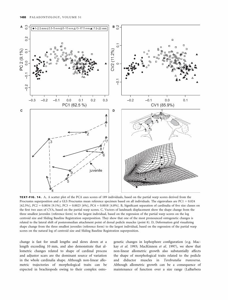
TEXT -F IG . 14 . A, A scatter plot of the PCA axes scores of 189 individuals, based on the partial warp scores derived from the
Procrustes superposition and a GLS Procrustes mean reference specimen based on all individuals. The eigenvalues are PC1 = 0.024
[62.5%], PC2 = 0.0034 [9.1%], PC3 = 0.0023 [6%], PC4 = 0.0018 [4.8%]. B, Significant separation of cardinalia of five size classes on
the first two axes of CVA, based on the partial warp scores. C, Vectors of landmark displacement show the shape change from the
three smallest juveniles (reference form) to the largest individual, based on the regression of the partial warp scores on the log
centroid size and Sliding Baseline Registration superposition. They show that one of the most pronounced ontogenetic changes is
related to the lateral shift of posteromedian attachment point of dorsal pedicle muscles (point 8). D, Deformation grid visualizing
shape change from the three smallest juveniles (reference form) to the largest individual, based on the regression of the partial warp
scores on the natural log of centroid size and Sliding Baseline Registration superposition.
1488 P A L A E O N T O L O G Y , V O L U M E 5 1
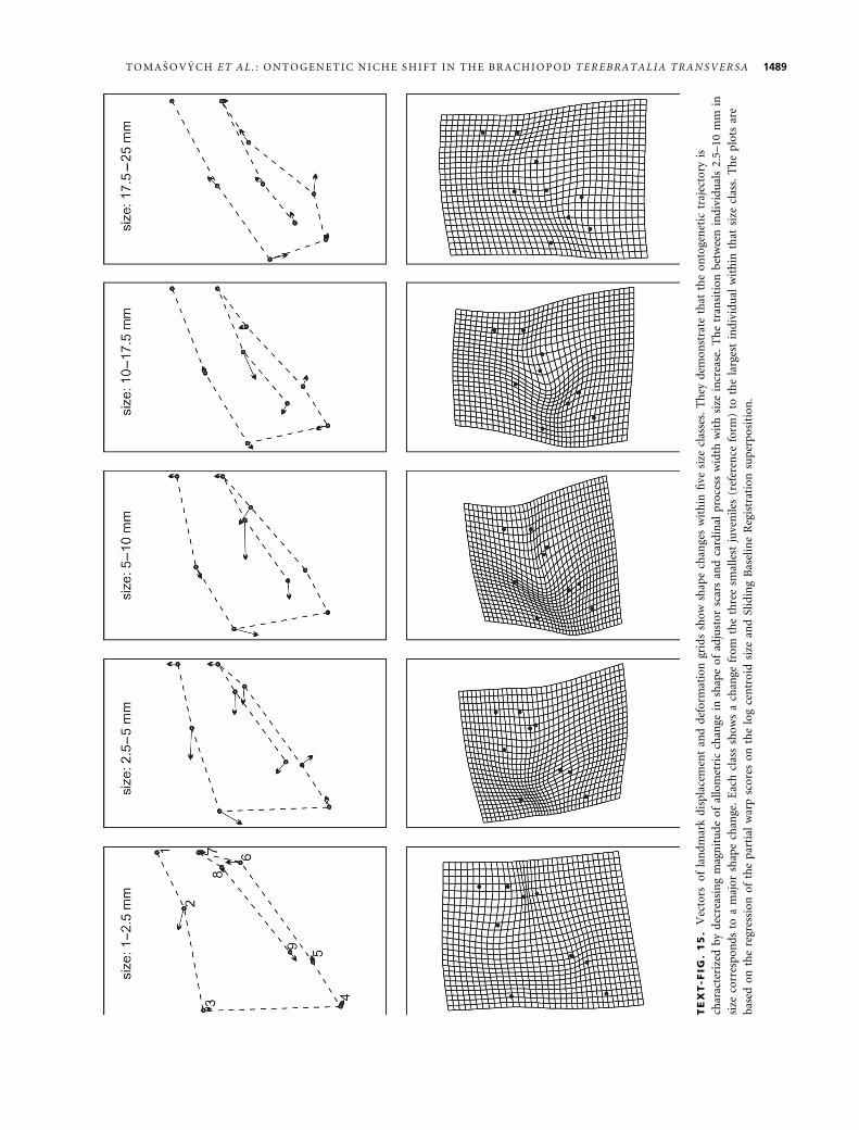
TE
XT
-FIG
.1
5.
Vec
tors
of
lan
dm
ark
dis
pla
cem
ent
and
def
orm
atio
ngr
ids
sho
wsh
ape
chan
ges
wit
hin
five
size
clas
ses.
Th
eyd
emo
nst
rate
that
the
on
toge
net
ictr
ajec
tory
is
char
acte
rize
db
yd
ecre
asin
gm
agn
itu
de
of
allo
met
ric
chan
gein
shap
eo
fad
just
or
scar
san
dca
rdin
alp
roce
ssw
idth
wit
hsi
zein
crea
se.
Th
etr
ansi
tio
nb
etw
een
ind
ivid
ual
s2.
5–10
mm
in
size
corr
esp
on
ds
toa
maj
or
shap
ech
ange
.E
ach
clas
ssh
ow
sa
chan
gefr
om
the
thre
esm
alle
stju
ven
iles
(ref
eren
cefo
rm)
toth
ela
rges
tin
div
idu
alw
ith
inth
atsi
zecl
ass.
Th
ep
lots
are
bas
edo
nth
ere
gres
sio
no
fth
ep
arti
alw
arp
sco
res
on
the
log
cen
tro
idsi
zean
dSl
idin
gB
asel
ine
Reg
istr
atio
nsu
per
po
siti
on
.
T O M A S O V Y C H E T A L . : O N T O G E N E T I C N I C H E S H I F T I N T H E B R A C H I O P O D T E R E B R A T A L I A T R A N S V E R S A 1489

1989), ontogenetic allometry of the pedicle muscle sys-
tem in Terebratalia transversa contributes to the
decrease in rotation ability.
The temporal correlation between the observed increase
of distance between adjustor scars, of width of cardinal
process, and of elevation of outer hinge plates above the
valve floor at a length of 2.5–10 mm on one hand, and
the observed decrease in rotation at a length of 2.5–5 mm
on the other hand, support the hypothesis that the
decrease in rotation angle observed in living Terebratalia
transversa is related to ontogenetic changes in the dorsal
attachment points of dorsal pedicle muscles. This agree-
ment between ontogenetic changes in the observed
reorientation ability and the ontogeny of internal mor-
phological structures implies that the rotation angle is
mechanically constrained in the adult stage because
contraction of dorsal pedicle muscles is functionally
handicapped. However, the misfit between the predicted
rotation based on the angle a (i.e. adults should show
rotations of about 20–25 degrees) and the observed
rotation on living individuals (i.e. complete absence of
rotation in adults) implies that some additional, morpho-
logical or ecological factors limit reorientation of the
adult Terebratalia transversa.
In addition to the ontogenetic change in reorientation,
several aspects of ecology substantially change over the
ontogeny of most brachiopods. Brachiopods change their
feeding flow configuration with size (Rudwick 1962,
1970), perceive their fluid environment at higher Rey-
nolds numbers with size increase (Vogel 1981; Carlson
1983, 1986), and emerge from the substrate-related
boundary layer with size increase (Carlson 1983). Terebra-
talia transversa juveniles possess trocholophe (<1 mm)
and schizolophe lophophore types (<2.5 mm), in contrast
to subadults and adults, which are characterized by zygol-
ophe (2.5–5 mm) and plectolophe lophophore types
(> 5 mm) (Atkins 1959; Dhar et al. 1997). At a length of
about 2.5 mm, the configuration of the lophophore
changes from a two-dimensional ring to a three-dimen-
sional folded ring and the direction of inhalant and exhal-
ant current flows, by active pumping, changes in
Terebratalia transversa (Carlson 1983, 1987a, 1987b). The
zygolophe-type configuration is characterized by an adult-
type current flow regime with median excurrent flow and
lateral incurrent flows (Rudwick 1962). At this stage,
pressure distributions act in concert with active pumping
under orientation with the anterior-posterior shell axis
perpendicular to the current direction (LaBarbera 1977).
Therefore, Terebratalia transversa achieves its preferred
‘adult’ orientation with respect to water currents only
after development of the zygolophe lophophore.
Small juveniles likely experience a different fluid envi-
ronment than adults owing to the dominance of viscous
forces instead of inertial forces (Vogel 1981), and high
flow resistance but low total flow rates (Grunbaum et al.
1998; Sherrard and LaBarbera 2005). Under a current
speed of 1–2 cm ⁄ s, juveniles smaller than 3 mm in length
are living within the substrate-associated boundary layer
(Carlson 1983, 1986, 1987a, 1987b). Terebratalia transver-
sa starts emerging into the free stream layer at these low
current velocities, approximately at the time when they
change their lophophore type to the adult configuration
(Carlson 1983, 1986). Terebratalia transversa thus reori-
ents to the preferred orientation with respect to water
currents at this stage because (1) the preferred pre-zygol-
ophe orientation differs from the orientation of adults,
and (2) pre-zygolophe stages probably do not detect free
stream velocity as do adults (Carlson 1983, 1986). Adult
Terebratalia transversa living in subtidal habitats are ori-
ented with the anterior-posterior shell axis perpendicular
to the current direction (LaBarbera 1978), in accord with
the preferred zygolophe and plectolophe orientation. We
note that lack of reorientation was observed for adult Ter-
ebratalia transversa individuals that live in habitats differ-
ing in depth, substrate and current velocity (Thayer 1975,
1977; LaBarbera 1977; Schumann 1991). Although rota-
tion can be inhibited by crowding or by growth within
crevices in topographically-complex habitats, unrestricted
individuals attached to flat substrates do not rotate in the
adult stage.
It is thus remarkable that the rotation loss proceeds
very rapidly after Terebratalia transversa can effectively
attain its preferred zygolophe-plectolophe orientation
with respect to water currents, and that this rotation loss
is especially well captured by the complex allometric
growth of dorsal pedicle muscle scars. This reorientation
loss in Terebratalia transversa can represents an ontoge-
netic niche shift (Werner and Giliam 1984) because reori-
entation can confer feeding efficiency (LaBarbera 1977)
and competition advantage of brachiopods (LaBarbera
1985). This shift can be associated with some substantial
gains related to growth and survivorship of subadult and
adult brachiopods that outweigh the disadvantages of sub-
stantially reduced reorientation ability at length of 5 mm.
The ontogenetic shift in rotation ability of Terebratalia
transversa is species-specific; many other brachiopod spe-
cies such as Terebratulina unguicula or Laqueus californi-
anus actively reorient in the adult stage (LaBarbera 1977,
1978; Richardson 1981a).
Morphological causes of reorientation loss
Allometric ontogenetic increase in between-scar distance,
increase in cardinal process width, and increase in eleva-
tion of scars above the valve floor closely correspond to
the observed ontogenetic change in rotation angle in liv-
ing individuals of Terebratalia transversa. The dorsal side
1490 P A L A E O N T O L O G Y , V O L U M E 5 1
of the angle a substantially changes its orientation from
vertical to nearly horizontal during the ontogeny of Tere-
bratalia transversa (i.e. outer hinge plates with attachment
sites of dorsal pedicle muscles move in a ventral and lat-
eral direction away from midline). The elevation of
attachments points of the dorsal pedicle muscles above
the valve floor and the allometric shift away from the
valve midline thus decrease the angle a. The ventral side
of the angle a has a relatively constant orientation
(approximately parallel with the commisural plane) in the
ontogeny of Terebratalia transversa because the pedicle
opening diameter shows isometric growth and ventral
attachment points of the dorsal pedicle muscles do not
change their position during ontogeny. Therefore, the
change in angle a is mainly driven by the change in its
dorsal side that is captured by morphological changes in
dorsal pedicle muscle scars and cardinalia.
In the adult stage, the rotation ability is effectively pro-
hibited not only by increased between-scar distance, but
by a very wide cardinal process that shows complex allo-
metric growth. The changes in size of scars of dorsal ped-
icle muscles measured by the adjustor scar width (i.e, scar
size decrease is accelerated later in ontogeny) and pedicle
opening width (i.e. isometric growth) seems to have a
weak relationship to the observed decrease in reorienta-
tion ability of Terebratalia transversa. Marked changes in
morphology of the loop of Terebratalia transversa (Atkins
1959) have little effect on the shape of cardinalia, and the
between-crural distance shows isometric growth. Simi-
larly, the width of cardinalia constrains inner socket
ridges, but cardinalia width shows more or less isometric
growth.
Other morphological factors that can reduce reorien-
tation loss can be related to the presence of stout pedi-
cle connectives – tissues that connect the pedicle with
inner socket ridges in adult individuals of Terebratalia
transversa (LaBarbera 1978). Pedicle connectives increase
in extent with age and can oppose the contraction of
dorsal pedicle muscles (LaBarbera 1978; Carlson 1986).
The highly depressed shape of the pedicle bulb can also
reduce the rotation potential of adults.
Ecological consequences of reorientation loss
On one hand, the ontogenetic reduction in reorientation
ability can affect ecological performance of Terebratalia
transversa by decreasing feeding efficiency (if current
directions are variable). On the other hand, loss of reori-
entation ability can decrease vulnerability to dislodgment
by currents and biotic disturbance (1) by minimizing tor-
que around the pedicle axis and ⁄ or by (2) shifting energy
investment into attachment strength at the expense of the
cost involved in reorientation. For example, under space-
and food-limitation and variable flow directions, activity
of pedicle muscles enhances the ability of brachiopods to
use ambient currents that augment lophophore-generated
flow through the mantle cavity (LaBarbera 1977, 1981),
and prevents crowding from other animals settled nearby
and aids in competion for space (LaBarbera 1977, 1985).
Under constant flow directions, it can be advantageous
for Terebratalia transversa to orient to the preferred ori-
entation in the juvenile stage, and then to reduce vulnera-
bility to physical or biotic dislodgement by reducing
torque around the pedicle and also to decrease energetic
costs of reorientation. Under high levels of disturbance,
the loss of dorsal adjustor torque around the pedicle can
be necessary to permit tight adhesion of the shell to the
substrate via strong ventral pedicle muscles; restricting
dorsal muscle activity in the adult stage increases shell
protection against dislodgement. The loss of reorientation
ability can thus be a consequence of changes that
strengthen substrate attachment. However, we also stress
that changes in the shape of cardinalia and the position
of dorsal-pedicle muscle attachment may be a conse-
quence of selection pressure on other morphological
traits, and the change in rotation ability may be related to
such hitchhiking effects (e.g. Wagner 1996). For example,
widening the cardinal process serves to increase the stabi-
lity of the valves during opening and closing by spreading
apart the diductor muscle origins. In addition, an onto-
genetic increase in shell convexity owing to maximizing
mantle-cavity volume will force elevation of hinge plates
above the dorsal valve floor, and thus can also reduce the
angle a.
Everything else being equal, the feeding efficiency of
immobile Terebratalia transversa would be diminished rel-
ative to brachiopods with active reorientation. However,
under high food supply, the ability to reduce vulnerability
to dislodgement may overwhelm negative effects caused
by the loss of reorientation ability. Submarine habitats of
San Juan Islands are characterized by seasonally-variable
but mostly nutrient-rich and plankton-rich waters (Mac-
kas and Harrison 1997; Strom et al. 2001; Zamon 2002).
A strong, tidally-induced mixing of open ocean and
estuarine waters ensures that waters are rich in nutrients
throughout the year, and Terebratalia transversa com-
monly occurs in San Juan Islands habitats with moderate
to high currents (LaBarbera 1977). Both high primary
productivity and high current speeds can thus ensure that
competition for food is not likely to play an important
role in the distribution of Terebratalia transversa. This
species also commonly occurs in crevices within topo-
graphically-complex substrates, formed either by boulder
undersides and caves or by solitary ascidians and giant
barnacles (Tomasovych 2008a, b). Such crevices are inevi-
tably affected by thicker benthic boundary layers, slower
and less predictable water currents, and increased risk of
T O M A S O V Y C H E T A L . : O N T O G E N E T I C N I C H E S H I F T I N T H E B R A C H I O P O D T E R E B R A T A L I A T R A N S V E R S A 1491

feeding current recirculation, and thus can limit suspen-
sion-feeders. In addition, the direction of water flow to
which juveniles reorient after they achieve the zygolophe
lophophore can change during their growth because they
are mostly surrounded by other organisms (e.g. Balanus
nubilus, solitary ascidians, fleshy algae, cheilostomid
encrusting and erect bryozoans). Therefore, it is interest-
ing to note that population density of Terebratalia trans-
versa increases from open and exposed substrates towards
crevices and cryptic habitats (Tomasovych 2008a, 2008b).
This observation might indicate that competition for food
is less limiting than other factors, such as disturbance,
that can reduce brachiopod density on exposed sub-
strates.
Although adult individuals Terebratalia transversa in
this area are rarely affected by carnivorous predators
(Mauzey et al. 1968; Thayer 1985), high consumer pres-
sure on juveniles and small-sized Terebratalia transversa is
probably mainly represented by the grazing activity of the
highly abundant sea urchins Strongylocentrotus francisc-
anus and S. droebachiensis in subtidal habitats, and by the
grazing activity of limpets and chitons in the intertidal
zone (Tomasovych 2008a). In such habitats with high
biotic disturbance and high food supply, it is possible that
the rapid loss of rotation ability in Terebratalia transversa
is related to higher shell protection (reduction of torques
around the pedicle axis) and ⁄ or to higher energy invest-
ment in attachment strength in the juvenile ⁄ subadult
stages rather than in reorientation. For example, trade-off
between foraging efficiency and predation risk predicts
that animals should reduce activity levels under high con-
sumer pressure or high resource availability (e.g. Dill
1987; Lima and Dill 1990; McNamara and Houston 1987;
1994; Abrams 1991; Anholt 1998; Anholt and Werner
1995; Werner and Anholt 1993).
Attachment strength of pedunculate brachiopods is
comparable to that of byssally-attached mussels in the
adult stage (Thayer 1975). However, in contrast to byssal-
ly-attached and byssally-cemented bivalves (Yonge 1977;
Yamaguchi 1998), juvenile brachiopods cannot re-attach
once they are detached from the substrate by physical or
biotic disturbances. This inability to re-attach makes them
more vulnerable to grazing pressure than epifaunal bival-
ves. Instead, Terebratalia transversa in San Juan Channel
seems to utilize several mechanisms that can enhance
their survivorship under intense consumer pressure such
as a possible evolutionary adoption of chemical defenses
(Thayer 1985), preferential occurrence in spatial refuges
such as rock crevices (Thayer 1977), or preferential asso-
ciation with predation-free organisms such as ascidians
(Young 1989). However, close adhesion to the substrate
with a shell-protected pedicle and resistance to torque
loads owing to the loss of rotation might substantially
aid in the resistance of small juveniles and subadults of
Terebratalia transversa to dislodgement by grazers or
other predators.
Terebratalia transversa attains smaller median length in
crevices than on exposed substrates because the propor-
tion of small-sized individuals is lower on exposed than
on protected substrates. Although brachiopod larvae pri-
marily prefer rugose substrates for settlement, such prefer-
ence should not lead to rarity of small-sized individuals
relative to large individuals on exposed substrates. We
note that these differences in body size refer to brachiopod
populations sampled among boulders along three 50–
100 m-long transects on the eastern side of San Juan
Channel. Thus, we believe it is likely that brachiopods in
crevices and on exposed substrates are derived from the
same larval pool; spatial or temporal variations in fecun-
dity should not affect these differences. Observed size dif-
ferences are more likely related to differences in mortality
and growth rates. Individuals on exposed substrates (i.e.,
those more likely affected by strong grazing effects and ⁄ or
higher current speeds owing to thinner benthic boundary
layer) are (1) either characterized by lower juvenile survi-
vorship than individuals in crevices, and ⁄ or (2) their
growth rate is lower in crevices. We suggest that lower
juvenile mortality contributes to the higher proportion of
small individuals in crevices because the population den-
sity of Terebratalia transversa also decreases from crevices
towards exposed flat surfaces (Tomasovych 2008a, 2008b).
CONCLUSIONS
The northeastern Pacific brachiopod Terebratalia
transversa undergoes major changes in ecology and
morphology during ontogeny. In addition to the onto-
genetic changes also seen in other pedunculate brachio-
pods, such as changes in lophophore configuration and
changes in fluid viscosity they experience with increas-
ing size, Terebratalia transversa shows a unique ontoge-
netic decrease in active rotation angle that correlates
with complex allometric shifts in position and size of
the dorsal pedicle scars and diductor scars. The tempo-
ral correlation between the observed increase of distance
between adjustor scars, of the width of the cardinal
process, and of the elevation of outer hinge plates
above the valve floor on one hand, and the observed
decrease in rotation on the other hand, support the
hypothesis that the decrease in rotation angle observed
in living Terebratalia transversa is mainly related to
ontogenetic changes in the dorsal attachment points of
dorsal pedicle muscles. The ontogenetic shift in rotation
ability is partly a consequence of the rapid ontogenetic
decrease in rotation angle owing to restriction of
muscle contraction, although other morphological
features can limit rotation in the adult stage.
1492 P A L A E O N T O L O G Y , V O L U M E 5 1

We hypothesize that reorientation loss can be a conse-
quence of morphological changes that strengthen sub-
strate attachment. This reorientation loss allows
individuals to increase their ability to protect themselves
by maximizing resistance to torque and ⁄ or by shifting
energy investments into attachment strength at the
expense of the cost involved in reorientation. The ontoge-
netic niche shift towards the loss of reorientation ability
is probably governed by the disturbance pressure on Tere-
bratalia transversa because this brachiopod attains smaller
median length in crevices than on exposed substrates –
small-sized individuals are rare on flat exposed substrates,
indicating higher juvenile mortality on flat substrates that
are less protected from dislodgement disturbances. Tere-
bratalia transversa is abundant in habitats with high pro-
ductivity, strong and predictable water currents and
relatively high grazing pressure, where low activity levels
can be predicted by a trade-off between resource acquisi-
tion and predation risk. In contrast, the maintenance of
reorientation ability in the adult stage can be more bene-
ficial for brachiopods living in habitats with variable or
food-poor water currents and ⁄ or high competition pres-
sure.
Acknowledgements. We thank the University of Washington and
Friday Harbor Laboratories for support of this research, and Fer-
nando Alvarez, Michał Kowalewski, and Melanie Hopkins for
critical comments. Adam Tomasovych also thanks Slovak
Research and Development Agency (APVV-0248-07), the Slova-
kian Scientific Grant Agency (2 ⁄ 0068 ⁄ 08), the National Science
Foundation (EAR-0345897), and acknowledges the support from
the University of Southern California Sea Grant Program,
National Oceanic and Atmospheric Administration, U.S. Depart-
ment of Commerce, under grant number NA07OAR4170008.
REFERENCES
A B E R H A N , M. 1994. Guild-structure and evolution of Meso-
zoic benthic shelf communities. Palaios, 9, 516–545.
—— KI E S S L I N G , W. and F U R S I CH , F. T. 2006. Testing the
role of biological interactions in the evolution of mid-Meso-
zoic marine benthic ecosystems. Paleobiology, 32, 259–277.
A B R A M S , P. A. 1991. The relationship between food availabil-
ity and foraging effort: effects of life history and time scale.
Ecology, 72, 1242–1252.
A L D R I D GE , A. E. 1998. Brachiopod outline and the impor-
tance of the logarithmic spiral. Paleobiology, 24, 215–226.
—— 1999. Brachiopod outline and episodic growth. Paleobiology,
25, 471–482.
A L L M ON , W. D. 1994. Patterns and processes of heterochrony
in Lower Tertiary turritelline gastropods, U.S. Gulf and Atlan-
tic Coastal Plains. Journal of Paleontology, 68, 80–95.
A L V A R E Z , F. 1990. Devonian athyrid brachiopods from the
Cantabrian Zone (NW Spain). Biostratigraphie du Paleozoique,
11, 1–311.
—— E M I G , C. C., R O L DA N , C. and V I E I T E Z , J. M. 2005.
Lophophorata: Phoronida, Brachiopoda. Fauna Iberica, 27,
1–273.
A N H O L T, B. R. and W E R N E R , E. E. 1995. Interaction
between food availability and predation mortality mediated by
adaptive behavior. Ecology, 76, 2230–2234.
—— 1998. Preditable changes in predation mortality as a conse-
quence of changes in food availability and predation risk.
Evolutionary Ecology, 12, 729–738.
A T K I N S , D. 1959. The growth stages of the lophophore and
loop of the brachiopod Terebratalia transversa. Journal of
Morphology, 105, 401–426.
A U CL A I R , A.-C., J O A CH I M S K I , M. M. and LE C U Y E R ,
C. 2003. Deciphering kinetic, metabolic and environmental
controls on stable isotope fractionations between seawater and
the shell of Terebratalia transversa (Brachiopoda). Chemical
Geology, 202, 59–78.
A Y A L A , F. J., V A L E N TI N E , J. W., D E L A CA , T. E. and
Z UM W A L T , G. S. 1975. Genetic variability of Antarctic bra-
chiopod Liothyrella notorcadensis and its bearing on mass
extinction hypotheses. Journal of Paleontology, 49, 1–9.
B A M B A C H , R. K. 1993. Seafood through time: changes in
biomass, energetics, and productivity in the marine ecosys-
tems. Paleobiology, 19, 372–397.
B A S S E T T , M. G. 1984. Life strategies of Silurian brachiopods.
Special papers in paleontology, 32, 237–263.
B O OK S T E I N , F. L. 1991. Morphometric tools for landmark
data. Cambridge University Press, Cambridge, 435 pp.
B O UR G E T , E., DE G UI S E , J. and D A I G L E , G. 1994.
Scales of substratum heterogeneity, structural complexity, and
the early establishment of a marine epibenthic community.
Journal of Experimental Marine Biology and Ecology, 181,
31–51.
B R OM L E Y , R. G. and S U R L Y K , F. 1973. Borings produced
by brachiopod pedicles, fossil and Recent. Lethaia, 6, 349–365.
B R UN T O N , C. H. C. 1996. The functional morphology of the
Recent brachiopod Bouchardia rosea. Acta Zoologica, 77,
23–240.
—— A L V A R E Z , F. and M a c K I N N O N , D. I. 1996. Morpho-
logical terms used to describe the cardinalia of articulate
brachiopods: Homologies and recommendations. Historical
Biology, 11, 9–41.
B UE N I N G , N. and C A R L S ON , S. J. 1992. Geochemical
investigation of growth in selected Recent articulate brachio-
pods. Lethaia, 25, 331–345.
B US H, A. M., B A M B A CH , R. K. and D A L E Y , G. M. 2007.
Changes in theoretical ecospace utilization in marine fossil
assemblages between the mid-Paleozoic and late Cenozoic.
Paleobiology, 33, 76–97.
C A R L S O N , S. J. 1983. Behavior of juvenile brachiopods: orien-
tation and flow patterns. Friday Harbor Laboratories Class
Papers, 1–39.
—— 1986. Ontogenetic and evolutionary trends in the articulate
brachiopod hinge mechanism. Unpublished PhD thesis, Uni-
versity of Michigan, Ann Arbor, 268 pp.
—— 1987a. Ontogenetic variation in articulate brachiopod hinge
mechanics: an example of Terebratalia transversa. Geological
Society of America, Abstracts with Programs, 19, 365.
T O M A S O V Y C H E T A L . : O N T O G E N E T I C N I C H E S H I F T I N T H E B R A C H I O P O D T E R E B R A T A L I A T R A N S V E R S A 1493

—— 1987b. Behavior of juvenile articulate brachiopods: orienta-
tion and flow patterns through the mantle cavity. Geological
Society of America, Abstracts with Programs, 19, 611.
—— 1989. The articulate brachiopod hinge mechanism: mor-
phological and functional variation. Paleobiology, 15, 364–386.
—— 1992. Evolutionary trends in the articulate brachiopod
hinge mechanism. Paleobiology, 18, 344–366.
C H A PM A N , B. E. and R I C HA RD S O N , J. R. 1981. Recent
species of Neothyris (Brachiopoda: Terebratellinae). New Zea-
land Journal of Zoology, 8, 157–161.
C OH E N , B. L., B A L F E , P., CO H E N , M. and C UR R Y , G.
B. 1991. Molecular evolution and morphological speciation in
North-Atlantic brachiopods (Terebratulina spp.). Canadian
Journal of Zoology, 69, 2903–2911.
—— 1993. Molecular and morphometric variation in European
populations of the articulate brachiopod Terebratulina retusa.
Marine Biology, 115, 105–111.
C UR R Y , G. B. 1981. Variable pedicle morphology in a popula-
tion of the Recent brachiopod Terebratulina septentrionalis.
Lethaia, 14, 9–20.
—— 1983. Ecology of the Recent deep-water rhynchonellid
brachiopod Cryptopora from the Rockall Trough. Palaeogeo-
graphy, Palaeoclimatology, Palaeoecology, 44, 93–102.
D A T TI L O, B. F. 2004. A New Angle on Strophomenid Paleo-
ecology: Trace-Fossil Evidence of an Escape Response for the
Plectambonitoid Brachiopod Sowerbyella rugosa from a Tem-
pestite in the Upper Ordovician Kope Formation (Edenian) of
Northern Kentucky. Palaios, 19, 332–348.
D HA R , S. R., L OG A N , A., M a c D ON A L D , B. A. and
W A R D, J. E. 1997. Endoscopic investigation of feeding
structures and mechanisms in two plectolophous brachiopods.
Invertebrate Biology, 116, 142–150.
D I L L , L. M. 1987. Animal decision making and its ecological
consequences: the future of aquatic ecology and behaviour.
Canadian Journal of Zoology, 65, 803–811.
E M I G , C. C. 1989. Distributional patterns along the Mediterra-
nean continental margin (upper bathyal) using Gryphus vitreus
(Brachiopoda) densities. Palaeogeography, Palaeoclimatology,
Palaeoecology, 71, 253–256.
F O OT E , M. 1993. Contributions of individual taxa to overall
morphological disparity. Paleobiology, 19, 403–419.
F U R S I C H, F. T. and HU R S T , J. M. 1974. Environmental
factors determining the distribution of brachiopods. Palae-
ontology, 17, 879–900.
G O UL D , S. J. and L E W ON TI N , R. C. 1979. The spandrels
of San Marco and the Panglossian paradigm: a critique of the
adaptationist programme. Proceedings of the Royal Society of
London, B205, 581–598.
G R UN BA U M , D., E Y R E , D. and F OG E L S O N , A. 1998.
Functional geometry of ciliated tentacular arrays in active sus-
pension-feeders. Journal of Experimental Biology, 201, 2575–
2589.
G U RA LN I C K, R. and K UR P I U S , J. 2001. Spatial and
temporal growth patterns in the phenotypically variable Lit-
torina saxatilis: surprising patterns emerge from chaos. 195–
227. In Z E L D I T C H, M. L. (ed.). Beyond heterochrony: the
evolution of development. John Wiley & Sons, New York,
371 pp.
J O L I C O E UR , P. 1963. The multivariate generalization of the
allometry equation. Biometrics, 19, 497–499.
K I M , K., S H E E T S , H. D., H A N E Y , R. A. and M I TC H E L L ,
C. E. 2002. Morphometric analysis of ontogeny and allometry
of the Middle Ordovician trilobite Triarthrus becki. Paleo-
biology, 28, 364–377.
K OE H L , M. A. R. 1996. When does morphology matter?
Annual Reviews of Ecology and Systematics, 27, 501–542.
K OW A L E W S K I , M., D Y R E S O N , E., M A R C O T , J. D.,
V A R G A S , J. A., FL E S S A , K. W. and H A L L M A N , D. P.
1997. Phenetic discrimination of biometric singletons: paleo-
biological implications of morphospecies in the lingulide
brachiopod Glottidia. Paleobiology, 23, 444–469.
—— H O F FM E I S T E R , A. P., B A UM I L L E R , T. K. and
BA M B A CH , R. K. 2005. Secondary evolutionary escalation
between brachiopods and enemies of other prey. Science, 308,
1774–1777.
K R A US E , R. A. 2004. An assessment of morphological fidelity
in the sub-fossil record of a terebratulide brachiopods. Palaios,
19, 460–476.
L A B A R B E R A , M. 1977. Brachiopod orientation to water
movement. 1. Theory, laboratory behavior, and field orienta-
tions. Paleobiology, 3, 270–287.
—— 1978. Brachiopod orientation to water movement: func-
tional morphology. Lethaia, 11, 67–79.
—— 1981. Water flow patterns in and around three species of
articulate brachiopods. Journal of experimental marine biology
and ecology, 55, 185–206.
—— 1985. Mechanisms of spatial competition of Discinisca strig-
ata (Inarticulata: Brachiopoda) in the intertidal of Panama.
Biological Bulletin, 168, 91–105.
—— 1989. Analyzing body size as a factor in ecology and
evolution. Annual Reviews in Ecology and Systematics, 20,
97–117.
L E E , D. E., M a c K I N N O N , D. I., S M I R N OV A , T. N.,
BA K E R , P. G., Y U - G A N , J. and DO N G L I , S. 2006. Tere-
bratulida. 1965–2255. In K A E S L E R , R. L. (ed). Treatise on
invertebrate paleontology, Part H, Brachiopoda. 5 (revised).
Geological Society of America, Boulder, Colorado, and Uni-
versity of Kansas Press, Lawrence, Kansas, 631 pp.
L E I G H T O N , L. R. 2000. Environmental distribution of spi-
nose brachiopods from the Devonian of New York: test of the
soft substrate hypothesis. Palaios, 15, 184–193.
L E S C I N S K Y , H. L. 1995. The life orientation of concavo-con-
vex brachiopods: overturning the paradigm. Paleobiology, 21,
520–551.
L I M A , S. L. and D I L L , L. M. 1990. Behavioral decision made
under the risk of predation: a review and prospectus. Cana-
dian Journal of Zoology, 68, 619–640.
L O G A N , A. 2007. Geographic distribution of extant articulated
brachiopods. 3082–3115. In S E L D E N , P. A. (ed). Treatise on
invertebrate paleontology, Part H, Brachiopoda. 6 (revised).
Geological Society of America, Boulder, Colorado, and Uni-
versity of Kansas Press, Lawrence, Kansas, 906 pp.
—— B I A N C H I , C. N., M OR R I , C. and Z I BR O W I U S , H.
2004. The present-day Mediterranean brachiopod fauna: diver-
sity, life habits, biogeography and paleobiogeography. Scientia
Marina, 68 (Supplement 1), 163–170.
1494 P A L A E O N T O L O G Y , V O L U M E 5 1

L U T E R , C. and CO H E N , B. 2002. DNA sequence evidence
for speciation, paraphyly and a Mesozoic dispersal of
cancellothyridid articulate brachiopods. Marine Biology, 141,
65–74.
M A CK A S , D. L. and H A R R I S ON , P. J. 1997. Nitrogenous
nutrient sources and sinks in the Juan de Fuca Strait ⁄ Strait of
Georgia ⁄ Puget Sound estuarine system: assessing the potential
for eutrophication. Estuarine, Coastal and Shelf Science, 44,
1–21.
M a c K A Y , S. and H E W I TT , R. A. 1978. Ultrastructural stud-
ies on the brachiopod pedicle. Lethaia, 11, 331–339.
—— M a c KI N N ON , D. I. and W I L L I A M S , A. 1993. Ultra-
structure of the loop of terebratulide brachiopods. Lethaia, 26,
367–378.
M a c K I N N O N , D. I. 1977. The formation of muscle scars in
articulate brachiopods. Philosophical Transactions of the Royal
Society of London, B280, 1–27.
—— S A I TO , M. and E N DO , K. 1997. Morphology and sys-
tematics of the Recent Japanese brachiopod Shimodaia pterygi-
ota gen. et sp. nov. (Laqueidae: Terebratulida). Paleontological
Research, 1, 225–233.
M A DI N , J. S., A L R O Y , J., A B E R HA N , M., F U R S I C H , F.
T., K I E S S L I N G , W., K O S N I K , M.A. and W A GN E R , P.
J. 2006. Statistical independence of escalatory ecological trends
in Phanerozoic marine invertebrates. Science, 312, 897–900.
M A N CE N I DO , M. O. and W A L L E Y , C. D. 1979. Functional
morphology and ontogenetic variation in the Callovian bra-
chiopod Septirhynchia from Tunisia. Palaeontology, 22, 317–
337.
M A UZ E Y , K. P., B I R KE LA N D, C. and DA Y T O N , P. K.
1968. Feeding behavior of asteroids and escape responses of
their prey in the Puget Sound region. Ecology, 49, 603–619.
M CG H E E , G. R. . JR 1980. Shell form in the biconvex articu-
late Brachiopoda – a geometric analysis. Paleobiology, 6, 57–
76.
M CK I N N E Y , M. L. 1984. Allometry and heterochrony in an
Eocene echinoid lineage: morphological change as a by-prod-
uct of size selection. Paleobiology, 10, 407–419.
M CN A M A R A , J. M. and H OU S T O N , A. I. 1987. Starvation
and predation as factors limiting population size. Ecology, 68,
1515–1519.
—— —— 1994. The effect of a change in foraging options on
intake rate and predation rate. American Naturalist, 144, 978–
1000.
N O V A C K- G O T T S HA LL , P. M. 2007. Using a theoretical
ecospace to quantify the ecological diversity of Paleozoic and
modern marine biotas. Paleobiology, 33, 273–294.
O S T R OW , D. R., W I N G , S. R., M L A D E N O V , P. V. and
R OY , M. S. 2001. Genetic differentiation of Terebratella san-
guinea in the New Zealand fiords: a dispersal barrier in the
marine environment? 150–158. In BR U N T ON , C. H. C.,
CO C KS , R. and L ON G , S. (eds). Brachiopods past and pres-
ent. Taylor and Francis, London, 441 pp.
P A I N E , R. T. 1969. Growth and size distribution of the bra-
chiopod Terebratalia transversa Sowerby. Pacific Science, 23,
337–343.
P E C K, L. S. 1996. Metabolism and feeding in the Antarctic bra-
chiopod Liothyrella uva: a low energy lifestyle species with
restricted metabolic scope. Proceedings of the Royal Society of
London, 263, 223–228.
P L OT N I CK , R. E. 1985. Lift-based mechanisms for swimming
in eurypterids and portunid crabs. Transactions of the Royal
Society of Edinburgh, 76, 325–337.
—— and B A UM I L L E R , T. K. 2000. Invention by evolution:
functional analysis in paleobiology. 305–323. In E R W I N ,
D. H. and W I N G , S. L. (eds.). Deep time. Supplement to
Paleobiology, 26. University of Chicago Press, Chicago, 371
pp.
R DEVELOPMENT CORE TEAM (2007) R: A language and
environment for statistical computing. R Foundation for Sta-
tistical Computing, Vienna, Austria. http://www.R-project.org.
R H O DE S , M. C. and T H O M P S ON , R. J. 1993. Comparative
physiology of suspension-feeding in living brachiopods and
bivalves: evolutionary implications. Paleobiology, 19, 322–334.
R I CH A R DS ON , J. R. 1975. Growth patterns of the loop and
cardinalia in five Recent terebratellid species. Proceedings of the
Royal Society of Victoria, 87, 197–205.
—— 1981a. Brachiopod and pedicles. Paleobiology, 7, 87–95.
—— 1981b. Distribution and orientation of six articulate bra-
chiopod species from New Zealand. New Zealand Journal of
Zoology, 8, 189–196.
—— 1987. Brachiopods from carbonate sands of the Australia
shelf. Proceedings of the Royal Society of Victoria, 99, 37–50.
—— 1994. Origins and dispersal of a brachiopod family – the
systematics, biogeography and evolution of the family Tere-
bratellidae. Proceedings of the Royal Society of Victoria, 106,
17–29.
—— 1997. Ecology of articulated brachiopods. 441–462. In
KA E S L E R , R. L. (ed.). Treatise on invertebrate paleontology,
Part H, Brachiopoda. 1 (revised). Geological Society of
America, Boulder, Colorado, and University of Kansas Press,
Lawrence, Kansas, 539 pp.
—— and W A T S O N , J. E. 1975. Form and function in a
Recent free living brachiopod Magadina cumingi. Paleobiology,
1, 379–387.
—— and M I N E U R , R. J. 1981. Differentiation of species of
Terebratella (Brachiopoda: Terebratellinae). New Zealand Jour-
nal of Zoology, 8, 163–167.
R I CK L E F S , R. E. and M I L E S , D. B. 1994. Ecological and
evolutionary inferences from morphology: an ecological per-
spective. 13–41. In W A I N W R I G H T , P. C. and R E I L L Y , S.
M. (eds). Ecological Morphology: integrative organismal biology.
The University of Chicago Press, Chicago-London, 384 pp.
R O OP N A R I N E , P. D. 2001. Testing the hypothesis of hetero-
chrony in morphometric data: lessons from a bivalved
mollusk. 271–303. In Z E L D I T C H, M. L. (ed.). Beyond
heterochrony: the evolution of development, John Wiley & Sons,
New York, 371 pp.
R U DW I CK , M. J. S. 1962. Filter feeding mechanisms in some
brachiopods from New Zealand. Journal of Linnean Society
(Zoology), 44, 592–615.
—— 1970. Living and fossil brachiopods. Hutchinson University
Library, London, 199 pp.
S C H UM A N N , D. 1991. Hydrodynamic influences in brachio-
pod shell morphology of Terebratalia transversa (Sowerby)
from the San Juan Islands, USA. 265–272. In M a cK I N N ON ,
T O M A S O V Y C H E T A L . : O N T O G E N E T I C N I C H E S H I F T I N T H E B R A C H I O P O D T E R E B R A T A L I A T R A N S V E R S A 1495
D. I., L E E , D. E. and CA M PB E L L , J. D. (eds). Brachiopods
Through Time. Proceedings of the 2nd International
Brachiopod Congress. Balkema, Rotterdam, 447 pp.
S E G E R , J. and S TU B BL E F I E L D, J. W. 1996. Optimization
and adaptation. 93–123. In R OS E , M. R. and L A UD E R ,
G. V. (eds.). Adaptation. Academic Press, San Diego, 511 pp.
S E P K OS K I , J. J. JR and M I L L E R , A. 1985. Evolutionary fau-
nas and the distribution of Paleozoic benthic communities in
space and time. 153–190. In V A L E N T I N E , J. W. (ed.).
Phanerozoic diversity patterns: profiles in macroevolution.
Princeton University Press, Princeton, 441 pp.
S H E R R A R D , K. M. and L A B A R BE R A , M. 2005. Form and
function in juvenile ascidians. I. Implications of early juvenile
morphologies for performance. Marine Ecology Progress Series,
287, 127–138.
S I M O E S , M. G., R O DR I G UE S , S. C. and KO W A L E W -
S K I , M. 2007. Comparative analysis of drilling frequencies in
Recent brachiopod-mollusk associations from the southern
Brazilian shelf. Palaios, 22, 143–154.
S T A N L E Y , S. M. 1970. Relation of shell form to life habits in
the Bivalvia (Mollusca). Geological Society of America, Memoir,
125, 1–296.
S T R I C K E R , S. A. and R E E D, C. G. 1985. Development of the
pedicle in the articulate brachiopod Terebratalia transversa
(Brachiopoda, Terebratulida). Zoomorphology, 105, 253–264.
S T R O M , S. L., B R A I N A R D , M. A., H O L M E S , J. L. and
OL S O N , M. B. 2001. Phytoplankton blooms are strongly
influenced by microzooplankton grazing in coastal North
Pacific waters. Marine Biology, 138, 355–368.
S U R L Y K , F. 1972. Morphological adaptations and population
structure of the Danish Chalk brachiopods (Maastrichtian,
Upper Cretaceous). Kongelige Danske Videnskabernes Selskab,
Biologiske Skrifter, 19, 2–57.
T H A Y E R , C. W. 1975. Strength of pedicle attachment in artic-
ulate brachiopods: ecologic and paleoecologic significance.
Paleobiology, 1, 388–399.
—— 1977. Recruitment, growth, and mortality of a living articu-
late brachiopod, with implications for the interpretation of
survivorship curves. Paleobiology, 3, 98–109.
—— 1983. Sediment-mediated biological disturbance and the
evolution of marine benthos. 479–625. In T E V E S Z, M. J. S.
and M CC A L L , P. L. (eds). Biotic interactions in recent and
fossil benthic communities. Plenum Press, New York, 837 pp.
—— 1985. Brachiopods versus mussels: competition, predation,
and palatability. Science, 228, 1527–1528.
T O M A S O V Y C H , A. 2008a. Substrate exploitation and resis-
tance to biotic disturbance in the brachiopod Terebratalia
transversa and the bivalve Pododesmus macrochisma. Marine
Ecology Progress Series, 363, 157–170.
—— 2008b. Evaluating neutrality and escalation hypothesis in
brachiopod communities from shallow, high-productivity hab-
itats. Evolutionary Ecology Research, 10, 667–698.
V A L E N T I N E , J. W. and J A B L ON S KI , D. 1983. Larval adap-
tations and patterns of brachiopod diversity in space and time.
Evolution, 37, 1052–1061.
V E R M E I J , G. J. 1977. The Mesozoic marine revolution: evi-
dence from snails, predators and grazers. Paleobiology, 37,
245–258.
V O G E L , S. 1981. Life in moving fluids: the physical biology of flow.
W. Grant Press, Boston, Massachusetts, 352 pp.
W A G N E R , P. 1996. Contrasting the underlying patterns of active
trends in morphological evolution. Evolution, 50, 990–1007.
W A I N W R I G H T , P. C. 1994. Functional morphology as a tool
in ecological research. 42–59. In W A I N W R I G H T, P. C. and
R E I L L Y , S. M. (eds). Ecological Morphology: integrative
organismal biology. The University of Chicago Press, Chicago-
London, 384 pp.
W A L T E R S , L. J. and W E TH E Y , D. S. 1996. Settlement and
early post-settlement survival of sessile marine invertebrates
on topographically complex surfaces: the importance of refuge
dimensions and adult morphology. Marine Ecology Progress
Series, 137, 161–171.
W E B S T E R , M., S H E E T S , H. D. and H UG H E S , N. C. 2001.
Allometric patterning in trilobite ontogeny: testing for hetero-
chrony in Nephrolenellus. 105–144. In Z E L D I T CH , M. L.
(ed.). Beyond heterochrony: the evolution of development. John
Wiley & Sons, New York, 371 pp.
—— 2007. Ontogeny and evolution of the early Cambrian trilo-
bite genus Nephrolenellus (Olenelloidea). Journal of Paleontol-
ogy, 81, 1168–1193.
W E R N E R , E. E. and G I L I A M , J. F. 1984. The ontoge-
netic niche and species interactions in size-structured popula-
tions. Annual Reviews of Ecology and Systematics, 15, 393–425.
—— and A N H O L T, B. R. 1993. Ecological consequences of
the trade-off between growth and mortality rates mediated by
foraging activity. American Naturalist, 142, 242–272.
W I L L I A M S , A. and H E W I TT , R. A. 1977. The delthyrial
covers of some living brachiopods. Proceedings of the Royal
Society of London, 197, 105–129.
—— and HU R S T , J. M. 1977. Brachiopod evolution. 79–121.
In H A L L A M , A. (ed.). Patterns of Evolution as Illustrated by
the Fossil Record, Elsevier, Amsterdam, 591 pp.
Y A M A G UC H I , K. 1998. Cementation vs mobility: develop-
ment of a cemented byssus and flexible mobility in Anomia
chinensis. Marine Biology, 132, 651–661.
Y O N G E , C. M. 1977. Form and Evolution in the Anomiacea
(Mollusca: Bivalvia)–Pododesmus, Anomia, Patro, Enigmonia
(Anomiidae): Placunanomia, Placuna (Placunidae Fam. Nov.).
Philosophical Transactions of the Royal Society of London, Series
B, Biological Sciences, 276, 453–523.
Y O U N G, C. M. 1989. Selection of predator-free settlement sites
by larval ascidians. Ophelia, 30, 131–140.
Z A M O N , J. E. 2002. Tidal changes in copepod abundance and
maintenance of a summer Coscinodiscus bloom in the south-
ern San Juan Channel, San Juan Islands, USA. Marine Ecology
Progress Series, 226, 193–210.
Z E L DI T CH , M. L., BO O KS T E I N , F. L. and L UN D R I -
G A N , B. L. 1993. Ontogeny of integrated skull growth in the
cotton rat Sigmodon fulviventer. Evolution, 46, 1164–1180.
—— S W I D E R S KI , D. L., S HE E TS , H. D. and F I N K , W. L.
2004. Geometric morphometrics for biologists: a Primer. Elsevier
Academic Press, San Diego, 443 pp.
Z E Z I N A , O. N. 2001. Global surface-water circulation and the
main features of brachiopod biogeography. 102–107. In
BR U N T ON , C. H. C., CO CK S , R. and L O N G , S. (eds). Bra-
chiopods past and present. Taylor and Francis, London, 441 pp.
1496 P A L A E O N T O L O G Y , V O L U M E 5 1