Only 40% of the Story - CUVS · Dens Invaginatus •Developmental anomaly •Enamel and dentin...
11
USE AND UTILITY OF DENTAL RADIOGRAPHS X-RAY, X-RAY, READ ALL ABOUT IT! The Use and Utility of Dental Radiographs in Practice Lisa Fink, DVM, DAVDC Dentistry & Oral Surgery Service October 4, 2015 Only 40% of the Story 204 Radiographs of teeth with clinical lesions had clinically important information in 22.6% of dogs Radiographs of teeth without clinical lesions had clinically important information in 27.8% of dogs Radiographs of teeth without clinical lesions had clinically important information in 41.7% of cats Radiographs of teeth with clinical lesions had clinically essential information in 32.2% of cats Source: “Clinical Canine Dental Radiography”, K. Bannon, VCNA 2013
Transcript of Only 40% of the Story - CUVS · Dens Invaginatus •Developmental anomaly •Enamel and dentin...
USE AND UTILITY OF DENTAL RADIOGRAPHS
X-RAY, X-RAY, READ ALL ABOUT IT!The Use and Utility of Dental
Radiographs in Practice
Lisa Fink, DVM, DAVDCDentistry & Oral Surgery Service
October 4, 2015
Only 40% of the Story
204
Radiographs of teeth withclinical lesions had clinically important information in 22.6% of dogs
Radiographs of teeth withoutclinical lesions had clinically important information in 27.8% of dogs
Radiographs of teeth withoutclinical lesions had clinically important information in 41.7% of cats
Radiographs of teeth withclinical lesions had clinically essential information in 32.2% of cats
Source: “Clinical Canine Dental Radiography”, K. Bannon, VCNA 2013
USE AND UTILITY OF DENTAL RADIOGRAPHS
Other Uses
*Small exotic animals whole body studies*Pediatric animals*Limb/Digit radiographs
TMJ Films
Nasal Films
Periodontal Disease
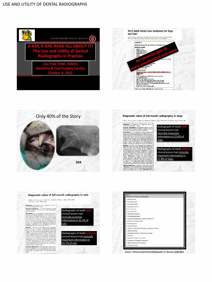
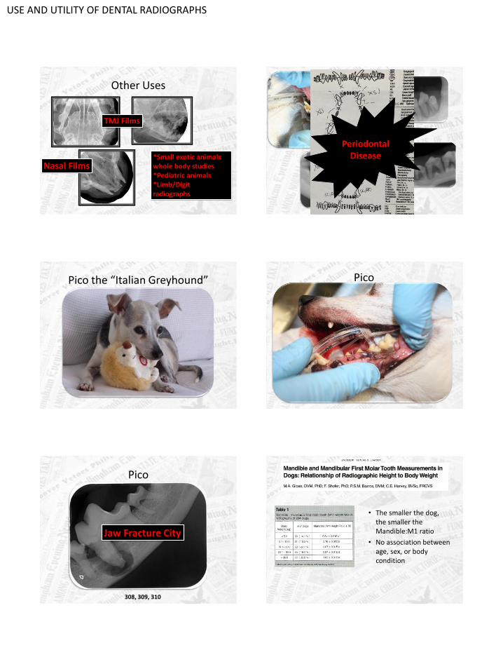
Pico the “Italian Greyhound” Pico
Pico
308, 309, 310
Jaw Fracture City
• The smaller the dog, the smaller the Mandible:M1 ratio
• No association between age, sex, or body condition
USE AND UTILITY OF DENTAL RADIOGRAPHS
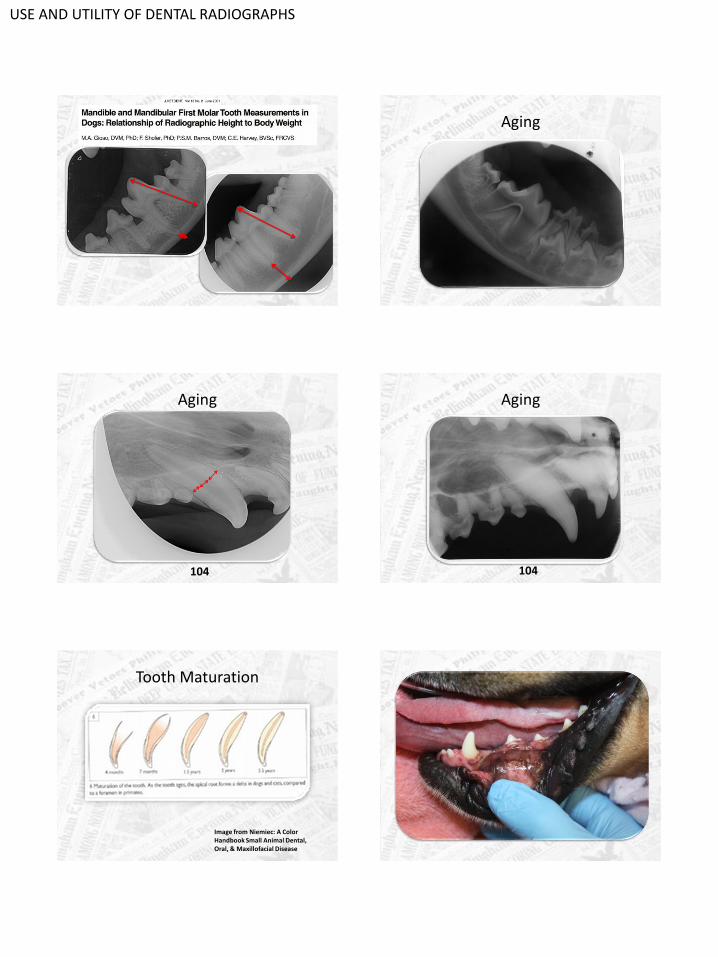
Aging
Aging
104
Aging
104
Tooth Maturation
Image from Niemiec: A Color Handbook Small Animal Dental, Oral, & Maxillofacial Disease
USE AND UTILITY OF DENTAL RADIOGRAPHS
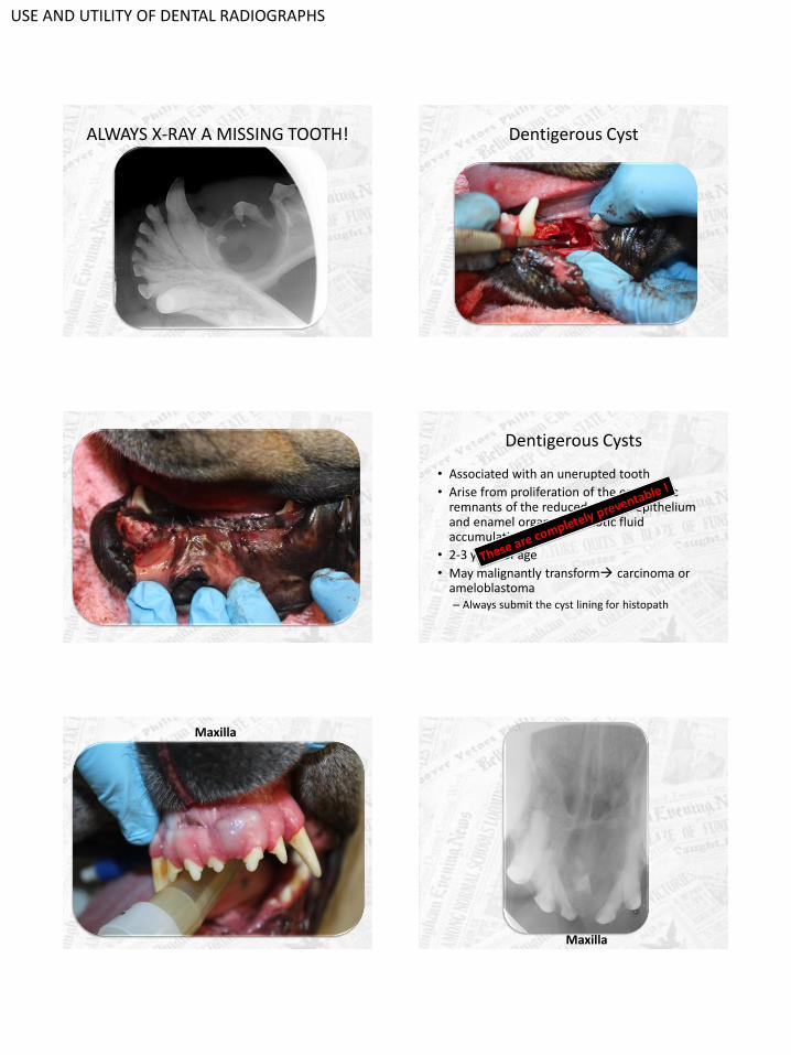
ALWAYS X-RAY A MISSING TOOTH! Dentigerous Cyst
Dentigerous Cysts
• Associated with an unerupted tooth
• Arise from proliferation of the embryonic remnants of the reduced enamel epithelium and enamel organ Osmotic fluid accumulation
• 2-3 years of age
• May malignantly transform carcinoma or ameloblastoma– Always submit the cyst lining for histopath
Maxilla
Maxilla
USE AND UTILITY OF DENTAL RADIOGRAPHS
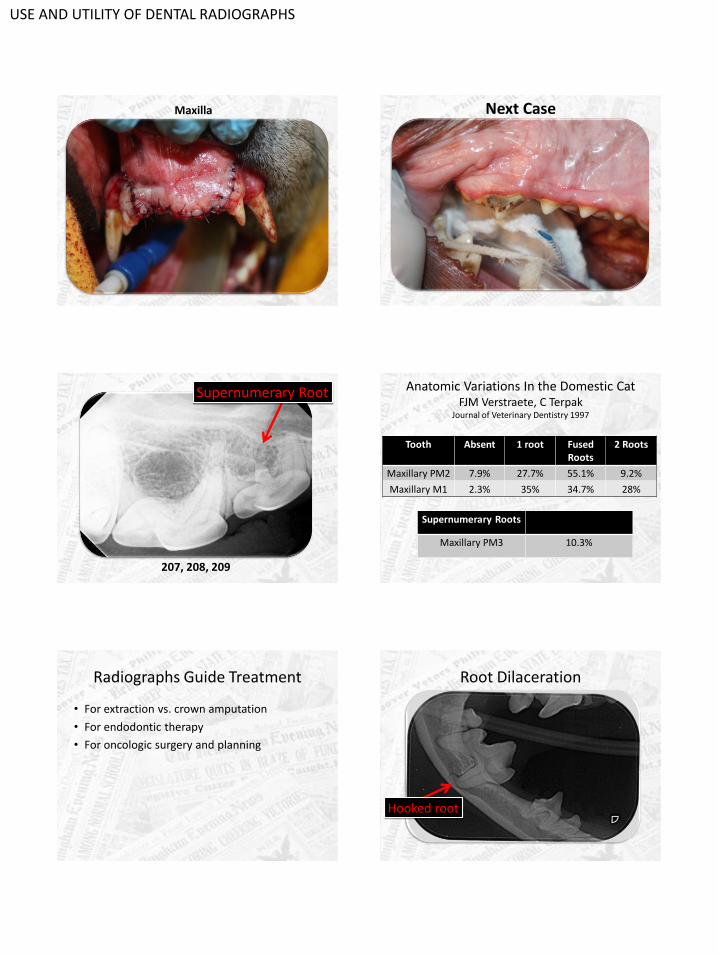
Maxilla Next Case
207, 208, 209
Supernumerary Root Anatomic Variations In the Domestic CatFJM Verstraete, C Terpak
Journal of Veterinary Dentistry 1997
Tooth Absent 1 root Fused Roots
2 Roots
Maxillary PM2 7.9% 27.7% 55.1% 9.2%
Maxillary M1 2.3% 35% 34.7% 28%
Supernumerary Roots
Maxillary PM3 10.3%
Radiographs Guide Treatment
• For extraction vs. crown amputation
• For endodontic therapy
• For oncologic surgery and planning
Root Dilaceration
Hooked root

USE AND UTILITY OF DENTAL RADIOGRAPHS
What is endodontic disease?
• Disease within the pulp cavity
• Via direct or indirect pulp exposure
– Tooth fracture
• Complicated vs. uncomplicated
– Concussive damage
• Irreversible pulpitis
• Can also be present with very little clinical evidence!!
Radiographic Signs of Endodontic Disease
• Periapical lucency
• Increased width of the apical PDL space
• Loss of the lamina dura
• Root tip resorption, internal resorption
• Arrested pulp cavity development
• Changes in the periapical bone
• Pulp canal obliteration
Reference: “Atlas of Dental Radiography in Dogs and Cats”, DuPont & DeBowes
Wider pulp cavity than neighboring teeth!
Periapical lucency!
What about this tooth? Chevron Effect
• Normal triangular lucency at the apex
• Created by the radiolucent trabecular bone and denser cortical bone
• Canine teeth, maxillary incisors, mandibular first molars
USE AND UTILITY OF DENTAL RADIOGRAPHS
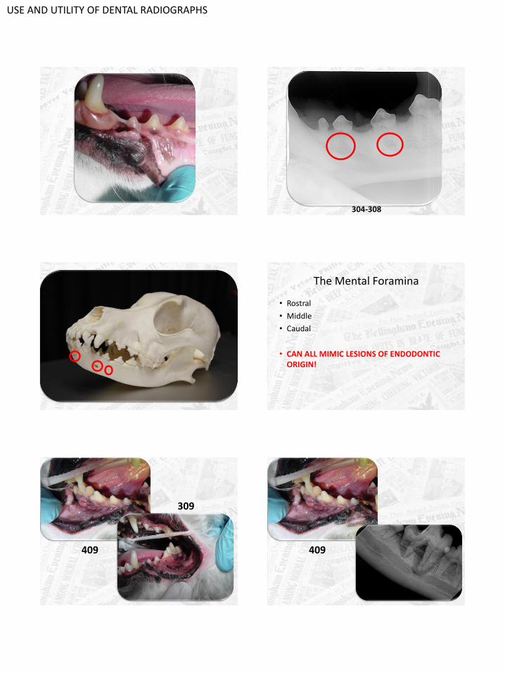
304-308
The Mental Foramina
• Rostral
• Middle
• Caudal
• CAN ALL MIMIC LESIONS OF ENDODONTIC ORIGIN!
409
309
409

USE AND UTILITY OF DENTAL RADIOGRAPHS
309
Dens Invaginatus
• Developmental anomaly
• Enamel and dentin invaginate into pulp
• Defect may be confined to crown only or involve the pulp
• The result is an increased susceptibility to endodontic and periodontal disease
USE AND UTILITY OF DENTAL RADIOGRAPHS
Congenital Generalized Odontodysplasia
• Generalized enamel and dentin defects
• Short roots
• Evidence of endodontic disease
• Genetic, infectious, nutritional factors
– Canine distemper virus
• Most likely infected at a young age during the formation of the tooth (bell stage)
What’s going on here?
304, 307, 308, 309
Tooth resorption in Cats
• Most common oral pathology in cats
• 28-75% show at least one lesion
• Osteoclasts/osteoblasts remodeling root
• Excess vitamin D proposed but hypothesis only
further follow-up prospective studies necessary to
truly determine if causative
Tooth resorption in dogs
• 53.6% of dogs examined have TR
• External replacement resorption most common
• Increased age = Increased TR
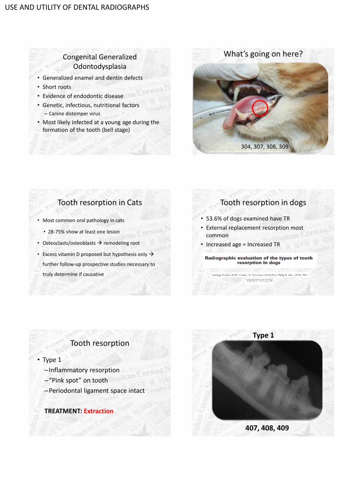
Tooth resorption
• Type 1
–Inflammatory resorption
–“Pink spot” on tooth
–Periodontal ligament space intact
TREATMENT: Extraction
407, 408, 409
Type 1

USE AND UTILITY OF DENTAL RADIOGRAPHS
Tooth resorption
• Type 2
–Root replacement resorption
–No periodontal ligament
–Density of tooth is less than density of surrounding bone
TREATMENT: Crown Amputation
Type 2
301- 304, 401- 404
Treatment for Tooth Resorption
• Crown Amputations
–NO signs of endodontic disease
–NO signs of a periodontal ligament
–NO stomatitis
–NO retrovirus +
104-107
Type 3: Mixed!A little bit of both
X-Rays are Essential to determine the
Course of Treatment in Animals
with Tooth Resorption!
Take Home Point
USE AND UTILITY OF DENTAL RADIOGRAPHS
Take Home Points
• Dental radiographs have a lot of uses!
• Dental radiographs can find surprises
• Dental radiographs guide your treatment plan and complete your oral examination
• Dental radiographs are the highest standard of care
Thank you!Questions?
Like Dentistry?Join the American Veterinary Dental
Society!