Onlay Uncemented Lateralized Reverse Shoulder Arthroplasty ...
10
Journal of Clinical Medicine Article Onlay Uncemented Lateralized Reverse Shoulder Arthroplasty for Fracture Sequelae Type 1 with Valgus/Varus Malunion: Deltoid Lengthening and Outcomes Alfonso Maria Romano 1,2 , Adriano Braile 3 , Pasquale Casillo 1 , Guglielmo Nastrucci 1 , Massimiliano Susanna 4 , Angelo Di Giunta 5 and Francesco Ascione 1,2, * 1 Orthopedics and Sport Medicine Unit, Campolongo Hospital, 84025 Salerno, Italy; [email protected] (A.M.R.); [email protected] (P.C.); [email protected] (G.N.) 2 Department of Orthopaedic and Trauma Surgery, Ospedale Buon Consiglio Fatebenefratelli, 80123 Naples, Italy 3 Dipartimento Multidisciplinare di Specialità Medico-Chirurgiche ed Odontoiatriche, Università degli Studi della Campania “Luigi Vanvitelli”, 80138 Naples, Italy; [email protected] 4 Orthopedic and Traumatology Unit, San Donà di Piave Hospital, 30027 Venice, Italy; [email protected] 5 Orthopaedic Division of Policlinico ‘G.B. Morgagni’, 95100 Catania, Italy; [email protected] * Correspondence: [email protected]; Tel.: +39-347-611-9973 Received: 6 August 2020; Accepted: 27 September 2020; Published: 1 October 2020 Abstract: The successful treatment of proximal humeral fractures remains challenging for shoulder surgeons, and failure rates are high, regardless of initial treatment. This study aimed to analyze the clinical and radiographic midterm results of onlay lateralized cementless stem reverse shoulder arthroplasty (RSA) in patients with valgus/varus malunion proximal humerus fracture sequelae without metaphyseal osteotomy. We retrospectively studied 35 cases with the diagnosis of fracture sequelae of the proximal part of the humerus with valgus/varus malunion. The mean duration of follow-up was 4.6 years (range, 2 to 7 years), and the mean time between fracture and arthroplasty was 6 years (1 to 32 years). Seventeen patients (48.6%) had initially been treated nonoperatively. The Constant score (CS), active range of motion, and radiographs of the affected shoulders, as well as the acromion to greater tuberosity (AGT) distance and deltoid length (DL), were analyzed before surgery and at their latest follow-up. A total of thirty-three patients (94.3%) rated their outcome as very good or good. Mean CS, forward flexion, and external rotation improved significantly (p < 0.0001), as did internal rotation and pain (p < 0.05). AGT distance significantly increased postoperatively from 14.7 to 43.3 mm, as did DL from 143 to 170 mm (p < 0.05). There was no correlation between the outcomes and valgus/varus deformity, previous surgeries, or AGT distance/DL. A total of four complications occurred (11.4%): two dislocations were detected (5.7%) and successfully revised with a longer cemented stem. Onlay lateralized uncemented stem RSA improves clinical outcomes and decreases complications when treating valgus/varus malunion fracture sequelae, avoiding intraoperative technical challenges, such as tuberosities osteotomy conscious of bone loss and proper deltoid tensioning. Keywords: reverse shoulder prosthesis; fracture sequelae; post-traumatic arthritis; onlay lateralized uncemented stem; complications; deltoid wrapping; humerus fracture; tuberosities J. Clin. Med. 2020, 9, 3190; doi:10.3390/jcm9103190 www.mdpi.com/journal/jcm
Transcript of Onlay Uncemented Lateralized Reverse Shoulder Arthroplasty ...
Journal of
Clinical Medicine
Article
Onlay Uncemented Lateralized Reverse ShoulderArthroplasty for Fracture Sequelae Type 1 withValgus/Varus Malunion: Deltoid Lengtheningand Outcomes
Alfonso Maria Romano 1,2, Adriano Braile 3, Pasquale Casillo 1, Guglielmo Nastrucci 1,Massimiliano Susanna 4, Angelo Di Giunta 5 and Francesco Ascione 1,2,*
1 Orthopedics and Sport Medicine Unit, Campolongo Hospital, 84025 Salerno, Italy;[email protected] (A.M.R.); [email protected] (P.C.);[email protected] (G.N.)
2 Department of Orthopaedic and Trauma Surgery, Ospedale Buon Consiglio Fatebenefratelli,80123 Naples, Italy
3 Dipartimento Multidisciplinare di Specialità Medico-Chirurgiche ed Odontoiatriche, Università degli Studidella Campania “Luigi Vanvitelli”, 80138 Naples, Italy; [email protected]
4 Orthopedic and Traumatology Unit, San Donà di Piave Hospital, 30027 Venice, Italy;[email protected]
5 Orthopaedic Division of Policlinico ‘G.B. Morgagni’, 95100 Catania, Italy; [email protected]* Correspondence: [email protected]; Tel.: +39-347-611-9973
Received: 6 August 2020; Accepted: 27 September 2020; Published: 1 October 2020�����������������
Abstract: The successful treatment of proximal humeral fractures remains challenging for shouldersurgeons, and failure rates are high, regardless of initial treatment. This study aimed to analyzethe clinical and radiographic midterm results of onlay lateralized cementless stem reverse shoulderarthroplasty (RSA) in patients with valgus/varus malunion proximal humerus fracture sequelaewithout metaphyseal osteotomy. We retrospectively studied 35 cases with the diagnosis of fracturesequelae of the proximal part of the humerus with valgus/varus malunion. The mean duration offollow-up was 4.6 years (range, 2 to 7 years), and the mean time between fracture and arthroplastywas 6 years (1 to 32 years). Seventeen patients (48.6%) had initially been treated nonoperatively.The Constant score (CS), active range of motion, and radiographs of the affected shoulders, as well asthe acromion to greater tuberosity (AGT) distance and deltoid length (DL), were analyzed beforesurgery and at their latest follow-up. A total of thirty-three patients (94.3%) rated their outcome as verygood or good. Mean CS, forward flexion, and external rotation improved significantly (p < 0.0001),as did internal rotation and pain (p < 0.05). AGT distance significantly increased postoperativelyfrom 14.7 to 43.3 mm, as did DL from 143 to 170 mm (p < 0.05). There was no correlation betweenthe outcomes and valgus/varus deformity, previous surgeries, or AGT distance/DL. A total of fourcomplications occurred (11.4%): two dislocations were detected (5.7%) and successfully revisedwith a longer cemented stem. Onlay lateralized uncemented stem RSA improves clinical outcomesand decreases complications when treating valgus/varus malunion fracture sequelae, avoidingintraoperative technical challenges, such as tuberosities osteotomy conscious of bone loss and properdeltoid tensioning.
Keywords: reverse shoulder prosthesis; fracture sequelae; post-traumatic arthritis; onlay lateralizeduncemented stem; complications; deltoid wrapping; humerus fracture; tuberosities
J. Clin. Med. 2020, 9, 3190; doi:10.3390/jcm9103190 www.mdpi.com/journal/jcm
J. Clin. Med. 2020, 9, 3190 2 of 10
1. Introduction
The successful treatment of complex humeral fractures remains challenging for shouldersurgeons, and failure rates are high, regardless of initial treatment. Soft tissue damage, scarring andcontracture, tuberosity and shaft bone deformity, malunion, non-union and loss, and humeral headavascular necrosis (caused by the fracture or previous surgery) are suspected to be responsible formassive functional impairment of the affected shoulder joint and rotator cuff. These heterogeneousfracture sequelae are the sum of failed conservative treatment, failed internal fixation, andfailed hemiarthroplasty.
Boileau [1,2] classified fracture sequelae of the proximal part of the humerus in four differentpathologies. Subsequently, Moineau [3] divided Type 1 sequelae into four groups (Table 1).
Table 1. Boileau and Moineau classification of humerus proximal part fracture sequelae.
Fracture Sequelae
Intracapsular Type 1
1a isolated post-traumatic osteonecrosis of the humeralhead without tuberosity malunion
1b isolated post-traumatic osteoarthritis withoutosteonecrosis or tuberosity malunion
1c proximal humeral deformity with valgus malunionsecondary to a valgus impacted fracture
1d varus malunion secondary to a varusimpacted fracture
Type 2 related to locked dislocation or fracture-dislocation
ExtracapsularType 3 surgical neck non-union
Type 4 severe tuberosity malunion
The therapeutic choice of anatomical shoulder arthroplasty (TSA) or reverse shoulder arthroplasty(RSA) for fracture sequelae of the proximal humerus must initially be made on the basis of the Boileauclassification [1–3] in an attempt to avoid secondary revision when possible since unsatisfactory resultsand high rates of complication for type 3 and 4 lesions have been reported after TSA. In patients inwhom conservative treatment has failed, TSA showed promising midterm outcomes [1–4]. However,in patients with varus malunion and fatty infiltration of the rotator cuff [3], results were worse.
Restoring the anatomy of the proximal humerus and the congruity of the joint through anatomicalarthroplasty can be difficult, especially in varus configurations. In proximal humerus valgus/varusmalunion, TSA is not suitable, and RSA may provide an alternative [5]. Where a prosthetic replacementis possible without an osteotomy, surgeons should accept the distorted anatomy of the proximalhumerus and adapt the prosthesis and their technique to the modified anatomy.
To our knowledge, neither a focus on the results of new design RSA (onlay, lateralized,and uncemented stems) in fracture sequelae without metaphyseal osteotomy nor a study restricted tovalgus/varus malunion sequelae have appeared in the literature.
The aim of the present study was to analyze the clinical and radiographic midterm results ofonlay lateralized cementless stem RSA in patients with type 1C and 1D proximal humerus fracturesequelae. We hypothesized that satisfactory functional results could be achieved while maintainingcomplication and revision rates manageable.
2. Material and Methods
Institutional review board approval was obtained to conduct a retrospective analysis of patientswho had undergone RSA at the participating institutions.
J. Clin. Med. 2020, 9, 3190 3 of 10
Inclusion criteria were the diagnosis of type 1C and 1D sequelae of a fracture of the proximalhumerus, according to Moineau [3], treatment with RSA, and a minimum follow-up of two years.Exclusion criteria were previous infection involving the affected shoulder, non-union, or previousarthroplasty of the shoulder. The results were retrospectively analyzed from prospectively gathereddatabases. Between 2012 and 2017, 42 patients were identified. Three were lost due to follow-up,and one died without adequate postoperative data, leaving 35 patients.
There were ten males and twenty-five were females. The dominant arm was affected in 31 patients.Their mean age at the time of arthroplasty was 71 years (54 to 87 years). The mean duration offollow-up was 4.6 years (range, 2 to 7 years). The mean time between fracture and arthroplasty was6 years (1 to 32 years). Seventeen patients (48.6%) had been initially treated nonoperatively, andeighteen had undergone open reduction and internal fixation (ORIF), involving a plate in eight (22.9%),intramedullary nail in two (5.7%), and Kirschner wires percutaneous pinning (K-wires) in eight (22.9%).The hardware had been removed in separate prior procedures in all patients. Twenty-two shouldershad a preoperative valgus deformity, and thirteen a varus deformity.
2.1. Surgical Technique
Surgical technique and postoperative patient management have been previously described indetail [6].
Three senior shoulder surgeons (AMR, ADG and MS) implanted RSAs using the EquinoxeShoulder System (Exactech Inc., Bloomington, MN, USA).
A deltopectoral approach was used in all cases. A subscapularis (SSC) tendon tenotomy wasperformed to access the joint. When possible (not excessive tissues and/or joint strain and acceptabletendon quality), the tenotomy was repaired with tendon-to-tendon sutures (28 RSAs had no SSCrepair). The long head of the biceps was tenodesed to the pectoralis major when present.
The humeral canal was sized and then compacted until rotational stability of the trials wasachieved. None required a cemented stem. Glenosphere diameters were 38 mm for females and 42 mmfor males. No metaphysis/tuberosity osteotomies were associated with RSA implant.
2.2. Clinical Evaluation
Patient pre- and post-operative active range of motion (ROM) assessments including forwardflexion (FF), external rotation (ER) with the arm at the side, internal rotation with the level of the thumbon the spine (IR), the Constant–Murley Score (CS) [7], perioperative data (implant sizes, intraoperativecomplications), and postoperative complications were collected. Severe stiffness, defined by externalrotation of < 0◦, was present preoperatively in 20 patients (57.1%).
The patients were asked to rate their outcome as very good, good, satisfactory, or unsatisfactory.The postoperative clinical evaluation was performed by two independent examiners not involved inthe surgery (FA and PC).
2.3. Radiographic Evaluation
Standardized pre- and post-operative radiographic films, including a true anterior-posterior (AP)view with three different rotations of the arm (internal, external, and neutral) and the scapular Y-view,and preoperative CT scans were obtained.
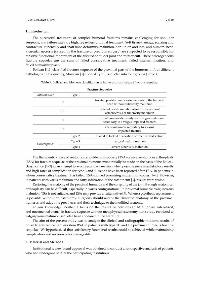
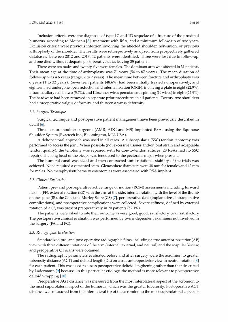
The radiographic parameters evaluated before and after surgery were the acromion to greatertuberosity distance (AGT) and deltoid length (DL) on a true anteroposterior view in neutral rotation [8]for each patient. This was used to assess postoperative deltoid lengthening rather than that describedby Ladermann [9] because, in this particular etiology, the method is more relevant to postoperativedeltoid wrapping [10].
Preoperative AGT distance was measured from the most inferolateral aspect of the acromion tothe most superolateral aspect of the humerus, which was the greater tuberosity. Postoperative AGTdistance was measured from the inferolateral tip of the acromion to the most superolateral aspect of
J. Clin. Med. 2020, 9, 3190 4 of 10
the humerus or humeral component. Deltoid length was measured from the inferolateral tip of theacromion to the deltoid tuberosity on the humerus (Figure 1). The position of the greater tuberosityrelative to the humeral diaphysis was used to define varus or valgus malunion of the proximal humerus,as described by Moineau [3]. CT images were analyzed for cuff muscle fatty infiltration grade.
Figure 1. Radiographic acromion to greater tuberosity distance (AGT) and deltoid length (DL);(a) preoperative measurement; (b) postoperative measurement.
Radiological examination included analysis of AP and Y-views at the final follow-up, documentingthe scapular notching grade, the inferior spur at the region of insertion of triceps, signs of loosening,and/or signs of ossification and osteolysis around the components.
2.4. Statistical Analysis
Statistical analysis was performed using the Social Science Statistics collaborative website (http://www.socscistatistics.com). Differences between preoperative and final follow-up data were comparedusing the Student t-test for paired data; the Fisher test or the Chi-square test were used to identifyrelationships between variables. Significance was set at p < 0.05.
3. Results
A total of twenty-one patients (60%) rated their outcome as very good, twelve (34.3%) as good,and two (5.7%) as unsatisfactory.
3.1. Clinical
The mean Constant score increased from 23.5 points (range, 5 to 40 points) preoperatively to66.7 points (14 to 87 points) postoperatively. The mean forward elevation of the shoulder increasedfrom 56.9◦ (range, 30◦ to 120◦) preoperatively to 134.8◦ (45◦ to 170◦) postoperatively. The clinicaloutcomes are shown in Table 2.
J. Clin. Med. 2020, 9, 3190 5 of 10
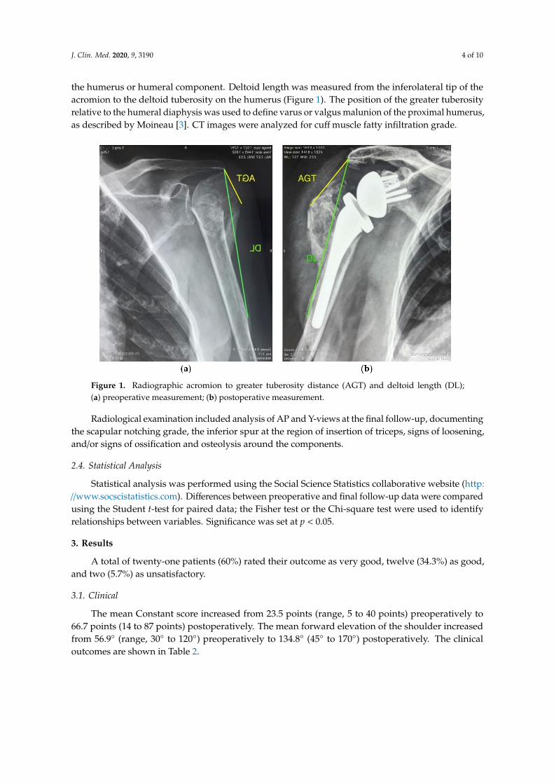
Table 2. Clinical outcomes.
Preoperative Postoperative ∆ p
FF (deg) 56.9 ± 22.4 134.8 ± 29.8 77.9 <0.0001
ER (deg) −4.5 ± 18 12.9 ± 15.1 17.4 <0.0001
IR (0–10) 2.1 ± 2 5.3 ± 2.3 3.4 <0.05
CS Pain/15 3.7 ± 1.8 12.2 ± 2.6 8.5 <0.05
CS% 23.5 ± 14.3 66.7 ± 19.7 43.2 <0.0001
FF: forward flexion; ER: external rotation with elbow at side; IR: internal rotation; level of the thumb on the spine;CS: Constant score.
The average Constant score, forward flexion, and external rotation improved significantly (allp < 0.0001), as well as the internal rotation and pain (both p < 0.05).
The mean postoperative Constant scores (p = 0.10) and range of motion were not significantlydifferent in patients with preoperative stiffness compared with those without. No differences werefound between patients who were initially treated nonoperatively and those who were treated surgicallywith regard to the Constant score (p = 0.30), shoulder flexion (p = 0.54), external rotation (p = 0.38),and internal rotation (p = 0.22).
There was no difference in the mean postoperative Constant score between those with a valgusdeformity and those with a varus sequela (p = 0.72).
3.2. Radiographic
There was no radiological evidence of loosening of either component. There was inferior scapularnotching of grade 1 in 4 patients (11.4%); no inferior spurs were found. There was no evidence ofosteolysis or progressive ossification.
AGT distance increased significantly postoperatively from 14.7 ± 13 to 43.3 ± 12 mm as well asDL from 143.1 ± 21.3 to 170 ± 11.6 mm (both p < 0.05). No significant differences between clinicaloutcomes (ROM and CS) and radiographic measurements of AGT distance or DL were observed.
3.3. Complications
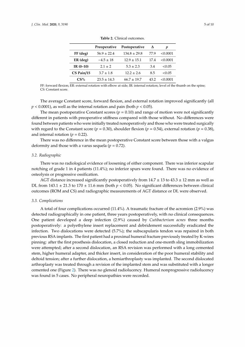
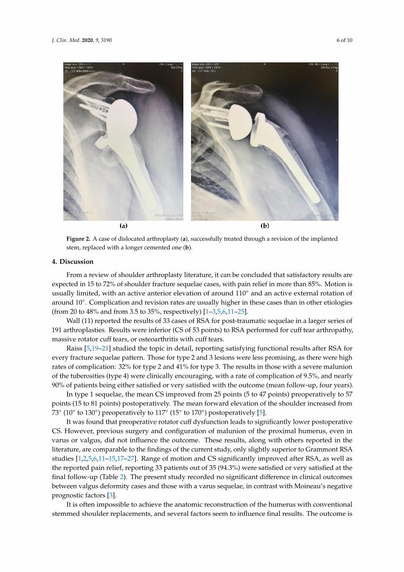
A total of four complications occurred (11.4%). A traumatic fracture of the acromion (2.9%) wasdetected radiographically in one patient, three years postoperatively, with no clinical consequences.One patient developed a deep infection (2.9%) caused by Cutibacterium acnes three monthspostoperatively: a polyethylene insert replacement and debridement successfully eradicated theinfection. Two dislocations were detected (5.7%); the subscapularis tendon was repaired in bothprevious RSA implants. The first patient had a proximal humeral fracture previously treated by K-wirespinning: after the first prosthesis dislocation, a closed reduction and one-month sling immobilizationwere attempted; after a second dislocation, an RSA revision was performed with a long cementedstem, higher humeral adapter, and thicker insert, in consideration of the poor humeral stability anddeltoid tension; after a further dislocation, a hemiarthroplasty was implanted. The second dislocatedarthroplasty was treated through a revision of the implanted stem and was substituted with a longercemented one (Figure 2). There was no glenoid radiolucency. Humeral nonprogressive radiolucencywas found in 5 cases. No peripheral neuropathies were recorded.
J. Clin. Med. 2020, 9, 3190 6 of 10
Figure 2. A case of dislocated arthroplasty (a), successfully treated through a revision of the implantedstem, replaced with a longer cemented one (b).
4. Discussion
From a review of shoulder arthroplasty literature, it can be concluded that satisfactory results areexpected in 15 to 72% of shoulder fracture sequelae cases, with pain relief in more than 85%. Motion isusually limited, with an active anterior elevation of around 110◦ and an active external rotation ofaround 10◦. Complication and revision rates are usually higher in these cases than in other etiologies(from 20 to 48% and from 3.5 to 35%, respectively) [1–3,5,6,11–25].
Wall (11) reported the results of 33 cases of RSA for post-traumatic sequelae in a larger series of191 arthroplasties. Results were inferior (CS of 53 points) to RSA performed for cuff tear arthropathy,massive rotator cuff tears, or osteoarthritis with cuff tears.
Raiss [5,19–21] studied the topic in detail, reporting satisfying functional results after RSA forevery fracture sequelae pattern. Those for type 2 and 3 lesions were less promising, as there were highrates of complication: 32% for type 2 and 41% for type 3. The results in those with a severe malunionof the tuberosities (type 4) were clinically encouraging, with a rate of complication of 9.5%, and nearly90% of patients being either satisfied or very satisfied with the outcome (mean follow-up, four years).
In type 1 sequelae, the mean CS improved from 25 points (5 to 47 points) preoperatively to 57points (15 to 81 points) postoperatively. The mean forward elevation of the shoulder increased from73◦ (10◦ to 130◦) preoperatively to 117◦ (15◦ to 170◦) postoperatively [5].
It was found that preoperative rotator cuff dysfunction leads to significantly lower postoperativeCS. However, previous surgery and configuration of malunion of the proximal humerus, even invarus or valgus, did not influence the outcome. These results, along with others reported in theliterature, are comparable to the findings of the current study, only slightly superior to Grammont RSAstudies [1,2,5,6,11–15,17–27]. Range of motion and CS significantly improved after RSA, as well asthe reported pain relief, reporting 33 patients out of 35 (94.3%) were satisfied or very satisfied at thefinal follow-up (Table 2). The present study recorded no significant difference in clinical outcomesbetween valgus deformity cases and those with a varus sequelae, in contrast with Moineau’s negativeprognostic factors [3].
It is often impossible to achieve the anatomic reconstruction of the humerus with conventionalstemmed shoulder replacements, and several factors seem to influence final results. The outcome is

J. Clin. Med. 2020, 9, 3190 7 of 10
better after initial conservative treatment (when compared to initial surgical treatment) in cases withoutany distortion of the tuberosities and post-traumatic arthritis with avascular necrosis (compared topost-traumatic arthritis with non-union, humeral head-metaphysis defect, and/or malunion). A greatertuberosity osteotomy is most likely responsible for the poor, unpredictable results and increasedcomplications after shoulder replacement arthroplasty in this context [1–4,12,17]. Therefore, our studydealt with an onlay lateralized stem consecutive arthroplasty without a tuberosity osteotomy, showingacceptable ouctomes and complications.
The constraints and torsional forces of Grammont-type RSA implants can lead to changes on thehumeral side, such as osteolysis, osteopenia, and the development of medial and lateral cortical bonenarrowing [16,28,29] due to the medialization of the implant protecting the glenoid from stress. Thisis greater on the humerus, leading to recurrent dislocations, stem loosening, prosthetic dissociation,and humeral component unscrewing and consequentially to various types of RSA failures [11,16].Ascione [28] reported 3.3% of humeral complications after RSA in a long-term follow-up multicenterstudy of 1035 Grammont arthroplasties: complications were more common following fracture sequelae(or difficult cases such as revision for failed arthroplasty), and postoperative humeral fractures/humeralloosening were exclusive relevance of cemented stems, that were not needed to implant in our cases.No humeral stem-related problems and/or reinterventions were reported in our series (fractures,loosening/migration, and disassembly), despite what the literature generally attributes to componentcomplications (1.5–10%) and reinterventions (12–27%) [2,16,28,30,31].
In recent years, alternative stemless or short-stemmed implants with metaphyseal fixation, withan onlay design, have been developed specifically for fracture sequelae to position the prosthetic headindependently of the humeral shaft [14,32–34]. Only a limited number of reports on the outcomes oftreatment using these new implants have been published but with promising early results.
The complications reported in RSA after fracture sequelae included humeral fracture, nerve palsy,dislocation, and loosening. Our complication rate was 11.4%, in particular, dislocation (5.7%) andrevision (5.7%). Data were consistent with the literature, with a relevant improvement in revisionsurgeries: all reported fracture sequelae complication rates ranged from 16 to 41%, and revisionrates ranged from 11 to 41% [2,3,18,23,24,35]. Interestingly, RSA instability appeared to be themain complication in fracture sequelae case series, ranging from 9.5 to 17%. In fact, a significantincrease in rotational micromotion with proximal bone loss, such as fracture sequelae cases, wasdemonstrated [5,36].
The scapular notching rate was reported to be greater than 50% in most series [11,13,15,16,23,37].This current study recorded 11.4% of grade 1 scapular notching, despite the specific etiology, but wasconsistent with the onlay lateralized RSA case series [38–40].
Greiner [22] evaluated prognostic factors after RSA in fracture sequelae, highlighting that lossof pretensioning of the deltoid or weakening of the deltoid insertion in bone loss (especially largerthan 3 cm), in addition to previous operations and an incompetent teres minor, may be contributingfactors in the poorer results. This was confirmed by two dislocation cases in the present series, bothwith humeral proximal bone loss and insufficient deltoid tensioning, subsequently successfully revisedusing longer components. Sabesan [8] found that deltoid lengthening and increase in AGT distancenegatively affected postoperative forward elevation, whereas Ladermann [9] found no correlationbetween deltoid lengthening and improvements in the range of motion, but significant improvementin patients’ postoperative forward flexion.
The aforementioned studies discussed Grammont inlay medialized RSA. Werner [41] studied56 lateralized onlay stem RSAs, and found a correlation between arm lengthening exceeding 2.5 cmand decrease in anterior elevation, and a relationship between arm lengthening averaging 2.2 cm andincreased outcomes, concluding that 1 to 2.5 cm of arm lengthening is the best compromise on thepostoperative range of motion. In our series, we found no significant differences between the clinicaloutcomes and AGT distance or DL, despite a mean increase of 2.9 cm and 2.7 cm, respectively.

J. Clin. Med. 2020, 9, 3190 8 of 10
Nevertheless, a defect of tensioning of deltoid can lead to RSA instability, especially in a particularpathology, such as post-traumatic osteoarthritis, with heterogeneous former treatments of fractureand different periods elapsed to RSA implant (1 to 32 years in the present series), in which deltoid hasbecome hypotrophic.
Limitations
Shortcomings inherent to a nonrandomized and retrospective study were the major limitations.In addition, the retrospective design may have resulted in an under-reporting of complications, andthere was an inadequate follow-up to assess long-term clinical and radiographic outcomes. The rate ofcomplication or need for reoperation surgeries may also increase over time.
However, the study was conducted over a short period, and selected surgical indications, basedon clinical and radiological assessments, surgical techniques, and rehabilitation protocols werestandardized for both surgeons. Still, a two-surgeon study might remain a bias.
Despite the relatively small number of cases in the current study, the results were quite promising.The cohort of patients was small but consistent with other studies of RSA for fracture sequelae.
Moreover, to our knowledge, this is the largest series available reporting on the results of type 1sequelae of a fracture of the proximal humerus treated only by onlay lateralized RSA.
5. Conclusions
In conclusion, RSA is an effective form of treatment for patients with type 1 sequelae of afracture of the proximal humerus, in association with valgus/varus malunion, leading to high rates ofsatisfaction. Deltoid lengthening does not influence the outcome, but a defect in deltoid tensioningand humeral proximal bone loss can lead to instability, which remains the most worrying complicationin this etiology.
Onlay lateralized stem improves clinical outcomes, reduces complications, and minimizesintraoperative technical challenges, such as tuberosities osteotomy; the need for revision is dramaticallyreduced by onlay lateralized uncemented RSA, provided that the metaphyseal bone stock is preserved.
Author Contributions: Conceptualization, A.M.R. and F.A.; Data curation, A.D.G.; Investigation, P.C.;Methodology, G.N.; Project administration, F.A.; Resources, A.D.G.; Supervision, A.M.R. and F.A.; Visualization,M.S. and A.D.G.; Writing—original draft, A.B.; Writing—review and editing, F.A. All authors have read andagreed to the published version of the manuscript.
Funding: No external funds were received for publishing the study.
Conflicts of Interest: Alfonso M. Romano, Angelo Di Giunta and Massimiliano Susanna receive fees for consultingfrom Exactech Inc. The funders had no role in the design of the study; in the collection, analyses, or interpretationof data; in the writing of the manuscript, or in the decision to publish the results. The other authors declare noconflict of interest.
References
1. Boileau, P.; Trojani, C.; Walch, G.; Krishnan, S.G.; Romeo, A.; Sinnerton, R. Shoulder arthroplasty for thetreatment of the sequelae of fractures of the proximal humerus. J. Shoulder Elbow Surg. 2001, 10, 299–308.[CrossRef]
2. Boileau, P.; Trojani, C.; Chuinard, C.; Lehuec, J.-C.; Walch, G. Proximal Humerus Fracture Sequelae: Impactof A New Radiographic Classification on Arthroplasty. Clin. Orthop. 2006, 442, 121–130. [CrossRef]
3. Moineau, G.; McClelland, W.B.; Trojani, C.; Rumian, A.; Walch, G.; Boileau, P. Prognostic Factors andLimitations of Anatomic Shoulder Arthroplasty for the Treatment of Posttraumatic Cephalic Collapse orNecrosis (Type-1 Proximal Humeral Fracture Sequelae). J. Bone Jt. Surg-Am. Vol. 2012, 94, 2186–2194.[CrossRef]
4. Dines, D.M.; Warren, R.F.; Altchek, D.W.; Moeckel, B. Posttraumatic changes of the proximal humerus:Malunion, nonunion, and osteonecrosis. Treatment with modular hemiarthroplasty or total shoulderarthroplasty. J. Shoulder Elbow Surg. 1993, 2, 11–21. [CrossRef]
J. Clin. Med. 2020, 9, 3190 9 of 10
5. Raiss, P.; Alami, G.; Bruckner, T.; Magosch, P.; Habermeyer, P.; Boileau, P.; Walch, G. Reverse shoulderarthroplasty for type 1 sequelae of a fracture of the proximal humerus. Bone Jt. J. 2018, 100-B, 318–323.[CrossRef] [PubMed]
6. Romano, A.M.; Oliva, F.; Nastrucci, G.; Casillo, P.; Di Giunta, A.; Susanna, M.; Ascione, F. Reverse shoulderarthroplasty patient personalized rehabilitation protocol. Preliminary results according to prognostic groups.Muscle Ligaments Tendons J. 2019, 7, 263. [CrossRef]
7. Constant, C.R.; Murley, A.H. A clinical method of functional assessment of the shoulder. Clin. Method Funct.Assess. Shoulder 1987, 214, 160–164. [CrossRef]
8. Sabesan, V.J.; Lombardo, D.; Josserand, D.; Buzas, D.; Jelsema, T.; Petersen-Fitts, G.R.; Wiater, J.M. The effectof deltoid lengthening on functional outcome for reverse shoulder arthroplasty. Musculoskelet Surg. 2016,100, 127–132. [CrossRef] [PubMed]
9. Lädermann, A.; Walch, G.; Lubbeke, A.; Drake, G.N.; Melis, B.; Bacle, G.; Collin, P.; Edwards, T.B.; Sirveaux, F.Influence of arm lengthening in reverse shoulder arthroplasty. J. Shoulder Elbow Surg. 2012, 21, 336–341.[CrossRef]
10. Roche, C.; Diep, P.; Hamilton, M.; A Crosby, L.; Flurin, P.-H.; Wright, T.W.; Zuckerman, J.D.; Routman, H.D.Impact of inferior glenoid tilt, humeral retroversion, bone grafting, and design parameters on muscle lengthand deltoid wrapping in reverse shoulder arthroplasty. Bull. Hosp. Jt. Dis. 2013, 71, 284–293.
11. Wall, B.; Nové-Josserand, L.; O’Connor, D.P.; Edwards, T.B.; Walch, G. Reverse Total Shoulder Arthroplasty:A Review of Results According to Etiology. J. Bone Jt. Surg. 2007, 89, 1476–1485. [CrossRef]
12. Mansat, P.; Bonnevialle, N. Treatment of fracture sequelae of the proximal humerus: Anatomical vs reverseshoulder prosthesis. Int. Orthop. 2015, 39, 349–354. [CrossRef] [PubMed]
13. Zumstein, M.A.; Pinedo, M.; Old, J.; Boileau, P. Problems, complications, reoperations, and revisions inreverse total shoulder arthroplasty: A systematic review. J. Shoulder Elbow Surg. 2011, 20, 146–157. [CrossRef][PubMed]
14. Ascione, F.; Bugelli, G.; Domos, P.; Neyton, L.; Godeneche, A.; Bercik, M.J.; Walch, G. Reverse ShoulderArthroplasty with a New Convertible Short Stem: Preliminary 2- to 4-year Follow-up Results. J. ShoulderElbow Arthroplast. 2017, 1. [CrossRef]
15. Ascione, F.; Kilian, C.M.; Laughlin, M.S.; Bugelli, G.; Domos, P.; Neyton, L.; Godeneche, A.; Edwards, B.;Walch, G. Increased scapular spine fractures after reverse shoulder arthroplasty with a humeral onlay shortstem: An analysis of 485 consecutive cases. J. Shoulder Elbow Surg. 2018, 27, 2183–2190. [CrossRef]
16. Boileau, P. Complications and revision of reverse total shoulder arthroplasty. Orthop. Traumatol. Surg. Res.2016, 102, S33–S43. [CrossRef]
17. Mansat, P.; Guity, M.R.; Bellumore, Y.; Mansat, M. Shoulder arthroplasty for late sequelae of proximalhumeral fractures. J. Shoulder Elbow Surg. 2004, 13, 305–312. [CrossRef]
18. Schliemann, B.; Theisen, C.; Kösters, C.; Raschke, M.J.; Weimann, A. Reverse total shoulder arthroplasty fortype I fracture sequelae after internal fixation of proximal humerus fractures. Arch. Orthop. Trauma Surg.2017, 137, 1677–1683. [CrossRef]
19. Raiss, P.; Edwards, T.B.; da Silva, M.R.; Bruckner, T.; Loew, M.; Walch, G. Reverse Shoulder Arthroplastyfor the Treatment of Nonunions of the Surgical Neck of the Proximal Part of the Humerus (Type-3 FractureSequelae). J. Bone Jt. Surg-Am. Vol. 2014, 96, 2070–2076. [CrossRef]
20. Raiss, P.; Edwards, T.B.; Collin, P.; Bruckner, T.; Zeifang, F.; Loew, M.; Boileau, P.; Walch, G. Reverse ShoulderArthroplasty for Malunions of the Proximal Part of the Humerus (Type-4 Fracture Sequelae). J. Bone Jt. Surg.2016, 98, 893–899. [CrossRef]
21. Raiss, P.; Edwards, T.B.; Bruckner, T.; Loew, M.; Zeifang, F.; Walch, G. Reverse arthroplasty for patients withchronic locked dislocation of the shoulder (type 2 fracture sequela). J. Shoulder Elbow Surg. 2017, 26, 279–287.[CrossRef]
22. Greiner, S.; Uschok, S.; Herrmann, S.; Gwinner, C.; Perka, C.; Scheibel, M. The metaphyseal bone defectpredicts outcome in reverse shoulder arthroplasty for proximal humerus fracture sequelae. Arch. Orthop.Trauma Surg. 2014, 134, 755–764. [CrossRef] [PubMed]
23. Alentorn-Geli, E.; Guirro, P.; Santana, F.; Torrens, C. Treatment of fracture sequelae of the proximal humerus:Comparison of hemiarthroplasty and reverse total shoulder arthroplasty. Arch. Orthop. Trauma Surg. 2014,134, 1545–1550. [CrossRef]

J. Clin. Med. 2020, 9, 3190 10 of 10
24. Martinez, A.A.; Calvo, A.; Bejarano, C.; Carbonel, I.; Herrera, A. The use of the Lima reverse shoulderarthroplasty for the treatment of fracture sequelae of the proximal humerus. J. Orthop. Sci. 2012, 17, 141–147.[CrossRef] [PubMed]
25. Kilic, M. Anatomic and reverse shoulder prostheses in fracture sequelae of the humeral head. Acta Orthop.Traumatol. Turc. 2010, 44, 417–425. [CrossRef] [PubMed]
26. Neyton, L.; Erickson, J.; Ascione, F.; Bugelli, G.; Lunini, E.; Walch, G. Grammont Award 2018: Scapularfractures in reverse shoulder arthroplasty (Grammont style): Prevalence, functional, and radiographic resultswith minimum 5-year follow-up. J. Shoulder Elbow Surg. 2019, 28, 260–267. [CrossRef]
27. Ascione, F.; Braile, A.; Romano, A.M.; Di Giunta, A.; Masciangelo, M.; Senorsky, E.H.; Samuelsson, K.;Marzano, N. Experience-optimised fast track improves outcomes and decreases complications in total kneearthroplasty. Knee 2019, 500–508. [CrossRef]
28. Ascione, F.; Domos, P.; Guarrella, V.; Chelli, M.; Boileau, P.; Walch, G. Long-term humeral complications afterGrammont-style reverse shoulder arthroplasty. J. Shoulder Elbow Surg. 2018, 27, 1065–1071. [CrossRef]
29. Melis, B.; DeFranco, M.; Lädermann, A.; Molé, D.; Favard, L.; Nérot, C.; Maynou, C.; Walch, G. An evaluationof the radiological changes around the Grammont reverse geometry shoulder arthroplasty after eight to 12years. J. Bone Jt. Surg. Br. 2011, 93-B, 1240–1246. [CrossRef]
30. Wright, T.; Alentorn-Geli, E.; Samitier, G.; Torrens, C. Reverse shoulder arthroplasty. Part 2: Systematicreview of reoperations, revisions, problems, and complications. Int. J. Shoulder Surg. 2015, 9, 60. [CrossRef]
31. Andersen, J.R.; Williams, C.D.; Cain, R.; Mighell, M.; Frankle, M. Surgically Treated Humeral Shaft FracturesFollowing Shoulder Arthroplasty. J. Bone Jt. Surg-Am. Vol. 2013, 95, 9–18. [CrossRef] [PubMed]
32. Langohr, G.D.G.; Reeves, J.; Roche, C.P.; Faber, K.J.; Johnson, J.A. The effect of short-stem humeral componentsizing on humeral bone stress. J. Shoulder Elbow Surg. 2020, 29, 761–767. [CrossRef] [PubMed]
33. Levy, O.; Narvani, A.; Hous, N.; Abraham, R.; Relwani, J.; Pradhan, R.; Bruguera, J.; Sforza, G.; Atoun, E.Reverse shoulder arthroplasty with a cementless short metaphyseal humeral implant without a stem: Clinicaland radiologic outcomes in prospective 2- to 7-year follow-up study. J. Shoulder Elbow Surg. 2016, 25,1362–1370. [CrossRef] [PubMed]
34. Ballas, R.; Béguin, L. Results of a stemless reverse shoulder prosthesis at more than 58 months mean withoutloosening. J. Shoulder Elbow Surg. 2013, 22, e1–e6. [CrossRef]
35. Grubhofer, F.; Wieser, K.; Meyer, D.C.; Catanzaro, S.; Schürholz, K.; Gerber, C. Reverse total shoulderarthroplasty for failed open reduction and internal fixation of fractures of the proximal humerus. J. ShoulderElbow Surg. 2017, 26, 92–100. [CrossRef]
36. Cuff, D.; Levy, J.C.; Gutiérrez, S.; Frankle, M.A. Torsional stability of modular and non-modular reverseshoulder humeral components in a proximal humeral bone loss model. J. Shoulder Elbow Surg. 2011, 20,646–651. [CrossRef]
37. Farshad, M.; Gerber, C. Reverse total shoulder arthroplasty—from the most to the least common complication.Int. Orthop. 2010, 34, 1075–1082. [CrossRef]
38. Simovitch, R.; Flurin, P.-H.; Wright, T.W.; Zuckerman, J.D.; Roche, C. Impact of scapular notching on reversetotal shoulder arthroplasty midterm outcomes: 5-year minimum follow-up. J. Shoulder Elbow Surg. 2019, 28,2301–2307. [CrossRef]
39. Merolla, G.; Walch, G.; Ascione, F.; Paladini, P.; Fabbri, E.; Padolino, A.; Porcellini, G. Grammont humeraldesign versus onlay curved-stem reverse shoulder arthroplasty: Comparison of clinical and radiographicoutcomes with minimum 2-year follow-up. J. Shoulder Elbow Surg. 2017. [CrossRef]
40. Erickson, B.J.; Frank, R.M.; Harris, J.D.; Mall, N.; Romeo, A.A. The influence of humeral head inclination inreverse total shoulder arthroplasty: A systematic review. J. Shoulder Elbow Surg. 2015, 24, 988–993. [CrossRef]
41. Werner, B.S.; Ascione, F.; Bugelli, G.; Walch, G. Does arm lengthening affect the functional outcome in onlayreverse shoulder arthroplasty? J. Shoulder Elbow Surg. 2017, 26, 2152–2157. [CrossRef] [PubMed]
© 2020 by the authors. Licensee MDPI, Basel, Switzerland. This article is an open accessarticle distributed under the terms and conditions of the Creative Commons Attribution(CC BY) license (http://creativecommons.org/licenses/by/4.0/).