Oncologic emergencies -...
11
2212 Crit Care Med 2012 Vol. 40, No. 7 C ancer is the second leading cause of death in the United States with over 500,000 deaths annually (1). Despite improvements in survival and decreased prevalence of certain malignancies (2), the overall prevalence of cancer is expected to rise (3). Individuals with malignancy may present with a cancer-related emergency; for many, this will be their initial manifes- tation of cancer (4). Efficient diagnosis and proper management of life-threatening complications may facilitate either defini- tive treatment of the underlying malig- nancy or palliation. Five conditions frequently associated with malignancy that may require emer- gent treatment in an intensive care unit are reviewed. Some complications (e.g., febrile neutropenia) are too broad to be covered in this review, and have been recently covered (5, 6). Superior Vena Cava Syndrome Scottish surgeon William Hunter in 1757 first described superior vena cava (SVC) obstruction from syphilic aorti- tis (7, 8). The syndrome resulting from the physiologic effects of obstruction is now associated more with malignancy than with mediastinal infection (8). Since the mid-20th century, malignancy has accounted for over 90% of cases of SVC syndrome (SVCS) (9, 10) (Fig. 1). However, implantable intravenous (IV) devices (e.g., tunneled central venous catheters, port catheters, pacemaker leads) have increased the prevalence of thrombosis-related SVCS and now account for up to 40% of cases of SVCS (11). Fibrosing mediastinitis due to histoplas- mosis (11–14) and pulmonary tuberculosis (9, 15) still occur infrequently. The SVC is a thin-walled vessel, about 4–6 cm in length and 1.5–2 cm in width in adults, extending from the confluence of the brachiocephalic veins and terminat- ing in the superior right atrium. Confined by the chest wall and surrounding struc- tures, a mediastinal mass impinging upon the SVC can easily obstruct blood flow (8). Over a period of 1–2 wks, the resul- tant high venous pressures and upstream vessel engorgement promote collateral vein dilatation to reduce this pressure (Fig. 2). In diseases associated with pleural thickening and adherence of the surfaces (e.g., tuberculosis), bridging veins can develop across the pleural space produc- ing significant systemic-to-pulmonary venous shunting and hypoxemia (15). Although the signs and symptoms of SVCS vary, the most common finding is facial edema (9–11, 16–18) (Fig. 3). The frequency of findings differ between malignant and benign etiologies, with dyspnea at rest, cough, chest and shoulder pain, and hoarseness more frequent in the former (19). This suggests some signs and symptoms of SVCS are due not solely to hydrostatic effects of venous obstruction but also direct effects of tumor compres- sion or invasion of the airways or nerves. Similarly, neurological findings (which occur in a minority of SVCS cases) should not be assumed to result from increased venous pressure, but should raise suspi- cion for brain metastases (17). Classically, SVCS has been considered an emergency requiring immediate treat- ment. Diagnostic procedures were assumed to be hazardous due to the risk of exces- sive bleeding or airway obstruction (16, 17, 20). Yet many diagnostic studies (e.g., cytology of sputum, bronchial washings, pleuraleffusion,palpablelymphnodebiopsy) are minimally invasive and have good Objectives: To provide an up-to-date review of current lit- erature on the pathophysiology, diagnosis, and management of five key malignancy-related complications: superior vena cava syndrome, malignant pericardial effusion, malignant spi- nal cord compression, hypercalcemia, and acute tumor lysis syndrome. Data Sources: Database searches and review of relevant medi- cal literature. Data Synthesis: Malignancy-related complications demand increased attention from intensivists due to their frequency and increasing cancer prevalence. Although such complications por- tend a poor prognosis, proper acute management can improve short-term outcomes by facilitating either definitive care of the underlying malignancy or the institution of appropriate palliative measures. Conclusions: Knowledge of malignancy-induced complica- tions in critically ill patients expedites the ability of the intensivist to properly manage them. Five complications commonly requiring emergency management are addressed in this review. Specifically, superior vena cava syndrome may warrant radiation, chemother- apy, vascular stenting, or surgical resection. Malignant pericardial effusion may require emergency pericardiocentesis if cardiac tam- ponade develops. Malignant spinal cord compression demands im- mediate spinal imaging, glucocorticoids, and either surgery or radia- tion. Hypercalcemia requires aggressive intravenous hydration and a bisphosphonate. Acute tumor lysis syndrome necessitates intrave- nous hydration, rasburicase, and management of associated elec- trolyte abnormalities. (Crit Care Med 2012; 40: 2212–2222) KEY WORDS: adult; hypercalcemia; malignancy; spinal cord com- pression; superior vena cava syndrome; tumor lysis syndrome Concise Definitive Review Series Editor, Jonathan E. Sevransky, MD, MHS Oncologic emergencies Michael T. McCurdy, MD; Carl B. Shanholtz, MD From the Department of Medicine, Division of Pulmonary and Critical Care Medicine (MTM, CBS) and Department of Emergency Medicine (MTM), The University of Maryland School of Medicine, Baltimore, MD Dr. Shanholtz consulted for Sanofi-Synthelabo (ras- buricase) and received honoraria/speaking fees from Sanofi-Synthelabo (rasburicase). Dr. McCurdy has not disclosed any potential conflicts of interest. For information regarding this article, E-mail: [email protected] Copyright © 2012 by the Society of Critical Care Medicine and Lippincott Williams & Wilkins DOI: 10.1097/CCM.0b013e31824e1865
Transcript of Oncologic emergencies -...
2212 Crit Care Med 2012 Vol. 40, No. 7
C ancer is the second leading cause of death in the United States with over 500,000 deaths annually (1). Despite
improvements in survival and decreased prevalence of certain malignancies (2), the overall prevalence of cancer is expected to rise (3). Individuals with malignancy may present with a cancer- related emergency; for many, this will be their initial manifes-tation of cancer (4). Efficient diagnosis and proper management of life- threatening complications may facilitate either defini-tive treatment of the underlying malig-nancy or palliation.
Five conditions frequently associated with malignancy that may require emer-gent treatment in an intensive care unit are reviewed. Some complications (e.g., febrile neutropenia) are too broad to be
covered in this review, and have been recently covered (5, 6).
Superior Vena Cava Syndrome
Scottish surgeon William Hunter in 1757 first described superior vena cava (SVC) obstruction from syphilic aorti-tis (7, 8). The syndrome resulting from the physiologic effects of obstruction is now associated more with malignancy than with mediastinal infection (8). Since the mid-20th century, malignancy has accounted for over 90% of cases of SVC syndrome (SVCS) (9, 10) (Fig. 1). However, implantable intravenous (IV) devices (e.g., tunneled central venous catheters, port catheters, pacemaker leads) have increased the prevalence of thrombosis- related SVCS and now account for up to 40% of cases of SVCS (11). Fibrosing mediastinitis due to histoplas-mosis (11–14) and pulmonary tuberculosis (9, 15) still occur infrequently.
The SVC is a thin- walled vessel, about 4–6 cm in length and 1.5–2 cm in width in adults, extending from the confluence of the brachiocephalic veins and terminat-ing in the superior right atrium. Confined by the chest wall and surrounding struc-tures, a mediastinal mass impinging upon the SVC can easily obstruct blood flow (8). Over a period of 1–2 wks, the resul-tant high venous pressures and upstream
vessel engorgement promote collateral vein dilatation to reduce this pressure (Fig. 2). In diseases associated with pleural thickening and adherence of the surfaces (e.g., tuberculosis), bridging veins can develop across the pleural space produc-ing significant systemic- to- pulmonary venous shunting and hypoxemia (15).
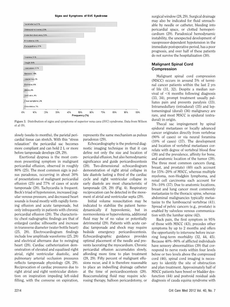
Although the signs and symptoms of SVCS vary, the most common finding is facial edema (9–11, 16–18) (Fig. 3). The frequency of findings differ between malignant and benign etiologies, with dyspnea at rest, cough, chest and shoulder pain, and hoarseness more frequent in the former (19). This suggests some signs and symptoms of SVCS are due not solely to hydrostatic effects of venous obstruction but also direct effects of tumor compres-sion or invasion of the airways or nerves. Similarly, neurological findings (which occur in a minority of SVCS cases) should not be assumed to result from increased venous pressure, but should raise suspi-cion for brain metastases (17).
Classically, SVCS has been considered an emergency requiring immediate treat-ment. Diagnostic procedures were assumed to be hazardous due to the risk of exces-sive bleeding or airway obstruction (16, 17, 20). Yet many diagnostic studies (e.g., cytology of sputum, bronchial washings, pleural effusion, palpable lymph node biopsy) are minimally invasive and have good
Objectives: To provide an up- to- date review of current lit-erature on the pathophysiology, diagnosis, and management of five key malignancy- related complications: superior vena cava syndrome, malignant pericardial effusion, malignant spi-nal cord compression, hypercalcemia, and acute tumor lysis syndrome.
Data Sources: Database searches and review of relevant medi-cal literature.
Data Synthesis: Malignancy- related complications demand increased attention from intensivists due to their frequency and increasing cancer prevalence. Although such complications por-tend a poor prognosis, proper acute management can improve short- term outcomes by facilitating either definitive care of the underlying malignancy or the institution of appropriate palliative measures.
Conclusions: Knowledge of malignancy- induced complica-tions in critically ill patients expedites the ability of the intensivist to properly manage them. Five complications commonly requiring emergency management are addressed in this review. Specifically, superior vena cava syndrome may warrant radiation, chemother-apy, vascular stenting, or surgical resection. Malignant pericardial effusion may require emergency pericardiocentesis if cardiac tam-ponade develops. Malignant spinal cord compression demands im-mediate spinal imaging, glucocorticoids, and either surgery or radia-tion. Hypercalcemia requires aggressive intravenous hydration and a bisphosphonate. Acute tumor lysis syndrome necessitates intrave-nous hydration, rasburicase, and management of associated elec-trolyte abnormalities. (Crit Care Med 2012; 40: 2212–2222)
KEY WORDS: adult; hypercalcemia; malignancy; spinal cord com-pression; superior vena cava syndrome; tumor lysis syndrome
Concise Definitive Review Series Editor, Jonathan E. Sevransky, MD, MHS
Oncologic emergencies
Michael T. McCurdy, MD; Carl B. Shanholtz, MD
From the Department of Medicine, Division of Pulmonary and Critical Care Medicine (MTM, CBS) and Department of Emergency Medicine (MTM), The University of Maryland School of Medicine, Baltimore, MD
Dr. Shanholtz consulted for Sanofi- Synthelabo (ras-buricase) and received honoraria/speaking fees from Sanofi- Synthelabo (rasburicase). Dr. McCurdy has not disclosed any potential conflicts of interest.
For information regarding this article, E- mail: [email protected]
Copyright © 2012 by the Society of Critical Care Medicine and Lippincott Williams & Wilkins
DOI: 10.1097/CCM.0b013e31824e1865
Crit Care Med 2012 Vol. 40, No. 7 2213
yield (10, 11, 20). Furthermore, recent series have confirmed the safety of invasive procedures (16, 17, 20). Diagnosing under-lying disease is essential before choosing a course of treatment because many causes of SVCS are benign. Additionally, some malignancies (e.g., small- cell lung cancer, non- Hodgkin lymphoma) require systemic chemotherapy (11, 21, 22). Radiation prior to biopsy causes tissue necrosis and can make histology uninterpretable. Consequently, SVCS becomes a true emer-gency requiring empiric treatment only in the setting of airway obstruction or cere-bral edema.
Management of SVCS includes symp-tomatic relief, treatment of complications, and therapy for the underlying condition. Symptoms usually improve over a period of 1–2 wks with radiation or chemother-apy, although they can improve just as quickly without treatment. Furthermore, autopsy series suggest collateral venous blood flow provides most of the
benefit (17). Elevation of the head of the bed and supplemental oxygen are stan-dard. Although commonly prescribed, glucocorticosteroids are of unclear ben-efit except in the cases of lymphoma or thymoma where indicated for the under-lying malignancy (9). Thrombosis- related SVC obstruction is treated with antico-agulation, intravascular device removal, and balloon dilatation or stenting if sig-nificant fibrosis remains (11).
In small- cell lung cancer and lymphoma, chemotherapy and exter-nal beam radiotherapy are equally effective in relieving SVC obstruction (21). Combination therapy is no more effective than either treatment alone (10). Radiotherapy only treats disease within the radiation fields, and is therefore best for patients with well- defined tumors that are less chemotherapy- sensitive, such as non– small- cell lung cancer and certain metastatic cancers causing focal obstruc-tion (23). Chemotherapy is preferred
for extensive chemo- sensitive tumors, such as small- cell lung cancer and lym-phoma. Intravascular stenting provides symptomatic relief within 24–48 hrs and can safely precede tissue diagnosis. This may be the most appropriate initial therapy for patients with the most severe symptoms such as airway obstruction or cerebral edema (23). Stenting may provide the only treatment option in patients with recurrent obstruction (24). Patients with relatively chemo- and radiotherapy- resistant tumors such as thymoma and residual germ- cell mass may benefit from surgical resection (9, 23). Median survival of patients with cancer- induced SVCS is roughly 6 months after presentation, but many patients have survived over 2 yrs with treatment (9, 10, 20, 23, 24).
Malignant Pericardial Disease
Pericardial effusion is common in malig-nancy with up to 34% of cancer patients having involvement of pericardium (8, 25, 26). Neoplastic etiology was reported in 7% of all acute pericardial disease, and in roughly half of these cases it was the first manifestation of previously undiagnosed malignancy (27). Most malignant pericar-dial disease is due to metastasis from sites of disease outside of the heart and peri-cardium, primarily from lung, breast, and hematologic sources (8, 25–27).
The pericardium is composed of a vis-ceral (or serous) layer formed by a single layer of mesothelial cells adhered to the surface of the heart and a fibrous parietal layer formed by the pericardium reflecting back on itself. The space between these two layers contains up to 50 mL of fluid serv-ing as a lubricant (8, 28). Fluid filling the pericardial sac initially has a flat pressure response until reaching the pericardial reserve volume, i.e., the volume that begins to distend the pericardium. Pressure then begins to rise abruptly due to the relative inextensibility of the parietal pericardium (28, 29). The steep rise in pressure with minimal increment in pericardial fluid volume eventually leads to a critical intra-pericardial pressure, which in turn results in impaired filling of the cardiac chambers and hemodynamic compromise. Thus, car-diac tamponade has been termed a “last drop” phenomenon (29). The amount of the pericardial fluid that causes tampon-ade is related to the rate of fluid formation. Rapidly accumulating effusions can cause symptoms with as little as 200 of pericardial fluid (28). However, if fluid accumulates
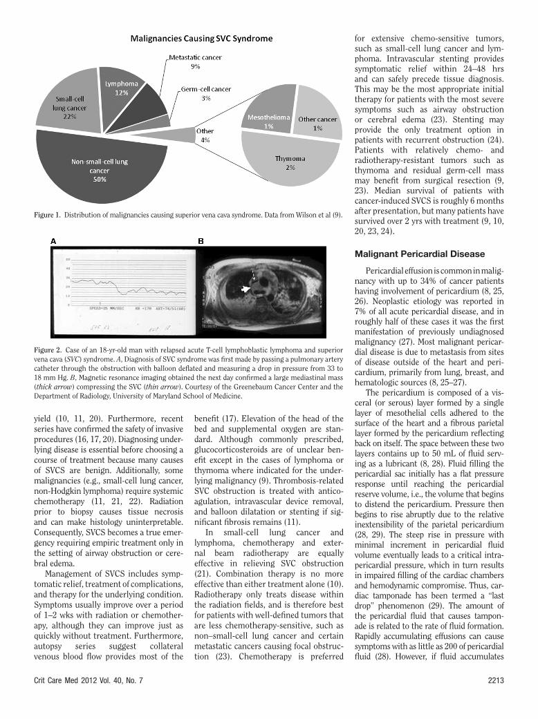
Figure 2. Case of an 18- yr- old man with relapsed acute T- cell lymphoblastic lymphoma and superior vena cava (SVC) syndrome. A, Diagnosis of SVC syndrome was first made by passing a pulmonary artery catheter through the obstruction with balloon deflated and measuring a drop in pressure from 33 to 18 mm Hg. B, Magnetic resonance imaging obtained the next day confirmed a large mediastinal mass (thick arrow) compressing the SVC (thin arrow). Courtesy of the Greenebaum Cancer Center and the Department of Radiology, University of Maryland School of Medicine.
Figure 1. Distribution of malignancies causing superior vena cava syndrome. Data from Wilson et al (9).
2214 Crit Care Med 2012 Vol. 40, No. 7
slowly (weeks to months), the parietal peri-cardial tissue can stretch. With this “stress relaxation” the pericardial sac becomes more compliant and can hold 2 L or more before tamponade develops (28, 29).
Exertional dyspnea is the most com-mon presenting symptom in malignant pericardial effusion, observed in roughly 80% (25). The most common sign is pul-sus paradoxus, occurring in about 30% of presentations of malignant pericardial effusion (25) and 77% of cases of acute tamponade (28). Tachycardia is frequent. Beck’s triad of hypotension, increased jug-ular venous pressure, and decreased heart sounds is found mostly with rapidly form-ing effusion and acute tamponade, but only infrequently in patients with chronic pericardial effusion (28). The characteris-tic chest radiographic findings are that of enlarged cardiac silhouette and increase in transverse diameter (water bottle heart) (25, 28). Electrocardiogram findings include low amplitude waveforms (25, 28) and electrical alternans due to swinging heart (28). Cardiac catheterization dem-onstration of elevated and equalized right atrial, right ventricular diastolic, and pulmonary arterial occlusion pressures reflects tamponade physiology (28, 29). Reciprocation of cardiac pressures due to right atrial and right ventricular disten-tion on inspiration impeding left- sided filling, with the converse on expiration,
represents the same mechanism as pulsus paradoxus (29).
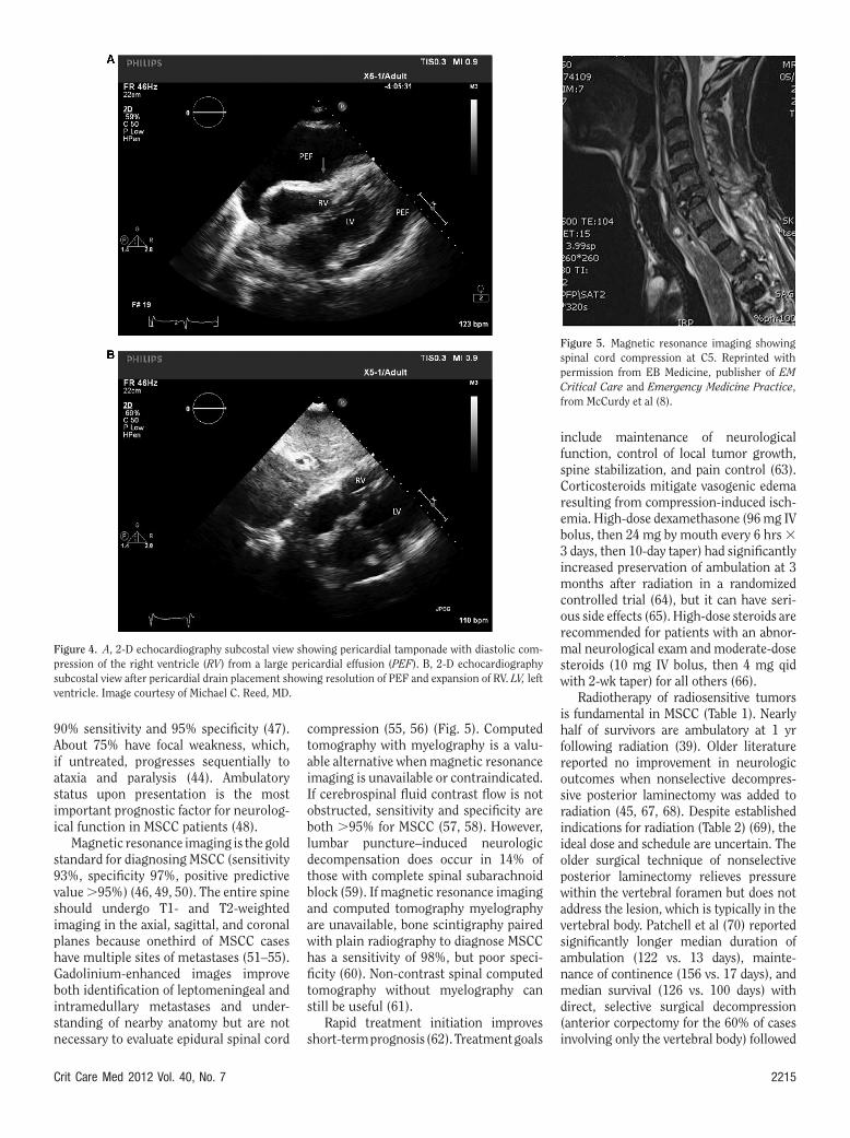
Echocardiography is the preferred diag-nostic imaging technique in that it can define not only the size and location of pericardial effusion, but also hemodynamic significance and guide pericardiocentesis (28). Two- dimensional ech ocardiogram demonstration of right atrial collapse in late diastole lasting a third of the cardiac cycle and right ventricular collapse in early diastole are most characteristic of tamponade (28, 29) (Fig. 4). Respiratory reciprocation can be detected in the move-ment of atrial and ventricular septa (29).
Initial volume resuscitation may be indicated to stabilize the patient hemo-dynamically if hypovolemic, but in normovolemia or hypervolemia, additional fluid may be of no value or potentially detrimental (29). Patients with acute car-diac tamponade and shock may require bedside emergency pericardiocentesis. Echocardiographic guidance provides optimal placement of the needle and pre-vents lacerating the myocardium. Chronic pericardial effusion accumulates slowly, affording more time to plan treatment (28, 29). Fifty percent of malignant effu-sions recur, and it is therefore reasonable to place an indwelling drainage catheter at the time of pericardiocentesis (28). Reaccumulating fluid may require scle-rosing therapy, balloon pericardotomy, or
surgical window (28, 29). Surgical drainage may also be indicated for fluid unreach-able by needle or catheter, bleeding into pericardial space, or clotted hemoperi-cardium (29). Paradoxical hemodynamic instability, the unexpected development of vasopressor- dependent hypotension in the immediate postoperative period, has a poor prognosis, and over half of these patients do not survive the hospitalization (30).
Malignant Spinal Cord Compression
Malignant spinal cord compression (MSCC) occurs in around 5% of termi-nal cancer patients within the last 2 yrs of life (31, 32). Despite a median sur-vival of 6 months following diagnosis (33, 34), prompt treatment usually pal-liates pain and prevents paralysis (33). Intramedullary (intradural) (35) and lep-tomeningeal (dural) (36) malignancy are rare, and most MSCC is epidural (extra-dural) in origin.
Thecal sac impingement by spinal epidural metastases or locally advanced cancer originates directly from vertebrae (90% of cases) or via neural foramina (10% of cases) (37). The development and location of vertebral metastases cor-relate with degree of vertebral blood flow (38) and the prevalence, affinity for bone, and anatomic location of the tumor (39). The three most common cancers (lung, breast, and prostate) (40) each account for 15%–20% of MSCC, whereas multiple myeloma, non- Hodgkin lymphoma, and renal cell carcinoma each account for 5%–10% (37). Due to anatomic locations, breast and lung cancer most commonly metastasize to the thoracic spine, whereas abdominal malignancies typically metas-tasize to the lumbosacral vertebrae (41). Spread of pelvic cancers (e.g., prostate) is enabled by valveless venous communica-tion with the lumbar spine (42).
Back pain, the first symptom in 95% of those with MSCC (43), precedes other symptoms by up to 2 months and offers the opportunity to intervene before incur-ring long- term morbidity (32, 44, 45). Because 40%–90% of afflicted individuals have sensory abnormalities (39) that cor-respond to nerve roots within four levels below or two levels above the compressed cord (46), spinal cord imaging is neces-sary to identify the site of obstruction and plan treatment. Approximately half of MSCC patients have bowel or bladder dys-function (44) and postvoid residual aids diagnosis of cauda equina syndrome with
Figure 3. Distribution of signs and symptoms of superior vena cava (SVC) syndrome. Data from Wilson et al (9).
Crit Care Med 2012 Vol. 40, No. 7 2215
90% sensitivity and 95% specificity (47). About 75% have focal weakness, which, if untreated, progresses sequentially to ataxia and paralysis (44). Ambulatory status upon presentation is the most important prognostic factor for neurolog-ical function in MSCC patients (48).
Magnetic resonance imaging is the gold standard for diagnosing MSCC (sensitivity 93%, specificity 97%, positive predictive value 95%) (46, 49, 50). The entire spine should undergo T1- and T2-weighted imaging in the axial, sagittal, and coronal planes because onethird of MSCC cases have multiple sites of metastases (51–55). Gadolinium- enhanced images improve both identification of leptomeningeal and intramedullary metastases and under-standing of nearby anatomy but are not necessary to evaluate epidural spinal cord
compression (55, 56) (Fig. 5). Computed tomography with myelography is a valu-able alternative when magnetic resonance imaging is unavailable or contraindicated. If cerebrospinal fluid contrast flow is not obstructed, sensitivity and specificity are both 95% for MSCC (57, 58). However, lumbar puncture–induced neurologic decompensation does occur in 14% of those with complete spinal subarachnoid block (59). If magnetic resonance imaging and computed tomography myelography are unavailable, bone scintigraphy paired with plain radiography to diagnose MSCC has a sensitivity of 98%, but poor speci-ficity (60). Non- contrast spinal computed tomography without myelography can still be useful (61).
Rapid treatment initiation improves short- term prognosis (62). Treatment goals
include maintenance of neurological function, control of local tumor growth, spine stabilization, and pain control (63). Corticosteroids mitigate vasogenic edema resulting from compression- induced isch-emia. High- dose dexamethasone (96 mg IV bolus, then 24 mg by mouth every 6 hrs 3 3 days, then 10-day taper) had significantly increased preservation of ambulation at 3 months after radiation in a randomized controlled trial (64), but it can have seri-ous side effects (65). High- dose steroids are recommended for patients with an abnor-mal neurological exam and moderate- dose steroids (10 mg IV bolus, then 4 mg qid with 2-wk taper) for all others (66).
Radiotherapy of radiosensitive tumors is fundamental in MSCC (Table 1). Nearly half of survivors are ambulatory at 1 yr following radiation (39). Older literature reported no improvement in neurologic outcomes when nonselective decompres-sive posterior laminectomy was added to radiation (45, 67, 68). Despite established indications for radiation (Table 2) (69), the ideal dose and schedule are uncertain. The older surgical technique of nonselective posterior laminectomy relieves pressure within the vertebral foramen but does not address the lesion, which is typically in the vertebral body. Patchell et al (70) reported significantly longer median duration of ambulation (122 vs. 13 days), mainte-nance of continence (156 vs. 17 days), and median survival (126 vs. 100 days) with direct, selective surgical decompression (anterior corpectomy for the 60% of cases involving only the vertebral body) followed
Figure 5. Magnetic resonance imaging showing spinal cord compression at C5. Reprinted with permission from EB Medicine, publisher of EM Critical Care and Emergency Medicine Practice, from McCurdy et al (8).
Figure 4. A, 2-D echocardiography subcostal view showing pericardial tamponade with diastolic com-pression of the right ventricle (RV) from a large pericardial effusion (PEF). B, 2-D echocardiography subcostal view after pericardial drain placement showing resolution of PEF and expansion of RV. LV, left ventricle. Image courtesy of Michael C. Reed, MD.
2216 Crit Care Med 2012 Vol. 40, No. 7
by postoperative radiation compared to radiotherapy alone, and this approach is generally recommended for patients meet-ing surgical criteria (Table 3) (71).
Malignancy- Associated Hypercalcemia
Malignancy- associated hypercalce-mia (MAH) occurs in a quarter of cancer patients (72) and comprises over a third of all cases of hypercalcemia present-ing to the emergency department (73). Antihypercalcemic therapy alone does not improve mortality, and half of all patients diagnosed with MAH die within a month of diagnosis (74). Yet temporizing clini-cal deterioration allows definitive cancer therapy or palliation.
Calcium homeostasis is maintained by intestinal absorption, bone resorption, and renal excretion. Parathyroid hormone (PTH) modulates calcium levels by increas-ing bone resorption, promoting renal calcium absorption and phosphate excre-tion, and converting calcidiol to calcitriol, the active form of vitamin D. Calcitriol promotes intestinal calcium absorption
and, to a lesser extent, bone resorp-tion. MAH results from: 1) PTH- related protein–induced humoral hypercalcemia of malignancy, 2) local osteolysis from bone metastasis, 3) lymphoma- associated calcitriol production, or 4) ectopic PTH secretion (75). The mechanism varies with each malignancy (Table 4). Humoral hypercalcemia of malignancy accounts for 80% of cases of MAH (72), and PTH- related protein is the most common mediator (76). Local osteolysis from bone metastasis (due to the cytokines inter-leukin-1, interleukin-6, interleukin-8, and locally produced PTH- related protein chemokines) accounts for nearly 20% of cases (72). Excess calcitriol production by activated mononuclear cells causes 1% of cases (72) but accounts for almost all Hodgkin and one third of non- Hodgkin lymphoma–induced hypercalcemia (77).
The protean symptoms and signs of hypercalcemia include lethargy, confu-sion, constipation, hypovolemia, and cardiac dysrhythmias. The degree of hyper-calcemia can be classified by total serum calcium level as mild (10.5–11.9 mg/dL), moderate (12.0–13.9 mg/dL), or severe (≥14.0 mg/dL) (72, 78). Clinical effects of hypercalcemia are related more to the rate of rise in serum calcium and the under-lying volume depletion resulting from osmotic diuresis than the absolute serum calcium value (72, 78, 79). Half of serum calcium is protein- bound, and formulas to correct for hypoalbuminemia are impre-cise; therefore, ionized calcium more accurately represents true serum calcium levels (80–82). Notably, due to competition
between hydrogen and calcium ions for serum protein binding sites, ionized cal-cium levels decrease 1.44 mg/dL with every pH unit increase (83). Serum phos-phorus, magnesium, and potassium are often low and should be monitored (84). Intact PTH level should be obtained to rule out primary hyperparathyroidism, which has an increased prevalence in cancer patients (85, 86). An elevated PTH- related protein confirms a diagnosis of humoral hypercalcemia of malignancy and moni-tors treatment response (87, 88), but is unnecessary for acute management (89).
Electrocardiographic findings in clini-cally significant hypercalcemia include prolonged PR interval, widened QRS com-plex, shortened QT interval, bundle branch block, Brugada syndrome (in predisposed individuals) (90), and bradydysrhythmias leading to cardiac arrest when serum cal-cium exceeds 15 mg/dL (91) (Fig. 6).
Restoring adequate intravascular vol-ume is fundamental to improve glomerular filtration rate and decrease passive sodium–calcium reabsorption from the proximal tubule (92). Normal saline infusion is recom-mended at 200–500 mL/hr (72) and adjusted for a urine output of 100–150 mL/hr, absent any contraindications. However, fluids only modestly decrease serum calcium levels, and 30% of patients achieve normocalce-mia with fluids alone (93). Loop diuretics to inhibit calcium reabsorption in the ascend-ing loop of Henle (72, 94) risk worsening electrolyte abnormalities and volume loss, and should only be used in volume overload (95). Hemodialysis is generally indicated for congestive heart failure, severe kidney injury (glomerular filtration rate 10–20 mL/min), clinically significant neurologi-cal findings, or calcium concentration 18 mg/dL (72, 96).
Management of calcium metabolism includes eliminating medications (e.g., thiazides) that increase intestinal absorp-tion of calcium and glucocorticosteroids (Prednisone, 40–100 mg PO; or hydrocor-tisone, 200–400 mg IV daily for 3–5 days) to decrease extra- renal calcitriol produc-tion in lymphoma (97, 98) or myeloma (79, 95, 99), increase renal excretion (100), and inhibit osteoclastic resorption from bone (101). However, effects may not be realized for 4 days, and glucocor-ticosteroids may precipitate tumor lysis syndrome (TLS) in these malignancies.
The bisphosphonates, pamidronate and zoledronate, are first line therapy for MAH. These pyrophosphate analogues bind to hydroxyapatite and inhibit bone crystal dis-solution and osteoclastic resorption (102).
Table 1. Radiosensitivity of tumors
SensitiveLymphomaMyelomaBreastProstate Small- cell lung cancers
ResistantMelanomaSarcomaRenal cell carcinoma
Table 2. Indications for radiation alone (48, 69)
Prior radical spinal decompressionNo spinal compression or instabilitySubclinical cord compressionPoor surgical candidate (e.g., anticipated survival
3 mos)
Table 3. Indications for surgery (71)
Spinal instability (e.g., spinal deformity, bony retropulsion into canal, pathologic fractures)
Previous radiation therapy to areaDisease progression despite radiationRadioresistant tumorUnknown primary tumorParaplegia for 48 hrsSingle area of cord compression
Table 4. Mechanism of hypercalcemia and type of malignancy (72)
Humoral hypercalcemia of malignancySquamous (head and neck, esophagus, cervix,
lung)RenalOvarianBreastEndometrialHuman T-lymphotropic virus-associated
lymphomaLocal osteolysis from bone metastasis
BreastMultiple myelomaLymphoma
Calcitriol productionHodgkin lymphoma Non- Hodgkin lymphoma
Ectopic parathyroid hormone secretionParathyroidOvaryLungPrimitive neuroectoderm
Crit Care Med 2012 Vol. 40, No. 7 2217
Calcium levels decrease 2–4 days after administration, reach their nadir between 4 and 7 days, and usually normalize for 1–4 wks, affording time to treat the underly-ing malignancy (72). Potential side effects include hypophosphatemia, hypomag-nesemia, hypocalcemia, influenza- like symptoms, nephrotoxicity (103), and jaw osteonecrosis (104). Zoledronate, either 8 mg or 4 mg given over 5 mins, has been proven more effective than pamidronate (90 mg given over a 2-hr period) at nor-malizing and maintaining serum calcium within 10 days (105). However, the higher dose of zoledronate is associated with slightly increased risk of kidney injury and is only recommended for relapsed or refractory MAH.
Calcitonin has negligible toxicity and rapid onset of action, but short duration of action and potential for tachyphylaxis (106) has caused it to be mostly abandoned for newer drugs. Less commonly used agents include gallium nitrate and plicamycin (mithramycin). Gallium nitrate lowers serum calcium by inhibiting osteoclastic bone resorption (107, 108), and particu-larly benefits patients with epidermoid cancers, calcium levels 13.5 mg/dL, or a poor response to bisphosphonates (108). Prolonged administration time (5 days) and side effects (e.g., nephrotoxicity) preclude its widespread acceptance. Plicamycin, an antineoplastic antibiotic formerly used for hypercalcemia, has less efficacy and toler-ability than newer alternatives (109).
The receptor activator of nuclear factor-B ligand induces osteoclast dif-ferentiation, activation, and survival. Denosumab is a monoclonal antibody that binds receptor activator of nuclear factor-B ligand and has been approved
for prevention of skeletal- related events in patients with bone metastases from solid tumors (110–112). Denosumab for MAH is experimental.
Acute TLS
Acute TLS is a potentially life- threatening emergency characterized by metabolic derangements resulting from the death of malignant cells and release of intracellular contents (113). The subject has been recently reviewed (114). TLS is primarily associated with aggressive hema-tologic malignancies due to the large tumor burden and rapid cell lysis with treatment, particularly high- grade non- Hodgkin lym-phomas and acute lymphocytic leukemia (115, 116). Acute TLS has been also been reported with low- and intermediate- grade hematologic malignancies (e.g., multiple myeloma, acute myeloid leukemia, chronic lymphocytic leukemia), solid tumors with high proliferative rates and rapid response to cytotoxic therapy (e.g., testicular cancer, small- cell lung cancer) (116), neuroblas-toma, and breast cancer, but is otherwise uncommon in solid tumors (117, 118). Preexisting renal dysfunction and elevated serum lactate dehydrogenase, commonly associated with lymphoid malignancies, are risk factors for TLS (113, 116, 119, 120).
Almost all types of cancer treatment can cause TLS, including systemic che-motherapy (115, 119–122), intrathecal met hotrexate, glucocorticosteroids for lymphoma, biological agents (e.g., ritux-imab, interferon ), ionizing radiation, and tamoxifen (115). However, sponta-neous TLS in the absence of treatment is well- recognized in rapidly proliferat-ing tumors, especially Burkitt lymphoma
(119), large T- cell lymphoma (123), and acute lymphocytic leukemia (115, 124).
Metabolic derangements of TLS incl ude hyperkalemia, hyperphosphatemia, hypocal-cemia, and hyperuricemia (Table 5). Coiffier et al (124) published consensus definitions, grading its severity based upon laboratory derangements and clinical manifesta-tions, and guidelines for management of TLS. Hyperkalemia, the most serious manifestation of acute TLS, occurs 6–72 hrs after the initiation of cytotoxic therapy and can be exacerbated by acute kidney injury (AKI) (115). Hyperphosphatemia occurs 24–48 hrs after chemotherapy. Malignant cells contain up to four times the amount of inorganic phosphorus as normal cells (115, 124), and chemo-therapy prevents phosphate reutilization (125). Hypocalcemia results from binding of excess phosphate to ionized calcium when the calcium phosphate solubility product of 70 is exceeded (115, 116, 124). Acute phosphate loads can cause AKI from calcium phosphate crystalluria and obstructive uropathy, followed by intratu-bular nephrocalcinosis (126).
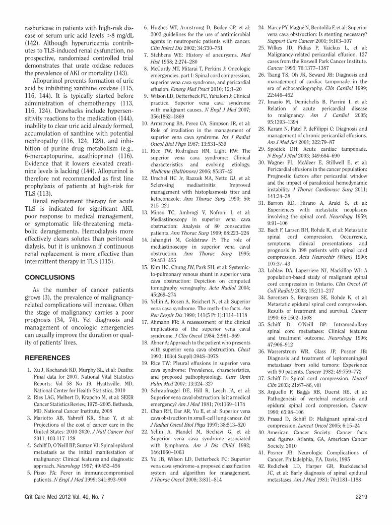
Hyperuricemia results from the catabolism of purine nucleic acids to hypoxanthine, xanthine, and then uric acid by xanthine oxidase (Fig. 7). Most uric acid is renally excreted, but poor sol-ubility in urine (pKa1 of 5.4–5.7) (116) can cause uric acid crystal precipitation in the collecting tubules. The resultant obstruc-tive nephropathy and uric acid–mediated glomerular and tubular injury from oxi-dant stress and inflammation can further injure the kidneys (117).
Management of acute TLS consists of prophylactic measures to reduce the risk of renal impairment and treatment of metabolic abnormalities. Although metabolic abnormalities may be more immediately life- threatening, AKI in the setting of acute TLS independently and significantly increases the risk of mortal-ity (127). Volume depletion predisposes AKI (117). The primary intervention for high- risk patients is volume loading with fluid at roughly twice the maintenance rate to increase glomerular filtration rate, urine flow, and uric acid and calcium phosphate solubility (113, 124). The prac-tice of alkalinizing urine to increase the solubility of uric acid (115) is no longer supported because of the nephropathy risk resulting from xanthine (116, 128) and calcium phosphate crystal precipita-tion (124, 126, 128).
Hyperkalemia can usually be managed with enteral sodium polystyrene. Because
Figure 6. Short QT interval; Ca 16 mg/dL. Courtesy of Amal Mattu, MD, Department of Emergency Medicine, University of Maryland School of Medicine.
2218 Crit Care Med 2012 Vol. 40, No. 7
ion exchange resins work slowly, symptom-atic patients require more rapid treatment such as insulin and dextrose or sodium bicarbonate to shift potassium intracel-lularly, and calcium gluconate to stabilize
cardiac cell membranes (115, 124). Sodium bicarbonate should be administered cau-tiously to avoid inadvertent hypervolemia (115), metabolic alkalosis (124), or precipi-tation of calcium either in the IV tubing
(124) or in the kidney (115, 117). Primary prevention of hyperphosphatemia includes phosphate removal from fluids, volume loading, and phosphate binders. For more severe cases, and when medical manage-ment has failed, renal replacement therapy has been employed (124). Hypocalcemia is treated only if symptomatic because of the risk of nephropathy (124).
Recombinant urate oxidase (rasburi-case) works by cata bolizing uric acid to more soluble allantoin (113, 116, 121, 122, 124, 129–132). Whether given thera-peutically or prophylactically, this agent is highly effective at rapidly normalizing uric acid levels (121, 122, 130–132), and has greater efficacy than allopurinol (133). It is recommended as first line treatment for high- risk patients with tumors prone to rapid lysis or the presence of preexisting kidney injury and elevated uric acid levels (113, 124). Because hydrogen peroxide is a byproduct of uric acid catabolism to allantoin, rasburicase can cause hemolytic anemia or methemoglobinemia in patients with glucose-6-phosphate dehydrogenase deficiency, and is therefore contraindicated in these patients (129). The recommended dose (0.2 mg/kg IV daily given for 5 days) is based on predicted body weight even in patients with morbid obesity (134). Because of the drug’s expense, alterna-tive dosing regimens using single doses or shorter courses have been reported (135–142). Our institution has pub-lished its experience with 6 mg fixed- dose
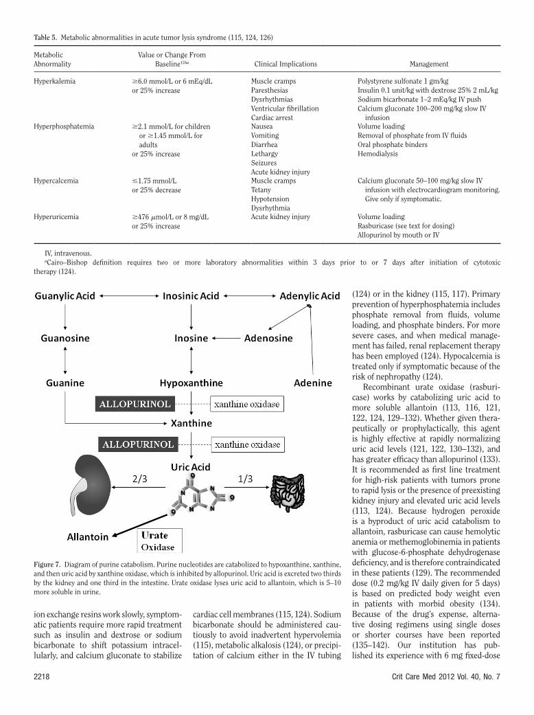
Table 5. Metabolic abnormalities in acute tumor lysis syndrome (115, 124, 126)
Metabolic Abnormality
Value or Change From Baseline124a Clinical Implications Management
Hyperkalemia 6.0 mmol/L or 6 mEq/dLor 25% increase
Muscle crampsParesthesiasDysrhythmiasVentricular fibrillationCardiac arrest
Polystyrene sulfonate 1 gm/kgInsulin 0.1 unit/kg with dextrose 25% 2 mL/kgSodium bicarbonate 1–2 mEq/kg IV pushCalcium gluconate 100–200 mg/kg slow IV
infusionHyperphosphatemia 2.1 mmol/L for children
or 1.45 mmol/L for adults
or 25% increase
NauseaVomitingDiarrheaLethargySeizuresAcute kidney injury
Volume loadingRemoval of phosphate from IV fluidsOral phosphate binders Hemodialysis
Hypercalcemia 1.75 mmol/Lor 25% decrease
Muscle crampsTetanyHypotensionDysrhythmia
Calcium gluconate 50–100 mg/kg slow IV infusion with electrocardiogram monitoring. Give only if symptomatic.
Hyperuricemia 476 mmol/L or 8 mg/dLor 25% increase
Acute kidney injury Volume loadingRasburicase (see text for dosing)Allopurinol by mouth or IV
IV, intravenous.aCairo–Bishop definition requires two or more laboratory abnormalities within 3 days prior to or 7 days after initiation of cytotoxic
therapy (124).
Figure 7. Diagram of purine catabolism. Purine nucleotides are catabolized to hypoxanthine, xanthine, and then uric acid by xanthine oxidase, which is inhibited by allopurinol. Uric acid is excreted two thirds by the kidney and one third in the intestine. Urate oxidase lyses uric acid to allantoin, which is 5–10 more soluble in urine.
Crit Care Med 2012 Vol. 40, No. 7 2219
rasburicase in patients with high- risk dis-ease or serum uric acid levels 8 mg/dL (142). Although hyperuricemia contrib-utes to TLS- induced renal dysfunction, no prospective, randomized controlled trial demonstrates that urate oxidase reduces the prevalence of AKI or mortality (143).
Allopurinol prevents formation of uric acid by inhibiting xanthine oxidase (115, 116, 144). It is typically started before administration of chemotherapy (113, 116, 124). Drawbacks include hypersen-sitivity reactions to the medication (144), inability to clear uric acid already formed, accumulation of xanthine with potential nephropathy (116, 124, 128), and inhi-bition of purine drug metabolism (e.g., 6-mercaptopurine, azathioprine) (116). Evidence that it lowers elevated creati-nine levels is lacking (144). Allopurinol is therefore not recommended as first line prophylaxis of patients at high- risk for TLS (113).
Renal replacement therapy for acute TLS is indicated for significant AKI, poor response to medical management, or symptomatic life- threatening meta-bolic derangements. Hemodialysis more effectively clears solutes than peritoneal dialysis, but it is unknown if continuous renal replacement is more effective than intermittent therapy in TLS (115).
CONCLUSIONS
As the number of cancer patients grows (3), the prevalence of malignancy- related complications will increase. Often the stage of malignancy carries a poor prognosis (34, 74). Yet diagnosis and management of oncologic emergencies can usually improve the duration or qual-ity of patients’ lives.
REFERENCES
1. Xu J, Kochanek KD, Murphy SL, et al: Deaths: Final data for 2007. National Vital Statistics Reports; Vol 58 No 19. Hyattsville, MD, National Center for Health Statistics, 2010
2. Ries LAG, Melbert D, Krapcho M, et al: SEER Cancer Statistics Review, 1975–2005. Bethesda, MD, National Cancer Institute, 2008
3. Mariotto AB, Yabroff KR, Shao Y, et al: Projections of the cost of cancer care in the United States: 2010-2020. J Natl Cancer Inst 2011; 103:117–128
4. Schiff D, O’Neill BP, Suman VJ: Spinal epidural metastasis as the initial manifestation of malignancy: Clinical features and diagnostic approach. Neurology 1997; 49:452–456
5. Pizzo PA: Fever in immunocompromised patients. N Engl J Med 1999; 341:893–900
6. Hughes WT, Armstrong D, Bodey GP, et al: 2002 guidelines for the use of antimicrobial agents in neutropenic patients with cancer. Clin Infect Dis 2002; 34:730–751
7. Stehbens WE: History of aneurysms. Med Hist 1958; 2:274–280
8. McCurdy MT, Mitarai T, Perkins J: Oncologic emergencies, part I: Spinal cord compression, superior vena cava syndrome, and pericardial effusion. Emerg Med Pract 2010; 12:1–20
9. Wilson LD, Detterbeck FC, Yahalom J: Clinical practice. Superior vena cava syndrome with malignant causes. N Engl J Med 2007; 356:1862–1869
10. Armstrong BA, Perez CA, Simpson JR, et al: Role of irradiation in the management of superior vena cava syndrome. Int J Radiat Oncol Biol Phys 1987; 13:531–539
11. Rice TW, Rodriguez RM, Light RW: The superior vena cava syndrome: Clinical characteristics and evolving etiology. Medicine (Baltimore) 2006; 85:37–42
12. Urschel HC Jr, Razzuk MA, Netto GJ, et al: Sclerosing mediastinitis: Improved management with histoplasmosis titer and ketoconazole. Ann Thorac Surg 1990; 50: 215–221
13. Mineo TC, Ambrogi V, Nofroni I, et al: Mediastinoscopy in superior vena cava obstruction: Analysis of 80 consecutive patients. Ann Thorac Surg 1999; 68:223–226
14. Jahangiri M, Goldstraw P: The role of mediastinoscopy in superior vena caval obstruction. Ann Thorac Surg 1995; 59:453–455
15. Kim HC, Chung JW, Park SH, et al: Systemic- to- pulmonary venous shunt in superior vena cava obstruction: Depiction on computed tomography venography. Acta Radiol 2004; 45:269–274
16. Yellin A, Rosen A, Reichert N, et al: Superior vena cava syndrome. The myth–the facts. Am Rev Respir Dis 1990; 141(5 Pt 1):1114–1118
17. Ahmann FR: A reassessment of the clinical implications of the superior vena caval syndrome. J Clin Oncol 1984; 2:961–969
18. Abner A: Approach to the patient who presents with superior vena cava obstruction. Chest 1993; 103(4 Suppl):394S–397S
19. Rice TW: Pleural effusions in superior vena cava syndrome: Prevalence, characteristics, and proposed pathophysiology. Curr Opin Pulm Med 2007; 13:324–327
20. Schraufnagel DE, Hill R, Leech JA, et al: Superior vena caval obstruction. Is it a medical emergency? Am J Med 1981; 70:1169–1174
21. Chan RH, Dar AR, Yu E, et al: Superior vena cava obstruction in small- cell lung cancer. Int J Radiat Oncol Biol Phys 1997; 38:513–520
22. Yellin A, Mandel M, Rechavi G, et al: Superior vena cava syndrome associated with lymphoma. Am J Dis Child 1992; 146:1060–1063
23. Yu JB, Wilson LD, Detterbeck FC: Superior vena cava syndrome–a proposed classification system and algorithm for management. J Thorac Oncol 2008; 3:811–814
24. Marcy PY, Magné N, Bentolila F, et al: Superior vena cava obstruction: Is stenting necessary? Support Care Cancer 2001; 9:103–107
25. Wilkes JD, Fidias P, Vaickus L, et al: Malignancy- related pericardial effusion. 127 cases from the Roswell Park Cancer Institute. Cancer 1995; 76:1377–1387
26. Tsang TS, Oh JK, Seward JB: Diagnosis and management of cardiac tamponade in the era of echocardiography. Clin Cardiol 1999; 22:446–452
27. Imazio M, Demichelis B, Parrini I, et al: Relation of acute pericardial disease to malignancy. Am J Cardiol 2005; 95:1393–1394
28. Karam N, Patel P, deFilippi C: Diagnosis and management of chronic pericardial effusions. Am J Med Sci 2001; 322:79–87
29. Spodick DH: Acute cardiac tamponade. N Engl J Med 2003; 349:684–690
30. Wagner PL, McAleer E, Stillwell E, et al: Pericardial effusions in the cancer population: Prognostic factors after pericardial window and the impact of paradoxical hemodynamic instability. J Thorac Cardiovasc Surg 2011; 141:34–38
31. Barron KD, Hirano A, Araki S, et al: Experiences with metastatic neoplasms involving the spinal cord. Neurology 1959; 9:91–106
32. Bach F, Larsen BH, Rohde K, et al: Metastatic spinal cord compression. Occurrence, symptoms, clinical presentations and prognosis in 398 patients with spinal cord compression. Acta Neurochir (Wien) 1990; 107:37–43
33. Loblaw DA, Laperriere NJ, Mackillop WJ: A population- based study of malignant spinal cord compression in Ontario. Clin Oncol (R Coll Radiol) 2003; 15:211–217
34. Sørensen S, Børgesen SE, Rohde K, et al: Metastatic epidural spinal cord compression. Results of treatment and survival. Cancer 1990; 65:1502–1508
35. Schiff D, O’Neill BP: Intramedullary spinal cord metastases: Clinical features and treatment outcome. Neurology 1996; 47:906–912
36. Wasserstrom WR, Glass JP, Posner JB: Diagnosis and treatment of leptomeningeal metastases from solid tumors: Experience with 90 patients. Cancer 1982; 49:759–772
37. Schiff D: Spinal cord compression. Neurol Clin 2003; 21:67–86, viii
38. Arguello F, Baggs RB, Duerst RE, et al: Pathogenesis of vertebral metastasis and epidural spinal cord compression. Cancer 1990; 65:98–106
39. Prasad D, Schiff D: Malignant spinal- cord compression. Lancet Oncol 2005; 6:15–24
40. American Cancer Society: Cancer facts and figures. Atlanta, GA, American Cancer Society, 2010
41. Posner JB: Neurologic Complications of Cancer. Philadelphia, F.A. Davis, 1995
42. Rodichok LD, Harper GR, Ruckdeschel JC, et al: Early diagnosis of spinal epidural metastases. Am J Med 1981; 70:1181–1188
2220 Crit Care Med 2012 Vol. 40, No. 7
43. Schiff D, Batchelor T, Wen PY: Neurologic emergencies in cancer patients. Neurol Clin 1998; 16:449–483
44. Helweg- Larsen S, Sørensen PS: Symptoms and signs in metastatic spinal cord compression: A study of progression from first symptom until diagnosis in 153 patients. Eur J Cancer 1994; 30A:396–398
45. Husband DJ: Malignant spinal cord compression: Prospective study of delays in referral and treatment. BMJ 1998; 317:18–21
46. Husband DJ, Grant KA, Romaniuk CS: MRI in the diagnosis and treatment of suspected malignant spinal cord compression. Br J Radiol 2001; 74:15–23
47. Deyo RA, Rainville J, Kent DL: What can the history and physical examination tell us about low back pain? JAMA 1992; 268:760–765
48. Maranzano E, Latini P: Effectiveness of radiation therapy without surgery in metastatic spinal cord compression: Final results from a prospective trial. Int J Radiat Oncol Biol Phys 1995; 32:959–967
49. Li KC, Poon PY: Sensitivity and specificity of MRI in detecting malignant spinal cord compression and in distinguishing malignant from benign compression fractures of vertebrae. Magn Reson Imaging 1988; 6:547–556
50. Kim JK, Learch TJ, Colletti PM, et al: Diagnosis of vertebral metastasis, epidural metastasis, and malignant spinal cord compression: Are T(1)-weighted sagittal images sufficient? Magn Reson Imaging 2000; 18:819–824
51. Cook AM, Lau TN, Tomlinson MJ, et al: Magnetic resonance imaging of the whole spine in suspected malignant spinal cord compression: Impact on management. Clin Oncol (R Coll Radiol) 1998; 10:39–43
52. Helweg- Larsen S, Hansen SW, Sørensen PS: Second occurrence of symptomatic metastatic spinal cord compression and findings of multiple spinal epidural metastases. Int J Radiat Oncol Biol Phys 1995; 33:595–598
53. van der Sande JJ, Kröger R, Boogerd W: Multiple spinal epidural metastases; an unexpectedly frequent finding. J Neurol Neurosurg Psychiatr 1990; 53:1001–1003
54. Schiff D, O’Neill BP, Wang CH, et al: Neuroimaging and treatment implications of patients with multiple epidural spinal metastases. Cancer 1998; 83:1593–1601
55. Khanna AJ, Shindle MK, Wasserman BA, et al: Use of magnetic resonance imaging in differentiating compartmental location of spinal tumors. Am J Orthop 2005; 34:472–476
56. Sze G, Krol G, Zimmerman RD, et al: Intramedullary disease of the spine: Diagnosis using gadolinium- DTPA- enhanced MR imaging. AJR Am J Roentgenol 1988; 151:1193–1204
57. Hagenau C, Grosh W, Currie M, et al: Comparison of spinal magnetic resonance imaging and myelography in cancer patients. J Clin Oncol 1987; 5:1663–1669
58. Helweg- Larsen S, Wagner A, Kjaer L, et al: Comparison of myelography combined with
postmyelographic spinal CT and MRI in suspected metastatic disease of the spinal canal. J Neurooncol 1992; 13:231–237
59. Hollis PH, Malis LI, Zappulla RA: Neurological deterioration after lumbar puncture below complete spinal subarachnoid block. J Neurosurg 1986; 64:253–256
60. Portenoy RK, Galer BS, Salamon O, et al: Identification of epidural neoplasm. Radiography and bone scintigraphy in the symptomatic and asymptomatic spine. Cancer 1989; 64:2207–2213
61. O’Rourke T, George CB, Redmond J 3rd, et al: Spinal computed tomography and computed tomographic metrizamide myelography in the early diagnosis of metastatic disease. J Clin Oncol 1986; 4:576–583
62. Rades D, Blach M, Bremer M, et al: Prognostic significance of the time of developing motor deficits before radiation therapy in metastatic spinal cord compression: One- year results of a prospective trial. Int J Radiat Oncol Biol Phys 2000; 48:1403–1408
63. Yeung SCJ, Escalante CP: Oncologic emergencies. In: Holland- Frei Cancer Medicine. Seventh Edition. Kufe DW, Bast Jr. RC, Hait W, et al (Eds). Hamilton, Ontario, BC Decker, 2006, pp xxiii, p. 2328
64. Sørensen S, Helweg- Larsen S, Mouridsen H, et al: Effect of high- dose dexamethasone in carcinomatous metastatic spinal cord compression treated with radiotherapy: A randomised trial. Eur J Cancer 1994; 30A:22–27
65. Heimdal K, Hirschberg H, Slettebø H, et al: High incidence of serious side effects of high- dose dexamethasone treatment in patients with epidural spinal cord compression. J Neurooncol 1992; 12:141–144
66. Loblaw DA, Perry J, Chambers A, et al: Systematic review of the diagnosis and management of malignant extradural spinal cord compression: The Cancer Care Ontario Practice Guidelines Initiative’s Neuro- Oncology Disease Site Group. J Clin Oncol 2005; 23:2028–2037
67. Young RF, Post EM, King GA: Treatment of spinal epidural metastases. Randomized prospective comparison of laminectomy and radiotherapy. J Neurosurg 1980; 53:741–748
68. Gilbert RW, Kim JH, Posner JB: Epidural spinal cord compression from metastatic tumor: Diagnosis and treatment. Ann Neurol 1978; 3:40–51
69. Sciubba DM, Petteys RJ, Dekutoski MB, et al: Diagnosis and management of metastatic spine disease. J Neurosurg Spine 2010; 13:94–108
70. Patchell RA, Tibbs PA, Regine WF, et al: Direct decompressive surgical resection in the treatment of spinal cord compression caused by metastatic cancer: A randomised trial. Lancet 2005; 366:643–648
71. Cole JS, Patchell RA: Metastatic epidural spinal cord compression. Lancet Neurol 2008; 7:459–466
72. Stewart AF: Clinical practice. Hypercalcemia associated with cancer. N Engl J Med 2005; 352:373–379
73. Lee CT, Yang CC, Lam KK, et al: Hypercalcemia in the emergency department. Am J Med Sci 2006; 331:119–123
74. Ralston SH, Gallacher SJ, Patel U, et al: Cancer- associated hypercalcemia: Morbidity and mortality. Clinical experience in 126 treated patients. Ann Intern Med 1990; 112:499–504
75. McCurdy MT, Mitarai T, Perkins J: Oncologic emergencies, part II: Neutropenic fever, tumor lysis syndrome, and hypercalcemia of malignancy. Emerg Med Pract 2010; 12:1–24
76. Ratcliffe WA, Hutchesson AC, Bundred NJ, et al: Role of assays for parathyroid- hormone- related protein in investigation of hypercalcaemia. Lancet 1992; 339: 164–167
77. Seymour JF, Gagel RF: Calcitriol: The major humoral mediator of hypercalcemia in Hodgkin’s disease and non-Hodgkin’s lymphomas. Blood 1993; 82: 1383–1394
78. Bilezikian JP: Clinical review 51: Management of hypercalcemia. J Clin Endocrinol Metab 1993; 77:1445–1449
79. National Cancer Institute: PDQ® Hypercalcemia. Available at: http://cancer.gov/cancertopics/pdq/supportivecare/hypercalcemia/healthprofessional. Accessed December 29, 2010
80. Ladenson JH, Lewis JW, Boyd JC: Failure of total calcium corrected for protein, albumin, and pH to correctly assess free calcium status. J Clin Endocrinol Metab 1978; 46: 986–993
81. Ijaz A, Mehmood T, Qureshi AH, et al: Estimation of ionized calcium, total calcium and albumin corrected calcium for the diagnosis of hypercalcaemia of malignancy. J Coll Physicians Surg Pak 2006; 16:49–52
82. Slomp J, van der Voort PH, Gerritsen RT, et al: Albumin- adjusted calcium is not suitable for diagnosis of hyper- and hypocalcemia in the critically ill. Crit Care Med 2003; 31:1389–1393
83. Wang S, McDonnell EH, Sedor FA, et al: pH effects on measurements of ionized calcium and ionized magnesium in blood. Arch Pathol Lab Med 2002; 126:947–950
84. Milionis HJ, Rizos E, Liamis G, et al: Acid- base and electrolyte disturbances in patients with hypercalcemia. South Med J 2002; 95:1280–1287
85. Farr HW, Fahey TJ Jr, Nash AG, et al: Primary hyperparathyroidism and cancer. Am J Surg 1973; 126:539–543
86. Fritchie K, Zedek D, Grenache DG: The clinical utility of parathyroid hormone- related peptide in the assessment of hypercalcemia. Clin Chim Acta 2009; 402:146–149
87. Wimalawansa SJ: Significance of plasma PTH- rp in patients with hypercalcemia of malignancy treated with bisphosphonate. Cancer 1994; 73:2223–2230
Crit Care Med 2012 Vol. 40, No. 7 2221
88. Rizzoli R, Thiébaud D, Bundred N, et al: Serum parathyroid hormone- related protein levels and response to bisphosphonate treatment in hypercalcemia of malignancy. J Clin Endocrinol Metab 1999; 84: 3545–3550
89. Behl D, Hendrickson AW, Moynihan TJ: Oncologic emergencies. Crit Care Clin 2010; 26:181–205
90. Antzelevitch C, Brugada P, Borggrefe M, et al: Brugada syndrome: Report of the second consensus conference: Endorsed by the Heart Rhythm Society and the European Heart Rhythm Association. Circulation 2005; 111:659–670
91. 2005 American Heart Association Guidelines for Cardiopulmonary Resuscitation and Emergency Cardiovascular Care. Circulation 2005; 112:IV1–203
92. Kvols LK: Neoplasms of the diffuse endocrine system. In: Cancer Medicine. Sixth Edition. Holland JF, Frei III E (Eds). Hamilton, Ontario, BC Decker, 2003, pp 1275–1323
93. Hosking DJ, Cowley A, Bucknall CA: Rehydration in the treatment of severe hypercalcaemia. Q J Med 1981; 50:473–481
94. Nkwuo N, Schamban N, Borenstein M: Selected oncologic emergencies. In: Rosen’s Emergency Medicine: Concepts and Clinical Practice. Sixth Edition. Marx JA, Hockberger RS, Walls RM (Eds). Philadelphia, PA, Mosby, 2006, pp 1914–1916
95. Body JJ: Hypercalcemia of malignancy. Semin Nephrol 2004; 24:48–54
96. Koo WS, Jeon DS, Ahn SJ, et al: Calcium- free hemodialysis for the management of hypercalcemia. Nephron 1996; 72:424–428
97. Davies M, Hayes ME, Yin JA, et al: Abnormal synthesis of 1,25-dihydroxyvitamin D in patients with malignant lymphoma. J Clin Endocrinol Metab 1994; 78:1202–1207
98. Mudde AH, van den Berg H, Boshuis PG, et al: Ectopic production of 1,25-dihydroxyvitamin D by B- cell lymphoma as a cause of hypercalcemia. Cancer 1987; 59:1543–1546
99. Chisholm MA, Mulloy AL, Taylor AT: Acute management of cancer- related hypercalcemia. Ann Pharmacother 1996; 30:507–513
100. Lukert BP, Raisz LG: Glucocorticoid- induced osteoporosis: Pathogenesis and management. Ann Intern Med 1990; 112:352–364
101. Strumpf M, Kowalski MA, Mundy GR: Effects of glucocorticoids on osteoclast- activating factor. J Lab Clin Med 1978; 92:772–778
102. Fleisch H: Bisphosphonates. Pharmacology and use in the treatment of tumour- induced hypercalcaemic and metastatic bone disease. Drugs 1991; 42:919–944
103. Layman R, Olson K, Van Poznak C: Bisphosphonates for breast cancer: Questions answered, questions remaining. Hematol Oncol Clin North Am 2007; 21:341–367
104. Woo SB, Hellstein JW, Kalmar JR: Narrative [corrected] review: Bisphosphonates and osteonecrosis of the jaws. Ann Intern Med 2006; 144:753–761
105. Major P, Lortholary A, Hon J, et al: Zoledronic acid is superior to pamidronate in the treatment of hypercalcemia of malignancy: A pooled analysis of two randomized, controlled clinical trials. J Clin Oncol 2001; 19:558–567
106. Silva OL, Becker KL: Salmon calcitonin in the treatment of hypercalcemia. Arch Intern Med 1973; 132:337–339
107. Chitambar CR: Apoptotic mechanisms of gallium nitrate: Basic and clinical investigations. Oncology (Williston Park, NY) 2004; 18(13 Suppl 10):39–44
108. Cvitkovic F, Armand JP, Tubiana- Hulin M, et al: Randomized, double- blind, phase II trial of gallium nitrate compared with pamidronate for acute control of cancer- related hypercalcemia. Cancer J 2006; 12:47–53
109. Thürlimann B, Waldburger R, Senn HJ, et al: Plicamycin and pamidronate in symptomatic tumor- related hypercalcemia: A prospective randomized crossover trial. Ann Oncol 1992; 3:619–623
110. Fizazi K, Lipton A, Mariette X, et al: Randomized phase II trial of denosumab in patients with bone metastases from prostate cancer, breast cancer, or other neoplasms after intravenous bisphosphonates. J Clin Oncol 2009; 27:1564–1571
111. Henry DH, von Moos R, Hungria V: Delaying skeletal- related events in a randomized phase III study of denosumab versus zoledronic acid in patients with advanced cancer. J Clin Oncol 2010; 28:A9133
112. Lipton A, Stopeck A, Von Moos R: A meta- analysis of results from two randomized, double- blind studies of denosumab versus zoledronic acid for treatment of bone metastases. J Clin Oncol 2010; 28:A9015
113. Cairo MS, Coiffier B, Reiter A, et al; TLS Expert Panel: Recommendations for the evaluation of risk and prophylaxis of tumour lysis syndrome (TLS) in adults and children with malignant diseases: An expert TLS panel consensus. Br J Haematol 2010; 149:578–586
114. Howard SC, Jones DP, Pui CH: The tumor lysis syndrome. N Engl J Med 2011; 364:1844–1854
115. Davidson MB, Thakkar S, Hix JK, et al: Pathophysiology, clinical consequences, and treatment of tumor lysis syndrome. Am J Med 2004; 116:546–554
116. Cairo MS, Bishop M: Tumour lysis syndrome: New therapeutic strategies and classification. Br J Haematol 2004; 127:3–11
117. Lameire NH, Flombaum CD, Moreau D, et al: Acute renal failure in cancer patients. Ann Med 2005; 37:13–25
118. Baeksgaard L, Sørensen JB: Acute tumor lysis syndrome in solid tumors–a case report and review of the literature. Cancer Chemother Pharmacol 2003; 51:187–192
119. Cohen LF, Balow JE, Magrath IT, et al: Acute tumor lysis syndrome. A review of 37 patients with Burkitt’s lymphoma. Am J Med 1980; 68:486–491
120. Hande KR, Garrow GC: Acute tumor lysis syndrome in patients with high- grade non-Hodgkin’s lymphoma. Am J Med 1993; 94:133–139
121. Coiffier B, Mounier N, Bologna S, et al; Groupe d’Etude des Lymphomes de l’Adulte Trial on Rasburicase Activity in Adult Lymphoma: Efficacy and safety of rasburi-case (recombinant urate oxidase) for the prevention and treatment of hyperuricemia during induction chemotherapy of aggres-sive non-Hodgkin’s lymphoma: Results of the GRAAL1 (Groupe d’Etude des Lymphomes de l’Adulte Trial on Rasburicase Activity in Adult Lymphoma) study. J Clin Oncol 2003; 21:4402–4406
122. Bosly A, Sonet A, Pinkerton CR, et al: Rasburicase (recombinant urate oxidase) for the management of hyperuricemia in patients with cancer: Report of an international compassionate use study. Cancer 2003; 98:1048–1054
123. Hsu HH, Huang CC: Acute spontaneous tumor lysis in anaplastic large T- cell lymphoma presenting with hyperuricemic acute renal failure. Int J Hematol 2004; 79:48–51
124. Coiffier B, Altman A, Pui CH, et al: Guidelines for the management of pediatric and adult tumor lysis syndrome: An evidence- based review. J Clin Oncol 2008; 26:2767–2778
125. Koduri PR: Hyperphosphatemia and tumor lysis syndrome. Ann Hematol 2005; 84:696
126. Desmeules S, Bergeron MJ, Isenring P: Acute phosphate nephropathy and renal failure. N Engl J Med 2003; 349:1006–1007
127. Darmon M, Guichard I, Vincent F, et al: Prognostic significance of acute renal injury in acute tumor lysis syndrome. Leuk Lymphoma 2010; 51:221–227
128. Ten Harkel AD, Kist- Van Holthe JE, Van Weel M, et al: Alkalinization and the tumor lysis syndrome. Med Pediatr Oncol 1998; 31:27–28
129. Pui CH, Jeha S, Irwin D, et al: Recombinant urate oxidase (rasburicase) in the prevention and treatment of malignancy- associated hyperuricemia in pediatric and adult patients: Results of a compassionate- use trial. Leukemia 2001; 15:1505–1509
130. Pui CH, Mahmoud HH, Wiley JM, et al: Recombinant urate oxidase for the prophylaxis or treatment of hyperuricemia in patients with leukemia or lymphoma. J Clin Oncol 2001; 19:697–704
131. Pui CH: Rasburicase: A potent uricolytic agent. Expert Opin Pharmacother 2002; 3:433–442
132. Jeha S, Kantarjian H, Irwin D, et al: Efficacy and safety of rasburicase, a recombinant urate oxidase (Elitek), in the management of malignancy- associated hyperuricemia in pediatric and adult patients: Final results of a multicenter compassionate use trial. Leukemia 2005; 19:34–38
133. Goldman SC, Holcenberg JS, Finklestein JZ, et al: A randomized comparison between rasburicase and allopurinol in children with
2222 Crit Care Med 2012 Vol. 40, No. 7
lymphoma or leukemia at high risk for tumor lysis. Blood 2001; 97:2998–3003
134. Arnold TM, Reuter JP, Delman BS, et al: Use of single- dose rasburicase in an obese female. Ann Pharmacother 2004; 38:1428–1431
135. Trifilio S, Gordon L, Singhal S, et al: Reduced- dose rasburicase (recombinant xanthine oxidase) in adult cancer patients with hyperuricemia. Bone Marrow Transplant 2006; 37:997–1001
136. Reeves DJ, Bestul DJ: Evaluation of a single fixed dose of rasburicase 7.5 mg for the treatment of hyperuricemia in adults with cancer. Pharmacotherapy 2008; 28:685–690
137. McDonnell AM, Lenz KL, Frei- Lahr DA, et al: Single- dose rasburicase 6 mg in the
management of tumor lysis syndrome in adults. Pharmacotherapy 2006; 26:806–812
138. Knoebel RW, Lo M, Crank CW: Evaluation of a low, weight- based dose of rasburicase in adult patients for the treatment or prophylaxis of tumor lysis syndrome. J Oncol Pharm Pract 2011; 17:147–154
139. Hutcherson DA, Gammon DC, Bhatt MS, et al: Reduced- dose rasburicase in the treatment of adults with hyperuricemia associated with malignancy. Pharmacotherapy 2006; 26:242–247
140. Hummel M, Reiter S, Adam K, et al: Effective treatment and prophylaxis of hyperuricemia and impaired renal function in tumor lysis syndrome with low doses of rasburicase. Eur J Haematol 2008; 80:331–336
141. Lee AC, Li CH, So KT, et al: Treatment of impending tumor lysis with single- dose rasburicase. Ann Pharmacother 2003; 37:1614–1617
142. Vines AN, Shanholtz CB, Thompson JL: Fixed- dose rasburicase 6 mg for hyperuricemia and tumor lysis syndrome in high- risk cancer patients. Ann Pharmacother 2010; 44:1529–1537
143. Cheuk DK, Chiang AK, Chan GC, et al: Urate oxidase for the prevention and treatment of tumor lysis syndrome in children with cancer. Cochrane Database Syst Rev 2010; CD006945
144. Smalley RV, Guaspari A, Haase- Statz S, et al: Allopurinol: Intravenous use for prevention and treatment of hyperuricemia. J Clin Oncol 2000; 18:1758–1763