Oncologic Emergencies: Part II - MD Anderson Cancer Center · (e.g. subdural hematoma) • Anemia...
14
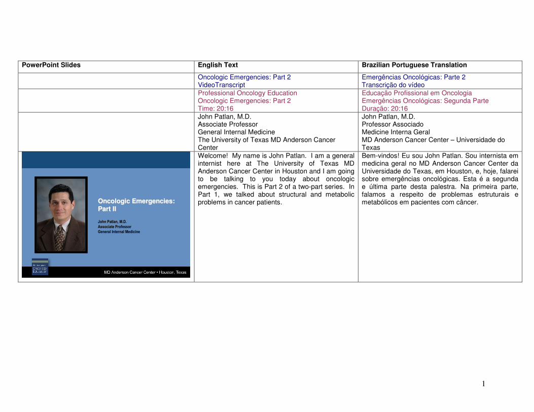
1 PowerPoint Slides English Text Brazilian Portuguese Translation Oncologic Emergencies: Part 2 VideoTranscript Emergências Oncológicas: Parte 2 Transcrição do vídeo Professional Oncology Education Oncologic Emergencies: Part 2 Time: 20:16 Educação Profissional em Oncologia Emergências Oncológicas: Segunda Parte Duração: 20:16 John Patlan, M.D. Associate Professor General Internal Medicine The University of Texas MD Anderson Cancer Center John Patlan, M.D. Professor Associado Medicine Interna Geral MD Anderson Cancer Center – Universidade do Texas Oncologic Emergencies: Part II Oncologic Emergencies: Part II Oncologic Emergencies: Part II Oncologic Emergencies: Part II Oncologic Emergencies: Oncologic Emergencies: Part II Part II John Patlan, M.D. Associate Professor General Internal Medicine Welcome! My name is John Patlan. I am a general internist here at The University of Texas MD Anderson Cancer Center in Houston and I am going to be talking to you today about oncologic emergencies. This is Part 2 of a two-part series. In Part 1, we talked about structural and metabolic problems in cancer patients. Bem-vindos! Eu sou John Patlan. Sou internista em medicina geral no MD Anderson Cancer Center da Universidade do Texas, em Houston, e, hoje, falarei sobre emergências oncológicas. Esta é a segunda e última parte desta palestra. Na primeira parte, falamos a respeito de problemas estruturais e metabólicos em pacientes com câncer.
Transcript of Oncologic Emergencies: Part II - MD Anderson Cancer Center · (e.g. subdural hematoma) • Anemia...
1
PowerPoint Slides English Text Brazilian Portuguese Translation
Oncologic Emergencies: Part 2 VideoTranscript
Emergências Oncológicas: Parte 2 Transcrição do vídeo
Professional Oncology Education Oncologic Emergencies: Part 2 Time: 20:16
Educação Profissional em Oncologia
Emergências Oncológicas: Segunda Parte Duração: 20:16
John Patlan, M.D. Associate Professor General Internal Medicine The University of Texas MD Anderson Cancer Center
John Patlan, M.D. Professor Associado Medicine Interna Geral MD Anderson Cancer Center – Universidade do Texas
Oncologic Emergencies: Part IIOncologic Emergencies: Part IIOncologic Emergencies: Part IIOncologic Emergencies: Part II
Oncologic Emergencies:Oncologic Emergencies:
Part IIPart II
John Patlan, M.D.
Associate Professor
General Internal Medicine
Welcome! My name is John Patlan. I am a general internist here at The University of Texas MD Anderson Cancer Center in Houston and I am going to be talking to you today about oncologic emergencies. This is Part 2 of a two-part series. In Part 1, we talked about structural and metabolic problems in cancer patients.
Bem-vindos! Eu sou John Patlan. Sou internista em medicina geral no MD Anderson Cancer Center da Universidade do Texas, em Houston, e, hoje, falarei sobre emergências oncológicas. Esta é a segunda e última parte desta palestra. Na primeira parte, falamos a respeito de problemas estruturais e metabólicos em pacientes com câncer.
2
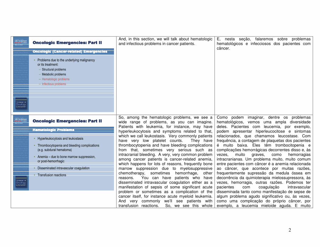
Oncologic Emergencies: Part IIOncologic Emergencies: Part IIOncologic Emergencies: Part IIOncologic Emergencies: Part II
Oncologic (CancerOncologic (CancerOncologic (CancerOncologic (Cancer----related) Emergenciesrelated) Emergenciesrelated) Emergenciesrelated) Emergencies
• Problems due to the underlying malignancy
or its treatment:
– Structural problems
– Metabolic problems
– Hematologic problems
– Infectious problems
And, in this section, we will talk about hematologic and infectious problems in cancer patients.
E, nesta seção, falaremos sobre problemas hematológicos e infecciosos dos pacientes com câncer.
Oncologic Emergencies: Part IIOncologic Emergencies: Part IIOncologic Emergencies: Part IIOncologic Emergencies: Part II
Hematologic ProblemsHematologic ProblemsHematologic ProblemsHematologic Problems
• Hyperleukocytosis and leukostasis
• Thrombocytopenia and bleeding complications
(e.g. subdural hematoma)
• Anemia – due to bone marrow suppression,
or post-hemorrhagic
• Disseminated intravascular coagulation
• Transfusion reactions
So, among the hematologic problems, we see a wide range of problems, as you can imagine. Patients with leukemia, for instance, may have hyperleukocytosis and symptoms related to that, which we call leukostasis. Very commonly patients have very low platelet counts. They have thrombocytopenia and have bleeding complications from that, sometimes very serious such as intracranial bleeding. A very, very common problem among cancer patients is cancer-related anemia, which happens for lots of reasons, frequently bone marrow suppression due to myelosuppressive chemotherapy, sometimes hemorrhage, other reasons. You can have patients who have disseminated intravascular coagulation either as a manifestation of sepsis of some significant acute problem or sometimes as a complication of the cancer itself, for instance acute myeloid leukemia. And very commonly we’ll see patients with transfusion reactions. So, we see this whole
Como podem imaginar, dentre os problemas hematológicos, vemos uma ampla diversidade deles. Pacientes com leucemia, por exemplo, podem apresentar hiperleucocitose e sintomas relacionados, que chamamos leucostase. Com frequência, a contagem de plaquetas dos pacientes é muito baixa. Eles têm trombocitopenia e complicações hemorrágicas decorrentes disso e, às vezes, muito graves, como hemorragias intracranianas. Um problema muito, muito comum entre pacientes com câncer é a anemia relacionada ao câncer, que acontece por muitas razões, frequentemente supressão da medula óssea em decorrência da quimioterapia mielossupressora, às vezes, hemorragia, outras razões. Podemos ter pacientes com coagulação intravascular disseminada tanto como manifestação de sepse de algum problema agudo significativo ou, às vezes, como uma complicação do próprio câncer, por exemplo, a leucemia mieloide aguda. E muito
3
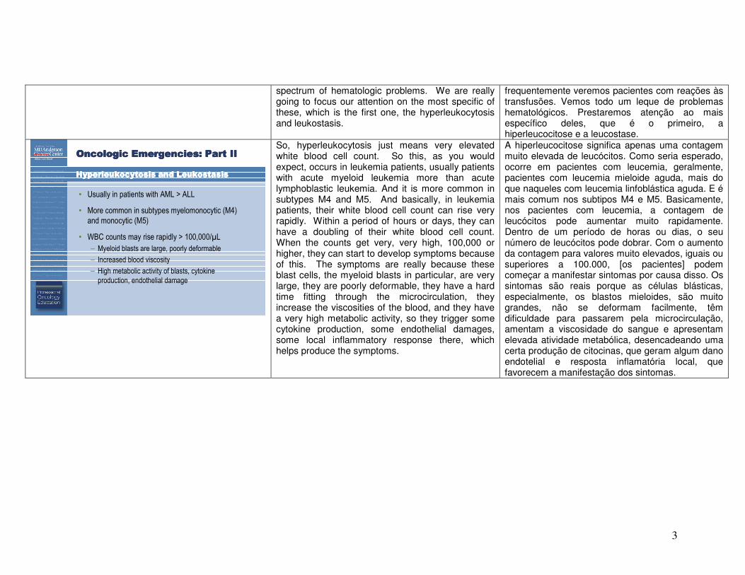
spectrum of hematologic problems. We are really going to focus our attention on the most specific of these, which is the first one, the hyperleukocytosis and leukostasis.
frequentemente veremos pacientes com reações às transfusões. Vemos todo um leque de problemas hematológicos. Prestaremos atenção ao mais específico deles, que é o primeiro, a hiperleucocitose e a leucostase.
Oncologic Emergencies: Part IIOncologic Emergencies: Part IIOncologic Emergencies: Part IIOncologic Emergencies: Part II
Hyperleukocytosis and LeukostasisHyperleukocytosis and LeukostasisHyperleukocytosis and LeukostasisHyperleukocytosis and Leukostasis
• Usually in patients with AML > ALL
• More common in subtypes myelomonocytic (M4)
and monocytic (M5)
• WBC counts may rise rapidly > 100,000/µL
– Myeloid blasts are large, poorly deformable
– Increased blood viscosity
– High metabolic activity of blasts, cytokine
production, endothelial damage
So, hyperleukocytosis just means very elevated white blood cell count. So this, as you would expect, occurs in leukemia patients, usually patients with acute myeloid leukemia more than acute lymphoblastic leukemia. And it is more common in subtypes M4 and M5. And basically, in leukemia patients, their white blood cell count can rise very rapidly. Within a period of hours or days, they can have a doubling of their white blood cell count. When the counts get very, very high, 100,000 or higher, they can start to develop symptoms because of this. The symptoms are really because these blast cells, the myeloid blasts in particular, are very large, they are poorly deformable, they have a hard time fitting through the microcirculation, they increase the viscosities of the blood, and they have a very high metabolic activity, so they trigger some cytokine production, some endothelial damages, some local inflammatory response there, which helps produce the symptoms.
A hiperleucocitose significa apenas uma contagem muito elevada de leucócitos. Como seria esperado, ocorre em pacientes com leucemia, geralmente, pacientes com leucemia mieloide aguda, mais do que naqueles com leucemia linfoblástica aguda. E é mais comum nos subtipos M4 e M5. Basicamente, nos pacientes com leucemia, a contagem de leucócitos pode aumentar muito rapidamente. Dentro de um período de horas ou dias, o seu número de leucócitos pode dobrar. Com o aumento da contagem para valores muito elevados, iguais ou superiores a 100.000, [os pacientes] podem começar a manifestar sintomas por causa disso. Os sintomas são reais porque as células blásticas, especialmente, os blastos mieloides, são muito grandes, não se deformam facilmente, têm dificuldade para passarem pela microcirculação, amentam a viscosidade do sangue e apresentam elevada atividade metabólica, desencadeando uma certa produção de citocinas, que geram algum dano endotelial e resposta inflamatória local, que favorecem a manifestação dos sintomas.
4
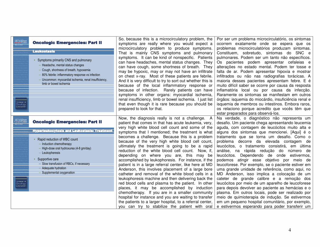
Oncologic Emergencies: Part IIOncologic Emergencies: Part IIOncologic Emergencies: Part IIOncologic Emergencies: Part II
LeukostasisLeukostasisLeukostasisLeukostasis
• Symptoms primarily CNS and pulmonary
– Headache, mental status changes
– Cough, shortness of breath, hypoxemia
– 80% febrile: inflammatory response vs infection
– Uncommon: myocardial ischemia, renal insufficiency,
limb or bowel ischemia
So, because this is a microcirculatory problem, the symptoms are really where you would expect a microcirculatory problem to produce symptoms. That is mainly CNS symptoms and pulmonary symptoms. It can be kind of nonspecific. Patients can have headaches, mental status changes. They can have cough, some shortness of breath. They may be hypoxic, may or may not have an infiltrate on chest x-ray. Most of these patients are febrile. And it is very difficult to try to sort out whether this is because of the local inflammatory response or because of infection. Rarely patients can have symptoms in other organs: myocardial ischemia, renal insufficiency, limb or bowel ischemia. I just list that even though it is rare because you should be prepared to look for that.
Por ser um problema microcirculatório, os sintomas ocorrem exatamente onde se espera que os problemas microcirculatórios produzam sintomas. Constituem, sobretudo, sintomas do SNC e pulmonares. Podem ser um tanto não específicos. Os pacientes podem apresentar cefaleias e alterações no estado mental. Podem ter tosse e falta de ar. Podem apresentar hipoxia e mostrar infiltrados ou não nas radiografias torácicas. A maioria desses pacientes apresentam febre. E é muito difícil saber se ocorre por causa da resposta inflamatória local ou por causa da infecção. Raramente os sintomas se manifestam em outros órgãos: isquemia do miocárdio, insuficiência renal e isquemia de membros ou intestinos. Embora raros, os relaciono porque acredito que vocês têm que estar preparados para observá-los.
Oncologic Emergencies: Part IIOncologic Emergencies: Part IIOncologic Emergencies: Part IIOncologic Emergencies: Part II
Hyperleukocytosis and Hyperleukocytosis and Hyperleukocytosis and Hyperleukocytosis and LeukostasisLeukostasisLeukostasisLeukostasis: Treatment: Treatment: Treatment: Treatment
• Rapid reduction of WBC count
– Induction chemotherapy
– High-dose oral hydroxurea (4-8 gm/day)
– Leukopheresis
• Supportive care
– Slow transfusion of RBCs, if necessary
– Adequate hydration
– Supplemental oxygenation
Now, the diagnosis really is not a challenge. A patient that comes in that has acute leukemia, very, very high white blood cell count and some of the symptoms that I mentioned; the treatment is what becomes a challenge. Because this is a problem because of the very high white blood cell count, ultimately the treatment is going to be a rapid reduction of the white blood cell count. Now, if, depending on where you are, this may be accomplished by leukophoresis. For instance, if the patient is in a large referral center, like here at MD Anderson, this involves placement of a large bore catheter and removal of the white blood cells in a leukophoresis machine and then delivering back the red blood cells and plasma to the patient. In other places, it may be accomplished by induction chemotherapy. If you are in a smaller community hospital for instance and you are waiting to transfer the patients to a larger hospital, to a referral center, you can try to stabilize the patient with oral
Na verdade, o diagnóstico não representa um desafio. Um paciente chega apresentando leucemia aguda, com contagem de leucócitos muito alta e alguns dos sintomas que mencionei. [Aqui] é o tratamento que se torna um desafio. Como o problema decorre da elevada contagem de leucócitos, o tratamento consistirá, em última análise, na rápida redução do número de leucócitos. Dependendo de onde estivermos, podemos atingir esse objetivo por meio de leucoforese. Por exemplo, se o paciente estiver em uma grande unidade de referência, como aqui, no MD Anderson, isso implica a colocação de um cateter de grande calibre e a remoção dos leucócitos por meio de um aparelho de leucoforese para depois devolver ao paciente as hemácias e o plasma. Em outros locais, pode ser realizado por meio de quimioterapia de indução. Se estivermos em um pequeno hospital comunitário, por exemplo, e estivermos esperando para poder transferir um
5
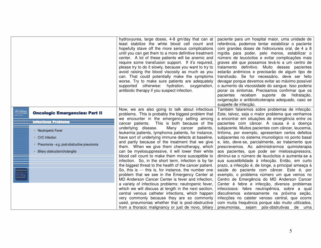
hydroxyurea, large doses, 4-8 gm/day that can at least stabilize the white blood cell count and hopefully stave off the more serious complications until you can get them to a more definitive treatment center. A lot of these patients will be anemic and require some transfusion support. If it’s required, please try to do it slowly, because you want to try to avoid raising the blood viscosity as much as you can. That could potentially make the symptoms worse. Try to make sure patients are adequately supported otherwise: hydration, oxygenation, antibiotic therapy if you suspect infection.
paciente para um hospital maior, uma unidade de referência, podemos tentar estabilizar o paciente com grandes doses de hidroxiureia oral, de 4 a 8 mg/dia para poder, pelo menos, estabilizar o número de leucócitos e evitar complicações mais graves até que possamos levá-lo a um centro de tratamento definitivo. Muito desses pacientes estarão anêmicos e precisarão de algum tipo de transfusão. Se for necessário, deve ser feito devagar porque devemos evitar ao máximo possível o aumento da viscosidade do sangue. Isso poderia piorar os sintomas. Precisamos confirmar que os pacientes recebam suporte de hidratação, oxigenação e antibioticoterapia adequado, caso se suspeite de infecção.
Oncologic Emergencies: Part IIOncologic Emergencies: Part IIOncologic Emergencies: Part IIOncologic Emergencies: Part II
Infectious ProblemsInfectious ProblemsInfectious ProblemsInfectious Problems
• Neutropenic Fever
• CVC Infection
• Pneumonia - e.g. post-obstructive pneumonia
• Biliary obstruction/cholangitis
Now, we are also going to talk about infectious problems. This is probably the biggest problem that we encounter in the emergency setting among cancer patients. This is both because of the underlying disease. Many cancer patients, leukemia patients, lymphoma patients, for instance, have sort of underlying immune defects at baseline, and partly because of the treatment that we give them. When we give them chemotherapy, which can be myelosuppressive, it will lower their white blood cell count to make them more susceptible to infection. So, in the short term, infection is by far the biggest threat to the health of the cancer patient. So, this is --- this is, for instance, the number one problem that we see in the Emergency Center at MD Anderson Cancer Center is fever and infection, a variety of infectious problems: neutropenic fever, which we will discuss at length in the next section, central venous catheter infections, which happen very commonly because they are so commonly used, pneumonias whether that is post-obstructive from a thoracic malignancy or just de novo, biliary
Também falaremos sobre problemas de infecção. Este, talvez, seja o maior problema que venhamos a encontrar em situações de emergência entre os pacientes com câncer. A causa é a doença subjacente. Muitos pacientes com câncer, leucemia, linfoma, por exemplo, apresentam certos defeitos subjacentes no sistema imunológico no ponto basal e, isto, deve-se, parcialmente, ao tratamento que prescrevemos. Ao administrarmos quimioterapia aos pacientes, que pode ser mielossupressora, diminui-se o número de leucócitos e aumenta-se a sua suscetibilidade à infecção. Então, em curto prazo, a infecção é, de longe, a principal ameaça à saúde do paciente com câncer. Este é, por exemplo, o problema número um que vemos no Centro de Emergência do MD Anderson Cancer Center é febre e infecção, diversos problemas infecciosos: febre neutropênica, sobre a qual discutiremos extensamente na próxima seção, infecções no cateter venoso central, que ocorre com muita frequência porque são muito utilizados, pneumonias, sejam pós-obstrutivas de uma
6
obstruction or cholangitis in patients with GI malignancies and every other kind of infectious complication that you can imagine happens in the cancer center.
neoplasia maligna torácica ou simplesmente "de novo", obstrução biliar ou colangite em pacientes com neoplasias malignas do TGI e todo tipo de complicações infecciosas que possam imaginar acontecem no centro de câncer.
Oncologic Emergencies: Part IIOncologic Emergencies: Part IIOncologic Emergencies: Part IIOncologic Emergencies: Part II
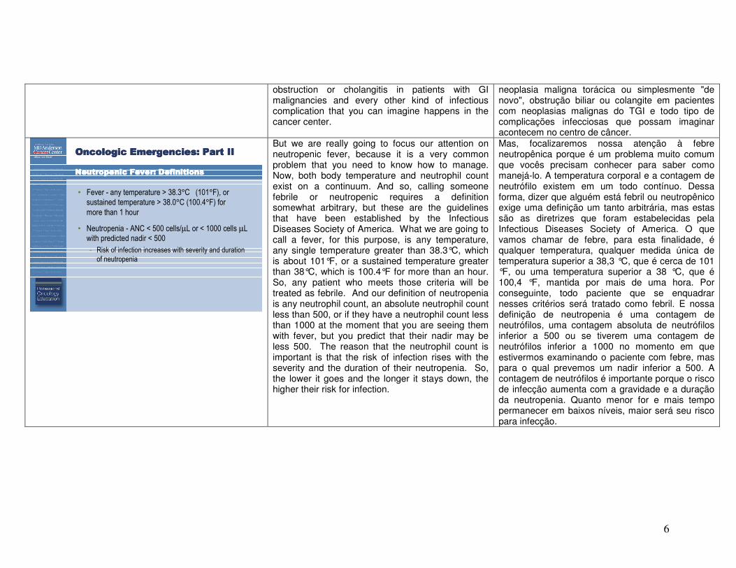
NeutropenicNeutropenicNeutropenicNeutropenic Fever: DefinitionsFever: DefinitionsFever: DefinitionsFever: Definitions
• Fever - any temperature > 38.3°C (101°F), or
sustained temperature > 38.0°C (100.4°F) for
more than 1 hour
• Neutropenia - ANC < 500 cells/µL or < 1000 cells µL
with predicted nadir < 500
- Risk of infection increases with severity and duration
of neutropenia
But we are really going to focus our attention on neutropenic fever, because it is a very common problem that you need to know how to manage. Now, both body temperature and neutrophil count exist on a continuum. And so, calling someone febrile or neutropenic requires a definition somewhat arbitrary, but these are the guidelines that have been established by the Infectious Diseases Society of America. What we are going to call a fever, for this purpose, is any temperature, any single temperature greater than 38.3°C, which is about 101°F, or a sustained temperature greater than 38°C, which is 100.4°F for more than an hour. So, any patient who meets those criteria will be treated as febrile. And our definition of neutropenia is any neutrophil count, an absolute neutrophil count less than 500, or if they have a neutrophil count less than 1000 at the moment that you are seeing them with fever, but you predict that their nadir may be less 500. The reason that the neutrophil count is important is that the risk of infection rises with the severity and the duration of their neutropenia. So, the lower it goes and the longer it stays down, the higher their risk for infection.
Mas, focalizaremos nossa atenção à febre neutropênica porque é um problema muito comum que vocês precisam conhecer para saber como manejá-lo. A temperatura corporal e a contagem de neutrófilo existem em um todo contínuo. Dessa forma, dizer que alguém está febril ou neutropênico exige uma definição um tanto arbitrária, mas estas são as diretrizes que foram estabelecidas pela Infectious Diseases Society of America. O que vamos chamar de febre, para esta finalidade, é qualquer temperatura, qualquer medida única de temperatura superior a 38,3 °C, que é cerca de 101 °F, ou uma temperatura superior a 38 °C, que é 100,4 °F, mantida por mais de uma hora. Por conseguinte, todo paciente que se enquadrar nesses critérios será tratado como febril. E nossa definição de neutropenia é uma contagem de neutrófilos, uma contagem absoluta de neutrófilos inferior a 500 ou se tiverem uma contagem de neutrófilos inferior a 1000 no momento em que estivermos examinando o paciente com febre, mas para o qual prevemos um nadir inferior a 500. A contagem de neutrófilos é importante porque o risco de infecção aumenta com a gravidade e a duração da neutropenia. Quanto menor for e mais tempo permanecer em baixos níveis, maior será seu risco para infecção.
7
Oncologic Emergencies: Part IIOncologic Emergencies: Part IIOncologic Emergencies: Part IIOncologic Emergencies: Part II
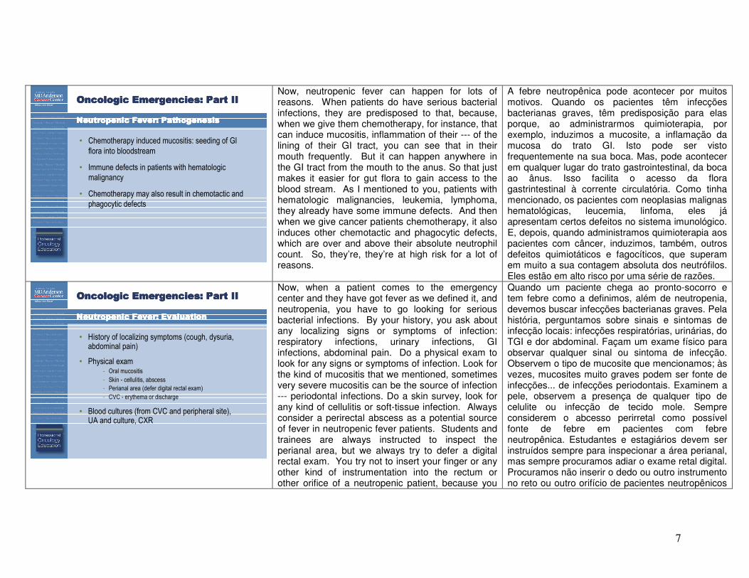
Neutropenic Fever: PathogenesisNeutropenic Fever: PathogenesisNeutropenic Fever: PathogenesisNeutropenic Fever: Pathogenesis
• Chemotherapy induced mucositis: seeding of GI
flora into bloodstream
• Immune defects in patients with hematologic
malignancy
• Chemotherapy may also result in chemotactic and
phagocytic defects
Now, neutropenic fever can happen for lots of reasons. When patients do have serious bacterial infections, they are predisposed to that, because, when we give them chemotherapy, for instance, that can induce mucositis, inflammation of their --- of the lining of their GI tract, you can see that in their mouth frequently. But it can happen anywhere in the GI tract from the mouth to the anus. So that just makes it easier for gut flora to gain access to the blood stream. As I mentioned to you, patients with hematologic malignancies, leukemia, lymphoma, they already have some immune defects. And then when we give cancer patients chemotherapy, it also induces other chemotactic and phagocytic defects, which are over and above their absolute neutrophil count. So, they’re, they’re at high risk for a lot of reasons.
A febre neutropênica pode acontecer por muitos motivos. Quando os pacientes têm infecções bacterianas graves, têm predisposição para elas porque, ao administrarmos quimioterapia, por exemplo, induzimos a mucosite, a inflamação da mucosa do trato GI. Isto pode ser visto frequentemente na sua boca. Mas, pode acontecer em qualquer lugar do trato gastrointestinal, da boca ao ânus. Isso facilita o acesso da flora gastrintestinal à corrente circulatória. Como tinha mencionado, os pacientes com neoplasias malignas hematológicas, leucemia, linfoma, eles já apresentam certos defeitos no sistema imunológico. E, depois, quando administramos quimioterapia aos pacientes com câncer, induzimos, também, outros defeitos quimiotáticos e fagocíticos, que superam em muito a sua contagem absoluta dos neutrófilos. Eles estão em alto risco por uma série de razões.
Oncologic Emergencies: Part IIOncologic Emergencies: Part IIOncologic Emergencies: Part IIOncologic Emergencies: Part II
NeutropenicNeutropenicNeutropenicNeutropenic Fever: EvaluationFever: EvaluationFever: EvaluationFever: Evaluation
• History of localizing symptoms (cough, dysuria, abdominal pain)
• Physical exam- Oral mucositis
- Skin - cellulitis, abscess
- Perianal area (defer digital rectal exam)
- CVC - erythema or discharge
• Blood cultures (from CVC and peripheral site), UA and culture, CXR
Now, when a patient comes to the emergency center and they have got fever as we defined it, and neutropenia, you have to go looking for serious bacterial infections. By your history, you ask about any localizing signs or symptoms of infection: respiratory infections, urinary infections, GI infections, abdominal pain. Do a physical exam to look for any signs or symptoms of infection. Look for the kind of mucositis that we mentioned, sometimes very severe mucositis can be the source of infection --- periodontal infections. Do a skin survey, look for any kind of cellulitis or soft-tissue infection. Always consider a perirectal abscess as a potential source of fever in neutropenic fever patients. Students and trainees are always instructed to inspect the perianal area, but we always try to defer a digital rectal exam. You try not to insert your finger or any other kind of instrumentation into the rectum or other orifice of a neutropenic patient, because you
Quando um paciente chega ao pronto-socorro e tem febre como a definimos, além de neutropenia, devemos buscar infecções bacterianas graves. Pela história, perguntamos sobre sinais e sintomas de infecção locais: infecções respiratórias, urinárias, do TGI e dor abdominal. Façam um exame físico para observar qualquer sinal ou sintoma de infecção. Observem o tipo de mucosite que mencionamos; às vezes, mucosites muito graves podem ser fonte de infecções... de infecções periodontais. Examinem a pele, observem a presença de qualquer tipo de celulite ou infecção de tecido mole. Sempre considerem o abcesso perirretal como possível fonte de febre em pacientes com febre neutropênica. Estudantes e estagiários devem ser instruídos sempre para inspecionar a área perianal, mas sempre procuramos adiar o exame retal digital. Procuramos não inserir o dedo ou outro instrumento no reto ou outro orifício de pacientes neutropênicos
8
can actually cause translocation of bacteria and serious infections. I will tell you, however, that, in real life, perirectal abscesses are not subtle. If you just ask the patient, do you have a lot of pain and swelling in that area, they will tell you, “Oh! Yes, Doctor.” Believe me they know it when they have got an abscess down there. If they have central venous catheters, be careful to inspect the area for exit site infections, erythema or discharge, and just ask, by history, if they have any history that sounds like a central venous catheter infection. A good history, for instance, would be, “You know, Doctor, every time my wife flushes the catheter, 30 minutes later I get these shaking chills and just cannot stop shaking, and my fever goes up”. That is a good story for a catheter-related infection. So, you will do your history, you will do your physical exam, and then you have to go looking for serious bacterial infection. You do blood cultures. Ideally, if the patient has a central venous catheter, you will obtain a culture from both the catheter itself and from a peripheral site. Do a urinalysis and culture. Do a chest x-ray even in the absence of any significant respiratory symptoms.
porque podemos causar a translocação de bactérias e infecções graves. No entanto, direi que, na vida real, os abcessos perirretais não são sutis. Se perguntarem ao paciente se sente muita dor e inchação nessa área, ele lhes dirá "Ah, sim, doutor!" Acreditem em mim. Eles sabem quando eles têm um abcesso lá embaixo. E se tiverem um cateter venoso central, tenham cuidado ao inspecionar a área para infecções nos locais de saída, eritema ou supuração, e simplesmente perguntem, pela história, se eles têm algum antecedente que se pareça a uma infecção no cateter venoso central. Uma boa história [clínica], por exemplo, seria: “Você sabe, doutor, toda vez que minha mulher limpa o cateter, 30 minutos depois começo a tremer de calafrios e não posso parar de tremer e a minha febre sobe.” Essa é uma boa história para uma infeção relacionada ao cateter. Vocês farão a história clínica, o exame físico e, depois, devem tentar descobrir uma infecção bacteriana grave. Vocês fazem cultivos de sangue. Se o paciente tiver um cateter venoso central, o ideal seria obter um cultivo do próprio local do cateter e de um local periférico. Façam uma urinálise e um cultivo. Façam uma radiografia torácica mesmo na ausência de quaisquer sintomas respiratórios significativos.
9
Oncologic Emergencies: Part IIOncologic Emergencies: Part IIOncologic Emergencies: Part IIOncologic Emergencies: Part II
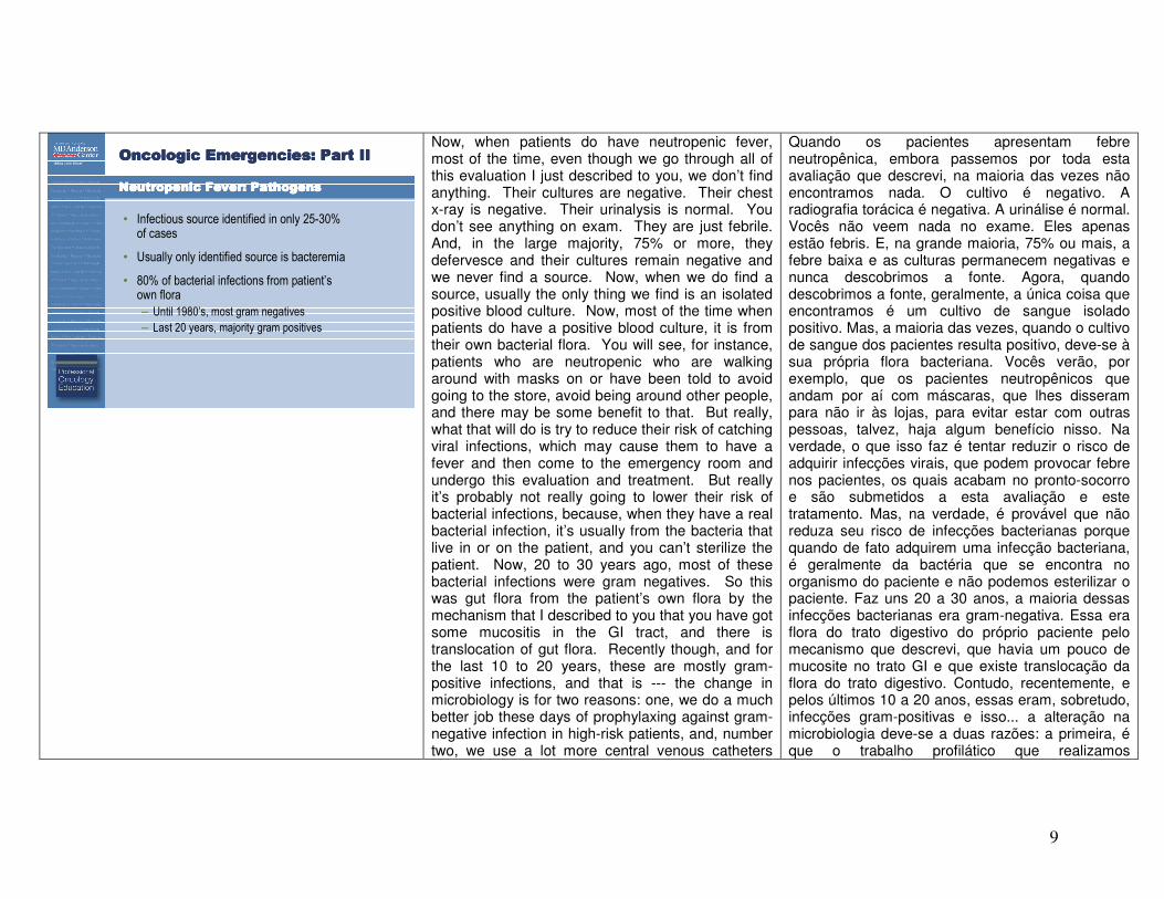
Neutropenic Fever: PathogensNeutropenic Fever: PathogensNeutropenic Fever: PathogensNeutropenic Fever: Pathogens
• Infectious source identified in only 25-30% of cases
• Usually only identified source is bacteremia
• 80% of bacterial infections from patient’s own flora
– Until 1980’s, most gram negatives
– Last 20 years, majority gram positives
Now, when patients do have neutropenic fever, most of the time, even though we go through all of this evaluation I just described to you, we don’t find anything. Their cultures are negative. Their chest x-ray is negative. Their urinalysis is normal. You don’t see anything on exam. They are just febrile. And, in the large majority, 75% or more, they defervesce and their cultures remain negative and we never find a source. Now, when we do find a source, usually the only thing we find is an isolated positive blood culture. Now, most of the time when patients do have a positive blood culture, it is from their own bacterial flora. You will see, for instance, patients who are neutropenic who are walking around with masks on or have been told to avoid going to the store, avoid being around other people, and there may be some benefit to that. But really, what that will do is try to reduce their risk of catching viral infections, which may cause them to have a fever and then come to the emergency room and undergo this evaluation and treatment. But really it’s probably not really going to lower their risk of bacterial infections, because, when they have a real bacterial infection, it’s usually from the bacteria that live in or on the patient, and you can’t sterilize the patient. Now, 20 to 30 years ago, most of these bacterial infections were gram negatives. So this was gut flora from the patient’s own flora by the mechanism that I described to you that you have got some mucositis in the GI tract, and there is translocation of gut flora. Recently though, and for the last 10 to 20 years, these are mostly gram-positive infections, and that is --- the change in microbiology is for two reasons: one, we do a much better job these days of prophylaxing against gram-negative infection in high-risk patients, and, number two, we use a lot more central venous catheters
Quando os pacientes apresentam febre neutropênica, embora passemos por toda esta avaliação que descrevi, na maioria das vezes não encontramos nada. O cultivo é negativo. A radiografia torácica é negativa. A urinálise é normal. Vocês não veem nada no exame. Eles apenas estão febris. E, na grande maioria, 75% ou mais, a febre baixa e as culturas permanecem negativas e nunca descobrimos a fonte. Agora, quando descobrimos a fonte, geralmente, a única coisa que encontramos é um cultivo de sangue isolado positivo. Mas, a maioria das vezes, quando o cultivo de sangue dos pacientes resulta positivo, deve-se à sua própria flora bacteriana. Vocês verão, por exemplo, que os pacientes neutropênicos que andam por aí com máscaras, que lhes disseram para não ir às lojas, para evitar estar com outras pessoas, talvez, haja algum benefício nisso. Na verdade, o que isso faz é tentar reduzir o risco de adquirir infecções virais, que podem provocar febre nos pacientes, os quais acabam no pronto-socorro e são submetidos a esta avaliação e este tratamento. Mas, na verdade, é provável que não reduza seu risco de infecções bacterianas porque quando de fato adquirem uma infecção bacteriana, é geralmente da bactéria que se encontra no organismo do paciente e não podemos esterilizar o paciente. Faz uns 20 a 30 anos, a maioria dessas infecções bacterianas era gram-negativa. Essa era flora do trato digestivo do próprio paciente pelo mecanismo que descrevi, que havia um pouco de mucosite no trato GI e que existe translocação da flora do trato digestivo. Contudo, recentemente, e pelos últimos 10 a 20 anos, essas eram, sobretudo, infecções gram-positivas e isso... a alteração na microbiologia deve-se a duas razões: a primeira, é que o trabalho profilático que realizamos
10
these days, and so, that is just a route for infection. Those are usually gram-positive infections.
atualmente contra infecções gram-negativas em pacientes de alto risco é muito melhor e a segunda é que, agora, usamos muito mais cateteres venosos centrais e esta é apenas uma rota de infecção. Geralmente, essas são infecções gram-positivas.
Oncologic Emergencies: Part IIOncologic Emergencies: Part IIOncologic Emergencies: Part IIOncologic Emergencies: Part II
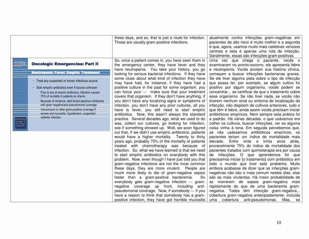
NeutropenicNeutropenicNeutropenicNeutropenic Fever: Empiric TreatmentFever: Empiric TreatmentFever: Empiric TreatmentFever: Empiric Treatment
• Treat any suspected or known infectious source
• Start empiric antibiotics even if source unknown
- Prior to era of empiric antibiotics, infection caused 75% of mortality in patients on chemo
- Because of virulence, start broad spectrum antibiotics
with gram negative/anti-pseudomonal coverage
- Vancomycin or other gram-positive coverage if
severe oral mucositis, hypotension, suspected
catheter infection
So, once a patient comes in, you have seen them in the emergency center, they have fever and they have neutropenia. You take your history, you go looking for serious bacterial infections. If they have some clues about what kind of infection they have may have had, for instance, if they have had a positive culture in the past for some organism, you can focus your --- make sure that your treatment covers that organism. If they don’t have anything, if you don’t have any localizing signs or symptoms of infection, you don’t have any prior cultures, all you have is fever, you still need to start empiric antibiotics. Now, this wasn’t always the standard practice. Several decades ago, what we used to do was, collect our cultures, go looking for infection, see if something showed up. Well, we soon figured out that, if we didn’t use empiric antibiotics, patients would have a higher mortality. Twenty to thirty years ago, probably 75% of the mortality of patients treated with chemotherapy was because of infection. So, what we have learned is that we need to start empiric antibiotics on everybody with this problem. Now, even though I have just told you that gram-negative infections are not the most common these days, they are more virulent. People are much more likely to die of gram-negative sepsis faster than a gram-positive bacteremia. So everybody gets gram-negative infection --- gram-negative coverage up front, including anti-pseudomonal coverage. Now, if somebody --- if you have a reason to think that somebody has a gram-positive infection, they have got horrible mucositis
Uma vez que chega o paciente, vocês o examinaram no pronto-socorro, ele apresenta febre e neutropenia. Vocês anotam sua história clínica, começam a buscar infecções bacterianas graves. Se ele tiver alguma pista sobre o tipo de infecção que possa ter, por exemplo, se algum cultivo foi positivo por algum organismo, vocês podem se concentrar... se certificar de que o tratamento cobre esse organismo. Se não tiver nada, se vocês não tiverem nenhum sinal ou sintoma de localização da infecção, não dispõem de cultivos anteriores, tudo o que têm é febre, ainda assim vocês precisam iniciar antibióticos empíricos. Nem sempre esta prática foi a padrão. Há várias décadas, o que usávamos era colher os cultivos, buscar infecções, ver se alguma coisa vinha à tona. Em seguida percebemos que, se não usássemos antibióticos empíricos, os pacientes teriam um índice de mortalidade mais elevado. Entre vinte e trinta anos atrás, provavelmente 75% do índice de mortalidade dos pacientes tratados com quimioterapia era por causa de infecções. O que aprendemos foi que precisamos iniciar [o tratamento] com antibiótico em todo o mundo que tiver este problema. Muito embora acabasse de dizer que as infecções gram-negativas não são o mais comum nestes dias, elas são as mais virulentas. Há maior probabilidade de as morrerem de sepsia gram-negativa mais rapidamente do que de uma bacteremia gram-negativa. Todos têm infecção gram-negativa... cobertura gram-negativa antecipadamente, incluída uma cobertura anti-pseudomonas. Mas, se
11
and most of the oral flora are gram positives, if you have got a reason to think they have a catheter-related infection, they told you a story like I suggested, some shaking chills after a catheter being flushed, something like that, then you would add gram-positive coverage. Or, if patients look septic, they are hypotensive or otherwise look bad, then you would, you would kind of throw the book at them. You would give them broad spectrum coverage, gram-negatives and gram-positives upfront.
alguém... se vocês tiverem alguma razão para pensar que uma pessoa tem uma infecção gram-positiva, eles apresentam uma mucosite terrível e a maior parte da flora é gram-positiva, se vocês tiverem um motivo para acreditar que há uma infecção relacionada ao cateter, ele lhes disse uma história como a que sugeri, com presença de calafrios depois de o cateter ser limpo, algo assim, depois, vocês incluiriam uma cobertura gram-positiva. Ou, se os pacientes parecerem sépticos, estiverem hipotensos ou tiverem parecerem doentes, vocês poderiam oferecer todas explicações possíveis. Vocês ofereceriam cobertura de amplo espectro, gram-negativas e gram-positivas já no início.
Oncologic Emergencies: Part IIOncologic Emergencies: Part IIOncologic Emergencies: Part IIOncologic Emergencies: Part II
NeutropenicNeutropenicNeutropenicNeutropenic Fever: Empiric TreatmentFever: Empiric TreatmentFever: Empiric TreatmentFever: Empiric Treatment
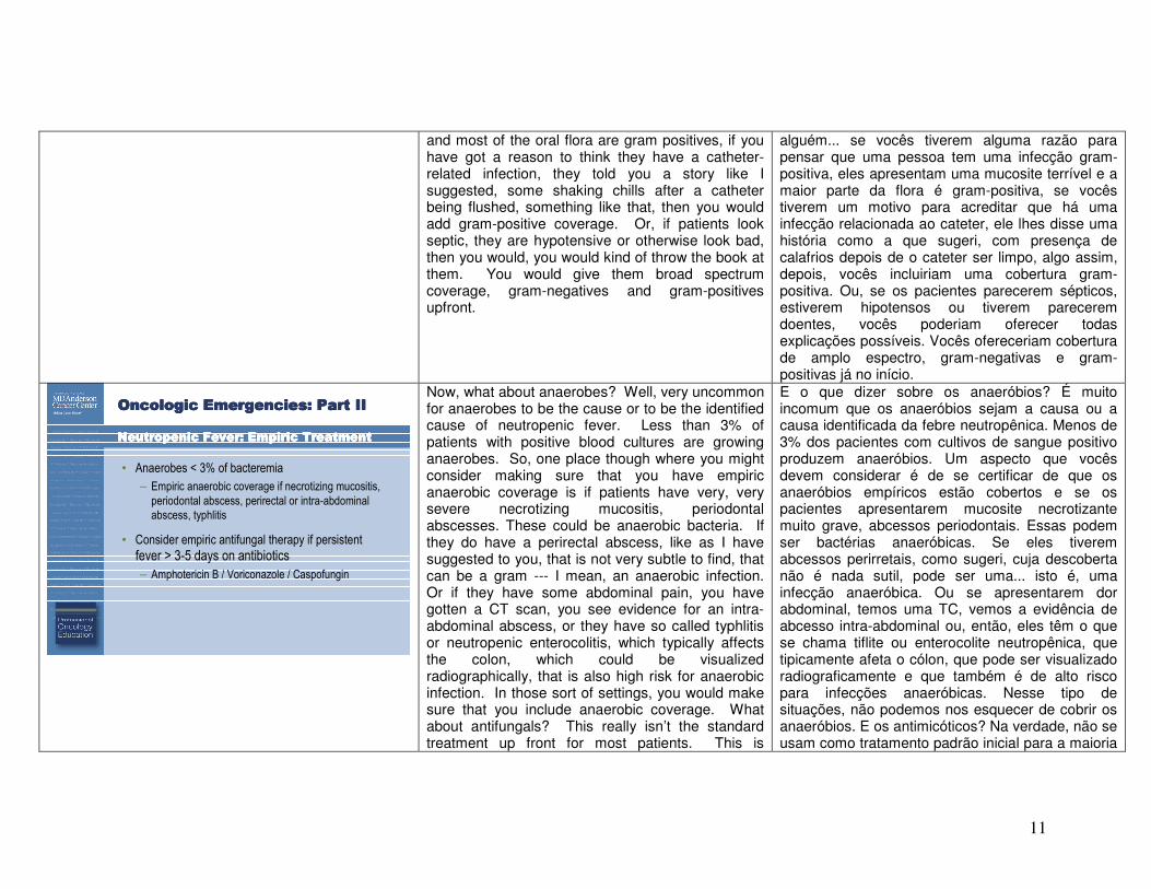
• Anaerobes < 3% of bacteremia
– Empiric anaerobic coverage if necrotizing mucositis,
periodontal abscess, perirectal or intra-abdominal
abscess, typhlitis
• Consider empiric antifungal therapy if persistent
fever > 3-5 days on antibiotics
– Amphotericin B / Voriconazole / Caspofungin
Now, what about anaerobes? Well, very uncommon for anaerobes to be the cause or to be the identified cause of neutropenic fever. Less than 3% of patients with positive blood cultures are growing anaerobes. So, one place though where you might consider making sure that you have empiric anaerobic coverage is if patients have very, very severe necrotizing mucositis, periodontal abscesses. These could be anaerobic bacteria. If they do have a perirectal abscess, like as I have suggested to you, that is not very subtle to find, that can be a gram --- I mean, an anaerobic infection. Or if they have some abdominal pain, you have gotten a CT scan, you see evidence for an intra-abdominal abscess, or they have so called typhlitis or neutropenic enterocolitis, which typically affects the colon, which could be visualized radiographically, that is also high risk for anaerobic infection. In those sort of settings, you would make sure that you include anaerobic coverage. What about antifungals? This really isn’t the standard treatment up front for most patients. This is
E o que dizer sobre os anaeróbios? É muito incomum que os anaeróbios sejam a causa ou a causa identificada da febre neutropênica. Menos de 3% dos pacientes com cultivos de sangue positivo produzem anaeróbios. Um aspecto que vocês devem considerar é de se certificar de que os anaeróbios empíricos estão cobertos e se os pacientes apresentarem mucosite necrotizante muito grave, abcessos periodontais. Essas podem ser bactérias anaeróbicas. Se eles tiverem abcessos perirretais, como sugeri, cuja descoberta não é nada sutil, pode ser uma... isto é, uma infecção anaeróbica. Ou se apresentarem dor abdominal, temos uma TC, vemos a evidência de abcesso intra-abdominal ou, então, eles têm o que se chama tiflite ou enterocolite neutropênica, que tipicamente afeta o cólon, que pode ser visualizado radiograficamente e que também é de alto risco para infecções anaeróbicas. Nesse tipo de situações, não podemos nos esquecer de cobrir os anaeróbios. E os antimicóticos? Na verdade, não se usam como tratamento padrão inicial para a maioria
12
frequently add-on therapy, if patients are persistently febrile for some period of time despite antibiotic --- antimicrobial coverage for bacteria. Sometimes you will suspect fungal infections in patients who are at high-risk, like leukemia patients who have some prior fungal infection or radiographic appearance suggestive of fungal pneumonia like a nodule or infiltrate, something like that.
dos pacientes. Geralmente, se usam como terapia suplementar, caso os pacientes continuem com febre por algum tempo apesar de receberem antibióticos.... antimicrobianos para combater as bactérias. Às vezes, suspeitamos de infecções fúngicas em pacientes de alto risco, como pacientes com leucemia, que já haviam tido infecções fúngicas ou aparência radiográfica sugestiva de pneumonia fúngica, como um nódulo ou infiltrado, algo similar.
Oncologic Emergencies: Part IIOncologic Emergencies: Part IIOncologic Emergencies: Part IIOncologic Emergencies: Part II
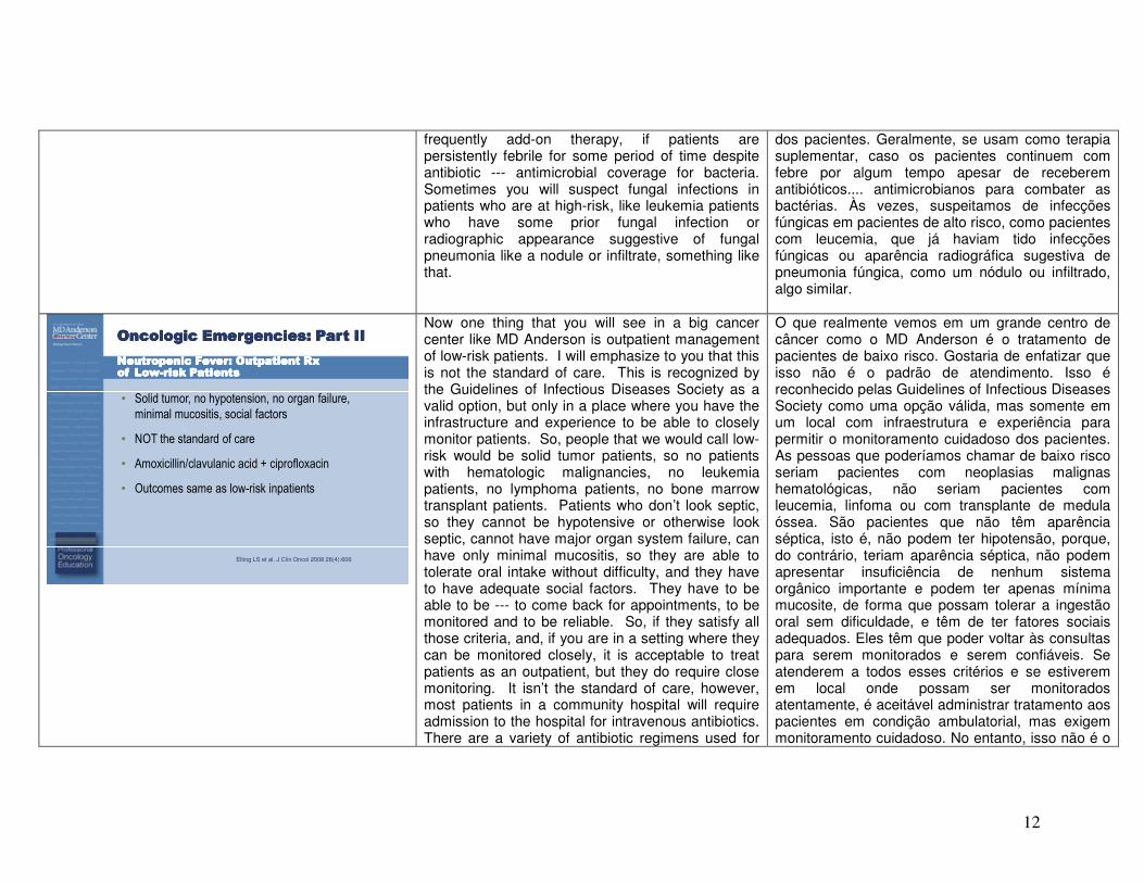
Neutropenic Fever: Outpatient Rx Neutropenic Fever: Outpatient Rx Neutropenic Fever: Outpatient Rx Neutropenic Fever: Outpatient Rx of Lowof Lowof Lowof Low----risk Patientsrisk Patientsrisk Patientsrisk Patients
• Solid tumor, no hypotension, no organ failure,
minimal mucositis, social factors
• NOT the standard of care
• Amoxicillin/clavulanic acid + ciprofloxacin
• Outcomes same as low-risk inpatients
Elting LS et al. J Clin Oncol 2008 26(4):606
Now one thing that you will see in a big cancer center like MD Anderson is outpatient management of low-risk patients. I will emphasize to you that this is not the standard of care. This is recognized by the Guidelines of Infectious Diseases Society as a valid option, but only in a place where you have the infrastructure and experience to be able to closely monitor patients. So, people that we would call low-risk would be solid tumor patients, so no patients with hematologic malignancies, no leukemia patients, no lymphoma patients, no bone marrow transplant patients. Patients who don’t look septic, so they cannot be hypotensive or otherwise look septic, cannot have major organ system failure, can have only minimal mucositis, so they are able to tolerate oral intake without difficulty, and they have to have adequate social factors. They have to be able to be --- to come back for appointments, to be monitored and to be reliable. So, if they satisfy all those criteria, and, if you are in a setting where they can be monitored closely, it is acceptable to treat patients as an outpatient, but they do require close monitoring. It isn’t the standard of care, however, most patients in a community hospital will require admission to the hospital for intravenous antibiotics. There are a variety of antibiotic regimens used for
O que realmente vemos em um grande centro de câncer como o MD Anderson é o tratamento de pacientes de baixo risco. Gostaria de enfatizar que isso não é o padrão de atendimento. Isso é reconhecido pelas Guidelines of Infectious Diseases Society como uma opção válida, mas somente em um local com infraestrutura e experiência para permitir o monitoramento cuidadoso dos pacientes. As pessoas que poderíamos chamar de baixo risco seriam pacientes com neoplasias malignas hematológicas, não seriam pacientes com leucemia, linfoma ou com transplante de medula óssea. São pacientes que não têm aparência séptica, isto é, não podem ter hipotensão, porque, do contrário, teriam aparência séptica, não podem apresentar insuficiência de nenhum sistema orgânico importante e podem ter apenas mínima mucosite, de forma que possam tolerar a ingestão oral sem dificuldade, e têm de ter fatores sociais adequados. Eles têm que poder voltar às consultas para serem monitorados e serem confiáveis. Se atenderem a todos esses critérios e se estiverem em local onde possam ser monitorados atentamente, é aceitável administrar tratamento aos pacientes em condição ambulatorial, mas exigem monitoramento cuidadoso. No entanto, isso não é o
13
outpatient management. Augmentin® and Ciprofloxacin® are probably the most commonly used and the ones for which we have the longest track record. All the regimens really are based on quinolones, so high-dose Levaquin® and other quinolones can be used as well, but I will refer you to the Infectious Diseases Society Guidelines for other treatment regimens. And, in general, if patients do qualify as low-risk, the outcomes are the same if they are managed as an outpatient or as an inpatient.
atendimento padrão. A maioria dos pacientes em hospitais comunitários precisarão ser ingressados para [receber] antibióticos por via intravenosa. Há diversos esquemas antimicrobianos utilizados no manejo do paciente ambulatorial. O Augmentin® e o Ciprofloxacin® talvez sejam os mais utilizados e dos quais dispomos o maior número de informações. Todos os esquemas terapêuticos são baseados em quinolonas. Então, o Levaquin® e outras quinolonas em altas doses podem ser utilizadas, mas recomendaria que consultassem as Infectious Diseases Society Guidelines para se informarem sobre outros esquemas terapêuticos. E, em geral, se os pacientes atenderem à condição de baixo risco, os resultados serão os mesmos independentemente de os pacientes serem ambulatoriais ou internados.
Oncologic Emergencies: Part IIOncologic Emergencies: Part IIOncologic Emergencies: Part IIOncologic Emergencies: Part II
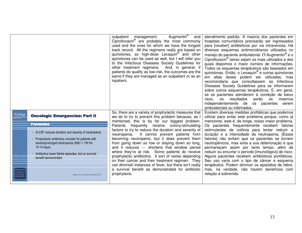
PreventionPreventionPreventionPrevention
• G-CSF reduces duration and severity of neutropenia
• Prophylactic antibiotics consider for patients with
severe/prolonged neutropenia (ANC < 100 for
10-14 days)
• Antibiotics lower febrile episodes, but no survival
benefit demonstrated
Rolston KV. Annu Rev Med 2004 55:519
So, there are a variety of prophylactic measures that we do to try to prevent this problem because, as I mentioned, this is by far our biggest problem. Patients frequently receive colony-stimulating factors to try to reduce the duration and severity of neutropenia. It cannot prevent patients from becoming neutropenic, but it does prevent them from going down so low or staying down so long, and it reduces --- shortens that window period where they’re at risk. Some patients do receive prophylactic antibiotics. It sort of varies depending on their cancer and their treatment regimen. They can diminish instances of fever, but there isn’t really a survival benefit as demonstrated for antibiotic prophylaxis.
Existem diversas medidas profiláticas que podemos utilizar para evitar este problema porque, como já mencionei, este é, de longe, nosso maior problema. Os pacientes frequentemente recebem fatores estimulantes de colônia para tentar reduzir a duração e a intensidade da neutropenia. [Esses fatores] não evitam que os pacientes se tornem neutropênicos, mas evita a sua deterioração e que permaneçam assim por tanto tempo, além de reduzir ou encurtar o período [imunológico] de risco. Alguns pacientes recebem antibióticos profiláticos. Seu uso varia com o tipo de câncer e esquema terapêutico. Podem diminuir os episódios de febre, mas, na verdade, não trazem benefícios com relação à sobrevida.

14
Oncologic Emergencies: Part IIOncologic Emergencies: Part IIOncologic Emergencies: Part IIOncologic Emergencies: Part II
SummarySummarySummarySummary
• Remember that not all emergencies in cancer
patients are related to the underlying malignancy
or its treatment
• Signs and symptoms may be blunted – have a
high pre-test probability
• Prompt recognition and management of these
conditions are essential to preventing morbidity
and mortality
So, in summary, I will remind you that not all emergency problems in cancer patients are related to the underlying malignancy and its treatment. Remember to keep in mind all the same kind of non-cancer emergency problems that other patients can get. I want you to remember that the signs and symptoms of disease may be blunted or masked in cancer patients for a variety of reasons, which we have discussed in the first lecture. So have a high pre-test probability for whatever problem it is that you are considering and a lower threshold for ordering whatever test is required to try to figure that out. And I want you to remember that --- to try to recognize and respond to these kinds of cancer --- these kinds of emergency problems promptly, because that is essential to try to lowering the morbidity and mortality that we see among cancer patients. And I thank you for your attention.
Resumindo, gostaria de lembrá-los que nem todos os problemas de emergência em pacientes com câncer são relacionados à neoplasia maligna subjacente e a seu tratamento. Procurem lembrar todos os mesmos tipos de problemas emergenciais não relacionados ao câncer que outros pacientes possam ter. Gostaria que lembrassem que os sinais e sintomas das doenças podem estar enfraquecidas ou mascaradas nos pacientes com câncer por diversas razões, o que discutimos na primeira palestra. Façam um teste de alta probabilidade para o problema que estiverem considerando e um baixo limiar para pedir qualquer teste que seja necessário para tentar descobrir o problema. E gostaria que lembrassem que... tentem reconhecer e responder a estes tipos de câncer, estes tipos de problemas emergenciais imediatamente, porque é essencial procurar a redução da morbidade e a mortalidade que observamos nos pacientes com câncer. Obrigado pela atenção.