Oncologic Emergencies and Paraneoplastic Syndromes · hypocalcemia hyperuricemia ......
64
Oncologic Emergencies and Paraneoplastic Syndromes Internal Medicine Board Review Amy M. Weise D.O. Assistant Professor of Medicine
Transcript of Oncologic Emergencies and Paraneoplastic Syndromes · hypocalcemia hyperuricemia ......

Oncologic Emergencies
and Paraneoplastic
SyndromesInternal Medicine Board Review
Amy M. Weise D.O.
Assistant Professor of Medicine

Oncologic Emergencies
Why should I learn this?
Cancer therapy is largely outpatient, so these patients
are likely to walk into your office.
Treatment should not be delayed while attempting to
contact the oncologist.
May be the presenting symptom
Board Exams

Oncologic Emergencies
Definition: Acute condition caused by cancer or
the treatment of cancer where early recognition
and intervention would result in the avoidance
of mortality or permanent morbidity.
Oncologic Emergencies
Initial Evaluation:
Brief focused history
Focused Exam
Lab/radiograph review
Initial Intervention and Additional Studies
Then comprehensive history and exam

Oncologic Emergencies
Categories:
Metabolic
Hematologic
Structural
Treatment related

Metabolic: Tumor Lysis Syndrome
Definition: acute lysis of tumor cells as a result
of chemotherapy or radiation therapy, usually
within 72 hours of therapy
Tumor Types:
Leukemia
High-grade lymphoma
SCLC

Metabolic: Tumor Lysis Syndrome
Features:
azotemia
acidosis
hyperphosphatemia
hyperkalemia
hypocalcemia
hyperuricemia
ARF
Symptoms:
Nausea
Vomiting
Weakness
Fatigue
Neuromuscular irritability
Arrhythmia

Metabolic: Tumor Lysis Syndrome
Tx/Prevention:
fluids
allopurinol
rasburicase
urine alkalinization
HD
kayexalate
phos-lo
Metabolic: Hypercalcemia
Seen in 20 to 30% of cancer patients
Rate of calcium mobilization from bone exceeds
renal calcium excretion
Tumor Types:
Multiple myeloma
Squamous Cell: lung, H&N, esophagus
Breast
Kidney
Metabolic: Hypercalcemia
Mechanisms:
Bone Mets- osteoclast activity
Parathyroid hormone-related peptide (binds PTH
receptors)
Calcitriol production- lymphoma
Co-existing Primary Hyperparathyroidism
Osteoclast activating factor (OAF)- Multiple
Myeloma
Metabolic: Hypercalcemia
Treatment:
Calcitonin — increases renal calcium excretion and
by decreasing bone reabsorption
Bisphosphonates — inhibit calcium release from
bone by interfering with the metabolic activity of
osteoclasts, and is cytotoxic to osteoclasts.
(Pamidronate, zoledronic acid, ibandronate and
etidronate)

Metabolic: Hypercalcemia
Treatment
Glucocorticoids (eg, prednisone in a dose of 20 to 40 mg/day) will usually reduce serum calcium concentrations within two to five days by decreasing calcitriol production and OAF
Volume expansion with isotonic saline
Loop diuretic will also contribute to increased calcium excretion by inhibiting calcium reabsorption in the loop of Henle
Hemodialysis
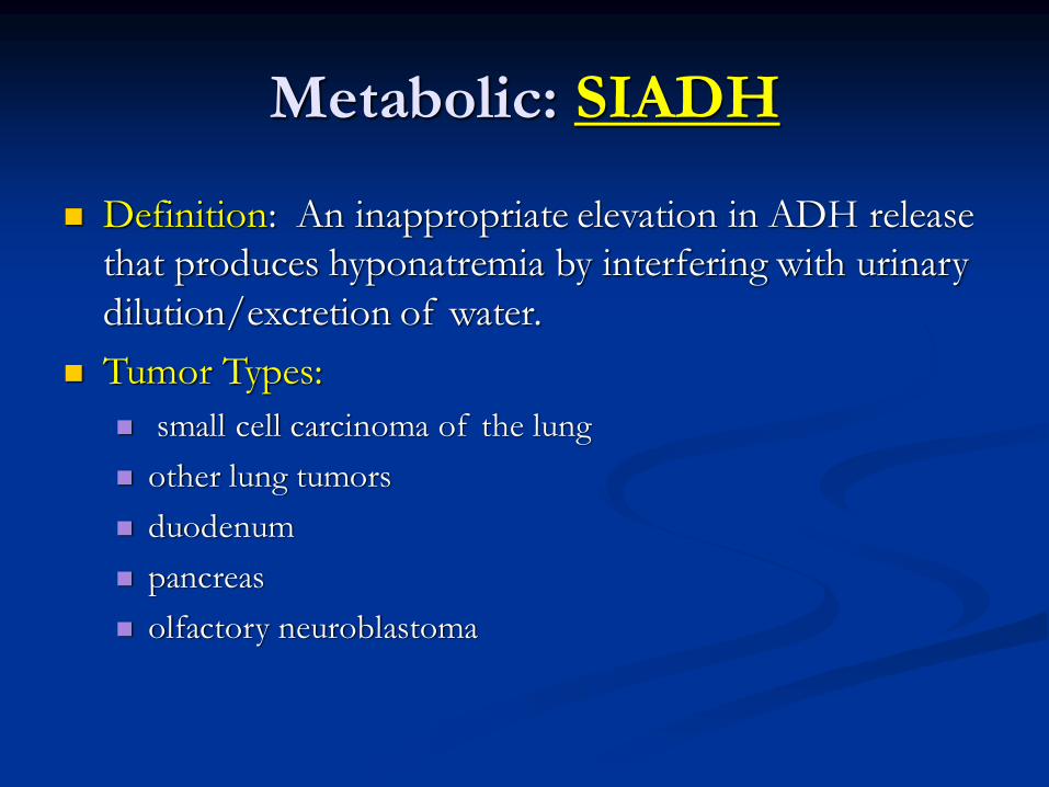
Metabolic: SIADH
Definition: An inappropriate elevation in ADH release
that produces hyponatremia by interfering with urinary
dilution/excretion of water.
Tumor Types:
small cell carcinoma of the lung
other lung tumors
duodenum
pancreas
olfactory neuroblastoma
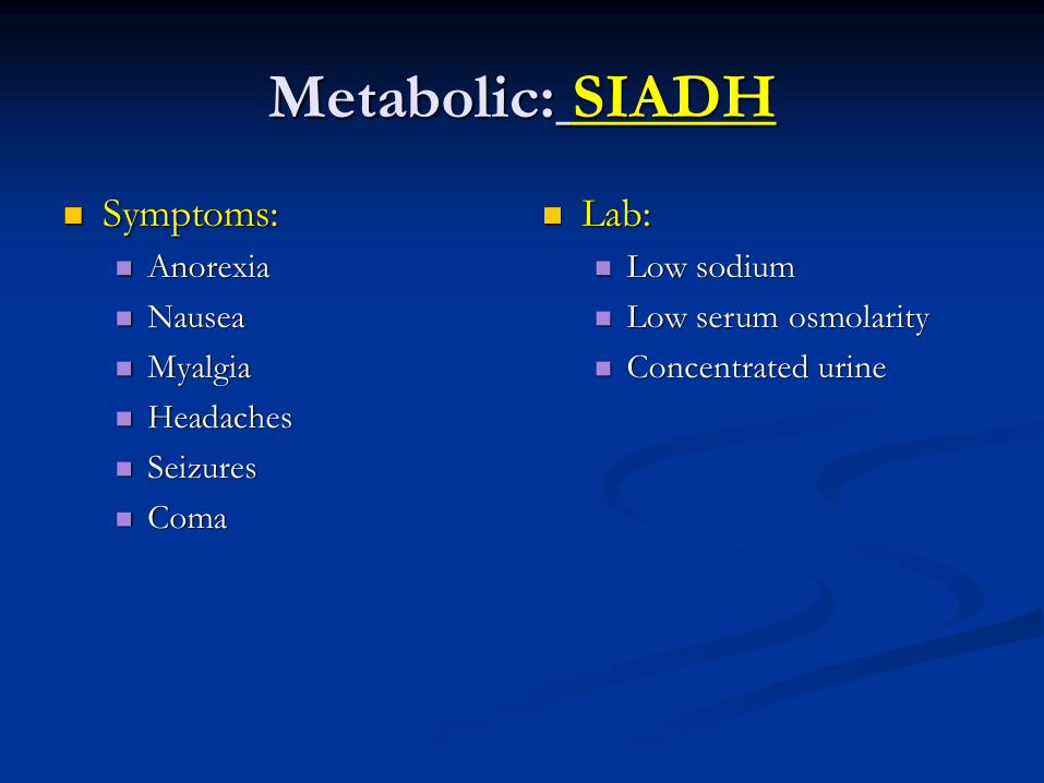
Metabolic: SIADH
Symptoms:
Anorexia
Nausea
Myalgia
Headaches
Seizures
Coma
Lab:
Low sodium
Low serum osmolarity
Concentrated urine

Metabolic: SIADH
Treatment:
Water restriction is the mainstay of therapy in
asymptomatic hyponatremia
Hypertonic saline (or salt tablets)
Loop Diuretic- lowers the urine osmolality and
increases water excretion by impairing the renal
responsiveness to ADH
Demeclocycline and lithium act on the collecting
tubule cell to diminish its responsiveness to ADH
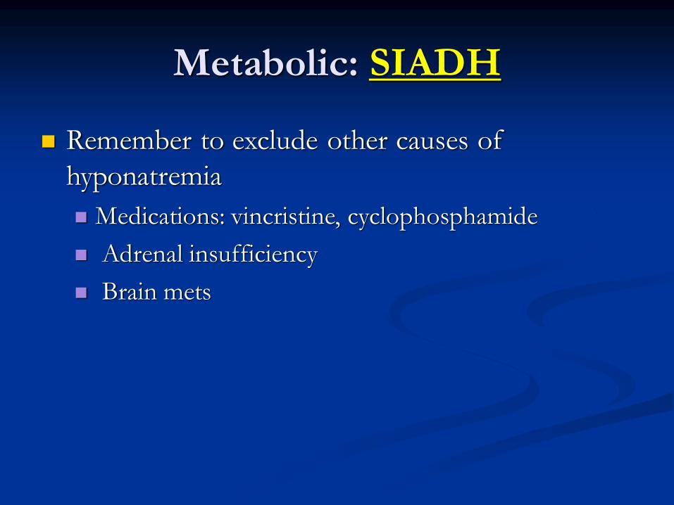
Metabolic: SIADH
Remember to exclude other causes of
hyponatremia
Medications: vincristine, cyclophosphamide
Adrenal insufficiency
Brain mets

Hematologic: Febrile Neutropenia
Neutropenia and Risk:
Risk of an occult infection increases with an ANC
<1,000 cells/microL
It is substantially higher for those with an ANC
<500 cells/microL
And is highest for those with an ANC <100
cells/microL
A rapid decline in ANC
Prolonged duration of neutropenia (>7 to 10 days)

Hematologic: Febrile Neutropenia
Defined as a single temperature of >38.3ºC
(101.3ºF), or a sustained temperature >38ºC
(100.4ºF) for more than one hour
Patients may present with hypothermia,
hypotension, or clinical deterioration as the
initial signs of occult infection.
Consequently there should be a low threshold
for starting empiric antibiotics

Hematologic: Febrile Neutropenia
Most common pathogens:
S. aureus
S. epi
Streptococcal species
Others: Candida albicans, aspergillus, Fusarium,
viral
Most often the source goes unidentified
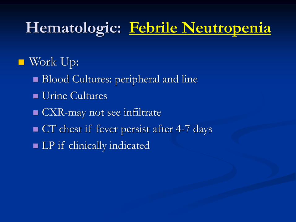
Hematologic: Febrile Neutropenia
Work Up:
Blood Cultures: peripheral and line
Urine Cultures
CXR-may not see infiltrate
CT chest if fever persist after 4-7 days
LP if clinically indicated

Hematologic: Febrile Neutropenia
Monotherapy
Beta-lactam carbapenem- meropenem, imipenem,
piperacillin tazobactam
4th generation cephalosporin-ceftazidime, cefepime
Dual Therapy
Aminoglycoside Plus Beta-lactam
Fluoroquinolone Plus Beta-lactam

Hematologic: Febrile Neutropenia
Vancomycin- if line associated, cellulitis, severe
mucositis, hypotension, history of MRSA
colonization, or recent quinolone prophylaxis,
patients with clinical deterioration or persistent
fever despite empiric antibiotics.
Anti-fungal- fever persists after 4-7 days

Hematologic: Febrile Neutropenia
70% mortality rate if
initiation of
antibiotics is delayed
Hematologic: Hyperviscosity
Syndrome
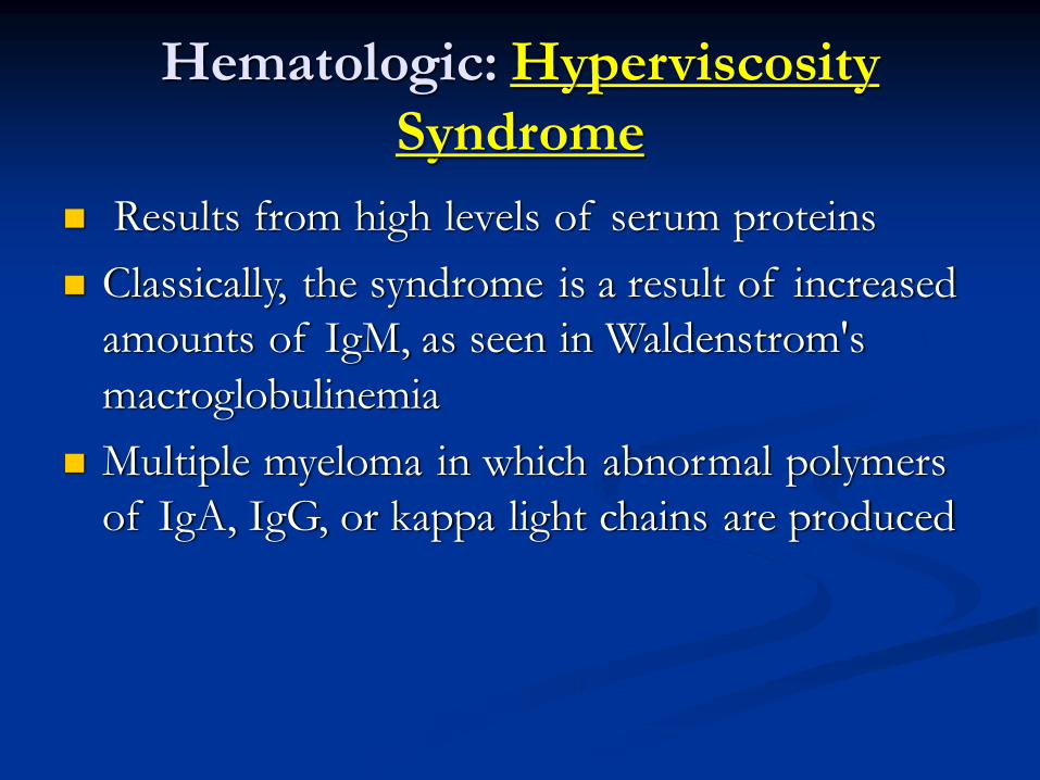
Results from high levels of serum proteins
Classically, the syndrome is a result of increased
amounts of IgM, as seen in Waldenstrom's
macroglobulinemia
Multiple myeloma in which abnormal polymers
of IgA, IgG, or kappa light chains are produced
Hematologic: Hyperviscosity
Syndrome
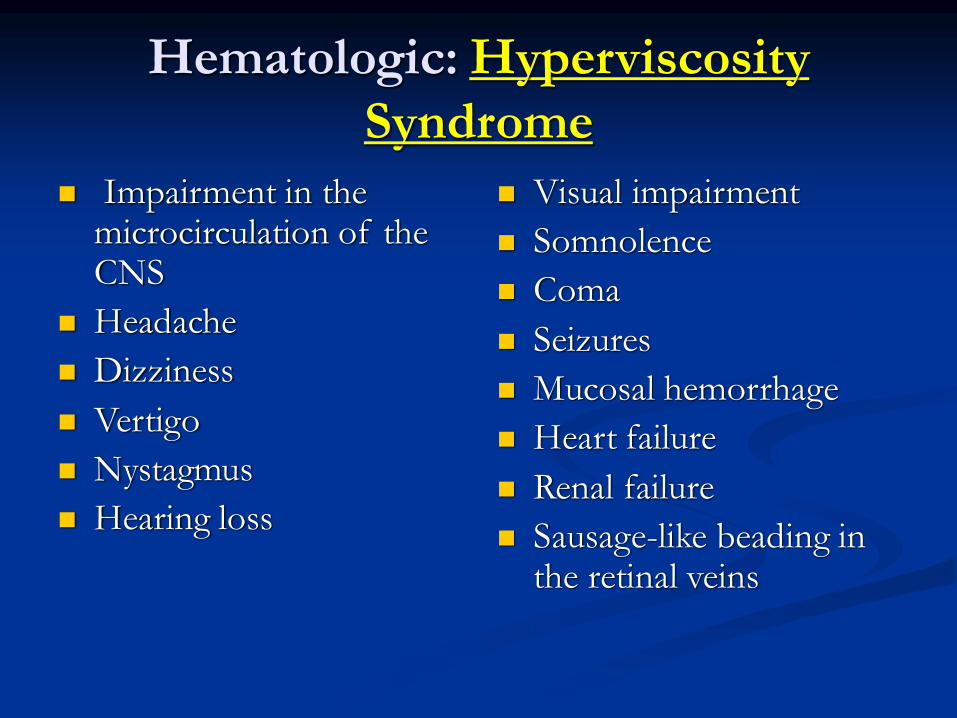
Impairment in the microcirculation of the CNS
Headache
Dizziness
Vertigo
Nystagmus
Hearing loss
Visual impairment
Somnolence
Coma
Seizures
Mucosal hemorrhage
Heart failure
Renal failure
Sausage-like beading in the retinal veins

Hematologic: Hyperviscosity
Syndrome

Hematologic: Hyperviscosity
Syndrome
Severe neurologic impairment, such as stupor
or coma, should be treated with plasma
exchange on an emergency basis
Chemotherapy for the underlying cause
AVOID pRBC TRANSFUSIONS
Structural: SVC Syndrome
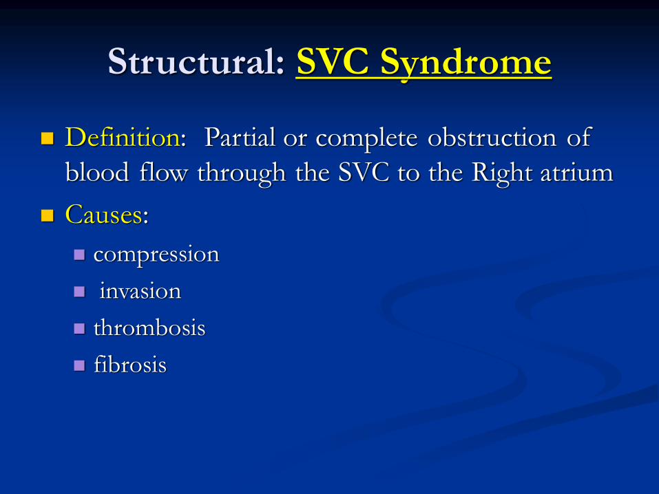
Definition: Partial or complete obstruction of
blood flow through the SVC to the Right atrium
Causes:
compression
invasion
thrombosis
fibrosis

Structural: SVC Syndrome
Tumor Types:
Lung
NHL
Thymoma
Germ cell
Breast

Structural: SVC Syndrome
Signs:
JVD
Venous dilation of chest
wall
Facial edema
Upper extremity edema
Papilledema if onset is
rapid
Symptoms
Head fullness or pressure
Cough
Dyspnea
Chest pain
dysphagia

Structural: SVC Syndrome
Treatment:
SVC stent
XRT
Chemotherapy
Anti-coagulants or thrombolytics
Pearls:
Secure an accurate diagnosis with biopsy
Oxygen
Elevate HOB
Diuretics sparingly if at all
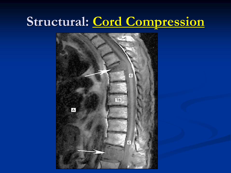
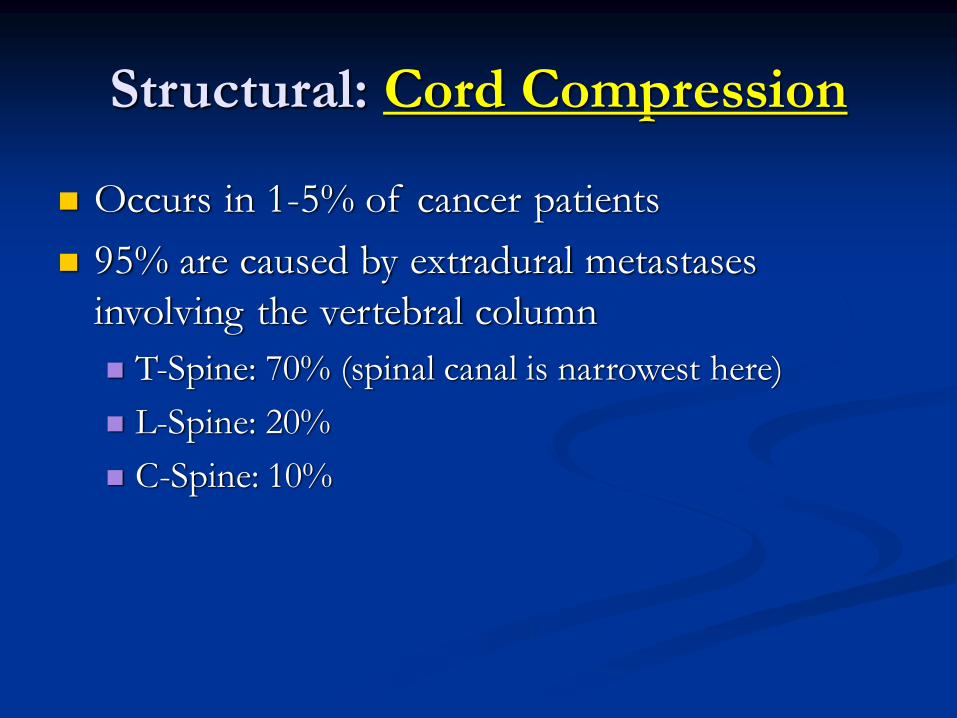
Structural: Cord Compression
Structural: Cord Compression
Occurs in 1-5% of cancer patients
95% are caused by extradural metastases
involving the vertebral column
T-Spine: 70% (spinal canal is narrowest here)
L-Spine: 20%
C-Spine: 10%

Structural: Cord Compression
Tumor Types:
Lung
Breast
Unknown primary
Prostate
Renal

Structural: Cord Compression
Symptoms:
Pain, Pain, Pain- localized or radicular, worse with
movement, cough, sneeze
Urinary retention/incontinence (late)
Constipation (late)
Poor prognostic features: paralysis or urinary
retention

Structural: Cord Compression
Dx: MRI
Treatment:
Narcotics
Dexamethasone: dose is controversial
Initial: 20-100mg IV bolus
Maintenance: 6-16mg q 6 hours
XRT
Neurosurgery- spine stabilization

Structural: Cord Compression
Permanent neurologic impairment can result if
treatment is delayed by only a few hours
Major quality of life issue that might be
preventable


Paraneoplastic Syndromes
Paraneoplastic neurologic syndromes are a
heterogeneous group of neurologic disorders
caused by mechanisms other than metastases,
metabolic and nutritional deficits, infections,
coagulopathy or side effects of cancer
treatment.
These syndromes may affect any part of the
nervous system from cerebral cortex to
neuromuscular junction and muscle.

Paraneoplastic Syndromes
The more common syndromes
Lambert-Eaton myasthenic syndrome affects 3% of
patients with small-cell lung cancer
Myasthenia Gravis affects 15% of all patients with
thymoma
<1% incidence for other solid tumors
Paraneoplastic Syndromes
Review Article: “Paraneoplastic neurological
syndromes” in Orphanet Journal of Rare
Diseases May 2007, 2:22
Occur in < 0.01% of cancer patients
Paraneoplastic Syndromes
Pathogenesis:
Onconeural antibodies: antibodies directed at tumor
antigens that cross-react with components of the
nervous system
50% of patients with PNS do not have detectable
antibodies
Antibodies may be present in the absence of a PNS

Paraneoplastic Syndromes
Definite PNS Criteria:
Classical neuro syndrome and cancer develops within 5 years of the neuro disorder
Non-classical neuro syndrome improves after treatment of underlying cancer
Non-classical neuro syndrome with onconeural antibodies AND cancer develops within 5 years
Neuro syndrome with well-characterized onconeural antibodies (Hu, Yo, CV2, Ri, Ma2, anphiphysin) AND no cancer

Paraneoplastic Syndromes
Possible Criteria:
Classic neuro syndrome, No Ab, No cancer, but is at
high risk for an underlying tumor
Neuro syndrome, partially characterized Ab, No
cancer
Non-classical neuro syndrome, No Ab, cancer
diagnosis within 2 years

Paraneoplastic Syndromes
Anti-Hu (antineuronal nuclear antibody ANNA-1)
Encephalomyelitis- asymmetric paresthesias SCLC
Limbic encephalitis- memory and mood
Anti-Yo: Cerebellar degeneration- dysarthria
and ataxia
Anti-Tr: Cerebellar degeneration (Hodgkin’s)

Paraneoplastic Syndromes
Anti-Ta: Limbic encephalitis (testicular/breast)
Anti-amphiphysin: Stiff-person syndrome
Anti-GAD: Stiff-person syndrome (breast)
LEMS Ab: Lambert-Eaton, SCLC
PNS: Lambert-Eaton myasthenic
syndrome
Disorder of reduced acetylcholine (ACh) release
from the presynaptic nerve terminals
Antibodies directed against the voltage-gated
calcium channel (VGCC) interfere with the
normal calcium flux required for the release of
acetylcholine

PNS: Lambert-Eaton myasthenic
syndrome
Symptoms
slowly progressive
proximal muscle
weakness, particularly
involving the legs
Deep tendon reflexes are
typically depressed or
absent
Ptosis and diplopia
Dry mouth
Erectile Dysfunction
Respiratory failure may
occur late in the course.
Hallmark: Recovery of lost deep tendon reflexes or improvement
in muscle strength with vigorous, brief muscle activation

PNS: Lambert-Eaton myasthenic
syndrome
Diagnosis:
Post-exercise facilitation: Maximal isometric
contraction the limb for 10 to 15 seconds may lead
to temporary reappearance of previously depressed
or absent DTR’s, and temporary improvement of
muscle weakness.
Ab to voltage-gated calcium channel (VGCC)
Electrodiagnostic studies-EMG

PNS: Lambert-Eaton myasthenic
syndrome
Occurs much less frequently than myasthenia
gravis
Approximately one-half of LEMS cases are
associated with a malignancy, usually SCLC
PNS: Myasthenia gravis
Mediated by autoantibodies against the
acetylcholine receptor (AChR-Ab)
The majority of patients AChR-Ab positive
myasthenia gravis have thymic abnormalities:
hyperplasia in 60 to 70% and thymoma in 10 to
12%
PNS: Myasthenia gravis
Symptoms
fluctuating skeletal muscle weakness, worse later in the day
or evening
fatigue- meaning worsening contractile force of the muscle
50% of patients present with ocular symptoms of ptosis
and/or diplopia
15% present with bulbar symptoms (dysarthria, dysphagia,
and fatigable chewing)
Less than 5% present with proximal limb weakness alone
Respiratory insufficiency

PNS: Myasthenia gravis
Diagnosis
Tensilon test- Edrophonium chloride is an
acetylcholinesterase inhibitor prolongs the presence
of Ach in the neuromuscular junction and results in
an immediate increase in muscle strength
Serologic tests for autoantibodies
Electrophysiological studies

PNS: Myasthenia gravis
Treatments:
Symptomatic treatments (anticholinesterase agents)
Chronic immunomodulating treatments
(corticosteroids and other immunosuppressive
drugs)
Rapid immunomodulating treatments (plasma
exchange and intravenous immune globulin)
Surgical treatment (thymectomy)

Others
Carcinoid Syndrome
Carcinoid tumors of foregut derivatives (small
intestine, stomach, bronchus, pancreas, thyroid)
Overproduce 5-HT (5-hydroxytryptamine)
serotonin, histamine, tachykinins, kallikrein and
prostaglandins
Urine excretion of 5-HIAA

Carcinoid Syndrome
Symptoms:
Episodic flushing
Fall in blood pressure and rise in pulse rate
Secretory diarrhea
Wheezing and dyspnea
Carcinoid heart disease- right side

Carcinoid Syndrome
Treatment:
Somatostatin analog octreotide
Cyproheptadine, a serotonin antagonist
Chemotherapy effectiveness is questionable
Isolated lesions: cryo, RFA (treat with octreotide
prior to anesthesia to prevent crisis)
Others
Zollinger-Ellison Syndrome- excessive gastrin
secretion from a gastrinoma
Develop peptic ulcers, most are in the first
portion of the duodenum
Diarrhea can also be a prominent feature

ZES
Diagnosis:
Fasting serum gastrin concentration
Secretin stimulation test
Gastric acid secretion studies

50 yo female, presents to
ER c/o falling, difficulty
arising from a chair.
Weakness improved
during the day. Also c/o
dyspnea on exertion. 45
pack years.
Proximal muscle
weakness on exam
What is the most
appropriate
management?
A. Follow up with PCP
B. Physical Therapy
C. Admit for EMG and
possible treatment
D. Smoking cessation
classes
Questions obtained from The John Hopkins Internal Medicine Board Review 2004

56yo female presents to
ER c/o epistaxis, blurry
vision, confusion.
PMHx: HTN
Vs all normal, blood at
nares and gums, bibasilar
crackles, no edema
Labs: Hgb-8, plts-350,
WBC-8.0, normal diff.,
T. ptn 10.2, albumin 3.0,
lytes all normal
What is the most likely
diagnosis?
A. Essential
thrombocythemia
B. CML
C. AML
D. Hyperviscosity
Questions obtained from The John Hopkins Internal Medicine Board Review 2004

What is the most
appropriate therapy?
A. Platelet transfusion
B. pRBC transfusion
C. Plasmapheresis
D. Prednisone
What tests or physical
exam findings will assist
in finding the diagnosis
in the ER?
A. CT brain
B. Serum viscosity
C. Fundoscopic exam
D. B and C
Questions obtained from The John Hopkins Internal Medicine Board Review 2004

65yo man presents to the
ER with confusion,
increased thirst, and
increased urination.
PMHx: HTN
Vs normal , dry mm,
skin tenting
Labs: Cr=5.4, BUN=46,
K=5, Ca=12.5, Alb=3.0,
T. protein=9
What is the most
appropriate initial
therapy?
A. Normal saline
B. Dialysis
C. Pamidronate
D. Diuretics
Questions obtained from The John Hopkins Internal Medicine Board Review 2004

Thank You and Best of Luck!