Onchocerciasis: the Role of Wolbachia Bacterial Endosymbionts in Parasite Biology, Disease
10
CLINICAL MICROBIOLOGY REVIEWS, July 2011, p. 459–468 Vol. 24, No. 3 0893-8512/11/$12.00 doi:10.1128/CMR.00057-10 Copyright © 2011, American Society for Microbiology. All Rights Reserved. Onchocerciasis: the Role of Wolbachia Bacterial Endosymbionts in Parasite Biology, Disease Pathogenesis, and Treatment Francesca Tamarozzi, 1 Alice Halliday, 1 Katrin Gentil, 2 Achim Hoerauf, 2 Eric Pearlman, 3 and Mark J. Taylor 1 * Molecular and Biochemical Parasitology, Liverpool School of Tropical Medicine, Pembroke Place, Liverpool L3 5QA, United Kingdom 1 ; Institute of Medical Microbiology, Immunology and Parasitology, University of Bonn, Sigmund Freud Str. 25, 53105 Bonn, Germany 2 ; and Department of Ophthalmology and Visual Sciences, Case Western Reserve University, 10900 Euclid Ave., Cleveland, Ohio 44106 3 INTRODUCTION .......................................................................................................................................................459 INTRODUCTION TO ONCHOCERCIASIS (RIVER BLINDNESS) .................................................................459 Parasitological and Epidemiological Features....................................................................................................459 Disease Manifestations ..........................................................................................................................................460 Immune Responses and Pathogenesis of Onchocerciasis in Humans ............................................................460 WOLBACHIA AND THE INFLAMMATORY PATHOGENESIS OF ONCHOCERCIASIS...............................461 Wolbachia and the Inflammatory Response ........................................................................................................461 Role of Wolbachia in a Murine Model of Onchocercal Keratitis .....................................................................461 Wolbachia and Toll-Like Receptors in the Cornea .............................................................................................461 Identification of a Wolbachia TLR2/TLR6 Ligand .............................................................................................462 Predicted Sequence of Events in O. volvulus/Wolbachia-Induced Keratitis .....................................................463 Alternative Roles for Wolbachia-Mediated Inflammation in Establishment of Infective Larvae ................463 WOLBACHIA AS A TARGET FOR CHEMOTHERAPY.......................................................................................464 In Vitro and In Vivo Animal Studies.....................................................................................................................464 Human Field Trials with Antiwolbachial Drugs ................................................................................................464 Future Prospects for Use of Antiwolbachial Treatment in the Control and Elimination of Onchocerciasis.....................................................................................................................................................465 REFERENCES ............................................................................................................................................................465 INTRODUCTION Most filarial nematodes, including those responsible for on- chocerciasis, harbor an intracellular bacterial symbiont, Wolba- chia. The presence of intracellular bacteria in Onchocerca volvulus was first reported by Kozek and Marroquin (45). In nematodes, Wolbachia bacteria are obligate mutualistic endosymbionts, in contrast to the case in most arthropod-Wolbachia associations, in which they are generally considered to be reproductive parasites due to their manipulation of host reproduction to enhance trans- mission (65). In Onchocerca volvulus all individual worms and all life cycle stages, contain the endosymbionts. They are present in all developmental stages and inhabit the lateral chords of adult worms and the reproductive system of females, where they are transmitted transovarially (68). Wolbachia bacteria are an impor- tant target for antifilarial therapy (66). Clearance of the endo- symbionts by antibiotic treatment causes inhibition of worm de- velopment, blocks embryogenesis and fertility, and reduces viability (36). Our understanding of the biological basis of the symbiotic relationship is limited and is based on comparative genomics of Wolbachia and host nematodes. This has suggested that various biochemical pathways which are intact in Wolbachia but absent or incomplete in the nematode, including heme, nu- cleotide, and enzyme cofactor biosynthesis, are candidates for Wolbachia’s contribution to nematode biology (61). INTRODUCTION TO ONCHOCERCIASIS (RIVER BLINDNESS) Parasitological and Epidemiological Features Onchocerciasis is caused by the filarial nematode On- chocerca volvulus, which is transmitted by Simulium sp. black flies, intermediate hosts that require fast-flowing wa- ter for their breeding and development; the disease is thus restricted to areas adjacent to river systems. An estimated 37 million people in 34 countries in sub-Saharan Africa and South America are infected with the disease (4). The large adult female worms are contained within fibrous nodules or onchocercomas in subcutaneous or deeper tissues. Males migrate between nodules to inseminate the females, which when fertilized give birth to 1,000 to 3,000 microfilariae per day that migrate into the skin to be transmitted to their black fly vectors. In communities where the infection is highly endemic, disease prevalence increases with age up to the 30- to 40-year age group and is accompanied by an increasing prevalence of troublesome itching and chronic papular onchodermatitis. Older age groups then begin to acquire skin depigmentation, impaired vision, and blindness (69). * Corresponding author. Mailing address: Molecular and Biochem- ical Parasitology Group, Liverpool School of Tropical Medicine, Pem- broke Place, L3 5QA Liverpool, United Kingdom. Phone: 44 (0)151 705 3112. Fax: 44 (0)151 705 3371. E-mail: [email protected]. 459 Downloaded from https://journals.asm.org/journal/cmr on 08 December 2021 by 45.238.132.136.
Transcript of Onchocerciasis: the Role of Wolbachia Bacterial Endosymbionts in Parasite Biology, Disease

CLINICAL MICROBIOLOGY REVIEWS, July 2011, p. 459–468 Vol. 24, No. 30893-8512/11/$12.00 doi:10.1128/CMR.00057-10Copyright © 2011, American Society for Microbiology. All Rights Reserved.
Onchocerciasis: the Role of Wolbachia Bacterial Endosymbionts inParasite Biology, Disease Pathogenesis, and Treatment
Francesca Tamarozzi,1 Alice Halliday,1 Katrin Gentil,2 Achim Hoerauf,2Eric Pearlman,3 and Mark J. Taylor1*
Molecular and Biochemical Parasitology, Liverpool School of Tropical Medicine, Pembroke Place, Liverpool L3 5QA, United Kingdom1;Institute of Medical Microbiology, Immunology and Parasitology, University of Bonn, Sigmund Freud Str. 25,
53105 Bonn, Germany2; and Department of Ophthalmology and Visual Sciences,Case Western Reserve University, 10900 Euclid Ave., Cleveland, Ohio 441063
INTRODUCTION .......................................................................................................................................................459INTRODUCTION TO ONCHOCERCIASIS (RIVER BLINDNESS) .................................................................459
Parasitological and Epidemiological Features....................................................................................................459Disease Manifestations ..........................................................................................................................................460Immune Responses and Pathogenesis of Onchocerciasis in Humans ............................................................460
WOLBACHIA AND THE INFLAMMATORY PATHOGENESIS OF ONCHOCERCIASIS...............................461Wolbachia and the Inflammatory Response ........................................................................................................461Role of Wolbachia in a Murine Model of Onchocercal Keratitis .....................................................................461Wolbachia and Toll-Like Receptors in the Cornea.............................................................................................461Identification of a Wolbachia TLR2/TLR6 Ligand .............................................................................................462Predicted Sequence of Events in O. volvulus/Wolbachia-Induced Keratitis.....................................................463Alternative Roles for Wolbachia-Mediated Inflammation in Establishment of Infective Larvae ................463
WOLBACHIA AS A TARGET FOR CHEMOTHERAPY.......................................................................................464In Vitro and In Vivo Animal Studies.....................................................................................................................464Human Field Trials with Antiwolbachial Drugs ................................................................................................464Future Prospects for Use of Antiwolbachial Treatment in the Control and Elimination of
Onchocerciasis.....................................................................................................................................................465REFERENCES ............................................................................................................................................................465
INTRODUCTION
Most filarial nematodes, including those responsible for on-chocerciasis, harbor an intracellular bacterial symbiont, Wolba-chia. The presence of intracellular bacteria in Onchocerca volvuluswas first reported by Kozek and Marroquin (45). In nematodes,Wolbachia bacteria are obligate mutualistic endosymbionts, incontrast to the case in most arthropod-Wolbachia associations, inwhich they are generally considered to be reproductive parasitesdue to their manipulation of host reproduction to enhance trans-mission (65). In Onchocerca volvulus all individual worms and alllife cycle stages, contain the endosymbionts. They are present inall developmental stages and inhabit the lateral chords of adultworms and the reproductive system of females, where they aretransmitted transovarially (68). Wolbachia bacteria are an impor-tant target for antifilarial therapy (66). Clearance of the endo-symbionts by antibiotic treatment causes inhibition of worm de-velopment, blocks embryogenesis and fertility, and reducesviability (36). Our understanding of the biological basis of thesymbiotic relationship is limited and is based on comparativegenomics of Wolbachia and host nematodes. This has suggestedthat various biochemical pathways which are intact in Wolbachiabut absent or incomplete in the nematode, including heme, nu-
cleotide, and enzyme cofactor biosynthesis, are candidates forWolbachia’s contribution to nematode biology (61).
INTRODUCTION TO ONCHOCERCIASIS(RIVER BLINDNESS)
Parasitological and Epidemiological Features
Onchocerciasis is caused by the filarial nematode On-chocerca volvulus, which is transmitted by Simulium sp.black flies, intermediate hosts that require fast-flowing wa-ter for their breeding and development; the disease is thusrestricted to areas adjacent to river systems. An estimated37 million people in 34 countries in sub-Saharan Africa andSouth America are infected with the disease (4). The largeadult female worms are contained within fibrous nodules oronchocercomas in subcutaneous or deeper tissues. Malesmigrate between nodules to inseminate the females, whichwhen fertilized give birth to 1,000 to 3,000 microfilariae perday that migrate into the skin to be transmitted to theirblack fly vectors. In communities where the infection ishighly endemic, disease prevalence increases with age up tothe 30- to 40-year age group and is accompanied by anincreasing prevalence of troublesome itching and chronicpapular onchodermatitis. Older age groups then begin toacquire skin depigmentation, impaired vision, and blindness(69).
* Corresponding author. Mailing address: Molecular and Biochem-ical Parasitology Group, Liverpool School of Tropical Medicine, Pem-broke Place, L3 5QA Liverpool, United Kingdom. Phone: 44 (0)151705 3112. Fax: 44 (0)151 705 3371. E-mail: [email protected].
459
Dow
nloa
ded
from
http
s://j
ourn
als.
asm
.org
/jour
nal/c
mr
on 0
8 D
ecem
ber
2021
by
45.2
38.1
32.1
36.

Disease Manifestations
The socioeconomic and public health importance of visualimpairment, blindness, and the more widespread dermatitisare profound (4). The spectrum of disease manifestationsranges from asymptomatic/paucisymptomatic infection, or gen-eralized onchocerciasis (GEO), to severe pathology presenting asvisual impairment and blindness and acute and chronic skin dis-ease. A large body of evidence supports host immunity to themicrofilarial stage as the cause of pathology, with a hyporespon-sive immunological state present in the majority of asymptomaticinfections (35). The type and magnitude of the immune responseand consequent clinical manifestations may be influenced by hostgenetic factors (48, 50).
Onchocerciasis is characterized by cutaneous and ocular pa-thology that occurs after the invasion and death of micro-filariae in the skin and eye, while adult worms are enclosed innodules (onchocercomas) in the subcutaneous and deeper tis-sues. Cutaneous pathology with troublesome itching is themost common manifestation in infected people, driving socialstigma due to skin appearance (82) and accounting for 60% ofthe 1 million disability-adjusted life years (DALYs) for on-chocerciasis (85). The spectrum of skin pathology manifesta-tions is broad. The more common generalized form presentswith subclinical or intermittent dermatitis (acute and chronicpapular dermatitis) that may progress to skin hyperpigmenta-tion or depigmentation (leopard skin) and atrophy with loss ofelasticity (hanging groin). A less common but severe hyperre-active form (lichenified onchodermatitis or sowda), a featureof onchocerciasis common in certain geographical areas suchas Yemen and Sudan, is characterized by pruritic hyperpig-mented hyperkeratotic plaques, often asymmetrical and local-ized, associated with local lymphadenopathy (49).
Visual impairment and blindness represent the most severepathological outcomes of onchocerciasis, with 500,000 and270,000 cases estimated, respectively (85). Their incidence hasbeen dramatically reduced in areas where control programs areimplemented (59). The occurrence of ocular pathology variesbetween geographical locations, being more common in savan-nah areas of West Africa and Central Africa and in LatinAmerica (6), and has been related to various factors, such aslocalization of nodules in the upper part of the body (60), vectorspecies (3), microfilarial burdens (47), and parasite strain (88),and more recently to a higher Wolbachia load in the more virulentsavannah strain (28). The most common ocular pathology in-volves the cornea, but other structures of the anterior segmentand the posterior segment can also be affected. Corneal pathologybegins with “fluffy” or “snow-flake” opacities (punctate keratitis),which later coalesce and may become hyperpigmented (sclerosingkeratitis). In the anterior chamber dead microfilariae can causeuveitis with formation of sinechiae, cataract, and glaucoma. Pos-terior segment lesions include atrophy of the retinal-pigment ep-ithelium, choroido-retinal scarring, subretinal fibrosis, and post-neuritic optical atrophy (17).
Immune Responses and Pathogenesis ofOnchocerciasis in Humans
The host inflammatory response to microfilariae and Wolba-chia is thought to be the driver of onchocercal keratitis and
dermatitis, while retinal lesions may result from autoimmuneprocesses driven by cross-reaction between retinal and parasiteproteins (53). Nematode-derived molecules such as proteasesmay also be involved (26). Consistent evidence shows thatpathology is caused by the immune response to the parasite, ina balance between pro- and anti-inflammatory regulation ofimmune responses, modulated by host factors (genetic back-ground and prenatal exposure). Studies on human onchocer-ciasis tend to classify patients into three groups. Patients withgeneralized onchocerciasis (GEO) represent the vast majorityof infected subjects and are characterized by having weak or noskin inflammation despite high parasite burdens, while patientswith severe chronic dermatitis (sowda) suffer from severesymptoms and present low microfilaria and adult burdens. Athird, small subgroup of people living in areas of endemicity donot acquire detectable patent infection despite exposure toinfective vector bites and have been termed “endemic nor-mals” (EN) or “putatively immune” (PI). EN have been stud-ied to shed light on the immune mechanisms involved in theirsupposed refractoriness to infection; however, their true clas-sification as uninfected is difficult to prove and has been de-bated (75, 81). The majority of studies investigating the rela-tionship between immune responses and pathogenesis ofonchocerciasis compared GEO and sowda patients, correlatingthe low microfilarial loads and the strong T helper 2 (Th2)response to greater severity of pathological manifestations, asseen in sowda (1, 70). Nevertheless, this comparison could bemisleading. Indeed, sowda is relatively rare compared to GEO,it is geographically confined, and it has been correlated withspecific genetic polymorphisms (2, 31). Sowda is characterizedby strong Th2 cytokine responses, high levels of anti-O. volvu-lus immunoglobulin G1 (IgG1) and IgG3 (isotypes involved inEN resistance to infection) and IgE, pronounced eosinophilia,increased circulating levels of eosinophil cationic protein, andincreased delayed-type hypersensitivity (7, 9, 70, 72). Eosino-phils are probably the effector cells involved in microfilarialkilling in sowda. Eosinophils from these patients show highchemotactive responsiveness in vitro, and they were the majoreffector cells of immunity to Onchocerca microfilaria in amouse model of onchocerciasis (18, 55). Taken together, thesecharacteristics suggest that the mechanisms behind the patho-genesis of sowda and GEO might not lie on a continuum, withsowda being instead the result of a hyperreactive Th2 immunereaction to microfilaria, mirroring tropical pulmonary eosino-philia observed in lymphatic filariasis.
GEO is characterized by weak proliferative responses tofilarial antigens, low levels of gamma interferon (IFN-�), andincreasing generalized Th2 responses with increasing severityof pathology, but the correlation between low microfilarialloads and severity of pathology in these patients is not clear-cut(70). IgG4 and IgE are the prominent antibody responses, withIgG4 possibly acting as a blocking isotype (20, 40). Vital non-degenerating microfilariae are not attacked by effector cells(granulocytes and mast cells) except after death either throughnatural attrition or following microfilaricidal treatment (12, 25,86). In comparison to EN, who show a mixed Th1/Th2 re-sponse, both responses appear to be downregulated in infectedpeople and partially restored after ivermectin (IVM) therapy(15, 27).
The majority of infected patients clearly have mechanisms
460 TAMAROZZI ET AL. CLIN. MICROBIOL. REV.
Dow
nloa
ded
from
http
s://j
ourn
als.
asm
.org
/jour
nal/c
mr
on 0
8 D
ecem
ber
2021
by
45.2
38.1
32.1
36.

modulating the host response to prevent immune-mediateddamage while allowing high parasite burdens. The fact thatboth arms (Th1 and Th2) of the immune response are restoredafter antimicrofilarial treatment shows that the hyporespon-siveness in generalized onchocerciasis is not simply due to ashift from Th1 to Th2 (15). Various mechanisms appear to beinvolved in the immune downregulation. Production of inter-leukin-10 (IL-10) has been repeatedly associated with hypore-sponsiveness in onchocerciasis (27, 62). The additional neu-tralization of transforming growth factor � (TFG-�) enhancedbut did not completely restore proliferation of peripheralblood mononuclear cells (PBMC) from microfilaridermic pa-tients (15). Antigen-specific regulatory T cells (Tr1), first de-scribed in human infectious diseases in onchocerciasis, seem toplay a key role in inducing peripheral tolerance by productionof IL-10 and TGF-� and expression of cytotoxic T-lymphocyte-associated protein 4 (CTLA-4) (57). Alternatively activatedmacrophages have been studied extensively in mouse modelsof filariasis (35), and recently macrophages with an alternativeactivation phenotype in onchocercomas have been described(44). Moreover, the antigen-presenting functions of dendriticcells have been found to be impaired by exposure to filarialparasites in vitro (58). This complex immune modulation hasbeen attributed to molecules secreted by the parasite, such asantioxidants, proteases, cytokine homologues (TGF-�), glyco-proteins (ES-62), and lipid mediators (prostaglandin E2
[PGE2]) (reviewed in reference 7), and to in utero exposure tothe parasite (16). Genetic background has also been implicatedin the diverse clinical outcomes after exposure, with polymor-phisms of several immune response genes, including those forFc gamma RIIa (CD32), IL-13, and HLA, associated withchronic and hyperreactive skin lesions (2, 13, 50, 71).
WOLBACHIA AND THE INFLAMMATORYPATHOGENESIS OF ONCHOCERCIASIS
Wolbachia and the Inflammatory Response
Wolbachia and Wolbachia-derived molecules, as demon-strated in another filarial species, Brugia malayi, can be re-leased from worms and come in contact with the host immunesystem after parasite death or release of excretory/secretoryproducts (5, 8, 43). The characteristics of this interaction canbe replicated in vitro by exposing innate immune cells toWolbachia-containing parasite extracts, which, in contrast toextracts from aposymbiotic species (Acanthocheilonema viteaeand Loa loa) or extracts from worms depleted of Wolbachia byantibiotic treatment of the host, mount a potent proinflamma-tory cytokine response (67, 76). Figure 1 illustrates Wolbachia-induced responses in different immune cell types. Experimentsusing Wolbachia-containing extracts of O. volvulus in a mousemodel of onchocercal keratitis demonstrated that the presenceof the bacteria was essential for neutrophil-mediated inflam-mation, opacity, and corneal haze (Fig. 2) (see “Role of Wolba-chia in a Murine Model of Onchocercal Keratitis” below).Neutrophil activation is also observed during adverse reactionsfollowing treatment with diethylcarbamazine (DEC) and IVM,which include fever, generalized body pain, pruritus, edema,lymphoadenopathy, and in the case of DEC, which kills micro-filaria much more rapidly than IVM, ocular inflammation (10).
The occurrence and severity of adverse reactions correlate withmicrofilarial load (19) and the presence of Wolbachia DNA,proinflammatory cytokines, and neutrophil-derived antibacte-rial calprotectin levels in the blood after microfilaricidal treat-ment (43). Neutrophils are an abundant inflammatory compo-nent of the nodule tissues and are most numerous adjacent toadult worms. Following doxycycline depletion of Wolbachia,the neutrophil infiltrate of nodules is drastically reduced (8).This depletion of neutrophils does not depend on the presenceof dead worms after treatment and suggests that intact viableparasites are a source of Wolbachia inflammatory productsreleased via secretory or excretory processes. The role ofWolbachia in the pathogenesis of river blindness is illustratedin Fig. 2.
Role of Wolbachia in a Murine Model ofOnchocercal Keratitis
Microfilariae invade both the anterior and the posteriorsegments of the eye (17). In the latter case, they cause uveitisand chorioretinitis, resulting in loss of vision. Due to ethicalrestrictions on the availability of ocular tissue from humancases of onchocerciasis, tissues from Onchocerca dermatitishave been studied, and these show microfilariae surrounded byneutrophils, eosinophils, or macrophages (12, 25). The likelyexplanation is that neutrophils surround recently dead anddegenerating worms, whereas macrophages and eosinophilsmigrate to the site at later time points. Our findings in a murinemodel (23) show that neutrophils surround microfilariae in thecornea within 24 h, and immunogold labeling of the majorWolbachia surface protein shows neutrophils in close proximityto Wolbachia (Fig. 3). Using a similar mouse model of O.volvulus keratitis in which filaria-Wolbachia extracts were in-jected into the corneal stroma (24, 56), we demonstrated thatendosymbiotic Wolbachia bacteria are essential for the patho-genesis of O. volvulus keratitis, as (i) O. volvulus from individ-uals depleted of Wolbachia by antibiotic treatment does notinduce corneal inflammation, (ii) related filarial species con-taining Wolbachia induce keratitis, in contrast to filarial specieslacking Wolbachia, and (iii) isolated Wolbachia bacteria induceneutrophil recruitment to the corneal stroma.
Wolbachia and Toll-Like Receptors in the Cornea
Toll-like receptors (TLR) are surface and endosomal recep-tors that are expressed in the cornea and respond to microbialproducts. TLR2 forms heterodimers with TLR1 or TLR6 toinitiate signaling through adaptor molecules to induce nuclearfactor kappa-light-chain enhancer of activated B cells (NF-�B)translocation to the nucleus and the production of proinflam-matory and chemotactic cytokines (42). Our findings usinggene knockout mice clearly demonstrate that O. volvulus ex-tracts containing Wolbachia or isolated nematode or insectWolbachia selectively activates TLR2 and TLR6 and the adap-tor molecules myeloid differentiation primary response gene88 (MyD88) and MyD88 adaptor-like molecule (Mal) (29).Figure 3 shows that corneal inflammation (neutrophil infiltra-tion and increased corneal haze) is entirely dependent on ac-tivating TLR2, and the use of chimeric mice also shows that
VOL. 24, 2011 WOLBACHIA BACTERIAL ENDOSYMBIONTS AND ONCHOCERCIASIS 461
Dow
nloa
ded
from
http
s://j
ourn
als.
asm
.org
/jour
nal/c
mr
on 0
8 D
ecem
ber
2021
by
45.2
38.1
32.1
36.

TLR2 expressed on bone marrow-derived cells has an impor-tant role (22).
Taken together, these findings indicate that Wolbachia in-duces TLR2 activation in resident macrophages in the cornealstroma and produces proinflammatory cytokines and CXCchemokines, which mediate neutrophil recruitment from pe-ripheral limbal vessels into the corneal stroma. Neutrophilresponses to Wolbachia are also dependent on TLR2/MyD88,which mediate cytokine production by these cells and maycontribute to degranulation and secretion of reactive oxygenspecies and matrix metalloproteinases, resulting in cell deathand loss of corneal clarity (22, 24).
In chronically infected, untreated individuals, there is alsoan ongoing adaptive immune response, due to repeated inva-sion of microfilariae into the corneal stroma and consistentworm degeneration and release of Wolbachia. Upon infiltra-tion, eosinophils and macrophages combine to cause perma-nent tissue damage, which manifests as corneal opacification,loss of vision, and blindness. We found TLR2-dependent
Wolbachia activation of dendritic cells and T-cell production ofIFN-� but not IL-4 or IL-5 (11). IFN-� also has an indirect rolein enhancing proinflammatory and chemotactic cytokine pro-duction and thereby increasing neutrophil recruitment to thecorneal stroma (21). Together, these findings demonstrate thatTLR2 governs the host response to Wolbachia at several levels,including systemic and corneal responses through activation ofinnate and adaptive immunity.
Identification of a Wolbachia TLR2/TLR6 Ligand
The TLR2/TLR6 heterodimer is activated by diacylatedlipoproteins. Biochemical removal of lipid and protein fromnative parasite extracts eliminates all inflammatory activity.Two lipoproteins were consistently predicated from databasemining as candidate TLR2/TLR6 ligands, Wolbachia pepti-doglycan-associated lipoprotein (wBmPAL) and type IV secre-tion system-VirB6. To examine the response of wBmPAL,corneas of C57BL/6, TLR1�/�, TLR2�/�, and TLR6�/� mice
FIG. 1. Wolbachia-induced responses of specific cell types. Wolbachia-exposed dendritic cells are activated via the TLR2/6-MyD88-Malpathway, as shown by enhanced expression of surface costimulatory molecules and produce proinflammatory cytokines, inducing a preferential type1 (Th1) immune response (11, 77). Macrophages stimulated with Wolbachia or Wolbachia-containing but not Wolbachia-depleted filarial extractsenhance their surface expression of costimulatory molecules and produce proinflammatory cytokines and oxidative products. Macrophages can behomo- and heterotolerized by a subsequent stimulation, contributing to the immune downregulation characterizing the majority of filarialinfections (29, 67, 76, 77). Neutrophils and corneal stromal cells are also able to interact with Wolbachia via the TLR2-MyD88 pathway, producingCXC chemokines and contributing to the inflammatory response to the parasite (22–24). Mast cells are stimulated by Wolbachia via TLR2 todegranulate and increase vascular permeability to facilitate establishment of infection (63). Abbreviations: TLR, Toll-like receptor; CD, cluster ofdifferentiation; MHCII, major histocompatibility complex class II; IL, interleukin; TNF-�, tumor necrosis factor alpha; RANTES, regulated uponactivation, normal T-cell expressed, and secreted; TGF-�, transforming growth factor �; NO, nitric oxide; MyD88, myeloid differentiation primaryresponse gene (88); Mal, MyD88 adaptor-like.
462 TAMAROZZI ET AL. CLIN. MICROBIOL. REV.
Dow
nloa
ded
from
http
s://j
ourn
als.
asm
.org
/jour
nal/c
mr
on 0
8 D
ecem
ber
2021
by
45.2
38.1
32.1
36.

were injected with synthetic diacylated peptides of wBmPAL,and corneal inflammation was examined as before. We foundthat TLR2�/� and TLR6�/� corneas had significantly im-paired neutrophil infiltration, indicating that the interactionbetween Wolbachia lipoproteins and TLR2/6 heterodimers onresident cells in the corneal stroma induces the early stages ofO. volvulus keratitis (77).
Predicted Sequence of Events in O. volvulus/Wolbachia-Induced Keratitis
Taken together, studies using the mouse model of ocularonchocerciasis are consistent with the following sequence ofevents (Fig. 3): (i) the inflammatory response to Wolbachia isinitiated after death and degeneration of microfilariae andrelease of bacteria into the corneal stroma; (ii) Wolbachia bac-teria activate TLR2/6 and MyD88 on resident cells in the cornea,including resident fibroblasts and bone marrow-derived macro-
phages and dendritic cells; (iii) these cells produce proinflamma-tory and chemotactic cytokines (21, 22, 29); (iv) neutrophils mi-grate in a CXCR2-dependent manner through the stromal matrixto the site of microfilarial degradation and release of Wolbachia(22); (v) as neutrophils also express TLR2/6 and MyD88, theyingest Wolbachia and produce proinflammatory and chemotacticcytokines (22), which stimulate further neutrophil infiltration; and(vi) neutrophil degranulation and secretion of cytotoxic productssuch as nitric oxide, myeloperoxidase, and oxygen radicals have acytotoxic effect on resident cells in the cornea, including fibro-blasts and corneal endothelium, resulting in corneal edema andfurther loss of corneal clarity.
Alternative Roles for Wolbachia-Mediated Inflammation inEstablishment of Infective Larvae
In addition to driving inflammatory components of diseasepathogenesis and inflammatory adverse reactions to drugs, the
FIG. 2. Role of Wolbachia in river blindness. Wolbachia release after microfilaria death in the cornea causes corneal edema and opacity by inducingneutrophil and macrophage infiltration and activation in the corneal stroma that are dependent on TLR2-MyD88 activation and production of CXCchemokines. Keratocytes and bone marrow-derived cells in the corneal stroma can initiate this response, which is then perpetuated by inflammatory cells(22–24, 29, 56, 77). When large loads of Wolbachia bacteria are released from microfilariae after microfilaricidal treatment, this induces cutaneous andsystemic side effects such as fever, tachycardia, hypotension, lymphadenopathy, and pruritus. In the skin, neutrophils are the first cells to be recruited andactivated, inducing dermal inflammation. At a systemic level, adverse events correlate with microfilarial loads and are associated with Wolbachia DNAand whole bacterial levels in blood, proinflammatory cytokines, neutrophilia, and antibacterial peptides (calprotectin and calgranulin) (25, 43, 52, 80). Thepresence of Wolbachia is associated with neutrophil infiltration in the cornea, skin, and onchocercomas (8, 22, 23). Abbreviations: TLR, Toll-like receptor;MyD88, myeloid differentiation primary response gene (88); TNF-�, tumor necrosis factor alpha; DEC, diethylcarbamazine.
VOL. 24, 2011 WOLBACHIA BACTERIAL ENDOSYMBIONTS AND ONCHOCERCIASIS 463
Dow
nloa
ded
from
http
s://j
ourn
als.
asm
.org
/jour
nal/c
mr
on 0
8 D
ecem
ber
2021
by
45.2
38.1
32.1
36.

stimulation of inflammation by Wolbachia appears to havebeen exploited by the rodent filaria Litomosoides sigmodontisto facilitate its infection of the mammalian host (63). Exposureof mice to infective bites from the mite intermediate hostresults in a chemokine (C-C motif) ligand 17 (CCL17)-depen-dent infiltration of mast cells into the skin, which degranulateand increase vascular permeability. The recruitment of mastcells and subsequent increase in vascular permeability facilitatethe entry of infective third-stage larvae into the host, leading tohigher worm burdens in mice deficient in CCL17 than in wild-type controls. Vascular permeability could be blocked bychemical inhibition of mast cell degranulation and was depen-dent upon the presence of Wolbachia and TLR2. Thus, in thiscase the parasite appears to exploit the Wolbachia-mediatedinduction of inflammatory cells to its own advantage to pro-mote its establishment in the mammalian host.
WOLBACHIA AS A TARGET FOR CHEMOTHERAPY
In Vitro and In Vivo Animal Studies
Following the resurgence of Wolbachia in filarial research,an obvious priority was to determine whether targeting thebacteria with antibiotics could provide an alternative approachto the treatment and control of onchocerciasis. Initial studiesscreened a series of antibiotics against Onchocerca spp. in vitroand in animal models, which showed that tetracyclines andrifamycins were the most potent drug classes to result in adultparasite death (73, 74). Macrofilaricidal activity was first dem-onstrated in a species infecting cattle, O. ochengi, in which aprotracted course of oxytetracycline resulted in the eliminationof Wolbachia, killing of adult worms, and resolution of nodulesat 9 months posttreatment (46).
Human Field Trials with Antiwolbachial Drugs
Currently the treatment and control of onchocerciasis relyon a single drug, ivermectin. This is used in mass drug admin-istration (MDA) either annually or biannually or for individualtreatment every 3 to 6 months. It is effective at reducing mi-crofilarial loads but is only marginally effective against adultworms and so requires sustained delivery for more than 15 to17 years in order to interrupt transmission (14, 69).
Doxycycline was the first drug used in trials in human on-chocerciasis to deplete Wolbachia. First, open trials performedin Ghana administered doxycycline at a daily dose of 100 mgfor 6 weeks. This treatment resulted in Wolbachia depletion ofmore than 90% (38), which in later studies has become anempirical threshold for a reduction in bacteria that will lead todeath of the worms (13, 78). After 4 to 6 months, embryogen-esis was interrupted (33, 38), and patients showed sustainedamicrofilaridermia after a single additional dose of the micro-filaricidal drug IVM, in contrast to patients who had receivedIVM only (32). This study was terminated too early to allowassessment of a macrofilaricidal effect, which is seen only afterapproximately 2 years following treatment with doxycycline.Such an effect was, however, observed in further double-blind, placebo-controlled, randomized trials in Ghana, afteradministration of 200 mg/day of doxycycline for 4 or 6 weeksor of 100 mg/day for 5 weeks, whereby the highest doseshowed a macrofilaricidal effect of up to 60% (70% if wormsthat had been newly acquired during the observation periodwere subtracted), in contrast to the 50% (60% after sub-traction of newly acquired worms) seen after the 4-week orthe 5-week dose (36, 37). The female worms that were stillalive at the time of nodulectomy (extirpation of the on-chocercomata) were sterile.
FIG. 3. Predicted sequence of events in O. volvulus/Wolbachia-induced keratitis. Wolbachia released from dying microfilariae in the cornealstroma (1) activates resident cells, including fibroblasts, dendritic cells, and macrophages (2). These cells produce chemokines MIP-2 andpredominantly KC (3). KC induces a CXCR2-dependent neutrophil migration to the corneal stroma, where neutrophils are also activated viaTLR2/6 by Wolbachia (4). Neutrophils produce additional chemokines, predominantly MIP-2, inducing further neutrophil migration (5). Neu-trophils degrade the corneal matrix, causing corneal haze and visual impairment (6). Abbreviations: TLR, Toll-like receptor; MyD88, myeloiddifferentiation primary response gene (88); Mal, MyD88 adaptor-like; KC, keratinocyte-derived chemokine; MIP, macrophage-inflammatoryprotein 2; TNF-�, tumor necrosis factor alpha; DC, dendritic cell.
464 TAMAROZZI ET AL. CLIN. MICROBIOL. REV.
Dow
nloa
ded
from
http
s://j
ourn
als.
asm
.org
/jour
nal/c
mr
on 0
8 D
ecem
ber
2021
by
45.2
38.1
32.1
36.

Importantly, both sterilizing and macrofilaricidal effects aredue to doxycycline alone, as could be shown in a parallel studyfrom Cameroon (79), where doxycycline monotherapy was ad-ministered without subsequent IVM, in contrast to trials inGhana that had used IVM after 200-mg doxycycline regimens.Doxycycline efficacy without IVM had also been observed inthe trial using doxycycline at 100 mg/day for 5 weeks (37).
Based on these trials, doxycycline at 200 mg/day for 6 weeksis recommended for patients in whom the highest possiblemacrofilaricidal activity is desired and who have moved awayfrom areas with ongoing transmission. In those areas wherereinfection will be frequent, single-dose IVM yearly is easier tocomply with. However, exceptions should be made if the pa-tient is suffering from severe skin disease, as this will resume afew months after IVM since this drug does not lead to asustained interruption of embryogenesis. If doxycycline treat-ment aims at permanently clearing microfilariae (the inducersof pathology) from the skin of a patient, a regimen of 4 weeksof doxycycline at 200 mg/day will be sufficient (30).
There has been some skepticism as to whether the currentdoxycycline regimens are deliverable via community-directedinterventions adopted for IVM-based control strategies. Wanjiand colleagues, however, proved the skeptics wrong when theyshowed that more than 97% of �13,000 people from villageswhere the infection is endemic who started doxycycline com-pleted a 6-week regimen after community-directed explanationand organization of the delivery (community-directed treat-ment) (84). Current follow-up studies are aimed at assessingthe impact of community-directed doxycycline administration(which comprised, on average, about 74% of the eligible pop-ulation having started the treatment) on community microfi-larial load and thus its impact on transmission.
Even if doxycycline should be administered in restrictedareas at the health district level, the problem of contraindica-tion for children and pregnant women remains. The best classof existing registered antibiotics would be macrolides, sincethey can be given during pregnancy and childhood. Therefore,following promising results by others (54) on the ability ofazithromycin to deplete Wolbachia, trials on onchocerciasis(and also lymphatic filariasis) were undertaken. Unfortunately,azithromycin did not fulfill its promise and led to neitherWolbachia depletion nor antifilarial effects (34). This is in con-trast to the case for rifampin, which had shown almost equiv-alent efficacy in the L. sigmodontis mouse model regardingWolbachia depletion and inhibition of larval development (83)and led to considerable depletion of Wolbachia and interrup-tion of embryogenesis when administered for 4 weeks to on-chocerciasis patients (64). However, this human trial coveredonly a small number of patients and is currently being repeatedin a placebo-controlled manner, from which more precise dataon the extent of Wolbachia copy number reduction and anti-filarial effects will be obtained. Importantly, since rifampin hasshown synergistic activity with doxycycline, allowing a reduc-tion of treatment time by 50% in the L. sigmodontis mousemodel (A. Hoerauf et al., unpublished results), the currenttrials also involve combination regimens with doxycycline andrifampin using reduced time frames and doses as part of theregimen refinement objective of the Anti-Wolbachia (A-WOL)Consortium program (www.A-WOL.com) (see below).
Future Prospects for Use of Antiwolbachial Treatment in theControl and Elimination of Onchocerciasis
Although human trials of antiwolbachial therapy have deliv-ered superior efficacy compared with existing antionchocercaldrugs through permanent sterilization and macrofilaricidal ac-tivity against adult O. volvulus, the prolonged treatment regi-mens (4 to 6 weeks) and contraindications for doxycycline(pregnancy and age �8 years) remain barriers to the wide-spread use of these approaches in current control strategyscenarios. The desire to determine whether alternative drugsor combinations could deliver regimens more compatible withMDA approaches led to the formation of the A-WOL Con-sortium. The A-WOL Consortium product portfolio seeks todeliver (i) optimized regimens of existing drugs for rapid de-ployment in restricted settings now, (ii) the identification ofalternative drugs and combinations which are effective inshorter time frames and safe for children and in pregnancy,and ultimately (iii) narrow-spectrum antiwolbachial drugswhich hopefully will ensure that the goal of eliminating on-chocerciasis can be ultimately achieved.
Regimen refinement trials are aimed at optimizing regimensof existing antiwolbachial drugs in combination and in reducedtime frames for delivery in restricted settings where the urgentdeployment of alternative treatment might be warranted. Thismight include areas where there is evidence for suboptimalefficacy and potential resistance to existing drugs may occur(51), areas where coendemicity of Loa loa and the risk ofserious adverse events prevent introduction of IVM-basedstrategies, or where sustained delivery of IVM is compromised.It may also provide a useful tool in program endgame situa-tions where a test-and-treat strategy to identify and treat re-sidual infection could be used if the aim is to achieve elimina-tion of the infection. The macrofilaricidal and permanentsterilization outcomes of antiwolbachial therapy should alsodrastically reduce the time frames of control programs com-pared with annual or biannual IVM delivery, which requires atleast 15 to 17 years before transmission is interrupted (14).
A comprehensive drug screening strategy has been usedagainst libraries of registered drugs, focused anti-infectivedrugs, and larger diversity-based synthetic and natural productlibraries to identify existing and novel compounds activeagainst Wolbachia, which conform to the A-WOL Consortiumtarget product profile for drugs compatible with MDA controlprograms. Target discovery approaches have also been ad-vanced to provide a core list of essential genes and enzymetargets in pathways validated as important to the Wolbachia-nematode symbiosis (39, 41, 61, 87).
The targeting of Wolbachia with antibiotics has already de-livered a superior efficacy for individual treatment and hasachieved the “holy grail” of a safe macrofilaricidal therapy thathas so far eluded the field of onchocerciasis chemotherapy.The goal of our ongoing research is to deliver an antiwolba-chial therapy that is compatible with MDA programs to sustainand enhance the achievements in onchocerciasis control anddeliver the means for elimination of onchocerciasis.
REFERENCES
1. Ali, M. M., et al. 2003. Immune responses directed against microfilariaecorrelate with severity of clinical onchodermatitis and treatment history. J.Infect. Dis. 187:714–717.
2. Ali, M. M., et al. 2007. Fc gamma RIIa (CD32) polymorphism and onchocer-
VOL. 24, 2011 WOLBACHIA BACTERIAL ENDOSYMBIONTS AND ONCHOCERCIASIS 465
Dow
nloa
ded
from
http
s://j
ourn
als.
asm
.org
/jour
nal/c
mr
on 0
8 D
ecem
ber
2021
by
45.2
38.1
32.1
36.

cal skin disease: implications for the development of severe reactive on-chodermatitis (ROD). Am. J. Trop. Med. Hyg. 77:1074–1078.
3. Baker, R. H., and O. M. Abdelnur. 1986. Onchocerciasis in Sudan: thedistribution of the disease and its vectors. Trop. Med. Parasitol. 37:341–355.
4. Basanez, M. G., et al. 2006. River blindness: a success story under threat?PLoS Med. 3:e371.
5. Bennuru, S., et al. 2009. Brugia malayi excreted/secreted proteins at thehost/parasite interface: stage- and gender-specific proteomic profiling. PLoSNegl. Trop. Dis. 3:e410.
6. Boatin, B. A., and F. O. Richards, Jr. 2006. Control of onchocerciasis. Adv.Parasitol. 61:349–394.
7. Brattig, N. W. 2004. Pathogenesis and host responses in human onchocer-ciasis: impact of Onchocerca filariae and Wolbachia endobacteria. MicrobesInfect. 6:113–128.
8. Brattig, N. W., D. W. Buttner, and A. Hoerauf. 2001. Neutrophil accumula-tion around Onchocerca worms and chemotaxis of neutrophils are depen-dent on Wolbachia endobacteria. Microbes Infect. 3:439–446.
9. Burchard, G. D., N. W. Brattig, T. F. Kruppa, and R. D. Horstmann. 1999.Delayed-type hypersensitivity reactions to Onchocerca volvulus antigens inexposed and non-exposed African individuals. Trans. R. Soc. Trop. Med.Hyg. 93:103–105.
10. Dadzie, K. Y., et al. 1987. Ocular findings in a double-blind study of iver-mectin versus diethylcarbamazine versus placebo in the treatment of on-chocerciasis. Br. J. Ophthalmol. 71:78–85.
11. Daehnel, K., et al. 2007. Filaria/Wolbachia activation of dendritic cells anddevelopment of Th1-associated responses is dependent on Toll-like receptor2 in a mouse model of ocular onchocerciasis (river blindness). ParasiteImmunol. 29:455–465.
12. Darge, K., R. Lucius, M. H. Monson, J. Behrendsen, and D. W. Buttner.1991. Immunohistological and electron microscopic studies of microfilariaein skin and lymph nodes from onchocerciasis patients after ivermectin treat-ment. Trop. Med. Parasitol. 42:361–367.
13. Debrah, A. Y., et al. 2006. Assessment of microfilarial loads in the skin ofonchocerciasis patients after treatment with different regimens of doxycy-cline plus ivermectin. Filaria J. 5:1.
14. Diawara, L., et al. 2009. Feasibility of onchocerciasis elimination with iver-mectin treatment in endemic foci in Africa: first evidence from studies inMali and Senegal. PLoS Negl. Trop. Dis. 3:e497.
15. Doetze, A., et al. 2000. Antigen-specific cellular hyporesponsiveness in achronic human helminth infection is mediated by T(h) 3/T(r) 1-type cyto-kines IL-10 and transforming growth factor-beta but not by a T(h) 1 to T(h)2 shift. Int. Immunol. 12:623–630.
16. Elson, L. H., et al. 1996. In utero exposure to Onchocerca volvulus: rela-tionship to subsequent infection intensity and cellular immune responsive-ness. Infect. Immun. 64:5061–5065.
17. Enk, C. D. 2006. Onchocerciasis—river blindness. Clin. Dermatol. 24:176–180.
18. Folkard, S. G., P. J. Hogarth, M. J. Taylor, and A. E. Bianco. 1996. Eosin-ophils are the major effector cells of immunity to microfilariae in a mousemodel of onchocerciasis. Parasitology 112:323–329.
19. Francis, H., K. Awadzi, and E. A. Ottesen. 1985. The Mazzotti reactionfollowing treatment of onchocerciasis with diethylcarbamazine: clinical se-verity as a function of infection intensity. Am. J. Trop. Med. Hyg. 34:529–536.
20. Garraud, O., C. Nkenfou, J. E. Bradley, F. B. Perler, and T. B. Nutman.1995. Identification of recombinant filarial proteins capable of inducingpolyclonal and antigen-specific IgE and IgG4 antibodies. J. Immunol. 155:1316–1325.
21. Gentil, K., and E. Pearlman. 2009. Gamma interferon and interleukin-1receptor 1 regulate neutrophil recruitment to the corneal stroma in a murinemodel of Onchocerca volvulus keratitis. Infect. Immun. 77:1606–1612.
22. Gillette-Ferguson, I., et al. 2007. Toll-like receptor 2 regulates CXC chemo-kine production and neutrophil recruitment to the cornea in Onchocercavolvulus/Wolbachia-induced keratitis. Infect. Immun. 75:5908–5915.
23. Gillette-Ferguson, I., et al. 2004. Wolbachia-induced neutrophil activation ina mouse model of ocular onchocerciasis (river blindness). Infect. Immun.72:5687–5692.
24. Gillette-Ferguson, I., et al. 2006. Wolbachia- and Onchocerca volvulus-in-duced keratitis (river blindness) is dependent on myeloid differentiationfactor 88. Infect. Immun. 74:2442–2445.
25. Gutierrez-Pena, E. J., J. Knab, and D. W. Buttner. 1996. Neutrophil granuleproteins: evidence for the participation in the host reaction to skin micro-filariae of Onchocerca volvulus after diethylcarbamazine administration.Parasitology 113:403–414.
26. Haffner, A., A. Z. Guilavogui, F. W. Tischendorf, and N. W. Brattig. 1998.Onchocerca volvulus: microfilariae secrete elastinolytic and males nonelas-tinolytic matrix-degrading serine and metalloproteases. Exp. Parasitol. 90:26–33.
27. Henry, N. L., M. Law, T. B. Nutman, and A. D. Klion. 2001. Onchocerciasisin a nonendemic population: clinical and immunologic assessment beforetreatment and at the time of presumed cure. J. Infect. Dis. 183:512–516.
28. Higazi, T. B., et al. 2005. Wolbachia endosymbiont levels in severe and mildstrains of Onchocerca volvulus. Mol. Biochem. Parasitol. 141:109–112.
29. Hise, A. G., et al. 2007. Innate immune responses to endosymbiotic Wolba-chia bacteria in Brugia malayi and Onchocerca volvulus are dependent onTLR2, TLR6, MyD88, and Mal, but not TLR4, TRIF, or TRAM. J. Immu-nol. 178:1068–1076.
30. Hoerauf, A. 2008. Filariasis: new drugs and new opportunities for lymphaticfilariasis and onchocerciasis. Curr. Opin. Infect. Dis. 21:673–681.
31. Hoerauf, A., et al. 2002. The variant Arg110Gln of human IL-13 is associatedwith an immunologically hyper-reactive form of onchocerciasis (sowda). Mi-crobes Infect. 4:37–42.
32. Hoerauf, A., S. Mand, O. Adjei, B. Fleischer, and D. W. Buttner. 2001.Depletion of Wolbachia endobacteria in Onchocerca volvulus by doxycyclineand microfilaridermia after ivermectin treatment. Lancet 357:1415–1416.
33. Hoerauf, A., et al. 2003. Doxycycline in the treatment of human onchocer-ciasis: kinetics of Wolbachia endobacteria reduction and of inhibition ofembryogenesis in female Onchocerca worms. Microbes Infect. 5:261–273.
34. Hoerauf, A., et al. 2008. Effects of 6-week azithromycin treatment on theWolbachia endobacteria of Onchocerca volvulus. Parasitol. Res. 103:279–286.
35. Hoerauf, A., J. Satoguina, M. Saeftel, and S. Specht. 2005. Immunomodu-lation by filarial nematodes. Parasite Immunol. 27:417–429.
36. Hoerauf, A., et al. 2008. Wolbachia endobacteria depletion by doxycycline asantifilarial therapy has macrofilaricidal activity in onchocerciasis: a random-ized placebo-controlled study. Med. Microbiol. Immunol. 197:295–311.
37. Hoerauf, A., et al. 2009. Efficacy of 5-week doxycycline treatment on adultOnchocerca volvulus. Parasitol. Res. 104:437–447.
38. Hoerauf, A., et al. 2000. Endosymbiotic bacteria in worms as targets for anovel chemotherapy in filariasis. Lancet 355:1242–1243.
39. Holman, A. G., P. J. Davis, J. M. Foster, C. K. Carlow, and S. Kumar. 2009.Computational prediction of essential genes in an unculturable endosymbi-otic bacterium, Wolbachia of Brugia malayi. BMC Microbiol. 9:243.
40. Hussain, R., R. W. Poindexter, and E. A. Ottesen. 1992. Control of allergicreactivity in human filariasis. Predominant localization of blocking antibodyto the IgG4 subclass. J. Immunol. 148:2731–2737.
41. Johnston, K. L., et al. 2010. Lipoprotein biosynthesis as a target for anti-Wolbachia treatment of filarial nematodes. Parasites Vectors 3:99.
42. Kawai, T., and S. Akira. 2010. The role of pattern-recognition receptors ininnate immunity: update on Toll-like receptors. Nat. Immunol. 11:373–384.
43. Keiser, P. B., et al. 2002. Bacterial endosymbionts of Onchocerca volvulus inthe pathogenesis of posttreatment reactions. J. Infect. Dis. 185:805–811.
44. Korten, S., J. T. Kaifi, D. W. Buttner, and A. Hoerauf. 2010. Transforminggrowth factor-beta expression by host cells is elicited locally by the filarialnematode Onchocerca volvulus in hyporeactive patients independently fromWolbachia. Microbes Infect. 12:555–564.
45. Kozek, W. J., and H. F. Marroquin. 1977. Intracytoplasmic bacteria inOnchocerca volvulus. Am. J. Trop. Med. Hyg. 26:663–678.
46. Langworthy, N. G., et al. 2000. Macrofilaricidal activity of tetracyclineagainst the filarial nematode Onchocerca ochengi: elimination of Wolbachiaprecedes worm death and suggests a dependent relationship. Proc. Biol. Sci.267:1063–1069.
47. Little, M. P., M. G. Basanez, L. P. Breitling, B. A. Boatin, and E. S. Alley.2004. Incidence of blindness during the onchocerciasis control programme inwestern Africa, 1971–2002. J. Infect. Dis. 189:1932–1941.
48. Meyer, C. G., et al. 1994. HLA-D alleles associated with generalized disease,localized disease, and putative immunity in Onchocerca volvulus infection.Proc. Natl. Acad. Sci. U. S. A. 91:7515–7519.
49. Murdoch, M. E., et al. 1993. A clinical classification and grading system ofthe cutaneous changes in onchocerciasis. Br. J. Dermatol. 129:260–269.
50. Murdoch, M. E., et al. 1997. HLA-DQ alleles associate with cutaneousfeatures of onchocerciasis. The Kaduna-London-Manchester Collaborationfor Research on Onchocerciasis. Hum. Immunol. 55:46–52.
51. Osei-Atweneboana, M. Y., J. K. Eng, D. A. Boakye, J. O. Gyapong, and R. K.Prichard. 2007. Prevalence and intensity of Onchocerca volvulus infectionand efficacy of ivermectin in endemic communities in Ghana: a two-phaseepidemiological study. Lancet 369:2021–2029.
52. Pearlman, E., et al. 1999. Temporal recruitment of neutrophils and eosino-phils to the skin in a murine model for onchocercal dermatitis. Am. J. Trop.Med. Hyg. 61:14–18.
53. Pearlman, E., and L. R. Hall. 2000. Immune mechanisms in Onchocercavolvulus-mediated corneal disease (river blindness). Parasite Immunol. 22:625–631.
54. Rao, R. U. 2005. Endosymbiotic Wolbachia of parasitic filarial nematodes asdrug targets. Indian J. Med. Res. 122:199–204.
55. Rubio de Kromer, M. T., C. E. Medina-De la Garza, and N. W. Brattig. 1995.Differences in eosinophil and neutrophil chemotactic responses in sowda andgeneralized form of onchocerciasis. Acta Trop. 60:21–33.
56. Saint Andre, A., et al. 2002. The role of endosymbiotic Wolbachia bacteria inthe pathogenesis of river blindness. Science 295:1892–1895.
57. Satoguina, J., et al. 2002. Antigen-specific T regulatory-1 cells are associatedwith immunosuppression in a chronic helminth infection (onchocerciasis).Microbes Infect. 4:1291–1300.
466 TAMAROZZI ET AL. CLIN. MICROBIOL. REV.
Dow
nloa
ded
from
http
s://j
ourn
als.
asm
.org
/jour
nal/c
mr
on 0
8 D
ecem
ber
2021
by
45.2
38.1
32.1
36.

58. Semnani, R. T., H. Sabzevari, R. Iyer, and T. B. Nutman. 2001. Filarialantigens impair the function of human dendritic cells during differentiation.Infect. Immun. 69:5813–5822.
59. Shibuya, K., C. Bernard, M. Ezzati, and C. D. Mathers. 2000. Global burdenof onchocerciasis in the year 2000: summary of methods and data sources.Epidemiology and Burden for Disease (EBD), Global Programme on Evi-dence for Health Policy (GPE), World Health Organization. http://www.who.int/healthinfo/statistics/bod_onchocerciasis.pdf.
60. Simonsen, P. E. 2009. Filariases, p. 1477–1513. In G. C. Cook (ed.), Man-son’s tropical diseases, 22nd ed. Saunders-Elsevier, Philadelphia, PA.
61. Slatko, B. E., M. J. Taylor, and J. M. Foster. 2010. The Wolbachia endo-symbiont as an anti-filarial nematode target. Symbiosis 51:55–65.
62. Soboslay, P. T., et al. 1999. Regulatory effects of Th1-type (IFN-gamma,IL-12) and Th2-type cytokines (IL-10, IL-13) on parasite-specific cellularresponsiveness in Onchocerca volvulus-infected humans and exposed en-demic controls. Immunology 97:219–225.
63. Specht, S., et al. 2011. CCL17 controls mast cells for the defense againstfilarial larval entry. J. Immunol. 186:4845–4852.
64. Specht, S., et al. 2008. Efficacy of 2- and 4-week rifampicin treatment on theWolbachia of Onchocerca volvulus. Parasitol. Res. 103:1303–1309.
65. Taylor, M. J., C. Bandi, and A. Hoerauf. 2005. Wolbachia bacterial endo-symbionts of filarial nematodes. Adv. Parasitol. 60:245–284.
66. Taylor, M. J., C. Bandi, A. M. Hoerauf, and J. Lazdins. 2000. Wolbachiabacteria of filarial nematodes: a target for control? Parasitol. Today 16:179–180.
67. Taylor, M. J., H. F. Cross, and K. Bilo. 2000. Inflammatory responsesinduced by the filarial nematode Brugia malayi are mediated by lipopoly-saccharide-like activity from endosymbiotic Wolbachia bacteria. J. Exp. Med.191:1429–1436.
68. Taylor, M. J., and A. Hoerauf. 1999. Wolbachia bacteria of filarial nema-todes. Parasitol. Today 15:437–442.
69. Taylor, M. J., A. Hoerauf, and M. Bockarie. 2010. Lymphatic filariasis andonchocerciasis. Lancet 376:1175–1185.
70. Timmann, C., et al. 2003. Cutaneous pathology in onchocerciasis associatedwith pronounced systemic T-helper 2-type responses to Onchocerca volvulus.Br. J. Dermatol. 149:782–787.
71. Timmann, C., et al. 2008. Human genetic resistance to Onchocerca volvulus:evidence for linkage to chromosome 2p from an autosome-wide scan. J.Infect. Dis. 198:427–433.
72. Tischendorf, F. W., N. W. Brattig, D. W. Buttner, A. Pieper, and M. Lintzel.1996. Serum levels of eosinophil cationic protein, eosinophil-derived neuro-toxin and myeloperoxidase in infections with filariae and schistosomes. ActaTrop. 62:171–182.
73. Townson, S., et al. 2000. Antibiotics and Wolbachia in filarial nematodes:antifilarial activity of rifampicin, oxytetracycline and chloramphenicolagainst Onchocerca gutturosa, Onchocerca lienalis and Brugia pahangi. Ann.Trop. Med. Parasitol. 94:801–816.
74. Townson, S., S. Tagboto, H. F. McGarry, G. L. Egerton, and M. J. Taylor.2006. Onchocerca parasites and Wolbachia endosymbionts: evaluation of aspectrum of antibiotic types for activity against Onchocerca gutturosa invitro. Filaria J. 5:4.
75. Turaga, P. S., et al. 2000. Immunity to onchocerciasis: cells from putativelyimmune individuals produce enhanced levels of interleukin-5, gamma inter-feron, and granulocyte-macrophage colony-stimulating factor in response toOnchocerca volvulus larval and male worm antigens. Infect. Immun. 68:1905–1911.
76. Turner, J. D., et al. 2006. Wolbachia endosymbiotic bacteria of Brugia malayimediate macrophage tolerance to TLR- and CD40-specific stimuli in aMyD88/TLR2-dependent manner. J. Immunol. 177:1240–1249.
77. Turner, J. D., et al. 2009. Wolbachia lipoprotein stimulates innate andadaptive immunity through Toll-like receptors 2 and 6 to induce diseasemanifestations of filariasis. J. Biol. Chem. 284:22364–22378.
78. Turner, J. D., et al. 2006. A randomized, double-blind clinical trial of a3-week course of doxycycline plus albendazole and ivermectin for the treat-ment of Wuchereria bancrofti infection. Clin. Infect. Dis. 42:1081–1089.
79. Turner, J. D., et al. 2010. Macrofilaricidal activity after doxycycline onlytreatment of Onchocerca volvulus in an area of Loa loa co-endemicity: arandomized controlled trial. PLoS Negl. Trop. Dis. 4:e660.
80. Turner, P. F., et al. 1994. Interleukin-6 and tumor necrosis factor in thepathogenesis of adverse reactions after treatment of lymphatic filariasis andonchocerciasis. J. Infect. Dis. 169:1071–1075.
81. Udall, D. N. 2007. Recent updates on onchocerciasis: diagnosis and treat-ment. Clin. Infect. Dis. 44:53–60.
82. Vlassoff, C., et al. 2000. Gender and the stigma of onchocercal skin diseasein Africa. Soc Sci. Med. 50:1353–1368.
83. Volkmann, L., K. Fischer, M. Taylor, and A. Hoerauf. 2003. Antibiotictherapy in murine filariasis (Litomosoides sigmodontis): comparative effectsof doxycycline and rifampicin on Wolbachia and filarial viability. Trop. Med.Int. Health 8:392–401.
84. Wanji, S., et al. 2009. Community-directed delivery of doxycycline for thetreatment of onchocerciasis in areas of co-endemicity with loiasis in Came-roon. Parasites Vectors 2:39.
85. WHO. 1995. Onchocerciasis and its control. Report of a WHO expert com-mittee on onchocerciasis control. World Health Organ. Tech. Rep. Ser.852:1–104.
86. Wildenburg, G., S. Korten, P. Mainuka, and D. W. Buttner. 1998. Ivermectininfluence on the mast cell activity in nodules of onchocerciasis patients.Trop. Med. Int. Health 3:918–925.
87. Wu, B., et al. 2009. The heme biosynthetic pathway of the obligate Wolba-chia endosymbiont of Brugia malayi as a potential anti-filarial drug target.PLoS Negl. Trop. Dis. 3:e475.
88. Zimmerman, P. A., et al. 1992. Onchocerca volvulus DNA probe classifica-tion correlates with epidemiologic patterns of blindness. J. Infect. Dis. 165:964–968.
Mark Taylor is Professor of Parasitologyand Head of Molecular and BiochemicalParasitology at the Liverpool School ofTropical Medicine and Director of the A-WOL Consortium.
Alice Halliday is a Ph.D. student (BBSRCCASE Award with Pfizer) in ProfessorMark Taylor’s laboratory, Molecular andBiochemical Parasitology Group, LiverpoolSchool of Tropical Medicine.
Katrin Gentil graduated from the MedicalSchool in Gottingen (Germany) in 2004.She worked under Eric Pearlman at CaseWestern Reserve University (Cleveland,OH) on ocular onchocerciasis before join-ing the Institute for Medical Microbiology,Immunology and Parasitology at the Uni-versity Hospital Bonn (Germany).
Achim Hoerauf is Full Professor (C4) andDirector of the Institute for Medical Micro-biology, Immunology and Parasitology(IMMIP), University Clinic of Bonn.
Continued next page
VOL. 24, 2011 WOLBACHIA BACTERIAL ENDOSYMBIONTS AND ONCHOCERCIASIS 467
Dow
nloa
ded
from
http
s://j
ourn
als.
asm
.org
/jour
nal/c
mr
on 0
8 D
ecem
ber
2021
by
45.2
38.1
32.1
36.
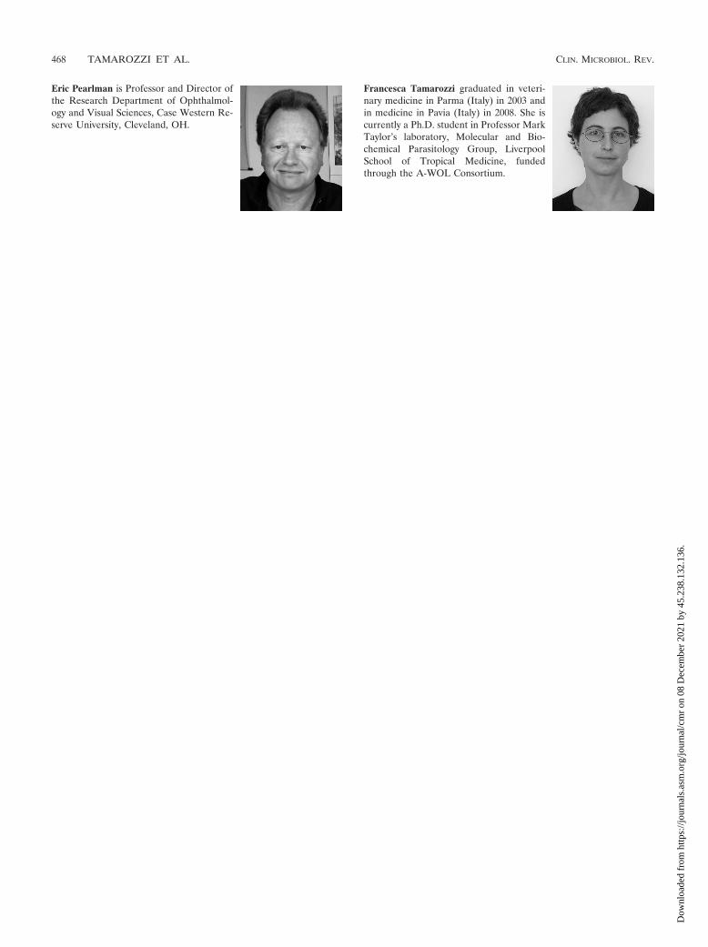
Eric Pearlman is Professor and Director ofthe Research Department of Ophthalmol-ogy and Visual Sciences, Case Western Re-serve University, Cleveland, OH.
Francesca Tamarozzi graduated in veteri-nary medicine in Parma (Italy) in 2003 andin medicine in Pavia (Italy) in 2008. She iscurrently a Ph.D. student in Professor MarkTaylor’s laboratory, Molecular and Bio-chemical Parasitology Group, LiverpoolSchool of Tropical Medicine, fundedthrough the A-WOL Consortium.
468 TAMAROZZI ET AL. CLIN. MICROBIOL. REV.
Dow
nloa
ded
from
http
s://j
ourn
als.
asm
.org
/jour
nal/c
mr
on 0
8 D
ecem
ber
2021
by
45.2
38.1
32.1
36.