Autogenous Dentin Graft in Bone Defects after Lower Third ...

On tissue reactions to dentin as a bone substitute material
Payam Farzad
Department of biomaterials
Institute of clinical sciences
Sahlgrenska Academy at University of Gothenburg
Göteborg 2017
On tissue reactions to dentin as a bone substitute material
© 4ff8 2017
http://hdl.handle.net/2077/52410
ISBN: 978-91-629-0209-4 (TRYCK)
ISBN: 978-91-629-0210-0 (PDF)
Printed in Gothenburg, Sweden 2017
Ineko AB

To my father-you would have been proud.

Content
Abstract 6
List of publications 10
Abbreviations 11
Introduction 13
-Clinical implications of dentin as a bone substitute material 15
Bone biology 17
Bone cells 19
-Osteoblast 20
-Osteocytes 20
-Bone lining cells 21
-Osteoclasts 22
Physiology of bone healing 23
-Osteogenesis 23
-Osteocunduction 24
-Osteoinduction 25
Healing of bone grafts 26
-Autogenous bone 26
-Vascularized bone grafts 30
-Allogenic bone 30
-Xenogenic grafts 33
-Alloplastic grafts 36
-Dentin as a bone substitute material 39
Analysis methods 42
-Bone histomorphometry 42

-Scanning electron microscopy 43
Aims 45
Material and methods 46
-Animals and anesthesia 46
-Implants 46
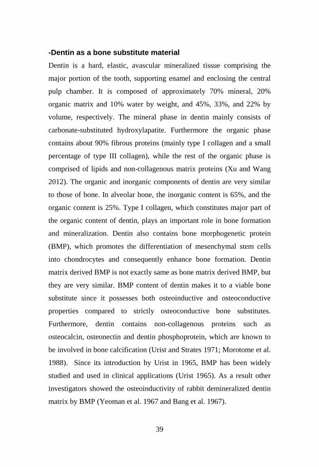
-Surgical protocols 47
-Specimen preparation 54
-Analysis and calculation 54
-Statistics 57
Results 58
-Study I 58
-Study II 60
-Study III 62
-Study IV 65
Discussion 67
-Study I 67
-Study II 71
-Study III 73
-Study IV 76
Conclusions 81
Acknowledgements 83
References 85

6
On tissue reactions to dentin as a
bone substitute material
Click here to enter text.
4ff8 Department of biomaterials, Institute of clinical sciences
Sahlgrenska Academy at University of Gothenburg
Göteborg, Sweden
Abstract
Background Reconstruction of the jaws due to resorption of the alveolar
crest may require bone augmentation prior to installation of endosseous
implants. Active research on new bone graft materials with bone
regeneration ability equivalent to autogenous bone but without the
limitations of allogenic, xenogenic and synthetic bone are constantly
ongoing. From clinical and experimental studies, it has been
demonstrated that replanted teeth without a viable periodontal membrane
will ankylose with the bone. The dentin of such teeth is fused with the
bone, and will be gradually replaced by bone, also called replacement
resorption or osseous replacement. In order to possibly modify treatment
protocols and also exploring possible cost-benefit alternatives to
commercially available bone replacement materials, there has been an
increased interest to explore the use of human dentin as a source for graft
material.
Aims The aim of the first study was to evaluate and compare the host
tissue response to autogenous and xenogenic non-demineralized dentin
blocks implanted in non-osteogenic areas, the abdominal connective

7
tissue and femoral muscle of rabbits. The objective of the second study
was primarily to evaluate the healing pattern of xenogenic non-
demineralized dentin granules and dentin blocks grafted to maxillary
bone of rabbits and secondarily to study integration of titanium micro-
implants installed in grafted areas. In paper III, we sought to evaluate the
healing pattern of xenogenic demineralized dentin granules and dentin
blocks grafted to cavities created in tibial bone of rabbits, secondarily to
study integration of titanium micro-implants installed in grafted areas and
thirdly to investigate the morphological appearances and differences
between demineralized and non-demineralized dentin by means of
Scanning Electron Microscopy (SEM) and Energy Dispersive X-ray
(EDX). Finally, the objective of study IV was to compare the host tissue
response and remodelling of onlay grafts of demineralized dentin in
comparison to onlay bone grafts transplanted to the native tibial cortical
bone wall.
Material and methods In study I, fifteen 6-month old New Zealand male
white rabbits were used. Dentin autografts taken from the same rabbit and
dentin xenografts taken from human premolars were implanted in
abdominal connective tissue and femoral muscles. All rabbits were
sacrificed after 12 weeks for light microscopic analysis.
In study II, fifteen 6-months old New Zealand male rabbits were used.
Dentin blocks and dentin granules from human premolars were implanted
in cavities prepared on either side of the maxilla (n=15x2). After a
healing period of 6 months, one micro implant (5 mm long, 2 mm in
diameter) was installed in each surgical site. All rabbits were sacrificed
24 weeks after implant installation. The specimens were studied by light
microscopic and histomorphometrical analysis. Study III included twelve
6-month old New Zealand male, white rabbits. Dentin blocks and dentin
granules from human premolars were implanted in cavities prepared on

8
both tibial bones. Twelve hours prior to grafting the dentin grafts were
rinsed in saline and demineralized on its surface by being placed in 24%
EDTA neutral, pH7, for 12 hours. After a healing period of 24 weeks, one
micro implant was installed in each surgical site.
To characterize the grafts, twelve additional dentin blocks were prepared
in standardized sizes. All samples were conditioned in 24% EDTA
neutral, pH7, for 12 hours followed by a second x-ray analysis. Four
samples were chosen for conventional SEM and energy dispersive X-ray
analysis (EDX), both image mode and element analysis mode. In study
IV, we used eight 6-months old New Zealand male rabbits. Standardized
sized dentin blocks from human premolars and similar autogenous bone
blocks, harvested from tibia were grafted as onlay blocks on each tibia
(n=8x2). All animals were sacrificed after a healing period of 12 weeks.
Descriptive histology as well as histomorphometrical analysis of the
remaining dentin, bone graft and soft tissue was determined using light
microscopy.
Results Study I showed only minor signs of heterotopic bone formation.
There were no significant differences between autografts and xenografts
or grafts implanted in connective tissue or muscle with regards to tissue
reactions except for a significant difference (P = 0.018) in findings of
more local inflammatory cells in relation to grafts placed in connective
tissue in the autograft group. In study II, no statistically significant
difference could be observed in BIC and BA between dentin and native
bone. Overall the BIC and percentage of new bone fill of the block
specimens were higher than the same parameters for the particulate
graft. Study III showed a tendency towards higher BIC and BA for the
EDTA conditioned dentin in conjunction with installed implants, but the
difference was not statistically significant. In addition, on the
demineralized dentin surface the organic marker element C dominated, as

9
revealed by EDX image mode. The hydroxyapatite constituents Ca, P and
O were close to devoid on the dentin surface. A similar pattern was
discerned from the semi-quantitative data analysis where the organic
markers C and N dominated. Study IV showed that in general, both the
dentin and bone block grafts were fused to the bone, resorbed and
replaced by bone and connective tissue to a varying degree. Resorption
cavities could be seen in the dentin with bone formation. Zones of
osseous replacement resorption of the dentin could be noted. In both graft
types, higher rate of bone formation was seen at the interface between
graft and recipient site.
Conclusion Non-demineralized dentin, whether autogenous or xenogenic
did not have the potential to induce bone formation when implanted in
non-osteogenic areas such as the abdominal wall and abdominal muscle
of rabbits. Limited or no bone contact between micro-implants and
xenogenic non-demineralized dentin grafts could be seen. Demineralized
xenogenic dentin onlay grafts showed similar resorption characteristics as
autogenous bone onlay grafts, being resorbed in a similar rate during 12
weeks. New bone formation occurred mainly in terms of replacement
resorption in the interface between dentin/bone graft and native bone. The
bone inductive capacity of the dentin material seemed limited although
demineralization by means of EDTA indicated a higher BIC and BA
value in conjunction with installed implants in the area.
Keywords Grafted dentin, tissue reaction, bone blocks, dental implants,
experimental study

10
LIST OF PUBLICATIONS
This dissertation is based on the following papers, which will be referred
to throughout by their Roman numerals I-IV:
I. Al-Asfour A*, Farzad P*, Andersson L, Joseph B, Dahlin C
Host tissue reactions of non-demineralized autogenic and xenogenic
dentin blocks implanted in a non-osteogenic environment. An
experimental study in rabbits. Dent Traumatol. 2014;30:198-203
*Equal contribution
II. Farzad P, Al-Asfour A, Dahlin A, Andersson L, Dahlin C
Integration of dental implants in conjunction with grafted dentin. An
experimental study in the rabbit maxilla. Oral Health Dent Manag
2015;5:289-293
III. Farzad P, Lundgren T, Al-Asfour A, Andersson L, Dahlin C
Integration and characterization of decalcified and non-decalcified dentin
in conjunction with dental implants. An experimental study in rabbit
tibia. In Manuscript
IV. Al-Asfour A*, Farzad P*, Al-Musawi A, Dahlin C, Andersson L.
Demineralized xenogenic dentin and autogenous bone as onlay grafts to
rabbit tibia. Implant Dent 2017;26:232-237.
*Equal contribution

11
Abbreviations
AB Autogenous bone
ALP Alkaline Phosphatase
BA Bone to implant area
BCP Biphasic calcium phosphate
BIC Bone to implant contact
BMP Bone morphogenetic protein
BMU Basic multicellular unit
CaPs Calcium phosphate ceramics
CSF Colony stimulating factor
DFDB Demineralized freeze-dried bone
EDS Energy dispersive spectroscopy
EDTA Ethylenediaminetetraacetic acid
FHA Flourohydroxyapatite
FPD Fixed partial denture (bridge)
HA Hydroxyapatite
IGF Insulin-like growth factor
IM Intramembranous
MSC Mesenchymal stem cell
MMP Matrix metalloproteinase
OC Osteocalcin
12
OP Osteopontin
OPG Osteoprotegrin
PDGF Platelet-derived growth factor
PDL Periodontal ligament
PTH Parathyroid hormone
RANK Receptor activator of nuclear factor
RANKL Receptor activator of nuclear factor-IB LIGAND
ROI Region of interest
RPD Removable partial denture
SEM Scanning electron microscopy
TCP Tricalcium phosphate
TGF Transforming growth factor
TRAP Tartrate-resistant acid phosphatase
TGF Transforming growth factor
VEGF Vascular endothelial growth factor

13
Introduction
Alveolar bone is a prerequisite for support of the teeth. Alveolar bone
atrophy can be caused by systemic disorders, endocrine imbalance, age,
mechanical forces or periodontal disease (Boyne 1982; Bays 1986) and is
also seen after loss of teeth (Schropp 2003; Carlson 2004).
These factors may act independently or concordantly and lead to reduced
chewing ability and morphological changes of the jaw bone. There are
several different methods of restoring the chewing function. Conventional
removable prostheses (RPD) retained by the remaining dentition and
supported by the residual alveolar bone is one way of solving this
functional problem. Another option is a fixed dental bridge (FPD), which
is cemented to the remaining teeth anterior and posterior to the
edentulous region. Both these options have drawbacks in that removable
prostheses are not accepted by all patients and tooth-supported fixed
bridges require a sufficient number of supporting teeth (Randow et
al.1986; Jepson et al.1995). Since the concept of osseointegration was
introduced back in 1969 by Brånemark and co-workers, endosseous
implants have been used successfully as an alternative treatment to
removable prosthesis and fixed dental bridges with good long-term
clinical results (Brånemark et al. 1969). One major advantage of
endosseous implants is that there is no need of engagement of remaining
teeth. Sufficient bone height and bone width is however a prerequisite for
achieving good results. If there is a bone deficiency in the maxilla, the
problem may be solved by using narrow implants (Hallman 2001), short
implants (Pohl 2017) or tilting the implants towards a new direction
where bone can be found (Mattson et al. 1999; Krekmanov 2000;
Aparicio et al. 2001). Other options include the use of specially designed
long implants (zygomatic implants) which are placed through the
14
maxillary sinus into the zygoma (Higuchi 2000; Malevez et al. 2000;
Farzad et al. 2006). In the mandible lateralization or transposition of the
inferior alveolar nerve enables installation of implants posterior to the
mental foramen when sufficient height bone superior to the nerve is not
available, however this is a less suitable method since sensory
disturbance might occur following this procedure (Hirsch and Brånemark
1995). Alveolar distraction osteogenesis is another method used to
increase the height of the available bone above the mandibular canal in
order to install dental implants. This technique avoids the sensory
disturbance problems associated with lateralization or transposition of the
inferior alveolar nerve (Felice et al. 2013).
In cases where atrophy of the alveolar bone is severe, there might be a
need for augmentation procedures prior to implant treatment. Most often,
a three dimensional lack of bone, i.e. lack of width and height, in the
desired position can be solved by reconstruction using veneer grafts and
allowing the graft to heal for a certain period prior to placement of dental
implants (Bahat and Fontanessi 2001). However, this augmentation
technique might not be applied in every bone deficiency situation. For
instance, the technique most often used in the posterior part of the maxilla
is augmenting vertically by grafting of the maxillary sinus floor (Boyne
1980; Wood and Moor 1988; Hallman et al. 2002; Hallman et al. 2002).
Autogenous bone grafts have been the gold standard to reconstruct bone
deficiency situations for many years (Bloomquist 1980; Sakkas et al.
2017). Their range of advantages includes early revascularization,
resistance to infections and evidence of immune activation (Burchardt
1983; Beirne 1986). Moreover, the autogenous bone graft possesses both
osteoinductive and osteoconductive properties (Urist 1965; Urist 1980).
However, a disadvantage is that this technique requires a second surgical
site to harvest the bone graft. Moreover, there are drawbacks such as

15
donor site morbidity, limitations in the quantity of available bone,
prolongation of surgery time and an increase of treatment cost (Dahlin et
al. 1988; Raghoebar et al. 2001; Andersson 2008). Several studies have
also shown that particularly onlay bone block grafts are prone to
resorption and a large part of the bone graft can be lost during the healing
period (Johansson et al. 2001; Nyström et al. 2002; Misch 2011). This has
encouraged research to find an acceptable bone substitute. The ideal bone
substitute should be readily available, well tolerated by the host, possess
both osteoinductive and osteoconductive properties and be able to be
resorbed gradually with the regeneration of new osseous tissue and
healing of the bone defect (Jensen et al. 1996; Schilling et al. 2004).
Available bone substitutes on the market are, either synthetic, inorganic
or biologically organic and may be associated with additional cost for the
patient. These materials are used solely to replace the bone grafting
procedure or used in combination with a minor amount of autogenous
bone to increase the volume of graft material. Since allogenic and
xenogenic bone substitute have a potential risk of disease transmission,
there has been an increasing demand for synthetic bone substitutes in
recent years (Sogal and Tofe 1999; Kim et al. 2016).
Clinical implications of dentin as a bone
substitute material
Dental trauma is one of the major causes for tooth loss. Alveolar bone
resorption is an inevitable consequence of tooth loss and may be
detrimental to long-term dental aesthetics and function. It is estimated
that the prevalence of dental trauma is 17, 5% in a global perspective
making it one of the most frequent traumas reported (Azami Aghdas et al.
2015). The tooth is physiologically connected to the alveolar bone via the
periodontal ligament attaching into the “bundle bone” portion of the

16
socket-associated bone. This part of the alveolar bone is always resorbed
following tooth loss as a normal physiological event (Araujo et al. 2006).
This can, in particular be observed in younger patients suffering from a
tooth loss. The change in dimensions is most pronounced in the anterior
maxilla and during the initial 6 months following tooth loss (Rodd et al.
2007). This study also reports a difference between genders. Hence
young women tend to have a more pronounced bone resorption compared
to male persons. Recently differences in the pattern of resorption with
regards to gingival biotype have also been described (Schappuis et al.
2013). It was demonstrated by means of CBCT analysis that a thin
biotype is associated with more pronounced resorption along the axis of
the socket while a thicker biotype tends to demonstrate more marginal
bone loss (Schappuis et al. 2013). Since a final restoration supported by
dental implants requires a completed skeletal growth, tooth loss in a
relatively young age will create a need for a detailed treatment planning
leading up to the final restoration. Based on these facts, researchers and
clinicians have become interested in the use of human dentin from
extracted teeth in the context of serving as graft material (Kim et al.
2010; Murata et al. 2011) since it is readily available, cheap and from
biological origin. Dentin has inorganic and organic contents that are very
similar to those of human bone. From clinical and experimental studies, it
has been well documented that replanted teeth without a viable
periodontal membrane will ankylose with the bone (Söder et al. 1977;
Andreasen JO 1981; Blomlöf et al. 1983; Andersson et al. 1984;
Andersson et al. 1989; Hammarström et al. 1989; Lindskog and Blomlöf
1992; Andreasen et al. 1995; Barrett and Kenny 1997; Trope 2011;
Maslamani et al. 2016). The dentin of such teeth is fused with the bone
(ankyloses), and will be gradually replaced by bone, also called
replacement resorption or osseous replacement (Andreasen and Hjörting-
17
Hansen 1966; Andersson 1988; Andersson et al. 1989). This is
considered to be mainly a bone remodelling process (Andreasen and
Hjorting-Hansen 1966; Andersson et al. 1984; Andersson 1988).
Furthermore, it has been suggested that dentin possesses not only
osteoinductive properties due to its content of bone morphogenic protein
(BMP) but also osteoconductive properties. These facts might indicate
that dentin might function as a bone substitute material (Pinholt et al.
1992; Ike and Urist 1998).
The increasing number of bone grafting procedures in the recent years
and the subsequent introduction of different bone substitutes to the
market require a better understanding of the bone biology and bone
grafts.
Bone biology
Human bone is biologically active connective tissue, which has its own
blood supply and consists of cells and extracellular matrix. This living
tissue has several important functions for the organism; (i) gives
mechanical support to the body; (ii) produces blood cells in the bone
marrow; (iii) functions as a reservoir of Ca-ions; (iv) provides protection
for internal organs and (v) serves as attachments for muscles, ligaments
and tendons. The bones in the human body can be assorted to as long
bones, short bones, irregular bones and flat bones. All bones are
composed by an outer dense structure called the cortical bone and an
inner layer of trabecular bone with lower density and a more porous
structure. About 80% of the skeletal mass is composed of cortical bone.
Mineralized bone appears in two forms, woven and lamellar. Woven
bone is seen during early bone formation i.e. during growth and healing.
18
Lamellar bone is the form of mature bone and is formed during modelling
and remodelling.
About 70% of the bone is composed of mineral, mainly hydroxyapatite,
Ca10(PO4)6(OH)2. The bone matrix consists of mainly type I collagen (up
to 90%), proteins such as osteocalcin (OC), bone sialoprotein,
osteopontin (OPN), osteonectin and a great number of growth factors,
e.g. BMPs. The remaining content consists of 5% to 10% water and <3%
of lipids (Buck and Dumanian 2012).
From an embryological standpoint, the craniofacial skeleton including
maxilla and mandible is formed from the neural crest cells. There are two
types of bone formation described: endochondral ossification (the most
common mechanism of primary bone formation) and intramembranous
ossification (Buck and Dumanian 2012; Makiewicz 2011).
In the regions of craniofacial skeleton, differentiation of mesenchymal
cells directly into osteoblasts initiates production of a trabecular pattern
of early bone matrix. Bone matrix matures through secretion of bone
matrix components and cellular synthesis. At his stage, calcium
phosphate, in the form of hydroxyapatite (HA) crystals are deposited at
the bone matrix site. This procedure is called intramembranous (IM) bone
formation and the flat bones of the skull, the clavicle and the mandible
are formed in this way (Makiewicz 2011; Buck and Dumanian 2012).
The endochondral ossification occurs in the long bones, pelvis, skull base
and vertebral column. In this type of ossification, mesenchymal cells
differentiate into chondrocytes, which produce a hyaline cartilaginous
framework. This cartilage is matured through hypertrophy of
chondrocytes followed by matrix erosion. The remaining cartilage matrix
mineralizes and the chondrocytes regress and die. Through invading
blood vessels, mesenchymal cells enter the calcified cartilage model
19
which may be differentiated into osteoblasts and subsequently start bone
formation (Zipfel et al.2003).
Bone remodelling refers to a continuous process throughout life where
old bone is replaced by new bone and during normal conditions equal
amount of bone is formed as the amount of bone resorbed keeping the
total bone mass unchanged. This phenomenon aims at maintaining
mechanical properties of the skeleton and support mineral homeostasis
and for maintaining a constant serum level of calcium (Zipfel et al. 2003;
Lerner 2006; Makiewicz 2011; Buck and Dumanian 2012). Bone
remodelling begins before birth and continues until the organism’s death.
In adults about 25% of trabecular and 3% of cortical bone is replaced
each year (Zipfel et al. 2003). The process of bone remodelling takes
place in a basic multicellular unit (BMU), which consists of bone
resorbing osteoclasts, the bone forming osteoblasts, osteocytes within the
bone matrix, bone lining cells on the bone surface, and the capillary
blood supply (Kular et al. 2012). The duration of the resorption process is
3 to 4 weeks and the subsequent bone formation takes about 3-4 months
to be completed. The bone remodelling process is shorter in cortical bone
than in cancellous bone where the length of the process is about 200 days
in human iliac bone (Kular et al. 2012).
Bone cells
The osteoblast, the bone lining cell, the osteocyte and the osteoclast are
the four cells types found in bone. In total, these cells make up around
10% of the total bone volume. The osteoclasts are formed by giant
multinucleated cells whereas the other three types are derived from
mesenchymal stem cells (Buck and Dumanian 2012).

20
-Osteoblast
Osteoblasts account for 4-6% of the bone cells and are estimated to have
a lifespan of three months in human bone. Osteoblasts are the only cells
with capability of bone formation through producing and secreting
proteins, thus forming the bone matrix. They line the surface of bone,
packed tightly against each other with a rounded, polyhedral form
(Rochefort 2010; Capulli et al. 2014). Osteoblasts are derived from
mesenchymal stem cells with a capability of differentiation into
fibroblasts, chondrocytes, myoblasts and adipocytes (Ducy et al. 2000).
Four maturational stages have been identified in osteoblast
differentiation: pre-osteoblast, osteoblast, osteocyte and bone lining cells
(non-active flattend osteoblast) (Kular et al. 2012). Several proteins such
as collagen type I, osteocalcin (OC), alkaline phosphatase (ALP),
osteonectin, osteopontin (OP), bone sioloprotein and a few other minor
matrix proteins are produced by osteoblasts (Manolagas 2000).
Fibers of type I collagen, which is the major protein in the matrix,
provide a structure on which mineral is deposited (Mackie 2003). At the
end of a bone formation cycle, mature osteoblasts face one of three fates:
approximately 50-70% undergoes apoptosis and the rest will either
develop into bone lining cells or osteocytes (Manolagas 2000, Kular et al.
2012).
Osteoblasts are also responsible for regulating the differentiation of the
bone resorbing osteoclasts by producing factors such as macrophage
colony-stimulating factor (M-CSF), osteoprotegerin (OPG) and cytokine
receptor activator of NF-KB ligand (RANKL). These factors play a major
role in osteoclast formation, activation and resorption (Kular et al. 2012).
-Osteocytes
Osteocytes account for more than 95% of all the bone cells. They
demonstrate a widely variable life expectancy, but a mean half-life time
21
of 25 years in human bone has been proposed, although it is probably less
due to a constant bone turnover of approximately 10% (Rochefort et al.
2010). Osteocytes have been differentiated from osteoblasts and are
entrapped in the bone matrix. The time span for a motile osteoblast to be
an entrapped osteocyte in the bone matrix is about 3 days. Osteocytes
demonstrate a size of 10 µm -20 µm in human bone, which is a reduction
to 30% of the size of the osteoblast origin (Knoteh et al. 2003; Bonewald
2011). They lie in lacunae embedded in the bone matrix and once there,
they start to extend projections through channels in the bone matrix called
canaliculi (Bonewald 2011). These channels aid the osteocytes to
communicate not only with each other but also with other bone cells on
the bone surface such as bone lining cells and osteoblasts (Dudley and
Spiro 1961; Tanaka-Kamioka et al. 1998; Bonewald 2011). Osteocytes
serve as mechanosensors, having the ability to detect mechanical pressure
and load through the interconnected network of fluid containing
canaliculi (Aarden et al. 1994; Burger and Klein-Nulend 1999). This
ability can induce bone repair following microdamage. Osteocytes are
also responsible for maintaining the bone matrix (Aarden et al. 1994;
Burger and Klein-Nulend 1999). By modulating secretion and expression
of insulin-like growth factor (IGF), osteocalcin (OC) and sclerostin, the
osteocytes are able to regulate skeletal homeostasis. Osteocytes also
provide the majority of RANKL that controls osteoclast formation in
cancellous bone (Robling 2008; Rochefort et al. 2010).
-Bone lining cells
The bone lining cells or surface osteoblasts are flattened, thin,
differentiated cells, mainly derived from osteoblasts. These cells are
located on top of a thin layer of unmineralized collagen matrix covering
the bone surface (Miller et al. 1989). They connect to the osteocytes
through gap junctions (Miller and Jee 1987). Lining cells can be activated
22
and differentiated into osteogenic cells and they also take part in the
homeostasis of mineral through control of bone fluids and ions e.g. by
immediate release of calcium from bone when the blood calcium level is
low (Miller et al. 1989). When exposed to PTH, bone lining cells secrete
collagenase to remove the collagen matrix so osteoclasts can attach to
bone (Recker 1992).
-Osteoclasts
In an adult organism, osteoclasts are derived from hematopoetic stem
cells and share precusrsors with macrophages and monocytes. They are
the only cell type that can resorb bone and are formed by multiple cellular
fusions of mononucleated cells (Vaananen and Laitala-Leinonen 2008).
The osteoclast is found and formed in much smaller numbers compared
to other bone cells on the surface of the bone. These cells are highly
motile, but since they are only formed on the bone surfaces, they ate
never encountered in the blood circulation (Lerner 2000). A differentiated
human osteoclast contains about five to eight nuclei in each cell and has a
diameter of 50-100 m. Bone resorption takes place in a finger shaped
extension of the ruffled border membrane. This is also the most
characteristic feature of the osteoclast (Manolagas 2000; Vaananen and
Laitala-Leinonen 2008). Osteoclast formation, activation and resoprtion
are regulated by the ratio of receptor activator of NF- ligand (RANKL,
which binds to RANK and activates osteoclastogenesis) to
osteoprotegerin (OPG, which inhibits osteoclastogenesis), IL-1 and IL-6,
colony stimulating factor (CSF), parathyroid hormone, 1,25-
dihydroxyvitamin D and calcitonin (Blair and Athanasou 2004).
Resorbing osteoclasts have a unique ability to create an acidic
environment in the resorption lacunae via secretion of hydrogen ions
through proton pumps and chloride channels. Hydroxyapatite is
23
dissoluted when the pH within the bone-resorbing space is lowered to
about 4,5. This is followed by secretion of tartrate-resistant acid
phosphatase (TRAP), cathepsin K, matrix metalloproteinases (MMPs)
and gelatinases from cytoplasmic lysosomes to digest the organic matrix.
The result is formation of Howship’s lacunae on the surface of trabecular
bone and Haversian canals in cortical bone. Degradation products such as
bicarbonate, calcium and phosphate ions are removed from the resoprtion
lacunae by transportation through the cells for secretion (Reddy 2004).
The resorption phase is completed by mononuclear cells after osteoclasts
undergo apoptosis. Resoprtion is followed by osteoblast activation and
formation of osteoid, which fills the cavities over a period of about three
months (Deal 2009).
Physiology of bone healing
The use of a bone graft for purposes of achieving increased bone volume
is affected by anatomical, histological, and biochemical principles.
Additionally, several physiological properties of bone grafts directly
affect the success or failure of graft incorporation. These properties are
osteogenesis, osteoinduction and osteoconduction (Prolo 1990).
-Osteogenesis
Osteogenesis is the ability of the graft to produce new bone, and this
process is dependent on the presence of live bone cells in the graft.
Osteogenic graft materials contain viable cells with the ability to form
bone (osteoprogenitor cells) or the potential to differentiate into bone-
forming cells (inducible osteogenic precursor cells). These cells, which
participate in the early stages of the healing process to unite the graft with
the host bone, must be protected during the grafting procedure to ensure
viability in order to produce osteoid. When new bone is formed by

24
osteoprogenitor cells within the wound defect, i.e. a bone fracture, it is
called spontaneous osteogenesis. Transplanted osteogenesis is when new
bone formation is related to presence of bone forming cells within the
bone graft (Muschler et al. 1990).
The role of osteogenesis as a mechanism of new bone formation during
nonvascularized bone graft healing, however, is thought to be of lesser
significance than that of osteoconduction (Burchardt 1983).
-Osteoconduction
Non-vascularized bone grafts heal through a predictable sequence of
events. In the first step, the graft will undergo partial necrosis, followed
by an inflammatory stage. During this phase, the graft is invaded slowly
by vessels, which in turn will deliver osteoclasts and osteoblasts to the
region. Interaction between these 2 cell lines will lead to replacement of
much of the grafted bone by new bone. The term creeping substitution is
used to describe this slow vessel invasion and bony replacement, a
process formally known as osteoconduction. The term refers to the
process where bone grows on a surface. An osteoconductive surface is
one that permits bone growth on its surface or down into pores, channels
or pipes (Albrektsson and Johansson 2001). In the context of bone
healing, the graft would serve as a scaffold on which new bone is
deposited (Muschler et al. 1990).
The second step in the process of healing is the formation of a hematoma.
Shortly after placement of the graft, a hematoma is formed around the
graft, which is due to the surgical disruption of host soft tissues and the
recipient bony bed. During this early stage, a small minority of cells on
the graft’s surface are able to survive, primarily as a result of plasmatic
imbibitions (Heslop et al. 1960; Muliken et al. 1984). The third step is the
start of an inflammatory reaction. The inflammatory reaction, which lasts
for 5 to 7 days is focused around the graft and ensues after hematoma

25
formation. A dense fibrovascular stroma is formed around the graft and
the onset of vascular invasion starts at 10 to 14 days (Gross et al. 1991).
Vascular invasion brings additional cells with osteogenic potential into
the graft, as the interstices of the old bone act as a directive matrix. As
osteoblasts deposit new bone, osteoclasts resorb necrotic bone and pave
the way for the graft to be penetrated by vascular tissue (Schmitz and
Hollinger 1996; Gross et al. 1991).
-Osteoinduction
The principle of osteoinduction was described by Urist and the
biochemical events by Reddi (Urist 1965; Bang and Urist 1967; Reddi
and Wientroub 1987). They described the inductive process in rodents as
ingrowth of vascular tissue and development of osteoprogenitor cells with
subsequent new bone formation by enchondral ossification (Urist 1965;
Bang and Urist 1967; Reddi and Wientroub 1987). Osteoinduction refers
to the process by which active factors released from the grafted bone
stimulate osteoprogenitor cells from the host to differentiate and form
new bone. This process is highly dependent of a soluble protein called
BMP. The BMP belongs to the family of transforming growth factors,
(TGF)-Three phases of osteoconduction have been described:
chemotaxis, mitosis, and differentiation. During chemotaxis, bone
inductive factors direct the migration and activity of osteogenic cells via
chemical gradients. The inductive factors then stimulate these
osteoprogenitor cells to undergo intense mitogenic activity, followed by
their differentiation into mature, osteoid-producing cellular elements (i.e.,
osteoblasts). Ultimately, the cells become revascularized by invading
blood vessels and are incorporated as new bone. The ultrastructural
character of the bone graft (i.e. cancellous versus cortical) determines the

26
ability of revascularization to take place and, therefore, significantly
impacts the process of incorporation (Muschler et al. 1990).
Healing of bone grafts
A graft is transplantation of tissue or cells. The most commonly used
materials for alveolar ridge augmentation purposes are:
-Autogenous bone (AB)
An autogenic graft is transplantation of tissue within the same individual
and is considered to be the ‘’gold standard’’ in reconstruction of defects
in the jaws. This is mainly due to its osteoinductive and osteoconductive
properties as well as low cost and minimal risk for disease transmission
(Burchart 1983). The healing of autogenous bone grafts is quite similar to
that of fracture repair. An important similarity in bone graft healing is
that a substantial portion of the biological activity originates from the
host. This occurs because most viable osteocytes within the graft itself
necrose shortly after transplantation. Nonetheless, substantial biological
interactions still remain between graft and host. This important biological
interplay contributes to the final outcome of graft take (Burchart 1983).
Most common donor sites in reconstruction of jaw defects prior to
implant surgery are various areas of the mandible, tibia and the iliac crest.
Cortical, cancellous or a combination of both can be obtained from these
different sites (Buser et al. 1996; Sjöström et al. 2007). Cancellous bone
is osteogenic providing vital osteoprogenitor cells, it is osteoinductive
and is completely replaced in time by osteoconduction because the graft
also is acting as a scaffold for bony ingrowth from the recipient site
(Burchart 1983). Cortical bone may be osteogenic but heals mainly by
osteoconduction. At the time of transplantation it provides more
mechanical support than cancellous grafts but the later are revascularized
more rapidly and completely than cortical grafts (Burchart 1983;

27
Sjöström et al. 2007). The graft consists partly of surviving cells
(preosteoblasts and preosteoclasts), but also proteins capable of
converting undifferentiated mesenchymal stem cells into bone producing
cells (Burchart 1983; Sjöström et al. 2007). Since the blood supply to the
bone graft is cut off at the time of harvest, revascularization needs to
occur for incorporation of the bone graft and resorption of cortical bone is
therefore a major part of bone graft healing (Urist 1980; Goldberg and
Stevenson 1987). Differences in revascularization time and pattern are
seen between trabecular and cortical bone. In trabecular bone,
revascularization is re-established through micro-anastomosis with
existing blood vessels. Since the cancellous bone is porous with marrow
tissue between the trabeculae, vascular ingrowth occurs more rapidly and
is completed after a few weeks. In contrast to cancellous bone grafts,
cortical bone graft is densely packed and revascularization proceeds
slowly and takes about almost two months to be completed (Albrektsson
1980).
The large spaces between trabeculae in cancellous grafts permit the
unobstructed invasion of vascular tissue and the facile diffusion of
nutrients from the host bed. This is thought to promote osteogenic cell
survival, imparting increased osteogenesis when compared with cortical
grafts. Osteoprogenitor cells, brought in by the invading vessels,
differentiate into osteoblasts and deposit a layer of new bone around the
necrotic trabeculae. An osteoclastic phase ensues, wherein the entrapped
cores of dead bone are resorbed. Cancellous bone grafts are completely
revascularized and ultimately replaced with new bone over several weeks
to months (Stevenson et al. 1996; Pinholt et al. 1994).
Revascularization of cortical bone grafts proceeds with initial osteoclastic
activity. Enlargement of the haversian and Volkmann’s canals must occur
before vessels are able to penetrate the graft. The dense lamellar structure

28
of cortical bone limits the vascular invasion, and the newly forming
vasculature is constrained to invade the graft along these preexisting
pathways. This process begins at the graft periphery and progress to the
interior of the graft (Burchardt 1983). Revascularization in cortical bone
grafts may also be restricted by the limited number of endosteal cells that
remain viable after transplantation. These cells are thought to contribute
to end-to-end vessel anastomosis during bone graft revascularization
(Heiple et al. 1987). Studies have shown that cortical grafts in the onlay
position show only superficial revascularization occurring in the first 10
to 21 days, and central revascularization by 8 to 16 weeks (Ozaki and
Buchman 1998). Once a graft has been placed, mesenchymal cells
recruited to the region will differentiate into fibroblasts, endothelial cells
or osteoblasts depending on the stimuli. These cells form new connective
tissue, vessels or osteoid respectively. One major factor of importance for
graft survival is the stability of the graft. This will improve both
revascularization and leads to a lower grade of resorption of the graft
(Phillips and Rahn 1988; Phillips and Rahn 1990). In the competition
between the soft and bone tissues, a cancellous bone graft may be more
prone to soft tissue ingrowth and resorption than a cortical graft (Körloff
et al. 1973; Gordh et al. 1998; Johansson et al. 2001).
Several different factors are considered important for the general bone
metabolism and survival of autogenous bone grafts. These factors can
either be systemic such as age, gender, physical activity, hormonal status
and drugs or local factors such as graft orientation, fixation of the graft,
recipient’s site, mechanical stress and revascularization. Certain
hormones such as calcitonin, insulin, vitamin D3 and parathyroid
hormones are also essential.
A fresh autogenous bone graft contains osteoinductive proteins (BMPs)
that stimulate the recruitment of mesenchymal stem cells and

29
osteogenesis and is therefore the golden standard in reconstructive
surgery (Sampath & Reddi 1983). Bone morphogenic proteins (BMPs)
are homodimeric proteins of approx. 30kD with two identical strands
linked by a cysteine binding group. Nearly 20 modifications of BMPs
with slightly different modifications in secondary structure elements have
been identified so far (Miyazono 2000). BMP2-BMP9 belong to the
TGF-β superfamily with a high degree of homology with the TGF-βs.
TGF-β and BMP have a common scaffold with the cysteine knot motif
and two double stranded beta sheets (Scheufler et al. 1999). BMP2,
BMP4 and BMP7 are considered to be osteogenic and have been tested in
experimental and clinical approaches. The content of BMPs in bone has
been estimated to be 1µg/g bone tissue (Kubler 1997).
In humans, three different transforming growth factor β (TGF-β) have
been identified and are primarily found in platelets. These growth factors
have been proven to enhance bone formation around titanium implants
(Clokie and Bell 2003). Other factors stimulating bone formation are
insulin-like growth factors. Insulin-like growth factors (IGFs) are single
chain peptides that exist in two isoforms (IGF-I and IGF-II). IGFs have
approximately 40-50% homology between themselves and with insulin.
Despite this significant homology between insulin and IGFs, all three
have unique binding sites to their receptors (O’Connor 1998). IGF-I has
been proven to be three times more efficient in bone cells than IGF-II
(Schmid 1993).
Platelet-derived growth factor (PDGF) is another factor that might
influence the speed of bone formation. PDGF is a highly basic dimeric
glycoprotein of 30 kD consisting of two disulphide bonded polypeptides
encoded by different genes (Cochran et al. 1993). There are three
isoforms characterized by the combination of A- and B-chains featuring
two homodimeric (PDGF-AA and PDGF-BB) and one heterodimeric

30
isoform (PDGF-AB) (Hock & Cannalis 1994). PDGF-BB and PDGF-AB
are systemically circulating isoforms contained in alpha granules of
platelets from where they are released after adhesion of platelets to
injured sites of vessel walls, whereas PDGF-AA is secreted by
unstimulated cells of the osteoblastic lineage (Cannalis 1992). Marx et al.
(1998) used platelet rich plasma (PRP) which is rich in PDGF, TGF- β1
and β2, IGF and fibrin in treating large mandibular defects with
autogenous bone as carrier and found increased bone maturation rate and
bone density compared with defects augmented with autogenous bone
only.
-Vascularized bone grafts
Free vascularized bone grafts are another option, widely used for
postablative reconstruction in irradiated recipient beds, where standard
bone grafts have been shown to be less viable. Bone flaps are in general
used in defects larger than 6 cm or when composite tissues are required.
Large bone segments from the fibula or iliac crest can be transplanted
together with various amount of soft tissue to restore form and function.
Instant blood circulation in the flap guaranties transfer of viable
osteocytes, thereby bypassing the need for new bone formation apart
from at the graft-host interfaces. Vascularized bone transfers are
technically challenging though and donor site morbidity is an issue in
some cases (Rohner et al. 2003; Jaquiery et al. 2004)
-Allogenic bone
An allogenic graft is obtained by transplanting tissue from one individual
to a genetically non-identical individual of the same species and contains
no viable cells (Urist 1965). For this reason allografts are considered to
be mainly osteoconductive and have very little or no osteoinductive
properties (Becker et al. 1995).

31
Different forms of allografts are available. Mineralized or demineralized
bone, frozen or freeze-dried bone, demineralized dentin and antigen-
extracted allogenic bone (AAA) are examples of allografts. All of the
components in bone are potentially immunogenic but bone minerals and
collagen are only weakly antigenic. For this reason, cortical bone is
preferable due to its high content of collagen compared to cancellous
bone, resulting in a weaker immunologic reaction (Dayi et al. 2002).
Transplantation of allogenic tissue initiates an immunological reaction in
the recipient of the cell-mediated type (Burwell et al. 1985). It is believed
that T-cell responses are the most significant in bone transplantation and
that the cell-mediated mechanisms are the same as those in skin graft
rejections. The immunologic responses result in impaired
revascularization of the graft and subsequent necrosis. Allografts also
carry the coincident risk of disease transmission. Extensive donor
screening protocols have been implemented worldwide in order to reduce
transmission of HIV and hepatitis B and C viruses (Buck et al. 1989). In
order to sterilize and lower the antigenicity of the allogenic graft different
processing methods have been tested. Freeze-drying, demineralization,
deep freezing (<-70° C) chemo sterilization or radiation, have all been
suggested (Chalmers 1959; Senn 1989; Lane & Sandhu 1987). The same
factors that reduce immunogenicity, however, also deactivate the
osteoinductive factors that are so critical to survival. In addition, deep
freezing (<-70° C) and freeze drying- the two most common methods of
preservation, may significantly alter the mechanical properties and
strength of the graft (Voggenreiter et al. 1994).
It was shown in dogs that the acceptance of a frozen allograft was
improved with histocompatibility matching or immunosuppression
(Goldberg et al. 1985). However clinical trials revealed no clear
relationship between the degree of histocompatibility of the donor and the
32
recipient and the incorporation of frozen bone allografts (Muscolo et al.
1987).
In maxillofacial surgery, frozen allogenic bank bone has mainly been
used in combination with autogenic bone (Sailer 1983; Plotnicov and
Nikitin 1985). The principles for incorporation of allografts follow the
same principles as for autogenous bone grafts but probably proceed more
slowly due to the absence of viable cells that are osteoinductive, although
allografts might have some osteoinductive properties. Osteoblasts from
the recipient generate bone as the transplanted bone is gradually resorbed.
Clinical trials have revealed that the incorporation is a slow and
incomplete process (Lane & Sandhu 1987). Pinholt et al. (1990) studied
demineralized and lyophilized dentin and bone implants in rats, and
demonstrated induction of new bone formation, however in two other
studies in rats and goats respectively, no osteoinduction was found
(Pinholt et al. 1991; Pinholt et al. 1992). Smiler et al. (1992) compared
autogenous bone (AB), deep frozen demineralized bone (DFDB), and
hydroxyapatite (HA) as grafting materials prior to implant placement,
with equally good results. However the healing time for the various grafts
differed significantly. In a human study by Boeck-Neto et al. (2002) bone
formation was evaluated in 10 patients who underwent maxillary sinus
floor augmentation using autogenous bone with DFDB or HA. They
concluded that both materials were still present after 10 months.
In a study by Lohman et al. (2001), bone harvested from patients and
processed by lyophilization, was divided into two portions, One of which
was used directly while the other was demineralized. They concluded that
the age of the patient played an important role in the osteoinductive
capacity of the bon study using mineralized cancellous bone allograft for
sinus augmentation a vital bone content of almost 26% was found after 9
months of graft healing (Froum et al. 2005). In a clinical study by Gapski

33
et al. (2006), human mineralized bone allografts were successfully used
for sinus lift procedures before placement of implants.
The use of an ideal graft material should result in high formation of vital
bone after graft maturation. The literature shows varying results for
different grafting materials. Vital bone content of 14% to 44% has been
reported in the literature.
-Xenogenic grafts
Tissue transplanted between individuals of different species is called a
xenogenic graft. Examples are bone-like minerals derived from corals or
algae, bovine bone and porcrine bone. One of the main tasks to overcome
using xenogenic bone grafts has been the immunological response and to
obtain safety of disease transmission (Enneking 1957; Nisbet 1977;
Burwell et al. 1985). The antigenicity of the graft initiates a T-cell
response and it is believed that the cell-mediated mechanisms are the
same as those seen in skin graft rejections. In order to avoid an
immunological rejection after implantation, the proteins have to be
extracted using various procedures. In the process of eliminating the
antigens the organic matrix is destroyed and thereby the osteoinductive
properties and as the osteoinductive capacity disappears the graft can
only act as an osteoconductive scaffold. Furthermore, the presence of
minerals in the graft impedes the transformation of fibroblasts to
osteoblasts (Urist 1971; Reddi and Huggins 1973). This leads to a
formation of new bone at lower pace compared to autogenous bone
grafts. Healing of xenografts follow the same principles as for allografts
and today they are frequently used for bone augmentation procedures in
implant dentistry due to their similarity to human bone.
Algipore®(Dentsply Friadent,Mannheim Germany) is a porous
fluorohydroxyapatite (FHA) derived from calcifying algae (Corallina
officinalis). Complete removal of organic components has been carried

34
out utilizing pyrolytical segmentation of native algae and hydrothermal
conversion of calcium carbonate into FHA in the presence of ammonium
phosphate at about 700°C (Thorwarth et al. 2007). Studies have shown
that Algipore® is a suitable biomaterial for periodontal treatment and for
sinus floor augmentation (Schopper et al. 2003; Roos-Jansaker et al.
2011, Scarano et al. 2012).
Deproteinized bovine bone (DPBB) is a natural bone mineral with
extreme similarities in structural and chemical composition to human
bone. DPBB consists of 100% deproteinized bovine hydroxyapatite. The
most documented DPBB used for reconstruction in implant surgery is
Bio-Oss®(Geistlich, Wolhausen, Switzerland). This material has been
investigated in numerous clinical and experimental studies by several
authors and since all proteins are claimed to be extracted, Bio-Oss®
works only as a 3-dimensional scaffold for ingrowth of blood vessels and
bone building cells (Maioranta et al. 2001; Hallman et al. 2002; Hallman
et al. 2002; Froum et al. 2006; Esposito et al 2009; Felice et al. 2009;
Esposito et al. 2010; Jensen et al. 2012; Lee et al. 2012; Lindgren et al.
2012). Bio-Oss® can either be used alone in various augmentation
procedures or in combination with autogenous bone which would add on
the osteoinductive properties of the autogenous bone to the transplant. In
a systematic review assessing augmentation of the maxillary sinus floor
with Bio-Oss® alone or Bio-Oss® combined with autogenous bone, it was
concluded that the hypothesis of no differences between the two
procedures could neither be confirmed nor rejected. They also concluded
that the addition of AB to bio-Oss® did not influence the biodegradation
of Bio-Oss® although long term studies were not available (Jensen et al.
2012).
There seem to be controversy in the literature whether DPBB is
resorbable, slowly degraded, phagocytated or non resorbable. True
35
resorption by means of osteoclastic activity of Bio-Oss® is however, one
topic where contradictory results have been found. In one study,
maxillary sinus augmentation using DPBB and AB was performed in 20
patients. Signs of resorption lacunas and the presence of osteoclasts on
the particle surface were observed in the specimens harvested four years
after initial surgery (Piatelli et al. 1999). In another report, there were no
signs of DPBB particles after 20 months of healing but it could not be
determined whether this was dependent on the biopsy technique or was
the result of a true resorption (Wallace et al. 1996). Other studies on
maxillary sinus grafting with Bio-Oss show no signs of resorption of the
material at all (Schlegel & Donatah 1998; Valentini et al. 1998, 2000).
The fact that this material does not resorb when used for maxillary sinus
grafting is highly important in comparisons to autogenous bone grafts
only, where in some cases the resorption is more than 50% of the original
volume (Körloff 1973; Johansson et al. 2001). The advantage of using a
non-resorbable bone substitute due to, its low-grade resorption has been
discussed in a paper by Cobb et al. (1990). They concluded that a 1:1
ratio is optimal and increased amounts of bone substitute result in more
fibrous encapsulation. Nevertheless, in some studies 100% of DPBB has
been used with results similar to those when it was used as an admixture
(Hising et al 2001; Yildrim et al. 2001).
Some researcher have claimed that Bio-Oss® after all contains certain
amount of both transforming growth factor β(TGF- β) and proteins,
whilst others have questioned these findings (Honig et al. 1999; Schwartz
et al. 2000; Benke et al. 2001). A systematic review by Kim and co-
workers indicates that bovine derived bone substitutes may theoretically
carry a risk of prion disease transmission to patients even though no
clinical reports of this complication have been published yet (Kim et al.
2013, 2016).

36
-Alloplastic grafts
Alloplastic bone grafts are derived synthetically and are alternatives to
autogenous, allogenous and xenogenic bone grafts in reconstructive
surgery. The development of alloplastic bone substitutes evolved out of
the operative morbidities of autografts and the limitation of allografts.
Alloplastic grafts follow the same principles as for the allografts except
that the materials contain no proteins and are only osteoconductive.
Formation of new bone can start if the material is placed in close contact
to bone, serving as a 3-dimensional scaffold for ingrowth of bone
building cells and blood vessels.
Calcium phosphate ceramics (CaPs) including HA and tricalcium
phosphate (TCP), calcium-sulphate, bioactive glasses and polymers are
all examples of alloplastic bone grafts materials with different
compositions. Furthermore, they exhibit different biological and
mechanical properties.
Calcium phosphates resemble the inorganic matrix of bone more closely
and therefore have greater utility as a bone substitute. Alloplastic bone
graft substitutes usually contain hydroxyapatite (HA) and different
calcium polymers such as, β-tricalcium phosphate or sintered calcium
phosphates, bioglass or sintered calcium sulphates. In contrast to HA,
pure calcium phosphate or calcium sulphate is generally weaker in its
composition and will presumably dissolve chemically in to its ions which
may stimulate bone formation (Daculci 1998).
There are two major varieties of hydrated calcium phosphate
(hydroxyapatite) preparations for use in bone applications: ceramics and
cements. Hydroxyapatite ceramic implants can be manufactured in a
variety of shapes, forms, structures and chemical compositions. They
possess different mechanical and biological properties and the time of
degradation may vary. Their similarity in composition to bone mineral,

37
their biodegradability and osteoconductivity were the rationales for
development of CaPs (Hannink and Arts 2011). Hydroxyapatite ceramic
implants have been extensively used within the field of oral implantology
(Kwon et al. 1986; El Deeb et al. 1991). Unlike ceramic forms,
hydroxyapatite cements are moldable, allowing for intraoperative
contouring, however their use has been limited to low-stress regions in
the craniofacial skeleton. Smiler and Holmes (1987) reported on a mean
bone ingrowth of 21,1% after 5 months when using porous
hydroxyapatite (HA) in sinus lifting procedures. In another study by
Wheeler et al. (1996), HA was used with and without autogenous bone
and the levels of newly formed bone ranged from 11 to 20% for the
different groups.
Active glass is another type of allograft that has been tested and shows
active bone formation and support for dental implants. In a study by
Cordioli et al. (2000), mixture in a ratio of 4:1 of bioactive glass and
autogenous bone was used as grafting material to the maxillary sinus.
Biopsies harvested after 9 to 12 months revealed a mean of 30,6±5,7%
bone tissue in grafted sites.
Tricalciumphosphate (TCP) has also been tested and has been found to be
suitable for use as a maxillary sinus floor graft (Szabo et al. 2001).
β-tricalcium phosphate has been developed to achieve a more organized
crystal structure in relation to pure calcium phosphate but will still
dissolve into calcium and phosphate ions. Cerasorb® (Curasan AG,
Kleinostheim, Germany) is the most used commercial β-tricalcium
phosphate today for dental implant surgery and the material has been
used for maxillary sinus floor augmentation. However drawbacks such as
rapid dissolution and volume reduction have been reported (Lu et al.
2002).

38
In order to combine different properties, materials are sintered together.
Bone Ceramic® (Straumann, Basel, Schweiz) is a novel, fully synthetic
bone substitute, aimed for the market of implant surgery. It consists of
60% HA and 40% β-TCP. The HA component is supposed to protect the
augmented area from resorption and the TCP component is supposed to
dissolve relatively quickly in to its ions and stimulate bone formation
(Lindgren et al. 2009).
In a randomized and controlled study, bilateral sinus floor augmentation
was performed in 11 patients with severe atrophy in the posterior maxilla
using biphasic calcium phosphate (Bone Ceramic®) at one side and
deproteinized bovine bone (Bio-Oss®) at the contra lateral side acting as
control. Micro implants were installed simultaneously and retrieved after
8 months with a surrounding bone core and analyzed by scanning
electron microscopy (SEM) and energy dispersive X-ray spectroscopy
(EDS). The conclusion was that the β-TCP component of BCP may be
gradually substituted by calcium deficient hydroxyapatite over the
healing period and might influence the progress of resorption and healing
(Lindgren et al. 2010).
In another prospective study with 9 patients undergoing bilateral sinus
floor augmentation with BoneCeramic® on one side and Bio-Oss® on the
other side, similar results were found after 1 & 3 years of functional
loading and implant success rate was not dependent on the biomaterial
used for augmentation (Lindgren et al. 2012a,b)
Other examples of alloplastic materials are Easygraft CRYSTAL®
(Degradable Solutions AG, Schiliern, Switzerland), Tricos® (Baxter
healthcare corp., USA) and calcium sulphate (CaSO4) (Surgiplaster,
Ghimas, Bologna, Italy) with different tissue response to their resorption
and dissolution (Hallman and Thor 2000, De Leonardis and Pecora
2000).
39
-Dentin as a bone substitute material
Dentin is a hard, elastic, avascular mineralized tissue comprising the
major portion of the tooth, supporting enamel and enclosing the central
pulp chamber. It is composed of approximately 70% mineral, 20%
organic matrix and 10% water by weight, and 45%, 33%, and 22% by
volume, respectively. The mineral phase in dentin mainly consists of
carbonate-substituted hydroxylapatite. Furthermore the organic phase
contains about 90% fibrous proteins (mainly type I collagen and a small
percentage of type III collagen), while the rest of the organic phase is
comprised of lipids and non-collagenous matrix proteins (Xu and Wang
2012). The organic and inorganic components of dentin are very similar
to those of bone. In alveolar bone, the inorganic content is 65%, and the
organic content is 25%. Type I collagen, which constitutes major part of
the organic content of dentin, plays an important role in bone formation
and mineralization. Dentin also contains bone morphogenetic protein
(BMP), which promotes the differentiation of mesenchymal stem cells
into chondrocytes and consequently enhance bone formation. Dentin
matrix derived BMP is not exactly same as bone matrix derived BMP, but
they are very similar. BMP content of dentin makes it to a viable bone
substitute since it possesses both osteoinductive and osteoconductive
properties compared to strictly osteoconductive bone substitutes.
Furthermore, dentin contains non-collagenous proteins such as
osteocalcin, osteonectin and dentin phosphoprotein, which are known to
be involved in bone calcification (Urist and Strates 1971; Morotome et al.
1988). Since its introduction by Urist in 1965, BMP has been widely
studied and used in clinical applications (Urist 1965). As a result other
investigators showed the osteoinductivity of rabbit demineralized dentin
matrix by BMP (Yeoman et al. 1967 and Bang et al. 1967).

40
The idea of using dentin as a bone substitute for clinical practice probably
originates from the principle of dentoalveolar ankylosis and replacement
resorption, which is seen in more severe types of dental trauma, after the
PDL has been severely damaged e.g. after tooth avulsion and delayed
replantation. During intrusion, lateral luxation or avulsion with
subsequent replantation, contusion or drying of the PDL is a common
occurrence. Wound healing is subsequently initiated when damaged
tissue is removed by macrophage or osteoclastic activity. During these
events, not only are necrotic PDL tissue remnants removed, but
sometimes also bone and cementum (Andreasen 1966; Andersson et al.
1984). When large areas of the PDL are traumatized, competitive wound
healing processes begin between bone marrow-derived stem cells
destined to form bone and PDL-derived cells which are programmed to
form PDL fibers and cementum resulting in fusion of the alveolar bone
with dentin (ankyloses) which is followed by replacement resorption
(Blomlöf & Lindskog 1994). IGF-1, TGF-β and BMPs in the dentin are
released slowly and may serve as stimulators for osteoclasts. If the area of
injury is limited healing with normal periodontal ligament can be seen
(Andreasen and Kristerson 1981). However, when the area of injury is
large ankyloses cannot be avoided (Andreasen and Kristerson 1981).
When ankylosis has been established the dentin is replaced by bone
(osseous replacement, replacement resorption) (Andreasen 1966,
Andersson et al. 1984, Andersson et al. 1989). The rate of replacement
resorption is related to the age of the patient with a higher rate in young
individuals (Andersson et al. 1989)
Yeomans and Urist showed back in 1967 bone induction by decalcified
dentin implanted into oral, osseous and muscle tissues in rabbits. Samples
of allogenic tendon from the lower leg, quadriceps muscle, and either
decalcified bone or decalcified dentin were implanted into three different

41
sites: (a) a pouch in the rectus abdominus muscle, (b) a drill-hole bone
defect in the mandible, and (c) an empty tooth socket. Samples were
recovered after 4, 8 and 12 weeks after operation. Tendon and muscle
were rapidly resorbed over a 4- to 12 week period and did not induce
osteogenesis. Samples of decalcified dentin in comparison were
relatively slowly resorbed and always positive for bone induction.
Bang et al. (1972) used allogenic demineralized dentin implants in jaw
defects of Java monkeys. Surgical defects of a certain diameter were
made bilaterally in the region below root apices of the mandibular third
molars in 16 Java monkeys. Allogenic demineralized dentin was placed
in the defects on the right side while the contra lateral side served as
controls. After a healing period of 1 week to 1 year histological studies
were undertaken. Dentin implants, were shown to be tolerated very well
and were gradually resorbed. They were both osteoinductive and
osteoconductive and secured complete osseous healing. The control
cavities exhibited incomplete osseous healing with persisting fibrous
defects.
Su-Gwan et al. (2001) used a combination of particulated dentin and
plaster as bone substitute material in calvarial bone defects in rats and
compared it with Bio-Oss. They concluded that the combination of
particulate dentin and plaster is an alternative bone substitute, although it
is less effective than Bio-Oss. Carvalho et al. 2004 used homogenous
demineralized dentin matrix as osteopromotive material in rabbit
mandibles. They concluded that demineralized dentin matrix were
biocompatible and were resorbed during the bone remodelling process.
Bone repair was accelerated in the bone defects treated with
demineralized dentin in comparison to the control group. Demineralized
dentin blocks implanted in the palatal connective tissue in rats did not
seem to induce any bone formation up to 4 weeks after implantation

42
(Miyaji et al. 2002). In a recent study in rats, it was concluded that
demineralization of dentin blocks in 24% EDTA for 2 or more hours
resulted in significantly higher rate of resorption and significantly lower
rate of encapsulation (Mordenfeld et al. 2011).
Furthermore, there is some evidence in the literature that non-
demineralized dentin may resorb fast with no signs of bone formation
when implanted in animal muscle (Bang 1972; Machado et al. 2006).
Hence, there is reason to believe that for dentin, besides the surgical
technique, the demineralization procedure and the implant environment
play important roles in osteoinduction and osteoconduction. However,
there is limited information in the literature systematically analyzing the
biological outcome of different clinically applicable preparations of
dentin for augmentative use.
Analysis methods
-Bone histomorphometry
Bone histomorphometry is a quantitative histological examination of an
undecalcified bone biopsy performed to obtain quantitative information
on bone remodelling and structure (Kulak and Dempster 2010). It is
considered a valuable and well-established clinical and research tool for
studying the pathogenesis of metabolic bone diseases as well as in order
to evaluate the healing process for different bone substitutes or implants
(Parfitt 1983; Dempster 2001). Histomorphometry has traditionally been
assessed in two dimensions by means of histology, where the structural
and remodelling parameters are measured on sections, and the third
dimension is extrapolated using standard stereology theory (Parfitt 1983).
In the last two decades, there have been significant advances in

43
histomorphometric techniques with coupled stereology software which
have largely substituted the manual techniques (Malluche et al. 1982).
Remarkable advances in bone histomorphometry were made in the 1950's
and 60's due to the discovery of plastic embedding allowing high quality
histologic sections of mineralized bone (Frost 1958) and the use of
labeling fluorochromes leading to a better understanding of the dynamic
process of bone formation (Frost 1969).
Placement of microimplants is a well-established method to create a
miniature model of the titanium-bone interface (Jensen and Sennerby
1998; Lundgren et al. 1999; Hallman et al. 2002). In brief, a titanium
threaded microimplant is installed in the graft penetrating the residual
bone. After a certain healing period the microimplant with surrounding
bone core is retrieved using a trephine burr. The specimens are fixed by
immersion in buffred formalin solution, dehydrated in alcohol and
embedded in plastic resin. A specialized laboratory prepares
undecalcified sections and histological and histomorphometrical analysis
is carried out after the samples have been stained with certain dyes.
-Scanning electron microscopy
Scanning electron microscopy (SEM) is a well-known technique used for
micro-anatomical imaging. The SEM is a microscope that uses electrons
instead of light to form an image. A beam of electrons is generated from a
scheelite cathode at the top of the microscope by an electron gun. The
electron beam follows a vertical path through the microscope, which is
held within a vacuum. The beam travels through electromagnetic fields
and lenses, which focus the beam down toward the sample. Once the
beam hits the sample, electrons and X-rays are ejected from the sample.
Detectors collect these X-rays, backscattered electrons, and secondary
electrons and convert them into a signal that is sent to a screen similar to
a television screen. This produces the final image (Sriamornsak and

44
Thirawong 2003). Since their development in the early 1950's, scanning
electron microscopes have developed new areas of study in the medical
and physical science communities. The SEM has allowed researchers to
examine a much bigger variety of specimens. The scanning electron
microscope has many advantages over traditional microscopes. The SEM
has a large depth of field, which allows more of a specimen to be in focus
at one time. The SEM also has much higher resolution, so closely spaced
specimens can be magnified at much higher levels. Because the SEM
uses electromagnets rather than lenses, the researcher has much more
control in the degree of magnification. SEM can exploit different types of
samples such as dried specimens non-embedded or embedded in resin,
frozen-wet tissue or damp-wet tissue. All of these advantages, as well as
the actual strikingly clear images, make the scanning electron microscope
one of the most useful instruments in research today (Slater et al. 2008).

45
AIMS
General aim:
The overall aim of the present thesis is to study dentin as a possible bone
replacement and augmentation material prior to implant treatment.
Specific aims
Paper I
To evaluate and compare the host tissue response to autogenous and
xenogenic non-demineralized dentin blocks implanted in non-osteogenic
areas, the abdominal connective tissue and femoral muscle of rabbits.
Paper II
To evaluate the healing pattern of xenogenic non-demineralized dentin
granules and dentin blocks grafted to maxillary bone of rabbits and
secondarily to study integration of titanium micro-implants installed in
grafted areas.
Paper III
To primarily investigate the morphological appearance and mineral
content in decalcified dentin grafts and secondarily to study the healing
pattern of xenogenic demineralized dentin blocks and granules grafted to
cavities created in tibial bone of rabbits and subsequent integration of
titanium micro-implants installed in the previously grafted areas.
Paper IV
To compare the host tissue response and remodelling of onlay grafts of
demineralized xenogenic dentin in comparison to onlay autogenous bone
grafts transplanted to the native tibial cortical bone wall.

46
Material and methods
Animals and anesthesia
New Zealand male white rabbits were used in all studies. The animals
were kept in specially designed rooms in separate cages and fed pellets
and water ad libitum throughout the duration of the study. The
experiments were carried out at the Animal Research Centre, Health
Sciences Centre, Kuwait University. The protocol for animal experiments
by the Animal Research Centre of the Health Sciences Center, Kuwait
was strictly adhered to.
Thirty minutes prior to the experimental surgery, the rabbits were sedated
with Xylazine HCl(Rompun, Bayer, Leverkusen, Germany) 5mg/kg by
intramuscular injection. Animals were anaesthetized by intravenous
injection of 35mg/kg of Ketamine HCl (Tekan, Hikma, Amman, Jordan).
A veterinarian was responsible for administering the sedation, anesthesia
and for the intra- and postoperative care of the animals. To compensate
for peri-operative and postoperative dehydration 10ml sterile saline
solution was injected subcutaneously immediately following surgery and
antibiotics (Pen-Hista-strep,Vetoquinol SA, Lure Cedex, France)
50mg/kg was administered by intramuscular injection. Antibiotic
administration was continued during the first 3 days after surgery. After
completion of healing, the animals were sacrificed by an overdose of
Ketamine.
Implants
Study II and III
Screw-shaped micro implants (5 mm long, 2 mm in diameter), which
were machined from medical grade Ti (grade IV) rods (Elos, Pinol,
Gørløse, Denmark) were used.

47
Fig.1 Screw-shaped micro implants used in study II & III
Study IV
Titanium fixation screws, 6-mm long, 1.5-mm-wide (DePuy Synthes,
Jonhsson & Johnsson) were used in this experiment.
Surgical Protocols
Study I
Fifteen 6-month old New Zealand male white rabbits were used in this
study. The surgical areas were shaved and washed with iodine 7.5%
solution and the animals were prepared for surgery. As a supplement to
general anesthesia and for vasoconstriction purposes, local anesthesia 1ml
Lidocainehydrochloride 1% + epinephrine 5µg/ml (Xylocain-adrenalin,
Astra Zeneca, Södertälje, Sweden) was administered in each
experimental area. Bilateral incisions, each 2,5cm long, on either side of
the abdominal midline were performed. In each wound, dissection was
carried out to create a pocket in the abdominal connective tissue. Bilateral
incisions, each 2.5 cm long over the femoral muscle in the groin region
on either side were performed. With the two pockets in the abdominal
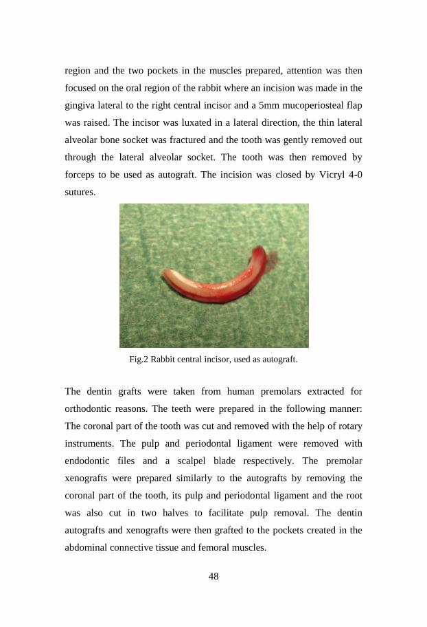
48
region and the two pockets in the muscles prepared, attention was then
focused on the oral region of the rabbit where an incision was made in the
gingiva lateral to the right central incisor and a 5mm mucoperiosteal flap
was raised. The incisor was luxated in a lateral direction, the thin lateral
alveolar bone socket was fractured and the tooth was gently removed out
through the lateral alveolar socket. The tooth was then removed by
forceps to be used as autograft. The incision was closed by Vicryl 4-0
sutures.
Fig.2 Rabbit central incisor, used as autograft.
The dentin grafts were taken from human premolars extracted for
orthodontic reasons. The teeth were prepared in the following manner:
The coronal part of the tooth was cut and removed with the help of rotary
instruments. The pulp and periodontal ligament were removed with
endodontic files and a scalpel blade respectively. The premolar
xenografts were prepared similarly to the autografts by removing the
coronal part of the tooth, its pulp and periodontal ligament and the root
was also cut in two halves to facilitate pulp removal. The dentin
autografts and xenografts were then grafted to the pockets created in the
abdominal connective tissue and femoral muscles.

49
Fig.3 Dentin graft implanted in femoral muscle of rabbit.
The incisions were closed in layers with Vicryl 4/0 (Ethicon, Cornelia,
USA). All rabbits were sacrificed after 12 weeks by an overdose of
Ketamine and block biopsies were prepared.
Study II
Fifteen 6-month old New Zealand male, white rabbits were used in this
study. As a supplement to general anesthesia and for vasoconstriction
purposes, local anesthesia 1ml lidocainehydrochloride 1% + epinephrine
5μg/ml (Xylocain-adrenalin, Astra Zeneca, Luton, UK) was administered
in each experimental area. The bilateral edentulous areas superior to and
between incisors and posterior teeth of the maxilla were used as
experimental sites. The bone surface was exposed via a 10 mm long
incision between buccal and palatal mucosa. A muco-periostal flap was
raised. A 5x5 mm wide and 3mm deep cavity was prepared penetrating
through the maxillary cortical bone wall with the use of round burr (3mm
in diameter) under irrigation with saline. Dentin grafts from human
premolars, which were extracted for orthodontic reasons, were prepared
in the following manner: The coronal part of the tooth was cut and
removed with the help of rotary instruments so no enamel remained. The
pulp and periodontal ligament were removed with endodontic files and a

50
scalpel blade respectively. With the help of a trephine burr (5mm
diameter) cylinder shaped block was harvested from the premolar and the
cylinder was sectioned into 3mm thick blocks. The rest of the premolar
was cut into granules in sizes of 1-3mm. Granules and blocks were
cleaned by being placed in 1% chlorhexidine and stored dry for one
month. They were rinsed in saline for one hour before being used as
grafts. The cavities on the right side were filled with dentin granules and
the cavities on the left side with dentin blocks. No membrane or any other
type of fixation was used. The incisions were closed with 4/0 Vicryl
(Ethicon, Bridgewater, NJ,USA). After a healing period of 24 weeks,
rabbits were anesthetized once again as described earlier. Surgical access
was accomplished in a similar way and one micro implant was installed
in each surgical site in such a way, that the apical half of the implant were
placed in native bone, serving as control site, and the coronal part in
dentin. All rabbits were sacrificed 24 weeks after the second surgery by
an overdose of Ketamine and block biopsies were prepared.
Fig.4 Micro-implant installed in grafted dentin

51
Study III
Twelve 6-month old New Zealand male, white rabbits were used in this
study. The surgical areas were shaved and washed with iodine 7.5%
solution and the animals were prepared for surgery. Local anesthesia 1ml
lidocaine hydrochloride 2% + epinephrine 1:100 000 (Lignospan
standard, Septodont, Saint Maur des Fosses Cedex, France) was
administered in each experimental area. Bilateral incisions, 2-3 cm long
over the lateral aspect of the tibia were performed and the tibial bone was
exposed by surgical dissection. On each side, two cavities of 5mm
diameter and 3mm depth were prepared penetrating through the tibial
cortex with the use of round burr (3mm in diameter) under irrigation with
saline. The dentin grafts were taken from human premolars, which had
been extracted on orthodontic indications. The teeth were prepared in the
following manner: The coronal part of the tooth was cut and removed
with the help of rotary instruments. The pulp and periodontal ligament
were removed with endodontic files and a scalpel blade respectively.
Cylindrical dentin block grafts were prepared in standardized sizes by the
use of a trephine bur, 5mm in diameter, and cut and trimmed to 2mm
thick dentin blocks by a diamond dish. The thickness of 2mm was
checked with a caliper. The rest of the premolar was cut into granules in
sizes of 1-3mm. The dentin blocks and granules were placed in
chlorhexidine to reduce bacterial growth. Twelve hours prior to grafting
the dentin grafts were rinsed in saline and demineralized on its surface by
being placed in 24% EDTA neutral, pH7, for 12 hours. On the right tibia,
the proximal cavity was filled with demineralized dentin block and the
distal cavity with demineralized dentin granules. On the left tibia non-
demineralized dentin was used in the same manner. No membrane or any
other type of fixation was used. The soft tissue was sutured in two layers,
muscle and dermis.

52
After a healing period of 24 wks, rabbits were anesthetized once again as
described earlier. Surgical access was accomplished in a similar way and
one micro implant was installed in each surgical site in such a way that
the coronal part of the micro implant was placed in dentin. All rabbits
were sacrificed 24 weeks after the second surgery by an overdose of
Ketamine and block biopsies were prepared.
Preparation and characterization of dentin grafts
Twelve dentin blocks were prepared as described (vide supra).
Conventional dental x-rays (0.06 sec, f 22.5 cm) were taken on all graft
samples prior to decalcification. All samples were conditioned in 24%
EDTA neutral, pH 7, for 12 hours followed by a second x-ray analysis.
Four samples were chosen for conventional SEM and energy dispersive
X-ray analysis (EDX), both image mode and element analysis mode.
Semi-quantitative data for the elements C, N, O, P, Ca and Au (100% in
total together) were sampled for 25 min.
Study IV
Eight 6-month old New Zealand male white rabbits were used in the
experiments. The surgical areas were shaved and washed with iodine
7.5% solution and the animals were prepared for surgery. Local
anesthesia 1ml lidocaine hydrochloride 2% + epinephrine 1:100 000
(Lignospan standard, Septodont, Saint Maur des Fosses Cedex, France)
was administered in each experimental area. Bilateral incisions, 2-3 cm
long over the lateral side tibia of the rabbit were performed and the tibial
bone was exposed by surgical dissection. With a 5mm diameter trephine
the tibial medial cortex was penetrated through its full thickness and a
bone block was harvested. The harvested bone block was measured and
trimmed by a diamond dish to a thickness of 2mm as controlled with the
help of a caliper. The dentin grafts were taken from human premolars,
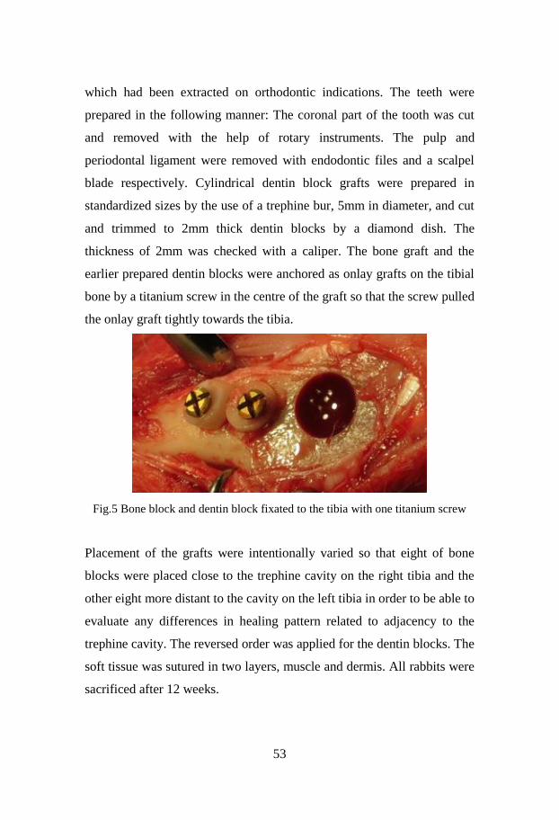
53
which had been extracted on orthodontic indications. The teeth were
prepared in the following manner: The coronal part of the tooth was cut
and removed with the help of rotary instruments. The pulp and
periodontal ligament were removed with endodontic files and a scalpel
blade respectively. Cylindrical dentin block grafts were prepared in
standardized sizes by the use of a trephine bur, 5mm in diameter, and cut
and trimmed to 2mm thick dentin blocks by a diamond dish. The
thickness of 2mm was checked with a caliper. The bone graft and the
earlier prepared dentin blocks were anchored as onlay grafts on the tibial
bone by a titanium screw in the centre of the graft so that the screw pulled
the onlay graft tightly towards the tibia.
Fig.5 Bone block and dentin block fixated to the tibia with one titanium screw
Placement of the grafts were intentionally varied so that eight of bone
blocks were placed close to the trephine cavity on the right tibia and the
other eight more distant to the cavity on the left tibia in order to be able to
evaluate any differences in healing pattern related to adjacency to the
trephine cavity. The reversed order was applied for the dentin blocks. The
soft tissue was sutured in two layers, muscle and dermis. All rabbits were
sacrificed after 12 weeks.

54
Specimen preparation
Study I
Following surgical removal, the samples were immersed and fixed for 48
h in 10% neutral buffered formalin. The samples were then decalcified in
neutral EDTA for 4-6 weeks, dehydrated in alcohol and embedded in
paraffin under vacuum using standard histological methods. Serial
sections were cut at 5µm thickness and were mounted on polylysine-
coated slides and then stained with hematoxylin and eosin, and examined
using light microscopy. The best sections including both dentin blocks
and the surrounding soft tissue were selected and evaluated for thickness
of fibrous capsule, number of inflammatory infiltrate and heterotopic
bone formation.
Study II-IV
Directly after the sacrifice of the animals, the implants and surrounding
tissue were removed en bloc and immediately fixed by an immersion of
10% neutral buffered formalin. The specimens were later dehydrated in a
graded series of ethanol, infiltrated with plastic resin and polymerized
prior to cutting along the long axis of the implant. A central ground
section was prepared by cutting and grinding, and was subsequently
stained with 1% toluidine blue.
Analysis and calculation
Study I
The thickness of the fibrous tissue surrounding the grafts was measured
using a software program called Leica Application Suite v3.1 (Leica,
Microsystems, Switzerland). Three areas were selected and the capsule
thickness was measured in µm by drawing a straight line across the
capsule. A magnification of 2.5 X was used for all the sections examined.
The average of the three readings, were taken as capsule thickness. Using

55
the same image analysis system, round cells consistent with inflammatory
cells were counted in the region of interest (ROI) close to the site where
the thickness of the capsule was measured with a magnification of 20x.
The area of this measurement was around 0.071mm2. Then using the cell
count tool, the cells were counted and the average of 3 readings, were
taken as the cell count. In sections where heterotopic hard tissue
formation was seen, the contact surface of heterotopic bone formation
related to the total graft circumference was calculated using the same
analysis system. The total circumference of the graft was measured as
well as the sites of hard tissue formation. Heterotopic bone formation was
expressed as a quotient of the sites of heterotopic hard tissue formation
related to the total circumference of the graft and expressed as a
percentage.
Study II
Two regions of interest were defined (ROI I and ROI II). ROI I (5
coronal threads) corresponded to the area where dentin blocks or granules
were placed.
Fig.6 ROI I corresponds to the coronal threads embedded in dentin and ROI II
corresponds to the apical threads embedded in host bone.

56
ROI II corresponded to the apical portion of the implants, which were
installed in the maxillary host bone only (serving as control). The
specimens were observed along their full length. The measurements of
bone-to-implant contact (BIC) and the bone fill area (BA) within the
threads were calculated on the mesial and distal aspect of each specimen.
A mean value was then calculated for each specimen (ROI I and II
respectively). The dentin and the bone-to-implant contact and the relative
amount of bone and dentin within the threads, were determined using
light microscopy (Nicon Eclipse E600) at 10 times magnification. The
specimens were assessed using NIS Elements Microscope Imaging
Software, Nikon.
Study III
One region of interest was defined (ROI). ROI (5 coronal threads)
corresponded to the area where dentin blocks or granules were placed.
The specimens were observed along their full length. The measurements
of bone-to-implant contact (BIC) and the bone fill area (BA) within the
threads were calculated on the mesial and distal aspect of each specimen.
A mean value was then calculated for each specimen. The dentin and the
bone-to-implant contact and the relative amount of bone and dentin
within the threads, were determined using light microscopy (Nicon
Eclipse E600) at 10 times magnification. The specimens were assessed
using NIS Elements Microscope Imaging Software, Nikon.
Study IV
A central ground section was prepared by cutting and grinding, and was
subsequently stained with toluidine blue. The specimens were observed
along their full length. A defined region of interest (ROI) was constructed
with equal dimensions as the respective grafts (5 x 2 mm). The presence

57
of the respective tissue types were calculated and presented as percentage
values of the respective graft area (ROI). Furthermore resorption pattern
of both graft types was assessed both on top and on the interface between
the grafts and native bone. Descriptive histology as well as
histomorphometric analysis of the remaining dentin, bone graft and soft
tissue was determined using light microscopy (Nicon Eclipse E600) at 10
times magnification. The specimens were assessed using NIS Elements
Microscope Imaging Software, Nikon.
Statistics
Study I
Host tissue reactions as expressed by the thickness of surrounding fibrous
connective tissue capsule and counts of inflammatory cells adjacent to the
grafted dentin were compared between the groups. The normal
distribution assumption for variables; thickness and counts, was
ascertained with Shapiro-Wilk test. The overall group differences were
compared using non-parametric Kruskal-Wallis test, whereas
Kolmogorov-Smirov Z test for comparing two independent variables. The
two-tailed probability value p < 0.05 was considered statistically
significant.
Study II
Results were reported as mean values and standard deviations. Bone-to-
implant contact (BIC) and bone fill area (BA) in ROI I and ROI II were
compared using the student’s t-test. P-values of < 0.05 were considered
statistically significant.
Study III
Results were reported as mean values and standard deviations. The
Wilkoxon signed rank test was used for comparing bone-to-implant
contact (BIC) and bone fill area (BA) between the groups. P-values of <

58
0.05 were considered statistically significant.
Study IV
The amount of resorption of dentin and bone on top versus on the
interface, the amount of newly formed bone on top of the graft versus on
the interface and the amount of newly formed soft tissue on top versus on
the interface were assessed. A pairwise Wilcoxon rank sum test with
Bonferroni correction for multiple testing was used to test for significant
differences between the test sites. P-values of < 0.05 were considered
statistically significant.
Results
Study I The aim of this study was to evaluate and compare the host tissue
response to autogenous and xenogenic non-demineralized dentin blocks
implanted in non-osteogenic areas, the abdominal connective tissue and
femoral muscle of rabbits. Clinical observation
All animals recovered uneventfully and gained weight. The soft tissue
healing in all 15 rabbits were uneventful and there were no signs of
infection.
Histological observation
The thickness of the fibrous tissue surrounding the grafts was measured.
There was no significant difference (p=0.388) in thickness between
capsules of autografts and xenografts or between capsules in muscle or
abdominal tissue.
Round cells consistent with inflammatory cells were counted in the
region of interest (ROI) close to the site where the thickness of the
capsule was measured. There were no significant differences between
59
autografts and xenografts. A significant difference (p=0.018) was seen
with more inflammatory cells in abdominal grafts than muscle grafts
among the autografts.
Fig.7 Connective tissue capsule surrounding the dentin with no evidence of any
inflammatory infiltrate
In sections where heterotopic hard tissue formation was seen, the contact
surface of heterotopic bone formation related to the total graft
circumference was calculated.
In the abdominal grafts no heterotopic bone formation was seen in any of
the sections except for one section in one autograft. Hard tissue formation
was seen in 3.9% of the dentin graft circumference in this section. In the
muscle grafts hard tissue formation was seen in 3 of the sections. The
surface area was estimated to 2.2 and 5.7% respectively in two xenograft
sections, and 3% of one section in the autograft group.

60
Fig.8 Dentin autograft in rabbit femoral muscle. Extensive resorption and
evidence of hard tissue formation is seen.
Study II
The aim of this study was primarily to evaluate the healing pattern of
xenogenic non-demineralized dentin granules and dentin blocks grafted
to maxillary bone of rabbits and secondarily to study integration of
titanium micro-implants installed in grafted areas.
Clinical observation
Three rabbits died during the healing period. The remaining 12 rabbits
recovered uneventfully and gained weight. The soft tissue healing in all
12 rabbits was uneventful and there were no signs of infection. Three
sites grafted with dentin granules and 3 sites grafted with dentin blocks
were encapsulated by loose connective tissue which did not allow any
implant installation due to lack of bone. Hence a total of 18 micro-
implants were installed (Block group n=9, granulae group n= 9).
Histological observation
In general, the incorporation of the dentin blocks and granulae varied. In
the block group, nine out of 12 available blocks were considered enough
fused to the surrounding bone and suitable for implant placement. In the
granulae group, less fusion to bone was seen. A common feature was that

61
granulae were encapsulated by means of fibrous tissue and only scarce
contact between the xenogenic dentin granulae and blocks and the
surrounding host bone was found. In general no or limited direct contact
between xenogenic dentin and the microimplant surface could be noted.
A few osteoclasts could be identified on the surface of the dentin, mostly
located adjacent to present native bone tissue.
Fig.9 Osteoclast adjacent to implanted dentin and subsequent bone deposition.
The dentin particles were otherwise surrounded by fibrous tissue with
scarce presence of cells. The dentin material per se, did not seem to
induce bone apposition on the implant surface. Instead newly formed
bone seemed to migrate into the microgap between the dentin and the
titanium surface.
Histomorphometric analysis
ROI I comprised of the first 5 threads and the border between native bone
(ROI II) and the dentin area was set at thread 5, where the interface could
be assessed. The dentin specimens (ROI I) revealed a mean BIC of 17.8%
and the native bone (ROI II) resulted in a BIC of 24.4% (p=0.188). The

62
percentages of new bone fill in the area (BA) within the threads (% bone
fill) for the dentin specimens were 31.6% and 42.6% (P=0.360) for the
native bone. Overall the BIC and percentage of new bone fill of the block
specimens were higher than the same parameters for the particulate graft.
Only fractional areas of direct contact between the dentin and the
titanium surface could be noted.
Study III
The aim of this study was primarily to evaluate the healing pattern of
xenogenic demineralized dentin granules and dentin blocks grafted to
cavities created in tibial bone of rabbits, secondarily to study integration
of titanium micro-implants installed in grafted areas and thirdly to
investigate the morphological appearances and differences between
decalcified and non-decalcified dentin by means of Scanning Electron
Microscopy (SEM) and Energy Dispersive X-ray (EDX).
Clinical observation
Four rabbits died during the healing period. The remaining 8 rabbits
recovered uneventfully
and gained weight. The soft tissue healing in all 8 rabbits was uneventful
and there were no signs of infection. Hence a total of 32 micro implants
were placed (demineralized group n=16, non-demineralized n=16).
Histological observation
In general no or limited direct contact between xenogenic dentin and the
microimplant surface could be noted. A few osteoclasts could be
identified on the surface of the dentin, mostly located adjacent to present
native bone tissue. The dentin particles were otherwise surrounded by
fibrous tissue with scarce presence of cells. The dentin material per se,
did not seem to induce bone apposition on the implant surface. Instead
newly formed bone seemed to have migrated into the microgap between
the dentin and the titanium surface.

63
Descriptive characterization of dentin block
In X-rays prior to decalcification, dentin grafts were clearly discernible.
After a 12 h EDTA surface conditioning no differences in image contrast
could be observed. After 4 weeks of decalcification, the dentin grafts
could not be discerned in X-ray images.
Fig.10 X-rays prior to, after 12h and after 4 weeks of decalcification
In longitudinal sections of dentin grafts densely packed tubules,
approximately 3 µm wide, were observed. In transversal sections, tubules
of approximately Ø 3 µm, devoid of odontoblast processes, were seen. In
transversal close ups the dense peritubular walls were discerned, between
which the demineralized collagenous intertubular dentin surface
appeared.
Fig.11 Close up of tubules with regular peritubular walls and irregular
intertubular dentin, x 19.8.103

64
On the demineralized dentin surface the organic marker element C
dominated, as revealed by EDX image mode. The hydroxyapatite
constituents Ca, P and O were close to devoid on the dentin surface.
Remnant of surface gold coating was seen. A similar pattern was
discerned from the semi-quantitative data analysis. The organic markers
C and N dominated, 55 and 27%, respectively. Ca, P and O constituted
less than 1% in the dentin surface layer.
C N O P Ca Au
Spectrum 1 57.95 23.68 14.70 -0.35 0.16 3.85
Spectrum 2 52.97 28.23 15.09 0.10 0.48 3.13
Spectrum 3 55.11 27.28 15.30 0.04 0.18 2.08
Spectrum 4 54.40 27.18 14.47 0.21 0.29 3.35
Mean 55.13 26.59 14.89 0.00 0.28 3.10
Std.deviation 2.08 2.00 0.38 0.24 0.14 0.75
Max 57.95 28.23 15.30 0.21 0.48 3.85
Min 52.97 23.68 14.47 -0.35 0.16 2.08
Table 1. EDX data. All elements analyzed and results shown in atomic %.
Histomorphometric analysis
After exclusion of specimen from the analysis due to the difficulty
encountered to show a visible screw during specimen preparation, a total
of 26 specimens were available for analysis. ROI comprised of the first 5
threads corresponding to the length placed in dentin. The non-
demineralized group revealed a mean BIC of 36.2% and the
demineralized group 40.4% (p=0.480). The percentages of new bone fill
in the area (BA) within the threads (% bone fill) for the non-
demineralized group were 67.4% and 72.4% (P=0.09) for the
demineralized group. Overall the BIC and percentage of new bone fill of
the demineralized group were higher than the same parameters for the
non-demineralized specimens. Only fractional areas of direct contact

65
between the dentin and the titanium surface could be noted.
Study IV
The aim of this study was to compare the host tissue response and
remodelling of onlay grafts of demineralized dentin in comparison to
onlay bone grafts transplanted to the native tibial cortical bone wall.
Clinical observation
All animals tolerated the experiments very well and gained weight during
the 12 weeks healing period. The soft tissue healing in all 8 rabbits was
uneventful and there were no signs of infection.
Histological observation
After exclusion of specimen from the analysis due to the difficulties
encountered in the histologic processing, a total of 14 and 12 specimens
were available for analysis respectively.
In general both the dentin and bone block grafts were to a varying degree
fused to the bone and replaced by bone and connective tissue. Both types
of grafts were still present after 12 weeks. Resorption had occurred to
approximately one third of the original sizes. Fusion of the graft to bone
was more pronounced in the bone graft group compared to dentin group
where some ingrowth of connective tissue was seen between the dentin
graft and host bone surface. Zones of osseous replacement resorption of
the dentin could be noted. New bone was formed on 4.55% of the surface
of the resorption cavities (range 1.5-9.9). There was no significant
difference with the connective tissue in contact with the dentin and bone
grafts. Dentin had induced bone only in the resorption cavities in the
interface and not on the surfaces facing the overlying soft tissues.
Connective tissue in contact with the bone graft was seen in 63.3% (range
60.0-73.0), while the connective tissue in contact with dentin was 72.6%
(range 54.4-89.6).
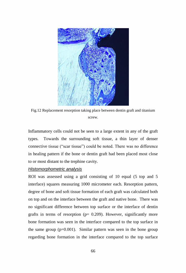
66
Fig.12 Replacement resorption taking place between dentin graft and titanium
screw.
Inflammatory cells could not be seen to a large extent in any of the graft
types. Towards the surrounding soft tissue, a thin layer of denser
connective tissue (“scar tissue”) could be noted. There was no difference
in healing pattern if the bone or dentin graft had been placed most close
to or most distant to the trephine cavity.
Histomorphometric analysis
ROI was assessed using a grid consisting of 10 equal (5 top and 5
interface) squares measuring 1000 micrometer each. Resorption pattern,
degree of bone and soft tissue formation of each graft was calculated both
on top and on the interface between the graft and native bone. There was
no significant difference between top surface or the interface of dentin
grafts in terms of resorption (p= 0.209). However, significantly more
bone formation was seen in the interface compared to the top surface in
the same group (p=0.001). Similar pattern was seen in the bone group
regarding bone formation in the interface compared to the top surface

67
(p=0.002). Finally, both groups showed significantly higher degree of
soft tissue formation towards the top compared to the interface (p=0.048
for dentin group and p=0.003 for the bone group).
Discussion
Study I
Comments on aim, material and methods
The aim of this study was to evaluate and compare the host tissue
response to autogenous and xenogenic non-demineralized dentin blocks
implanted in non-osteogenic areas, the abdominal connective tissue and
femoral muscle of rabbits.
Fifteen New Zealand male white rabbits were used in the experiments.
Dentin blocks of autogenous and xenogenic origin were implanted in
pockets created in the abdominal connective tissue and femoral muscle.
All animals survived throughout the healing period without any
macroscopic signs of infection. They were sacrificed after 12 weeks.
A similar model has previously been used to study bone induction by
implantation of human dentin and cementum into subcutaneous tissues of
rats (Morris 1957). The use of rabbits in bone induction studies has been
criticized since reactive ectopic bone formation is considered common in
this animal (Morris 1957). However these objections, are not supported
by others using the same animal model as in the present study (Gomes et
al. 2002; Carvalho et al. 2004). This was confirmed in our experiment
where we did not find any heterotopic bone formation in spite of using
the rabbit as experimental model.
Previous studies have shown the property of BMPs to induce bone
formation in a variety of models having many clinical applications in
orthopedics and in oral and maxillofacial areas (Herford et al. 2007;

68
Herford and Boyne 2008; Sing et al. 2011). Dentin contains BMP and has
been used in several experimental studies as bone substitute due to its
osteoinductive and osteoconductive properties (Pinholt et al. 1992;
Andersson et al. 2009; Andersson 2010). However, we do not know if
bone formation seen around dentin is solely due to osteoinductivity or if
the bone may also be due to osteoconductivity.
The reason for choosing non-demineralized dentin in our experimental
model is that we wanted to continue the studies based on results reported
from such grafts (Andersson 2010; Andersson et al. 2009), which has
been based on the findings from replantation of teeth, where dento-
alveolar ankylosis is a well-known phenomenon (Andreasen 1966,
Andersson at al 1984, Andersson et al 1989). This phenomenon is a bone
remodelling process but the possible role of active bone induction cannot
be ruled out and the mechanism is not fully understood. To further study
the mechanisms of dentin as an osteoinductive material, in our opinion, it
had to be implanted in areas where there is no bone tissue present
initially. Other factors of importance may be if the dentin is autograft or
xenograft, or whether there is rich or poor vascularity in the region where
the dentin is implanted.
Comments on results
The results of this experimental study showed that non-demineralized
dentin grafts of xenogenic or autogenic origin implanted in the rabbit
abdominal wall did not induce bone formation. It was further shown that
a fibrous capsule displaying none or very little signs of inflammatory
reactions surrounded all these grafts. This was probably due to graft
mobility since dentin blocks were implanted in sites surrounded by
mobile tissue.
Two specimens were lost during the healing period and could not be
retrieved. The most likely explanation is that they had been lost through

69
the wound early in the healing phase and less likely due to resorption
since resorption was a very uncommon finding in our study. Another five
specimens were not possible to evaluate due to difficulties in the
methodology when the histological sectioning was carried out. The
specimens separated from the connective tissue and hence histological
evaluation was not possible to carry out.
Apparently the bone inductive properties of dentin in the present
experimental model were weak. The reasons for this may be several.
Connective tissue of the rabbit abdominal wall has a sparse vascularity
with few blood vessels as shown in the histological samples of our
investigation. We believe that vascularity of the host bed plays a crucial
role in recruiting pluripotent cells to the grafted area and subsequent bone
formation.
A similar animal model had previously been used to study bone induction
by implantation of human dentin and cementum into subcutaneous tissues
of rats. In accordance to the findings in our study no bone formation was
observed and hence it was suggested that direct cellular contact between
osteocyte and mesenchymal cell is a prerequisite for bone induction and
mesenchymal cells may not have been able to reach the site of the graft
(Morris 1957). One could also discuss the role of implant environment
for any significant expression of the osteogenic properties of dentin as
recently demonstrated for different intrabony sites (Andersson
2010).When dentin has been experimentally implanted in direct contact
with bone, fusion between bone and dentin has been reported with
replacement resorption of the dentin by bone (Andersson et al. 2009).
However, this phenomenon is mainly due to osteoconductive properties
and may be a possible explanation why we could not find any heterotopic
bone formation in our experiment where dentin was implanted far away
from bone without assistance of osteoconductive properties.

70
The idea of using non-demineralized dentin blocks originates from
previous findings claiming that bone morphogenetic properties of dentin
are lost if the matrix is exposed to chemical solvents which denature or
otherwise derange the three dimensional framework of BMP (Urist
1971). Also we hypothesized that non-demineralized dentin may have
long term BMP releasing properties, possibly acting as a slow releasing
carrier.
Dentin blocks in our study were harvested after a period of 12 weeks. The
choice of healing time for this experiment was based on previous animal
studies (Yeomans and Urist 1967; Bang and Urist 1967). However, it has
been shown that resorption of non-demineralized dentin starts about 8-12
weeks later than totally demineralized dentin when implanted in
abdominal muscle of rat (Bang 1971; Urist 1973). This finding could be
another explanation to why we did not observe bone formation around
our grafts.
Host tissue reactions, i.e. number of inflammatory cells around
autogenous and xenogenic dentin blocks showed a statistically significant
difference in numbers. The fact that the xenogenic grafts showed less
inflammatory cells than autogenous grafts indicates that the inflammatory
cells may not be due to an immunogenic response. A more plausible
explanation may be an inflammatory reaction due to pulp remnants,
which were more difficult to completely eliminate in a direct
transplantation situation as compared to the xenografts in which the block
grafts had been stored dry between the time of extraction/preparation and
the time of implantation surgery.
We concluded that during the time frame of this study, non-demineralized
dentin, whether autogenous or xenogenic did not have the potential to
induce bone formation when implanted in the abdominal wall of rabbit

71
and did not induce inflammatory response suggesting that
immunogenicity is not an important factor in dentin.
Study II
Comments on aim, material and methods
The aim of this study was to evaluate the healing pattern of xenogenic
non-demineralized dentin granules and dentin blocks grafted to maxillary
bone of rabbits. A further purpose was to study integration of titanium
micro-implants installed in grafted areas. Limited data exists regarding
the interaction between dentin as a bone substitute material and
placement of dental implants in the same location. In vivo studies have
demonstrated that successful implant integration can be obtained in the
presence of intentionally retained root fragments (Schwartz et al. 2013).
These findings mainly comprise of the establishment of newly formed
root cementum and establishment of a periodontal ligament in the contact
areas (Buser et al. 1990; Warrer et al. 1993; Hurzeler et al. 2010;
Schwartz et al.2013). All these studies have in common that they involve
a root with the presence of a viable root cementum, periodontal ligament
and dentin with vascular support from the pulp.
In this experiment, fifteen New Zealand male white rabbits were used.
The edentulous area superior and between the incisors and posterior teeth
was chosen as experimental sites. One cavity on each side was prepared
and filled either with non-demineralized dentin block or non-
demineralized dentin granulae. No membrane or any type of fixation was
used. After a healing period of 24 weeks one micro implant was installed
in each grafted area. In order to optimize this study, it would have been
desirable to have a control group. However, our intention was to install
the micro-implants in such a way, that the apical half of the implants
were placed in native bone, serving as control site, and the coronal part in
dentin. Another aspect to think of would have been usage of a membrane

72
of to avoid in-growth of soft tissue in the grafted areas and particularly
restricting movement of the grafted particles.
Dentin blocks in our study were not demineralized in anyway, because
we wanted to use a similar experimental situation like in dentoalveolar
ankylosis after trauma to be able to compare our result to studies using
the same principle (Andersson et al. 1984; Andersson et al. 1989;
Andersson et al. 2009; Andersson 2010; Al-Asfour et al. 2013; Al-Asfour
et al. 2014).
Comments on results
The result of this experimental and descriptive study showed only limited
or no bone contact between micro-implants and xenogenic dentin grafts.
Furthermore, it was indicated that the granulae were encapsulated by
means of a fibrous connective tissue in the majority of cases, whereas
most dentin blocks were fused with the bone. One may speculate that
granules might have been subjected to more mobility in the experimental
cavity than a block and that this mobility could have promoted formation
of fibrous tissue rather than bone.
During the specimen processing the ground sections were stained with
toluidine blue. The staining procedure might present some problems, such
as possible differences in the resulting color for different samples and the
presence of artifacts, which sometimes are visible as stripes.
The result of the histomorphometric analysis revealed no significant
difference between BIC and BA for the block group compared to the
granulae group. This might be due to the limited number of micro-
implants analyzed (18/30). Fibrous encapsulation encountered in 6 cases
and death of another 3 rabbits rendered in additional six drop-outs.
However, BIC and BA indicated higher values for the block group
compared to the granulae group.
Since the retrieved biopsies contained micro-implants, the

73
osteoconductive properties of the titanium must also be considered. An
interesting observation in this study was that there was no statistically
significant difference between BIC and BA when comparing ROI I
(dentin) and ROI II (native bone). This indicates that dentin shows
comparable osteoconductive properties to bone.
Dentin grafts in our study were cleaned by being placed in chlorhexidine
and stored dry for one month before implantation. It has been
demonstrated that storage of dentin for more than 8-12 weeks may lead to
decreased bone inducing capacity, especially if stored in room
temperature (Bang & Johannessen 1972). In vitro studies have proven
chlorhexidine to be toxic to fibroblasts and odontoblast-like cells (Lessa
et al. 2010; Pucher and Daniel 1999). Since we cleaned our grafts with
chlorhexidine, one might speculate that this fact has affected the
integration of our grafts, however the very same processing protocol has
been used in previous studies, without any adverse effects on healing of
the dentin grafts (Andersson 2010; Al-Asfour et al. 2013).
The findings from our study raised the question for an alternative study
trying to find answers to whether demineralization of the dentin grafts
might affect the integration of the grafts to the surrounding bone and also
the integration of the micro-implants to the grafted dentin (Paper III).
Study III
Comments on aim, material and methods
The aim of this experimental and descriptive study was to evaluate the
morphological appearance and mineral content in decalcified dentin prior
to grafting by means of energy dispersive x-ray (EDX) and scanning
electron microscopy (SEM). A further purpose was to assess the
integration of dental implants in conjunction with grafted dentin.
It has been shown that dentin possesses both osteoinductive assets due to
its content of BMP among others as well as osteoconductive properties

74
(Bang 1967, Bang & Urist 1970). This may indicate that dentin might
function as a bone substitute in the clinical setting. Previous studies have
demonstrated that non–treated grafted dentin into non-osteogenic
environments possess only minimal amount of bone formation capacity
(Wedenberg 1994, Machado 2006, Al-Asfour 2014). When non-
demineralized dentin is placed in direct contact with native cortical bone,
the novel bone formation seems to be more of an osteoconductive
resorption character (Al-Asfour et al. 2016). Several studies propose a
preconditioning of the dentin block surface in order to facilitate release of
BMP from the dentin (Bang & Urist 1967, Urist 1971, Bang 1972,
Machado 2006, Mordenfeld et al. 2011).
EDTA at neutral pH was chosen since it selectively removes mineral
from the dentin surface without any adverse effects on the integrity of the
collagen matrix. Etchants operating at lower pH has been shown to have a
negative impact on the integrity of the collagen matrix thereby affecting
the bone formation capacity of dentin (Blomlöf 1997). It has also been
claimed that bone morphogenetic properties are lost if the matrix is
exposed to chemical solvents that denature or otherwise derange the
three-dimensional framework of the fibrous proteins (Urist 1971). EDTA
however seems to preserve the integrity of intercellular structures
(Kiviranta et al. 1980).
Energy dispersive x-ray provides information about the chemical
composition in a subject and changes in the chemical composition over
time can also be assayed. Scanning electron microscopy (SEM) is a well-
known technique used for micro-anatomical imaging. The scanning
electron microscope has many advantages over traditional microscopes.
The SEM has a large depth of field, which allows more of a specimen to
be in focus at one time and the resolution is much higher compared to
traditional light microscopy.

75
SEM and EDX used together is an easily affected method that requires
technical support and continuous calibration, which must be considered.
Comments on results
After exclusion of specimen from the analysis due to the difficulty
encountered to show a visible screw during specimen preparation, a total
of 26 micro-implants were available for analysis. The non-demineralized
group revealed a mean BIC of 36.2% and the demineralized group 40.4%
(p=0.480). The percentages of new bone fill in the area (BA) within the
threads (% bone fill) for the non-demineralized group were 67.4% and
72.4% (P=0.09) for the demineralized group. Overall, the BIC and
percentage of new bone fill of the demineralized group were higher than
the same parameters for the non-demineralized specimens.
Demineralization is a crucial step in dentin grafting. This process
increases the bioavailability of matrix associated noncollagenous proteins
such as osteocalcin, osteonectin, dentinphosphoprotein and BMP, which
may enhance new bone formation (Gomes et al. 2006, Kim et al. 2013,
Rezende et al. 20014). The purpose of the demineralization was not a
complete removal of minerals from the dentin. The 12 h EDTA
conditioning resulted in a surface decalcification, as seen from the EDX
analysis. Notably, no difference in radiograph contrast was observed after
the 12 h conditioning. It can be speculated about whether the surface
decalcification promotes the initial graft-host response. The graft surface
exposes its full organic composition to the wound area, possibly
contributing to the rapid initiation of the replacement resorption process,
where the initial inorganic tissue components are already devoid.
In radiographic images taken prior to decalcification, dentin grafts were
clearly discernible. After a 12 h EDTA surface conditioning no
differences in image contrast could be observed. After 4 weeks of
decalcification, the dentin grafts could not be discerned in the images. In
76
longitudinal sections of dentin grafts densely packed tubules,
approximately 3 µm wide, were observed by means of SEM. In
transversal sections, tubules of approximately Ø 3 µm, devoid of
odontoblast processes, were seen. In transversal close ups the dense
peritubular walls were discerned, between which the demineralized
collagenous intertubular dentin surface appeared. On the demineralized
dentin surface the organic marker element C dominated, as revealed by
EDX image mode. The hydroxyapatite constituents Ca, P and O were
close to devoid on the dentin surface. Remnant of surface gold coating
was seen. A similar pattern was discerned from the semi-quantitative data
analysis. The organic markers C and N dominated, 55 and 27%,
respectively. Ca, P and O constituted less than 1% in the dentin surface.
This study indicated that the bone inductive capacity of the dentin
material seemed limited although demineralization by means of EDTA
resulted in a higher BIC and BA value in conjunction with installed
implants in the area. No implants were found to be integrated in direct
contact with the dentin particles or blocks. The impact of dentin as a bone
substitute material seems mostly to rely on a replacement resorption
pattern.
Study IV
Comments on aim, material and methods
The aim of this study was to compare the host tissue response and
remodelling of onlay grafts of demineralized dentin in comparison to
onlay bone grafts transplanted to the native tibial cortical bone wall.
Autogenous bone is often used for grafting and bone augmentation prior
to implant installation (Kahnberg 2010). Bone block grafts of autogenous
origin seem to be more resistant to loss in bone volume than particulate
grafts but several studies have shown that also onlay bone block grafts
are prone to resorption and a large part of the bone graft can be lost
77
during the healing period (Gordh & Alberius 1999; Nyström et al. 2002).
For this reason various alternatives of less resorption prone material such
as deproteinized bovine bone, biphasic calcium phosphate and dentin,
have been attempted for clinical use (Lindgren et al. 2012; Kim et al.
2010).
It has been shown previously that dentin xenografts implanted in rabbits
had a potential to be incorporated in bone without inflammation and were
then gradually resorbed and replaced by new bone (Andersson 2009;
Andersson 2010). Based on these findings it was hypothesized that
transplanted dentin onlay block grafts will undergo a comparable host
integration as autogenous cortical bone blocks and hence may have a
clinical potential as bone replacement material for augmentation of
deficient alveolar bone.
Bone blocks and demineralized dentin blocks of certain thickness were
transplanted to the rabbit tibia and rigidly fixated. Placement of the grafts
were intentionally varied so that eight of bone blocks were placed close
to the trephine cavity from where the bone block was harvested and the
other eight more distant to the cavity in order to be able to evaluate any
differences in healing pattern related to adjacency to the trephine cavity.
The reversed order was applied for the dentin. After 12 weeks, all rabbits
were sacrificed. A defined region of interest (ROI) was constructed with
equal dimensions as the respective grafts (5 x 2 mm). The presence of the
respective tissue types were calculated and presented as percentage
values of the respective graft area (ROI). Furthermore resorption pattern
of both graft types was assessed both on top and on the interface between
the grafts and native bone. Descriptive histology as well as
histomorphometric analysis of the remaining dentin, bone graft and soft
tissue was determined using light microscopy (Nicon Eclipse E600) at 10

78
times magnification. The specimens were assessed using NIS Elements
Microscope Imaging Software, Nikon.
Comments on results
Demineralized xenogenic dentin onlay grafts showed similar resorption
patterns as autogenous bone onlay grafts and being resorbed in a similar
rate during 12 weeks. Resorption had occurred to approximately one third
of the original sizes. The replacement with connective tissue was more
pronounced in the interface zone for the dentin grafts as compared to the
bone grafts, which demonstrated a more marked fusion to the host
surface. Zones of osseous replacement resorption of the dentin could be
noted. New bone was formed on 4.55% of the surface of the resorption
cavities (range 1.5-9.9). Connective tissue in contact with the bone graft
was seen in 63.3% (range 60.0-73.0), while the connective tissue in
contact with dentin was 72.6% (range 54.4-89.6). Inflammatory cells
could not be seen to a large extent in any of the graft types. We could not
detect any significant difference with the connective tissue in contact
with the dentin and bone grafts.
Dentin had induced bone only in the resorption cavities in the interface
and not on the surfaces facing the overlying soft tissues.
There was no significant difference between top surface or the interface
of dentin grafts in terms of resorption (p= 0.209). However, significantly
more bone formation was seen in the interface compared to the top
surface in the same group (p=0.001). Similar pattern was seen in the
bone group regarding bone formation in the interface compared to the top
surface (p=0.002). Finally, both groups showed significantly higher
degree of soft tissue formation towards the top compared to the interface
(p=0.048 for dentin group and p=0.003 for the bone group).
Cortical bone blocks were characterized by an incomplete
revascularisation, thus resulting in a composition of nonvital necrotic and

79
newly formed vital bone. In some areas fusion of bone to dentin was seen
indicating a process of osseous replacement of the dentin by bone. This
has been reported in previous studies on grafted dentin (Murata et al.
2011; Al-Asfour et al. 2013) and in experiments with delayed
replantation of teeth (Andersson et al. 2009; Andersson 2010) and clinical
studies (Andersson et al. 1980; Andreasen et al. 1995).
As seen from our previous studies, no inflammatory reactions were
detected indicating that the use of xenogenic dentin was a factor of no or
minor importance.
The choice of demineralization time of 12 hours prior to implantation in
our study, was based on a previous study in rats where it was shown that
dentin blocks demineralized for this time frame showed significantly
higher rate of resorption and bone formation compared to
demineralization time of 1, 2 or 6 hours (Mordenfeld et al. 2011)
One concern prior to the experiments was that, a graft located close to a
harvest site could possibly be influenced by the vicinity to an open cavity,
with cells migrating from the bone marrow. For this reason the placement
of the grafts were intentionally varied so that some of the grafts were
placed close to the trephine cavity and other more distant to the cavity.
However, no difference was seen in healing pattern if the grafts were
located closest to or more distant to the trephine cavity indicating that the
nearby trephine cavity had little or no influence on the healing process.
This may indicate that the replacement resorption is rather a local bone
remodelling process than being under influence of specially recruited
cells from the marrow.
Assessment of the top surface and interface of the grafts with native bone
in terms of bone formation indicated significantly higher values in the
interface for both groups. This confirms the fact that newly formed bone
in dentin group is mainly due to replacement resorption and that dentin

80
per se does not induce bone formation (Al-Asfour et al. 2013; Al-Asfour
et al. 2014). In both groups the newly formed bone originated mainly
from the interface with native host tissue and not from the top surface of
the graft.

81
Conclusions
Paper I
We conclude that during the timeframe of this study, non-demineralized
dentin, whether autogenous or xeno- genic and regardless of being
implanted in a vascular rich or vascular sparse environment, did not have
the potential to induce bone formation and did not induce inflammatory
response suggesting that immunogenicity is not an important factor in
dentin when grafted. Furthermore, this study supports previous findings
that replacement resorption of dentin in contact with bone is the prime
mechanisms for bone formation and possible long-term BMP-releasing
properties of non- demineralized dentin seems to be of less importance in
the clinical setting.
Paper II
The result of this experimental study showed limited or no bone contact
between micro- implants and xenogenic dentin grafts. Furthermore, it was
indicated that the granulae were encapsulated by means of a fibrous
connective tissue in the majority of cases, whereas most dentin blocks
were fused with the bone.
Paper III
The bone inductive capacity of the dentin material seemed limited
although demineralization by means of EDTA indicated a higher BIC and
BA value in conjunction with installed implants in the area. The impact
of dentin as a bone substitute material seems mostly to rely on a
replacement resorption pattern. EDTA-conditioned dentin grafts exhibit
all surface characteristics of human dentin. The dentin grafts were cell
free, i.e. no discernible odontoblast processes. A 12 h EDTA treatment
did not fully decalcify the grafts, as revealed by X-ray and the dentin

82
graft surfaces were almost devoid of calcium and phosphorus, as revealed
by EDX.
Paper IV
Demineralized xenogenic dentin onlay grafts show similar resorption
characteristics as autogenous bone onlay grafts, being resorbed in a
similar rate during 12 weeks. Bone formation occurs mainly in terms of
replacement resorption in the interface between dentin and native bone.
Our findings indicate that dentin may be used as a bone grafting material,
replacing bone or adding to the bone as grafting material.

83
Acknowledgements
I would like to express my sincere gratitude to the following people.
Christer Dahlin, professor and main supervisor, sharing deep
experiences of research, guidance and encouragement throughout the
thesis. For always being available, accepting me as a doctoral student and
for all help in the planning of this dissertation.
Lars Andersson, professor and my co-supervisor, who introduced me to
the field of OMFS and research in Västerås, where this journey started
many years ago. Thanks for all support during these years and thank you
and Karin for opening up your home to me so generously during my
visits in Q8.
Adel Al-Asfour, Bobby Joseph, Ala Al-Musawi, my co-authors for all
help during this project.
Lena Emanuelsson, Birgitta Nordlindh in particular and all other staff
at the department of Biomaterials for technical assistance and support.
Severino Gabato, for taking good care of the animals throughout these
trials.
Anna Dahlin, co-author in paper II, for helping me with the preparation
and interpretation of the samples in this study.
Lovely James, for excellent technical assistance in paper I.

84
Ted Lundgren, associate professor and co-author in paper III, for
valuable contribution to this paper, interesting discussions and comments
on the project as a whole.
Anders Eriksson, Göran Gynther, Måns Jungner, former heads and
Daniel Danielsson current head of the department of OMFS, Karolinska
University hospital for providing me with leave of absence during
completion of this work.
Finally my family,
My mum, for indescribable and endless patience, for always being there
no matter what.
Bita & Neda, best sisters ever, for the support during the hard times…
Afshin, Neggi & Nav for being in my life and the later two for giving me
the honor of being their uncle.
Kian and Arvin, my two most precious things in this world. It is a
privilege to be your dad.
Last but not least, Shabnam, love of my life, for her endless support, for
her patience, for being a role model to our kids and for simply being the
wonderful person she is.

85
References Aarden E, Burger E, Nijweide P. Function of osteocytes in bone. J Cell
Biochem. 1994;55:287-299.
Al-Asfour A, Andersson L, Kamal M, Joseph B. New bone formation
around xenogenic dentin grafts to rabbit tibia marrow. Dent Traumatol
2013; 29:455-460.
Al-Asfour A, Farzad P, Andersson L, Joseph B, Dahlin C. Host tissue
reactions of non-demineralized autogenic and xenogenic dentin blocks
implanted in a non-osteogenic environment. An experimental study in
rabbits. Dent Traumatol 2014;30:198-203.
Albrektsson T, Johansson C. Osteoinduction, osteocunduction and
osseointegration. Eur Spine J 2001;10:96-101.
Albrektsson T. Repair of bone grafts. A vital microscopic and
histological investigation. Scand J plast Rec Surg 1980;14:1-12.
Andersson L, Blomlöf L, Lindskog S, Feiglin B, Hammarström L. Tooth
ankylosis. Clinical, radiographic and histological assessments. Int J Oral
Surg 1984;13:423-431.
Andersson L. Dentin xenografts to experimental bone defects in rabbit
tibia are ankylosed and undergo osseous replacement. Dent Traumatol
2010; 26:306-310.
Andersson L, Bodin I, Sörensen S. Progression of root resorption
following replantation of human teeth after extended extra-oral storage.
Endodontics and Dent Traumatol 1989;5:38-47.
Andersson L, Ramzi A, Joseph B. Studies on dentin grafts to bone defects
in rabbit tibia and mandible; development of an experimental model.
Dent Traumatol 2009;1:78-83.
Andersson L, Bodin I, Sörensen S. Progression of root resorption
following replantation of human teeth after extended extra-oral storage.
Endod Dent Traumatol 1989;5:38-47.
86
Andersson L. Dentoalveolar ankylosis and associated root resorption in
replanted teeth. Experimental and clinical studies in monkeys and man.
Swedish Dental Journal Suppl 1988: 56:1-75.
Andersson L. Patient self-evaluation of intra oral bone grafting treatment
to the maxillary frontal region. Dent Traumatol 2008;24:164-169.
Andreasen JO, Borum M, Jacosbsen H, Andreasen F. Replantation of 400
avulsed permanent incisors. Factors related to periodontal ligament
healing. Endod Dent Traumatol 1995;11:76-89.
Andreasen JO, Hjorting-Hansen E. Replantation of teeth. Radiographic
and clinical study of 110 human teeth replanted after accidental loss. Acta
Odontol Scand 1966; 3:263-286.
Andreasen JO, Kristerson L. The effect of limited drying or removal of
the periodontal ligament. Periodontal healing after replantation of mature
permanent incisors in monkeys. Acta Odontol Scand. 1981;39:1-13.
Andreasen JO. The effect of extra-alveolar period and storage media
upon periodontal and pulpal healing after replantation of mature
permanent incisors in monkeys. Int J Oral Surg 1981;10:43-51.
Aparicio C, Perales P, Rangert B. Tilted implants as an alternative to
maxillary sinus grafting: a clinical, radiological and prosthetic study. Clin
Implant Dent Relat Res 2001;3:39-49.
Araújo M, Wennström J, Lindhe J. Modeling of the buccal and lingual
bone walls of fresh extraction sites following implant installation.
Clin Oral Implants Res 2006;17:606-614.
Azami-Aghdash S, Ebadifard Azar F, Pournaghi Azar F, Rezapour
A, Moradi-Joo M, Moosavi A, Ghertasi Oskouei S. Prevalence, etiology,
and types of dental trauma in children and adolescents: systematic review
and meta-analysis. Med J Islam Repub Iran 2015;10:234-238.
Bahat O, Fontanessi R. Efficacy of implant placement after bone grafting
for three-dimensional reconstruction of the posterior jaw. Int J
Periodontics Restorative Dent 2001;21:220-223.
Bang G. Induction of heterotopic bone formation by demineralized dentin
in guinea pigs: antigenicity of the dentin matrix. J Oral Pathol.
1972;4:172-185.

87
Bang G. Induction of heterotopic bone formation by demineralized dentin
in guinea pigs: relation to time. Acta Path Microbiol Scand 1973;
236(Suppl): 60-70.
Bang G, Urist M. Bone induction in excavation chambers in matrix of
decalcified dentin. Arch Surg 1967;94:781-789.
Barrett E, Kenny D. Avulsed permanent teeth: a review of the literature
and treatment guidelines. Endod Dent Traumatol 1997;13:153-163.
Bays R. The pathophysiology and anatomy of edentulous bone loss. In:
Fonseca RJ, Davis HW, eds. Reconstructive preprosthetic oral and
maxillofacial surgery. Philadelphia: WB Saunders Company, 1986;1-17.
Becker W, Urist M, Tucker L, Becker B, Ochsenbein C. Human
demineralized freeze-dried bone: inadequate induced bone formation in
athymic mice. A preliminary report. J Periodontol 1995;66:822-828.
Beirne O. Comparison of complications after bone removal from lateral
and medial plates of the anterior ilium for mandibular augmentation. Int J
Oral & Maxillofac Surg 1986;15:269-272.
Benke D, Olah A, Möhler H. Protein-chemical analysis of Bio-Oss bone
substitute and evidence on its carbonate content. Biomaterials.
2001;9:1005-1012.
Blair H, Athanasou N. Recent advances in osteoclast biology and
pathological bone resorption. Histol Histopathol 2004;19:189-199.
Blomlöf L, Andersson L, Lindskog S, Hedström K-G, Hammarström L.
Periodontal healing of replanted monkey teeth prevented from drying.
Acta Odontol Scand 1983; 41:117-123.
Blomlöf L, Lindskog S. Quality of periodontal healing II. Dynamics of
reparative cementum formation. Swed Dent J 1994;18:131-138.
Bloomquist D.S. (1980) Bone grafting in dentofacial defects. In: Surgical
correction of dentofacial deformities: 1st
edn., pp 1502-1523.
Philadelphia: W.B Saunders Company.
Boëck-Neto R, Gabrielli M, Lia R, Marcantonio E, Shibli J.

88
Histomorphometrical analysis of bone formed after maxillary sinus floor
augmentation by grafting with a combination of autogenous bone and
demineralized freeze-dried bone allograft or hydroxyapatite. J
Periodontol. 2002;73:266-270.
Bonewald L. The amazing osteocyte. J Bone Miner Res 2011;26:229-
238.
Boyne P, James R. Grafting of the maxillary sinus floor with autogenous
marrow and bone. J Oral Surg 1980;38.613-616.
Boyne R. The science of alveolar ridge augmentation. Compend Contin
Educ Dent 1982;Suppl 2:49-51.
Brånemark P, Adell R, Breine U, Hansson B, Lindström J, Ohlsson A.
Intra-osseous anchorage of dental prostheses. I. Experimental studies.
Scand J Plast Reconstr Surg. 1969;3:81-100.
Buck B, Malinin T, Brown M. Bone transplantation and human
immunodeficiency virus: an estimate of risk of acquired
immunodeficiency syndrome (AIDS). Clin Orthop Realt Res
1989;240:129-136.
Buck D, Dumanian G. Bone biology and physiology: Part I. The
fundamentals. Plast Reconstr Surg. Jun 2012;129:1314-1320.
Burchardt H. The biology of bone graft repair. Clin Orthop Relat Res
1983;174: 28-42.
Burger E, Klein-Nulend J. Mechanotransduction in bone –role of the
lacunocanalicular network. FASEB 1999;13:101-112.
Burwell R, Friedlander G, Mankin H. Current perspectives and future
directions: The 1983 invitational conference on osteochondral allografts.
Clin Orthop 1985;197:141-157.
Buser D, Warrer K, Karring T. Formation of a periodontal ligament
around titanium implants. Journal of Periodontology 1990;61: 597–601.
Buser D, Dula K, Hirt H, Schenk R. Lateral ridge augmentation using
autografts and barrier membranes: a clinical study with 40 partially
edentulous patients. J Oral Maxillofac Surg. 1996;54:420-432.

89
Cannalis E, Varghese W, McCarthy T, Centrella M. Role of platelet
derived growth factor in bone cell function. Growth Regul 1992;2:151-
155.
Capulli M, Paone R, Rucchi N. Osteoblast and osteocyte: Games without
frontiers. Arch Biochem Biophys. May 14 2014.
Carlsson G. Responses of jawbone to pressure. Gerodontology.
2004;21:65-70.
Carvalho V, Tosello Dde O, Salgado M, Gomes M. Histomorphometric
analysis of homogenous demineralized dentin matrix as osteopromotive
material in rabbit mandibles. Int J Oral Maxillofac Impl 2004;5:679-686.
Chalmers J. Transplantation immunity in bone homografting. J Bone
Joint Surg 1959;41:160-179.
Clokie C, Bell R. Recombinant human transforming growth factor beta-1
and its effects on osseointegration. J Craniofac Surg. 2003;14:268-77.
Cochran D, Rouse C, Lynch S, Graves D. Effects of platelet-derived
growth factor isoforms on calcium release from neonatal mouse
calvariae. Bone. 1993;14:53-58.
Cobb C, Eick J, Barker B, Mosby E, Hiatt W. Restoration of mandibular
continuity defects using combinations of hydroxylapatite and autogenous
bone: microscopic observations. J Oral Maxillofac Surg. 1990;48:268-
275.
Cordioli G, Mazzocco C, Schepers E, Brugnolo E, Majzoub Z. Maxillary
sinus floor augmentation using bioactive glass granulaes and autogenous
bone with simultaneous implant placement. Clinical and histological
findings. Clin Oral Impl Res 2001;12:270-278.
Daculci G. Biphasic calcium phosphate concept applied to artificial bone,
implant coating and injectable bone substitute. Biomat 1998;19:1473-
1478.
Dahlin C, Linde A, Gottlow J, Nyman S. Healing of bone defects by
guided tissue regeneration. Plast Reconstr Surg. 1988;81:672-676
90
Dayi E, Aslan M, Simsek G, Yilmaz A. The effects of bone chips
dehydrated with solvent on healing bone defects. J Int Med Res
2002;30:168-173.
Deal C. Future therapeutic targets in osteoporosis. Curr Opin Rheumatol
2009;21:380-385.
De Leonardis, Pecora G. Prospective study on the augmentation of the
maxillary sinus with calcium sulphate: histological results. J Periodont
2000;71:940-947.
Dempster D, Shane E. Bone quantification and dynamics of bone
turnover by histomorphometric analysis In: Becker KL (ed.). Principles
and practice of endocrinology and metabolism, 3rd ed. Lippincott
Williams and Wilkins; 2001. p. 541-548.
Ducy P, Schinke T, Karsenty G. The osteoblast: a sophisticated fibroblast
under central surveillance. Science 2000:289;1501-1504.
Dudley H, Spiro D. The fine structure of bone cells. J Cell Biol.
1961;11:627-649.
El Deeb M, Tompach P, Morstad A, Kwon P. Long-term follow-up of the
use of nonporous hydroxyapatite for augmentation of the alveolar ridge. J
Oral Maxillofac Surg 1991;49:257-261.
Enneking W. Histological investigation of bone transplants in
immunologically prepared animals. J Bone Joint Surg 1957;39-A:597-
615.
Esposito M, Grusovin M, Felice P, Karatzopoulos G, Worthington H,
Coulthard P. Interventions for replacing missing teeth: horizontal and
vertical bone augmentation techniques for dental implant treatment.
Cochrane Database Syst Rev. 2009 Oct 7;(4):CD003607. doi:
10.1002/14651858.CD003607.pub4.
Esposito M, Grusovin MG, Rees J, Karasoulos D, Felice P, Alissa R,
Worthington HV, Coulthard P. Interventions for replacing missing teeth:
augmentation procedures of the maxillary sinus. Cochrane Database Syst
Rev. 2010 Mar 17;(3):CD008397. doi: 10.1002/14651858.CD008397.
Farzad P, Andersson L, Gunnarsson S, Johansson B. Rehabilitation of
severely resorbed maxillae with zygomatic implants: an evaluation of

91
implant stability, tissue conditions, and patients' opinion before and after
treatment. Int J Oral Maxillofac Implants. 2006;21:399-404
Felice P, Lizio G, Checchi L. Alveolar distraction osteogenesis in
posterior atrophic mandible: a case report on a new technical approach.
Implant Dent 2013;22:332-338.
Felice P, Marchetti C, Iezzi G, Piattelli A, Worthington H, Pellegrino G,
Esposito M. Vertical ridge augmentation of the atrophic posterior
mandible with interpositional bloc grafts: bone from the iliac crest vs.
bovine anorganic bone. Clinical and histological results up to one year
after loading from a randomized-controlled clinical trial. Clin Oral
Implants Res 2009;12:1386-1393.
Frost H. Preparation of thin undecalcified bone sections by rapid method.
Stain Technol. 1958;33:273-277.
Frost H. Tetracycline based analysis of bone dynamic. Calcif Tiss Res.
1969;3:211-237.
Froum S, Tarnow D, Wallace S, Jalbout Z, Cho S, Rohrer M, Prasad H.
The use of mineralized allograft for sinus augmentation: an interim
histological case report from a prospective clinical study. Compend
Countin Educ Dent 2005;26:259-260, 262-264,266-268, quiz 270-271.
Froum SJ, Wallace SS, Elian N, Cho SC, Tarnow DP. Comparison of
mineralized cancellous bone allograft (Puros) and anorganic bovine bone
matrix (Bio-Oss) for sinus augmentation: histomorphometry at 26 to 32
weeks after grafting. Int J Periodontics Restorative Dent 2006;26:543-
551.
Gapski R, Neiva R, Oh T, Wang H. Histologic analyze of human
mineralized bone grafting material in sinus elevation procedures: a case
series. Int J Periodont Res Dent 2006;26:59-69.
Goldberg V, Powell A, Shaffer J, Zika J, Bos G, Heiple K. Bone grafting:
Role of histocompatibility in transplantation. J Orthop Res 1985;3:389-
404.
Goldberg V, Stevenson S. Natural history of autografts and allografts.
Clin Orthop 1987;225:7-16.

92
Gomes M, dos Anjos M, NogueiraTde O, Catanzaro Guimarães
S.Autogenous demineralized dentin matrix for tissue engineering
applications: radiographic and histomorphometric studies. Int J Oral
Maxillofac Implants. 2002;17:488-497.
Gord M, Alberius P, Johnell O, Linde A. Osteopromotive membranes
enhance onlay integration and maintenance in the adult rat skull. J Oral
Maxillofac Surg 1998;27:67-73.
Gordh M, Alberius P. Some basic factors essential to autogeneic
nonvascularized onlay bone grafting to the craniofacial skeleton. Scand J
Plast Reconstr Surg Hand Surg. 1999;33:129-146.
Gross T, Jinnah R, Clarke H, Cox Q. The biology of bone grafting.
Orthopedics 1991;14:563-568.
Hallman M, Hedin M, Sennerby L, Lundgren S. A prospective 1-year
clinical and radiographic study of implants placed after maxillary sinus
floor augmentation with bovine hydroxyapatite and autogenous bone. J
Oral Maxillofac Surg 2002;60:277-284.
Hallman M, Sennerby L, Lundgren S. A clinical and histologic evaluation
of implant integration in the posterior maxilla after sinus floor
augmentation with autogenous bone, bovine hydroxyapatite, or a 20:80
mixture. Int J Oral Maxillofac Implants 2002;5:635-643.
Hallman M, Thor A. Bone substitutes and growth factors as an
alternative/complement to autogenous bone for grafting in implant
dentistry. Periodontology 2000 2008;47:172-192.
Hallman M. A prospective study of treatment of severely resorbed
maxillae with narrow non submerged implants: results after one year of
loading. J Oral Maxillofac Impl 2001;16:731-736.
Hammarstrom L, Blomlöf L, Lindskog S. Dynamics of dentoalveolar
ankylosis and associated root resorption. Endod Dent Traumatol
1989;5:163-175.
Hannink G, Arts J. Bioresorbability, porosity and mechanical strength of
bone substitutes: what is optimal for bone regeneration? Injury
2011;42:Suppl 2:S22-25.
93
Heiple K, Goldberg V, Powell A, Bos G, Zika J. Biology of cancellous
bone grafts. Orthop Clin North Am 1987;18:179-185.
Herford A, Boyne P, Rawson R, Williams R. Bone morphogenetic
protein-induced repair of the premaxillary cleft. J Oral Maxillofac Surg.
2007;11:2136-2141.
Herford A, Boyne P. Reconstruction of mandibular continuity defects
with bone morphogenetic protein-2 (rhBMP-2). J Oral Maxillofac Surg
2008;4:616-624.
Heslop B, Zeiss I, Nisbet N. Studies on transference of bone: A
comparison of autologous and homologous bone implants with reference
to osteocyte survival, osteogenesis, and host reaction. Br J Exp Pathol
1960;41:269-287.
Higuchi K. The zygomaticus fixture: an alternative approach for implant
anchorage in the posterior maxilla. Ann R Australas Coll Dent Surg
2000;15:28-33.
Hirsch J, Brånemark P. Fixture stability and nerve function after
transposition and lateralization of the inferior alveolar nerve and fixture
installation. Br J Oral Maxillofac Surg. 1995;33:276-281.
Hising P, Bolin A, Branting C. Reconstruction of severely resorbed
alveolar ridge crests with dental implants using a bovine bone mineral for
augmentation. Int J Oral Maxillofac Implants 2001;16:90-97.
Hock J, Cannalis E. Platelet-derived growth factor enhances bone cell
replication but not differentiated function of osteoblasts. Endocrinology
1994;134:1423-1428.
Honig J, Merten H, Heinemann D. Risk of transmission of agents
associated with Creutzfeldt-Jakob disease and bovine spongiform
encephalopathy. Plast Reconstr Surg. 1999;103:1324-1325.
Hurzeler M, Zuhr O, Schupbach P, Rebele S, Emmanouilidis N, Fickl S.
The socket-shield technique: a proof-of-principle report. Journ Clin
Periodontol 2010;37:855–862.
Ike M, Urist M. Recycled dentin root matrix for a carrier of recombinant
human bone morphogenetic protein. J Oral Implantol 1998;3:124-132.
94
Jaquiéry C, Rohner D, Kunz C, Bucher P, Peters F, Schenk RK, Hammer
B. Reconstruction of maxillary and mandibular defects using
prefabricated microvascular fibular grafts and osseointegrated dental
implants -- a prospective study. Clin Oral Implants Res 2004;15:598-606.
Jensen O, Sennerby L Histologic analysis of clinically retrieved titanium
microimplants placed in conjunction with maxillary sinus floor
augmentation. Int J Oral Maxillofac Implants 1998;4:513-521.
Jensen S, Aaboe M, Pinholt E, Hjørting-Hansen E, Melsen F, Ruyter IE.
Tissue reaction and material characteristics of four bone substitutes. Int J
Oral Maxillofac Implants 1996;11:55-66.
Jensen T, Schou S, Stavropoulos A, Terheyden H, Holmstrup P.Maxillary
sinus floor augmentation with Bio-Oss or Bio-Oss mixed with
autogenous bone as graft: a systematic review. Clin Oral Implants Res
2012;3:263-273.
Jepson N, Thomsson J, Steele J. The influence of denture and denture
design on patient acceptance of partial of partial dentures. Br dent J
1995;178:296-300.
Johansson B, Grepe A, Wannfors K, Hirsch J. A clinical study of changes
in the volume of bone grafts in the atrophic maxilla. Dentomaxillofac
Radiol 2001;30:157-161.
Kim Y, Kim S, Byeon J, Lee H, Um I, Lim S, Kim S. Development of a
novel bone grafting material using autogenous teeth. Oral Surg Oral Med
Oral Pathol Oral Radiol Endod. 2010;4:496-503.
Kim Y, Nowzari H, Rich S. Risk of prion disease transmission through
bovine-derived bone substitutes: a systematic review. Clin Implant Dent
Relat Res 2013;15:645-653.
Kim Y, Rodriguez A, Nowzari H. The risk of prion infection through
bovine grafting materials. Clin Implant Dent Relat Res 2016;18:1095-
1102.
Knothe M, Adamson J, Tami A, Bauer T. The osteocyte. Int J Biochem
Cell Biol 2003;36:1-8.

95
Krekmanov L. Placement of posterior mandibular and maxillary implants
in patients with severe bone deficiency: a clinical report of procedure. J
Oral Maxillofac Impl 2000;15:722-730.
Kubler N. Osteoinduktion und reparation. Mund Kiefer Gesichtschirurgie
1997;1:2-25
Kulak C, Dempster D. Bone histomorphometry: a concise review for
endocrinologists and clinicians. Arq Bras Endocrinol Metab 2010;2:87-
98.
Kular J, Tickner J, Chim S, Xu J. An overview of the regulation of bone
remodelling at the cellular level. Clin Biochem 2012;45:863-873.
Kwon H, El Deeb M, Morstad T, Waite D. Alveolar ridge maintenance
with hydroxyapatite ceramic cones in humans. J Oral Maxillofac Surg
1986;44:503-508.
Körloff B, Nylin B, Ritz K. Bone grafting of skull defects: A report of 55
cases. Plast Rec Surg 1973;52:378-383.
Lane J, Sandhu H. Current approaches to experimental bone grafting.
Orthop Clin North Am 1987;18:213-225.
Lee D, Chen S, Darby I. Maxillary sinus floor elevation and grafting with
deproteinized bovine bone mineral: a clinical and histomorphometric
study. Clin Oral Implants Res. 2012;8:918-924.
Lerner U. Osteoclast formation and resorption. Matrix Biol 2000;19:107-
120.
Lerner U. Bone remodelling in post –menopausal osteoporosis. J Dent
Res 2006;85:584-595.
Lessa, F, Aranha A, Nogueira I, Giro E, Hebling J, Costa C. Toxicity of
chlorhexidine on odontoblast-like cells. J Appl Oral Sci. 2010;18:50-58.
Lindgren C, Mordenfeld A, Johansson C, Hallman M. A 3-year clinical
follow-up of implants placed in two different biomaterials used for sinus
augmentation. Int J Oral Maxillofac Implants. 2012;27:1151-62.

96
Kahnberg K. Treatment of bone deficient ridges in implant rehabilitation.
In Andersson L, Kahnberg K, Pogrel M, ed. Oral & Maxillofacial
Surgery, Oxford, UK; Wiley-Blackwell: 2010:462-468.
Lindgren C, Hallman M, Sennerby L, Sammons R. Back-scattered
electron imaging and elemental analysis of retrieved bone tissue
following sinus augmentation with deproteinized bovine bone or biphasic
calcium phosphate. Clin Oral Implants Res 2010;21:924-930.
Lindgren C, Mordenfeld A, Hallman M. A prospective 1-year clinical and
radiographic study of implants placed after maxillary sinus floor
augmentation with synthetic biphasic calcium phosphate or deproteinized
bovine bone. Clin Implant Dent Relat Res 2012;14:41-50.
Lindgren C, Sennerby L, Mordenfeld A, Hallman M. Clinical histology
of microimplants placed in two different biomaterials. Int J Oral
Maxillofac Implants 2009;24:1093-1100.
Lindskog S, Blomlöf L. Mineralized tissue formation in periodontal
wound healing. J Clin Periodontol 1992;19:741-748.
Lohman C, Andreacchio D, Koster G, Carnes D, Cochran D, Dean D,
Boyan B, Schwartz Z. Tissue response and osteoinduction of human
bonegrafts in vivo. Arch orthop Trauma Surg 2001;121:583-590.
Lu J, Descamps M, Dejou J, Koubi G, Hardouin P, Lemaitre J, Proust J.
The biodegradation mechanism of calcium phosphate biomaterials in
bone. J Biomed Mater Res 2002;63:408-412.
Lundgren S, Rasmusson L, Sjöström M, Sennerby L. Simultaneous or
delayed placement of titanium implants in free autogenous iliac bone
grafts. Histological analysis of the bone graft-titanium interface in 10
consecutive patients. Int J Oral Maxillofac Surg. 1999;1:31-37.
Machado M, Souza A, Araújo V. Histological evaluation of the
osteoinduction capability of human dentine. Int Endod J. 2006;11:855-
859.
Mackie E.Osteoblasts: novel roles in orchestration of skeletal
architecture. Int J Biochem Cell Biol 2003;35:1301-1305.

97
Mackiewicz Z, Niklińska W, Kowalewska J, Chyczewski L. Bone as a
source of organism vitality and regeneration. Folia Histochem
Cytobiol. 2011;49:558-569.
Maiorana C, Santoro F, Rabagliati M, Salina S. Evaluation of the use of
iliac cancellous bone and anorganic bovine bone in the reconstruction of
the atrophic maxilla with titanium mesh: a clinical and histologic
investigation. Int J Oral Maxillofac Implants. 2001;16:427-432.
Malevez C, Daelemans P, Adriaenssens P, Durdu F. Use of zygomatic
implants to deal with resorbed posterior maxillae. Periodontol 2000.
2003;33:82-89.
Malluche H, Sherman D, Meyer W, Massry S. A new semiautomatic
method for quantitative static and dynamic bone histology. Calcif Tiss
Int. 1982;34:439-444.
Manolagas S. Birth and death of bone cells: basic regulatory mechanisms
and implications for the pathogenesis and treatment of osteoporosis.
Endocer Rev 2000;21:115-137.
Marx R, Carlson E, Eichstaedt R, Schimmele S, Strauss J, Georgeff K.
Platelet-rich plasma: Growth factor enhancement for bone grafts. Oral
Surg Oral Med Oral Pathol Oral Radiol Endod 1998;85:638-646.
Maslamani M, Almusawi A, Joseph B, Gabato S, Andersson L. An
experimental model for studies on delayed tooth replantation and
ankylosis in rabbits. Dent Traumatol 2016;32:443-449.
Mattson T, Köndell P-Å, Gynther G, Fredholm U, Bolin A. Implant
treatment without bone grafting in severely resorbed edentulous maxillae.
J Oral Maxillofac Surg 1999;57:281-287.
Miller S, de Saint-Georges L, Bowman B, Jee W. Bone lining cells;
structure and function. Scamming Microsc 1989;3:953-960.
Miller S, Jee W. The bone lining cell: a distinct phenotype? Calcif Tissue
Int. 1987;41:1-5.

98
Misch C. Maxillary autogenous bone grafting. Oral Maxillofac Surg Clin
North Am. 2011;23:229-238.
Miyaji H, Sugaya T, Miyamoto T, Kato K, Kato H. Hard tissue formation
on dentin surfaces applied with recombinant human bone morphogenetic
protein-2 in the connective tissue of the palate. J Periodontal Res
2002;37:204-209.
Miyazono K. Positive and negative regulation of TGF-beta signaling.
J Cell Sci. 2000;113:1101-1109. Review.
Mordenfeld A, Hallman M, Lindskog S. Tissue reactions to subperiosteal
onlays of demineralized xenogenous dentin blocks in rats. Dent
Traumatol 2011;27:446-451.
Morris M. The implantation of decalcified human dentin and cementum
into subcutaneous tissues of the rat. J Periodont Res 1957;2:284-288
Mulliken J, Kaban L, Glowacki J. Induced osteogenesis: the biological
principle and clinical applications. J Surg Res 1984;37:487-496.
Murata M, Akazawa T, Mitsugi M , In-Woong U, Kyung-Wook K,
Young-Kyun K. Human dentin as novel biomaterial for bone
regeneration. In: Rosario Pignatello R, ed. Biomaterials-Physics and
Chemistry, InTech; 2011:127-140.
Muschler G, Lane J, Dawson E. The biology of spinal fusion, in Cotler
JM, Cotler HP (eds): Spinal Fusion Science and Technique. Berlin:
Springer-Verlag, 1990, pp 9-21
Muscolo D, Caletti E, Schajowicz F, Araujo E, Makino A. Tissue-typing
in human massive allografts of frozen bone. J Bone Joint Surg 1987;69-
A:583-595
Nisbet N. Antigenicity of bone. J Bone Joint Surg 1977;59-B:263-266
Nyström E, Ahlqvist J, Legrell P, Kahnberg K. Bone graft remodelling
and implant success rate in the treatment of the severely resorbed maxilla:
a 5-year longitudinal study. Int J Oral Maxillofac Surg 2002;31:158-164.
O'Connor R. Survival factors and apoptosis. Adv Biochem Eng

99
Biotechnol. 1998;62:137-166.
Ozaki W, Buchman S. Volume maintenance of onlay bone grafts in the
craniofacial skeleton: micro-architecture versus embryologic origin. Plast
Reconstr Surg. 1998;102:291-299.
Parfitt A. Stereologic basis of bone histomorphometry; theory of
quantitative microscopy and reconstruction of the third dimension. In:
Recker RR (ed.). Bone histomorphometry: techniques and interpretation.
Boca Raton, FL: CRC Press, USA; 1983. p. 53-85.
Parfitt A. The physiologic and clinical significance of bone
histomorphometric data. In: Recker RR (ed.). Bone histomorphometry:
techniques and interpretation. Boca Raton, FL: CRC Press, USA; 1983. p.
143-244.
Phillips J, Rahn B. Fixation effects on membranous and endochondral
onlay bone-graft resorption. Plast Reconstr Surg. 1988;82:872-877.
Phillips J, Rahn B. Fixation effects on membranous and endochondral
onlay bone graft revascularization and bone deposition. Plast Reconstr
Surg. 1990;85:891-897.
Piattelli M, Favero GA, Scarano A, Orsini G, Piattelli A. Bone reactions
to anorganic bovine bone (Bio-Oss) used in sinus augmentation
procedures: a histologic long-term report of 20 cases in humans. Int J
Oral Maxillofac Implants 1999;6:835-840.
Pinholt E, Bang G, Haanaes H. Alveolar ridge augmentation by
osteocunduction in rats. Scand J Dent Res 1990;98:434-441.
Pinholt E, Bang G, Haanaes H. Alveolar ridge augmentation by
osteocunduction in rats by combined hydroxylapatitae and
osteoconductive material. Scand J Dent Res 1991;99:64-74.
Pinholt E, Haanaes H, Roservik M, Donath K, Bang G. Alveolar ridge
augmentation by osteoconductive materials in goats. Scand J Dent Res
1992;100:361-365.
Pinholt E, Solheim E, Talsnes O, Larsen T, Bang G, Kirkeby O.
Revascularization of calvarial, mandibular, tibial, and iliac bone grafts in
rats. Ann Plast Surg. 1994;33:193-197.

100
Pohl V, Thoma DS, Sporniak-Tutak K, Garcia-Garcia A, Taylor TD,
Haas R, Hämmerle CH. Short dental implants (6 mm) versus long dental
implants (11-15 mm) in combination with sinus floor elevation
procedures: 3-year results from a multicentre, randomized, controlled
clinical trial. J Clin Periodontol. 2017 Jan 12. doi: 10.1111/jcpe.12694.
[Epub ahead of print].
Plotnicov N, Nikitiin A. Reconstruction of the mandible by bone
alloplasty. 20th annual meeting, Scandinavian association oral
maxillofacial surgeons, Tampere, 1985;abstract 18.
Prolo D: Biology of bone fusion. Clin Neurosurg 1990;36:135-146.
Pucher J, Daniel J. The effects of chlorhexidine digluconate on human
fibroblasts in vitro. J Periodontol. 1992;63:526-532.
Raghoebar G, Louwerse C, Kalk W, Vissink A. Morbidity of chin bone
harvesting. Clinical Oral Implants Research 2001;5:503-507.
Randow K, Glanz P, Zoger B. Technical failures and some related
clinical complications in extensive fixed prosthodontics. An
epidemiological study of long-term clinical quality. 1986;44:241-255.
Recker R. Embryology, anatomy and microstructure of bone. In: Coe FL
and Favus M, ed. Disorders of bone and mineral metabolism. New York:
Raven press, 1992:219-240.
Reddi A, Huggins C. Influence of geometry of transplanted tooth and
bone on transformation of fibroblasts. Proc Soc Exp Biol Med
1973;143:634-647.
Reddi A, Wientroub S. Biologic principles of bone induction. Orthop
Clin North Am 1987;18:207-212.
Reddy S. Regulatory mechanisms operative in osteoclasts. Crit Rev
Eukaryot Gene Exp 2004;14:255-270.

101
Rodd H, Malhotra R, O'Brien C, Elcock C, Davidson L, North S. Change
in supporting tissue following loss of a permanent maxillary incisor in
children. Dent Traumatol 2007;23:328-332.
Robling A, Niziolek P, Baldridge L, Condon K, Allen M, Alam I,
Mantila S, Gluhak-Heinrich J, Bellido T, Harris S, Turner C. Mechanical
stimulation of bone in vivo reduces osteocyte expression of
Sost/sclerostin. J Biol Chem 2008;283:5866-5875.
Rochefort G, Pallu S, Benhamou C. Osteocyte: the unrecognized side of
bone tissue. Osteoporos Int 2010;21:115-137.
Rohner D, Jaquiéry C, Kunz C, Bucher P, Maas H, Hammer B.
Maxillofacial reconstruction with prefabricated osseous free flaps: a 3-
year experience with 24 patients. Plast Reconstr Surg. 2003;112:748-757.
Roos-Jansaker A, Lindahl C, Persson G, Renvert S. Long-term stability
of surgical bone regenerative procedures of peri-implantitits lesions in a
prospective case-control study over 3 years. J Clin Periodontol
2011;38:590-597.
Sailer H. Transplantation of lyophilized cartilage in maxilla-facial
surgery. Experimental foundations and clinical success. Basel: S Karger
1983;1-138.
Sakkas A, Wilde F, Heufelder M, Winter K, Schramm A.
Autogenous bone grafts in oral implantology-is it still a "gold standard"?
A consecutive review of 279 patients with 456 clinical procedures. Int J
Implant Dent. 2017;3:23-27.
Sampath T, Reddi A. Homology of bone-inductive proteins from human,
monkey, bovine, and rat extracellular matrix. Proc Natl Acad Sci U S A.
1983;80:6591-6595.
Scarano A, Perotti V, Degidi M, Piatelli A, Lezzi G. Bone regeneration
with algae-derived hydroxyapatite: a pilot histologic and
histomorphometric study in rabbit tibia defects. Int J Oral and maxillofac
Implants 2012;27:336-340.
Chappuis V, Engel O, Reyes M, Shahim K, Nolte L, Buser D. Ridge
alterations post-extraction in the esthetic zone: a 3D analysis with CBCT.
J Dent Res. 2013;92(12 Suppl):195S-201S.

102
Scheufler C, Sebald W, Hülsmeyer M. Crystal structure of human bone
morphogenetic protein-2 at 2.7 A resolution. J Mol Biol 1999
19;287:103-115.
Schilling A, Linhart W, Filke S, Gebauer M, Schinke T, Rueger J,
Amling M. Resorbability of bone substitute biomaterials by human
osteoclasts. Biomaterials 2004;25:3963-3972.
Schlegel A, Donath K. BIO-OSS-a resorbable bone substitute? J Long
Term Eff Med Implants 1998;8:201-209.
Schmid C. The regulation of osteoblast function by hormones and
cytokines with special reference to insulin-like growth factors and their
binding proteins. J Intern Med 1993;234:535-542.
Schmitz J, Hollinger J. The critical size defect as an experimental model
for craniomandibulofacial nonunions. Clin Orthop Relat Res
1996;324:66-74.
Schopper C, Moser D, Sabbas A, Lagogiannis G, Spassova E, könig F,
Donath K, Ewers R. The fluorohydroxyapatite (FHA) FRIOS® Algipore®
is a suitable biomaterial for the reconstruction of severely atrophic human
maxillae. Clin Oral Impl Res 2003;14:743-749.
Schropp L, Wenzel A, Kostopoulos L, Karring T. Bone healing and soft
tissue contour changes following single-tooth extraction: a clinical and
radiographic 12-month prospective study. Int J Periodontics Restorative
Dent 2003;23:313-323.
Schwartz Z, Weesner T, van Dijk S, Cochran D, Mellonig J, Lohmann C,
Carnes D, Goldstein M, Dean D, Boyan B. Ability of deproteinized
cancellous bovine bone to induce new bone formation. J Periodontol.
2000;71:1258-1269.
Schwarz F, Mihatovic I, Golubovic V, Becker J. Dentointegration of a
titanium implant: a case report. Oral and Maxillofacial Surgery
2013;3:235-241.
Sjöström M, Sennerby L, Nilson H, Lundgren S. Reconstruction of the
atrophic edentulous maxilla with free iliac crest grafts and implants: a 3-
year report of a prospective clinical study. Clin Implant Dent Relat Res
2007;9:46-59.

103
Senn M. On healing of aseptic bone cavities by implantation of antiseptic
decalcified bone. Am J Med Sci 1889;98:219-243.
Singh K, Dumonski M, Stanley T, Ponnappan R, Phillips F. Repeat Use
of Human Recombinant Bone Morphogenetic Protein-2 for Second Level
Lumbar Arthrodesis. Spine 2011; 3:192-196.
Slater N, Dasmah A, Sennerby L, Hallman M, Piattelli A, Sammons R.
Back-scattered electron imaging and elemental microanalysis of retrieved
bone tissue following maxillary sinus floor augmentation with calcium
sulphate. Clin Oral Implants Res. 2008;8:814-822.
Smiler D, Holmes R. Sinus lift procedure using porous hydroxylapatite:
A preliminary clinical report. J Oral Impl 1987;13:239-252.
Smiler D, Johnson P, Lozada J, Misch C, Rosenlicht J, Tatum O, Wagner
J. Sinus lift grafts and endosseous implants. Treatment of the atrophic
posterior maxilla. Dent Clin North Am 1992;36:151-186.
Söder P, Otteskog , Andreasen J, Modéer T. Effect of drying on viability
of periodontal membrane. Scand J Dent Res 1977;85:164-168.
Sogal A, Tofe A. Risk assessment of bovine spongiform encephalopathy
transmission through bone graft material derived from bovine bone used
for dental applications. J Periodontol. 1999;70:1053-1063.
Sriamornsak P, Thirawong N. Use of back-scattered electron imaging as
a tool for examining matrix structure of calcium pectinate. Int J Pharm
2003;267:151-156.
Stevenson S, Emery S, Goldberg V. Factors affecting bone graft
incorporation. Clin Orthop Relat Res 1996;324:66-74.
Su-Gwan K, Hak-Kyun K, Sung-Schul L. Combined implantation of
particulate dentine, plaster of Paris, and a bone xenograft (Bio-Oss ®) for
bone regeneration in rats. J Cranio-Maxillofac Surg 2001;29:282-288.
Szabo G, Suba Z, Hrabak K, Barabás J, Németh Z. Autogenous bone
versus beta-tricalcium phosphate graft alone for bilateral sinus elevations
(2- and 3-dimensional computed tomographic, histologic, and
histomorphometric evaluations): Preliminary results. J Oral Maxillofac
Impl 2001;16:681-692.

104
Tanaka-Kamioka K, Kamioka H, Ris H, Lim S-S. Osteocyte shape is
dependent on actin filaments and osteocyte processes are unique actin-
rich projections. J Bone Miner Res 1998;13:1555-1568.
Thorwarth M, Wehrhan F, Srour S, Schultze-Mosgau S, Felszeghy E,
Bader R, Schlegel K. Evaluation of substitutes for bone: comparison of
microradiographic and histological assessments. Br J Oral Maxillofac
Surg. 2007;1:41-47.
Trope M. Avulsion of permanent teeth: theory to practice. Dent
Traumatol 2011;27:281-94.
Urist M. Bone formation by autoinduction. Science 1965;150:893-899.
Urist M, Strates B. Bone morphogenetc protein. J Dent Res
1971;50:Suppl6:1392-1406.
Urist M. Bone histogenesis and morphogenesis in implants of
demineralised enamel and dentin. J Oral Surg 1971;29:88-102.
Urist M. Bone transplants and implants. In Urist MR (ed). Fundamental
and clinical physiology of bone. Philadephia, JB Lippicot Co. 1980;331-
368.
Vaananen H, Laitala-Leinonen T. Osteoclast lineage and function.
Archives of biochemistry and biophysics 2008;473:132-138.
Valentini P, Abensur D, Densari D, Graziani J, Hämmerle C. Histological
evaluation of Bio-Oss in a 2-stage sinus floor elevation and implantation
procedure. A human case report. Clin Oral Implants Res 1998;9:59-64.
Valentini P, Abensur D, Wenz B, Peetz M, Schenk R. Sinus grafting with
porous bone mineral (Bio-Oss) for implant placement: a 5-year study on
15 patients. Int J Periodontics Restorative Dent 2000;203:245-253.
Voggenreiter G, Ascherl R, Blumel G, Schmit-Neuerburg K. Effects of
preservation and sterilization on cortical bone grafts: a scanning electron
microscopic study. Arch Orthop Trauma Surg 1994;113:294-296.
Wallace S, Froum S, Tarnow D. Histologic evaluation of a sinus
elevation procedure: a clinical report. Int J Periodontics Restorative Dent
1996;1:46-51.

105
Warrer K, Karring T, Gotfredsen K. Periodontal ligament formation
around different types of dental titanium implants. The self-tapping screw
type implant system. Journal of Periodontology 1993;64:29–34.
Wheeler S, Holmes R, Calhoun C. Six-year clinical and histologic study
of sinus lift grafts. J Oral Maxillofac Impl 1996;1:26-34.
Wood R, Moor D. Grafting of the maxillary sinus with intraorally
harvested autogenous bone prior to implant placement. J Oral Maxillofac
Impl 1988;3:209-214.
Xu C, Wang Y. Chemical composition and structure of peritubular and
intertubular human dentine revisited. Arch Oral Biol. 2012;57:383-391.
Yeomans J, Urist M. Bone induction by decalcified dentine implanted
into oral, osseous and muscle tissues. Arch Oral Biol. 1967;8:999-1008.
Yildirim M, Spiekermann H, Handt S, Edelhoff D. Maxillary sinus
augmentation with the xenograft Bio-Oss and autogenous intraoral bone
for qualitative improvement of the implant site: a histologic and
histomorphometric clinical study in humans. Int J Oral Maxillofac
Implants. 2001;16:23-33.
Zipfel G, Guiot B, Fessler R. Bone grafting. Neuro Surg 2003; 15:14:e8.


![EffectsofCondylarElasticPropertiesto ...downloads.hindawi.com/journals/bmri/2009/509848.pdf · cancellous bone [24], cortical bone [21], dentate mandible [18], enamel and dentin [25],](https://static.fdocuments.in/doc/165x107/5f3e17f75ef2332a7542381d/effectsofcondylarelasticpropertiesto-cancellous-bone-24-cortical-bone-21.jpg)