ON THE VITELLINE MEMBRANE OF THE EGG …jeb.biologists.org/content/jexbio/9/1/93.full.pdf · ON THE...
14
93 ON THE VITELLINE MEMBRANE OF THE EGG OF PSAMMECHINUS MILIARIS AND OF TEREDO NORVEGICA BY A. D. HOBSON, M.A. (Lecturer in Experimental Zoology, University of Edinburgh, and Ray Lankester Investigator at the Marine Biological Laboratory, Plymouth.) (Received 8th September, 1931.) (With Eight Text-figures.) IT has been assumed very generally that the unfertilised eggs of many animals are surrounded by a clear surface layer, discontinuous with the general protoplasm, which is known as the vitelline membrane. In marine forms this layer is commonly supposed to be separated from the true surface of the egg at the time of, or soon after, fertilisation and then becomes the fertilisation membrane. There is no doubt that the eggs of very many, if not all, animals become en- closed after they have been fertilised in one or more layers or membranes which were not present, at any rate, in the same condition, before fertilisation. In some cases these, such as the ectoplasm or hyaline plasma layer of the Echinoderm egg, are formed relatively slowly and their development is not an immediate conse- quence of fertilisation. Other types, such as the fertilisation membrane of the Echinoderm egg, appear almost as soon as fertilisation is accomplished and form one of the most reliable criteria of this event. The type of structure to which the term "fertilisation membrane" is generally applied is a membrane usually separated by a considerable space from the egg surface. It can as a rule be detected very soon after fertilisation, although the rapidity of its formation in the sea-urchin egg is by no means typical. In the Echinoderms there is a well-developed mechanism for pushing the membrane away from the egg surface (Hobson, 1927). In some other eggs, however, such a mechanism is not present unless it is in an imperfect form. This is seen in Thalas- sema in which the fertilisation membrane, although separated from the egg surface, is frequently not spherical. In Teredo the fertilisation membrane is usually fairly spherical but often lies close to the surface of the egg and is not easily distinguishable until the polar bodies are formed. In the Echinoderms there has been for a long time much controversy con- cerning the origin of the fertilisation membrane. The majority of workers are of the opinion that it is present on the surface of the unfertilised egg as the vitelline membrane, and that this is merely pushed away from the surface of the egg when
Transcript of ON THE VITELLINE MEMBRANE OF THE EGG …jeb.biologists.org/content/jexbio/9/1/93.full.pdf · ON THE...

93
ON THE VITELLINE MEMBRANE OF THE EGGOF PSAMMECHINUS MILIARIS AND OF
TEREDO NORVEGICA
BY A. D. HOBSON, M.A.(Lecturer in Experimental Zoology, University of Edinburgh, and RayLankester Investigator at the Marine Biological Laboratory, Plymouth.)
(Received 8th September, 1931.)
(With Eight Text-figures.)
IT has been assumed very generally that the unfertilised eggs of many animals aresurrounded by a clear surface layer, discontinuous with the general protoplasm,which is known as the vitelline membrane. In marine forms this layer is commonlysupposed to be separated from the true surface of the egg at the time of, or soonafter, fertilisation and then becomes the fertilisation membrane.
There is no doubt that the eggs of very many, if not all, animals become en-closed after they have been fertilised in one or more layers or membranes whichwere not present, at any rate, in the same condition, before fertilisation. In somecases these, such as the ectoplasm or hyaline plasma layer of the Echinoderm egg,are formed relatively slowly and their development is not an immediate conse-quence of fertilisation. Other types, such as the fertilisation membrane of theEchinoderm egg, appear almost as soon as fertilisation is accomplished and formone of the most reliable criteria of this event.
The type of structure to which the term "fertilisation membrane" is generallyapplied is a membrane usually separated by a considerable space from the eggsurface. It can as a rule be detected very soon after fertilisation, although therapidity of its formation in the sea-urchin egg is by no means typical. In theEchinoderms there is a well-developed mechanism for pushing the membraneaway from the egg surface (Hobson, 1927). In some other eggs, however, sucha mechanism is not present unless it is in an imperfect form. This is seen in Thalas-sema in which the fertilisation membrane, although separated from the egg surface,is frequently not spherical. In Teredo the fertilisation membrane is usually fairlyspherical but often lies close to the surface of the egg and is not easily distinguishableuntil the polar bodies are formed.
In the Echinoderms there has been for a long time much controversy con-cerning the origin of the fertilisation membrane. The majority of workers are ofthe opinion that it is present on the surface of the unfertilised egg as the vitellinemembrane, and that this is merely pushed away from the surface of the egg when
94 A. D. HOBSON
fertilisation takes place. Others (e.g. Harvey, 1910; Gray, 1922) have supportedthe view that the fertilisation membrane is produced by the precipitation of asubstance secreted by the egg. It is not necessary to discuss here in detail theevidence for these views, as this has already been done in an earlier paper (Hobson,1927). It is sufficient to note that the conclusion was reached that the evidenceavailable was not sufficient to warrant unqualified acceptance of either opinion.
It is evident that the hypothesis that the fertilisation membrane of the Echino-derm egg is present before fertilisation depends entirely on the demonstrationof the presence of a vitelline membrane. The existence of this structure has beengenerally assumed, and it has been figured on several occasions as a distinct clearlayer on the surface of the unfertilised egg.
It is necessary to consider briefly the possible structure of the surface region ofan egg such as that of the sea urchin. There appear to be four possibilities: (1) Thegranular fluid cytoplasm extends to the surface of the egg which is bounded by anextremely thin membrane which may be fluid, i.e. a surface tension "membrane,"or may be solid. (2) The granular cytoplasm may extend to the surface of the eggbut may be solid at, and for some undefined distance below, the surface. (3) The sur-face may be bounded by a layer of solid material, free from granules, which hasno definite inner limit and is continuous with the fluid ground substance of thecytoplasm. (4) The surface may be bounded by a membrane which has a clearlydefined inner surface and is therefore discontinuous with the general cytoplasm.
The recognition of these possibilities is a matter of considerable importancein the study of living cells in general, and there appear to have been very fewcritical studies of the detailed morphology of the cell surface.
In view of these possibilities examination has been made of the surface structureof the eggs of two species of marine animals, Psammechinus miliaris and Teredonorvegica. In both of these a fertilisation membrane is formed. In the former it isseparated by a considerable distance from the egg. In the latter it lies typicallyclose to the egg surface from which it cannot be distinguished readily until thepolar bodies are formed. These push out the fertilisation membrane over a smallarea so that it becomes clearly visible.
THE UNFERTILISED EGG OF PSAMMECHINUS MILIARIS.
In this form the cytoplasm appears to be composed of a hyaline ground substancein which are granules. These granules are arranged irregularly, sometimes isolated,but generally in more or less well-marked clumps or rows. The grouping of thegranules gives to the cytoplasm an ill-defined alveolar appearance. Near the surfaceof the cytoplasm the granules are more numerous and more closely pressed to-gether. The spaces or "alveoli" are constantly changing in size and shape owingto the continual slow movements of the granules. I have not, however, been ableto detect the alveolar spheres described and figured by Wilson (1899, 1926) andother authors in the eggs of other species of Echinoderms. The appearance of thecytoplasm at the surface of the egg is represented in Fig. 1.
Vitelline Membrane of the Egg of P. miliaris and of T. norvegica 95
The surface of a cell is a peculiarly difficult object for study by direct obser-vation with the high powers of the microscope1. Even when care is taken to avoiddiffraction as far as possible, it is difficult to be certain that some of the apparentstructures seen under a 1/12 in. objective are not due to this cause. In view ofexperiences with the egg of Psammechinus miliaris and especially with that of Teredo,where a vitelline membrane is certainly present, the suspicion cannot be avoidedthat the conspicuous vitelline membranes figured by certain authors are diffractionrings.
The observations of Carter (1924) on Sphaerechinus granularis are of interestin this connection. He figures the vitelline membrane as a conspicuous zone freefrom granules on the surface of the egg. No definite inner surface to the vitellinemembrane is shown and this point is not mentioned in the text. His illustrations
Fig. 1.
Fig. 2.
Fig. 1. Psammechinus miliaris. Semi-diagrammatic view of surface region of living unfertilised eggas seen with 1/12 in. oil immersion objective and x 12 compensating ocular. Note the irregulararrangement of micromeres between which are clear spaces of various shapes and sizes presumablycorresponding to the macromeres of other authors. Note also the clear zone at the surface.
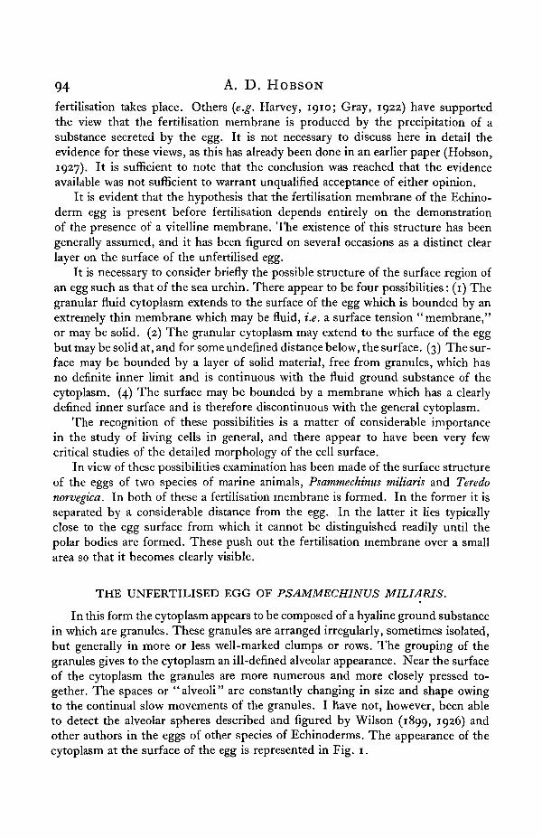
Fig. 2. Psammechinus miliaris. Unfertilised egg. Surface slightly torn with microdissection needle.Note bulging out of cytoplasm. New surface forms acute angle with old. Stippling is diagrammaticand does not represent true distribution and size of granules.
of the surface of the unfertilised egg agree with my own observations, except thatthe clear surface layer appears to be much thicker in Sphaerechinus than in Psamm-echinus. Carter figures and describes the fertilisation membrane arising outsidethe vitelline membrane. In Psammechinus I have never been able to satisfy myselfby direct observation whether or not this is so. The delicacy and similarity inoptical properties of the two structures in question is such that, in Psammechinus,it has so far proved impossible to distinguish them from one another even in eggsin which the fertilisation membrane is only partially formed.
That there is a solid layer at the surface of the unfertilised sea-urchin eggcannot be doubted. The wrinkling of the surface in hypertonic solutions couldscarcely be produced in any other way, unless the whole egg sets to a jelly. Thatthis is not the case is shown by the fact that, when the plasmolysis is slight, slowmovements of granules can still be seen in the cytoplasm.
1 In all the investigations recorded in this paper all high-power observations were made on eggssuspended in a hanging drop in order to avoid as far as possible any effects due to compression.
96 A. D. HOBSON
Prof. Chambers and the writer collaborated during the summer of 1930 in anendeavour to determine the structure of the surface of the unfertilised egg ofPsammechinus miliaris with the aid of the microdissection apparatus. After repeatedexamination with 1/12 in. objective (Leitz) and x 12 compensating eyepiece it wasconcluded that it was impossible to demonstrate with certainty the existence of adefinite vitelline membrane. Under the best optical conditions which we couldobtain, when diffraction was at a minimum, the granules could be seen to approachthe surface of the egg very closely. There remained, however, a thin superficialzone from which granules were absent (Fig. 1). The inner limit of this zone wasnot smooth or sharply marked. The appearance was not that of a differentiatedmembrane such as is implied by the term "vitelline membrane," but rather of apart of the clear matrix of the protoplasm from which granules were absent. Thisview was supported by experiments made with the microdissection needle. Pressingthe needle against the surface of the egg in the plane of optical section failed todispel the appearance of a very thin, clear surface layer. The tip of the needleremained separated by a short distance from the nearest cytoplasmic granules.When the needle was inserted immediately below the surface of the egg and then
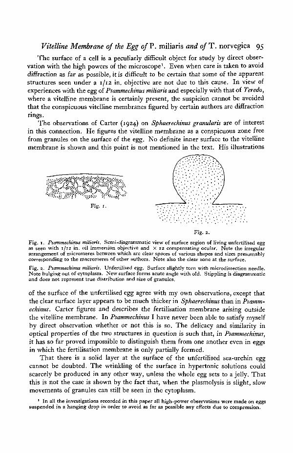
Fig. 3. Psammechinus miliaris. Unfertilised egg. Surface damaged, but not torn, with microdis-section needle. Damaged region of surface pressed out slightly, forming obtuse angle with normalsurface. Stippling is diagrammatic and does not represent true distribution and size of granules.
pulled outwards a cone of gelatinous material was drawn out, but there was nosign of the separation of a membrane as has been described by Chambers (1921,*93°) •
If the surface of the egg is slightly torn with the microdissection needle, thecytoplasm exudes and forms a bulge with a smooth surface attached to the mainpart of the egg by means of a relatively narrow neck (Fig. 2). If the damage hasnot been too great, the exuded portion of the cytoplasm is gradually withdrawnand the egg presents once more a spherical contour. If the damage caused by theneedle is greater, the whole of the exuded cytoplasm may not be incorporated inthe egg again. Whenever exudation of cytoplasm takes place, the undamagedsurface of the egg contracts so that it forms an acute angle with the surface of theoutflowing protoplasm. If, on the other hand, the egg surface is slightly damagedand not actually torn, a rounded elevation develops whose junction with the normalegg surface forms an obtuse angle (Fig. 3).
The results of damaging the egg surface with the microdissection needle showclearly the presence of a solid surface layer. This layer is elastic, as is shown, notonly by the spherical form of the normal egg, but also by the way in which itcontracts when torn, forcing out part of the egg contents, and by the sharp angle
Vitelline Membrane of the Egg of P. miliaris and of T. ftorvegica 97
formed with the newly exposed cytoplasm. Local damage without rupture weakensthe solid surface layer so that a slight protuberance is formed. In such a case thestrength of the damaged surface layer is still sufficient to prevent the formation ofa true exudation of cytoplasm.
The action of hypertonic and hypotonic solutions does not throw much lighton the structure of the surface of the unfertilised egg. As these will be consideredin more detail in a succeeding paper, they will be mentioned only briefly here.
When the eggs are placed in hypertonic solution shrinkage occurs. This processis divisible into two distinct phases. Firstly the egg contracts with a perfectlysmooth surface. Later the surface becomes covered with innumerable fine wrinkles.These become more marked as time goes on and vary, of course, with the strengthof the solution employed. The egg shrinks uniformly and remains approximatelyspherical.
The observed effects can be explained on the view that the egg is surroundedby an elastic, solid layer which is normally in a state of tension. When the volumeof the egg is reduced by removal of water this layer contracts until it is completelyrelaxed. After this, further diminution of the volume of the egg merely results inthrowing the attached surface layer into folds.
The response of the egg to a hypotonic solution such as tap water is characteristic.The egg swells and cytolyses. A slight bulge with a faintly irregular surface appearson one side of the egg. There is no outflow of the egg contents, but the cytoplasmbecomes clear and only slightly granular. When cytolysis is complete the surfaceof the egg is smooth, sharply defined and forms a perfect sphere. The appearancestrongly suggests the presence of a solid membrane which is not destroyed bythe swelling of the egg and which persists, surrounding that part of the contentswhich has been unable to diffuse through it into the surrounding medium.
THE UNFERTILISED EGG OF TEREDO NORVEGICA.
The microscopic examination of the surface of the egg of Teredo norvegicapresents difficulties similar to those encountered in that of Psammechinus. Theeffects of hypertonic and hypotonic solutions, however, enable a definite conclusionto be reached concerning the existence of a vitelline membrane on the unfertilisedegg. The results obtained serve to emphasise the need for great caution in describingthe structure of the surface in the sea-urchin egg.
The egg of Teredo norvegica is about 50 //. in diameter. When shed into seawater by an animal removed from the wood in which it lives, the nucleus is intact,sometimes spherical, but frequently more or less wrinkled. Maturation does notbegin until fertilisation takes place or some method of artificial activation is em-ployed.
Under the high power of the microscope an optical section of the surface regionof this egg presents an appearance somewhat similar to that of Psammechinusmiliaris. The granules of the cytoplasm are slightly larger and seem to be ratherless numerous in the peripheral part of the egg than in the zone round the nucleus.

98 A. D. HOBSON
Surrounding the egg is a narrow, clear layer somewhat wider than that found inthe egg of Psammechinus. A distinct inner surface to this layer could not be madeout. It appeared to be directly continuous with the granular cytoplasm.
A few attempts to separate the clear layer from the rest of the egg with themicrodissection needle failed. The only result was to draw out a cone of clearsubstance like that which has been described for Psammechinus.
When the egg of Teredo is fertilised no immediate visible change takes placein the structure of the surface. The fertilisation membrane is not separated fromthe rest of the egg as in the Echinoderms. The clear layer may be a little thicker,but it is difficult to be certain of this.
When maturation takes place, however, the fertilisation membrane can beseen distinctly at the point where it is stretched over the polar bodies (Fig. 4). Itthen appears as a clear, rather thick membrane which is, except in this part, soclosely adherent to the surface of the egg as to be almost indistinguishable as aseparate structure.
Fig. 4. Teredo norvegica. Surface of fertilised egg showing two polar bodiescausing local elevation of vitelline (fertilisation) membrane.
The existence of a true vitelline membrane on the egg before fertilisation hasbeen accomplished or maturation begun can be demonstrated by the action ofeither hypotonic or hypertonic solutions.
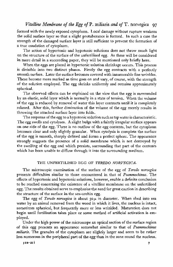
The hypotonic solution used was tap water. The Plymouth tap water is collectedfrom the granite district of Dartmoor and is almost free from dissolved salts.When unfertilised eggs are placed in this medium they cytolyse in about 2 min.as a rule. When cytolysis takes place the egg bursts at one point on its surface.A little jet of granular material emerges from the aperture and gradually dispersesin the surrounding water (Fig. 5a). Then the nucleus, which is now spherical andturgid is forced through the opening which becomes much enlarged by its passage(Fig. 56). Finally the greater part of the granular material of the cytoplasm alsopasses out of the opening (Fig. 5 c). All that remains of the egg is a collapsed andmuch crumpled, clear membrane to which are still adhering a few scattered granules
(Fig- 5<9-The hypertonic solutions employed were made either by evaporating sea water
to about 35 per cent, of its original volume or by the addition of 2-4 M NaCl tonormal sea water.
If unfertilised eggs are put into a hypertonic medium they assume a verycharacteristic form. The original spherical shape is lost. The surface becomesindented in one or more places and the appearance is that of a ball of plastic sub-stance, such as clay, squeezed between the finger tips. This shape is similar to the
Vitelline Membrane of the Egg of P. miliaris and of T . norvegica 99
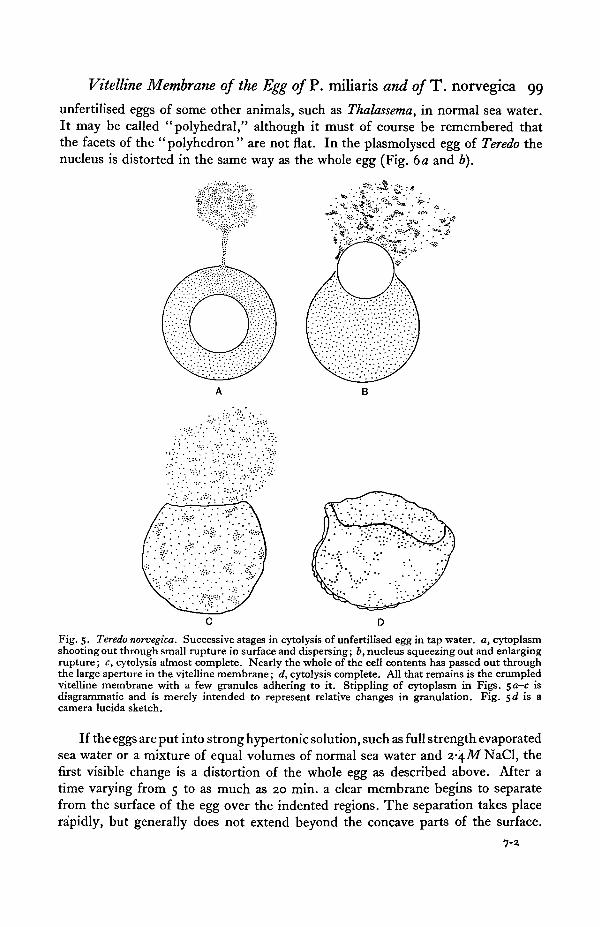
unfertilised eggs of some other animals, such as Thalassema, in normal sea water.It may be called "polyhedral," although it must of course be remembered thatthe facets of the "polyhedron" are not flat. In the plasmolysed egg of Teredo thenucleus is distorted in the same way as the whole egg (Fig. da and b).
Fig. 5. Teredo norvegica. Successive stages in cytolysis of unfertilised egg in tap water, a, cytoplasmshooting out through small rupture in surface and dispersing; b, nucleus squeezing out and enlargingrupture; c, cytolysis almost complete. Nearly the whole of the cell contents has passed out throughthe large aperture in the vitelline membrane; d, cytolysis complete. All that remains is the crumpledvitelline membrane with a few granules adhering to it. Stippling of cytoplasm in Figs. $a—c isdiagrammatic and is merely intended to represent relative changes in granulation. Fig. sd is acamera lucida sketch.
If the eggs are put into strong hypertonic solution, such as full strength evaporatedsea water or a mixture of equal volumes of normal sea water and z-\M NaCl, thefirst visible change is a distortion of the whole egg as described above. After atime varying from 5 to as much as 20 min. a clear membrane begins to separatefrom the surface of the egg over the indented regions. The separation takes placerapidly, but generally does not extend beyond the concave parts of the surface.

IOO A. D. HOBSON
The result is that, at first, the egg is irregular in shape and is enclosed within aspherical membrane. Gradually the protoplasmic surface becomes smooth androunded, so that finally the egg is more or less spherical but is still attached to themembrane over part of its surface. Occasionally the separation of the membraneis complete.
These facts can be explained on the assumption that a tough elastic membranesurrounds the unfertilised egg. This membrane is firmly attached to the surfaceof the protoplasm but is not directly continuous with it. The strain in the membraneis greatest in the concave areas where it is most distorted, and here it will separatemost readily and, once separated, it will assume its natural spherical form.
The firmness with which the membrane is attached to the surface of the proto-plasm can be seen by examining the process of separation. As the membranedraws itself away from the surface of the egg, strands of protoplasm are pulled outwith it. These may be slender threads or broad, flame-shaped processes. This isfollowed by slow retraction of these strands which seem as though composed of a
Fig. 6. Teredo norvegica. Two unfertilised eggs in hypertonic solution showing the type of plasmo-lysis. The indentations are often more numerous and complicated than in these examples.
viscous fluid. During retraction a strand often breaks in the middle, and in thiscase each half contracts rapidly, the one to form a small cone which graduallydisappears on the surface of the egg, and the other persists as a globule of cytoplasmattached to the inner surface of the separated membrane.
This type of behaviour is of particular interest, as it shows clearly that evenintense plasmolysis is not accompanied by gelation of the protoplasm. The appear-ance described could hardly be produced unless the consistency of the cytoplasmwas that of a somewhat viscous fluid. This is supported by the fact already men-tioned that slow movement of granules can be seen both in the normal and in theplasmolysed egg. The polyhedral form of the egg must therefore be due to theproperties of the vitelline membrane and not to the gelation of the protoplasm.
After fertilisation and even after cleavage the attachment of the membrane tothe surface of the egg or the blastomeres is hardly less firm than in the unfertilisedegg. Exactly the same phenomena occur in hypertonic solutions, although theseparation of the membrane takes place more rapidly.
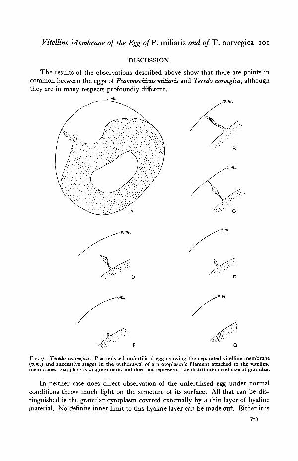
Figs. 7 and 8 illustrate typical examples of the cytoplasmic processes describedabove and their withdrawal.
Vitelline Membrane of the Egg of P. miliaris and of T. norvegica 101
DISCUSSION.
The results of the observations described above show that there are points incommon between the eggs of Psammechinus miliaris and Teredo norvegica, althoughthey are in many respects profoundly different.
v.m.
v. m.
v.m.
Fig. 7. Teredo norvegica. Plasmolysed unfertilised egg showing the separated vitelline membrane(v.m.) and successive stages in the withdrawal of a protoplasmic filament attached to the vitellinemembrane. Stippling is diagrammatic and does not represent true distribution and size of granules.
In neither case does direct observation of the unfertilised egg under normalconditions throw much light on the structure of its surface. All that can be dis-tinguished is the granular cytoplasm covered externally by a thin layer of hyalinematerial. No definite inner limit to this hyaline layer can be made out. Either it is
7-3

I O 2 A. D. HOBSON
continuous with the general cytoplasm or is so closely attached to the true cyto-plasmic surface that it cannot be distinguished from it. The behaviour of the eggsof Teredo in hypertonic solutions strongly suggests the latter possibility. Tap waterserves as an agent by means of which the solid, hyaline surface layer may beseparated from the rest of the egg.
v.m.
v.m.
B
v.m. v. m. v.m.
C D EFig. 8. Teredo norvegica. Plasmolysis of an egg which has been fertilised and has undergone cleavage,showing successive stages in the withdrawal of two protoplasmic filaments, a and b, attached to thevitelline membrane (v.m.). The filaments at c were attached to the upper part of the vitelline mem-brane. Filament b eventually broke in the middle and the outer part formed a small globule ofprotoplasm attached to the inner surface of the vitelline membrane. Note that the vitelline (ferti-lisation) membrane is more completely detached from the surface of the egg than in the unfertilisedegg. Stippling is diagrammatic and does not represent true distribution and size of granules.
The eggs of Psammechinus are such that they do not lend themselves to anequally clear demonstration of a separate surface layer. Chambers (1921) hasdescribed the removal of the vitelline membrane from Echinoderm eggs by meansof the microdissection needle. Lack of success in similar experiments on the eggof Psammechinus does not necessarily mean that such a membrane does not exist,since, as has been described above, the egg of Teredo also resisted efforts to dissectaway the vitelline membrane. It merely serves to emphasise the firmness withwhich the membrane, if present, is attached.
The behaviour of the egg of Psammechinus in hypertonic solutions and in tap

Vitelline Membrane of the Egg of P. miliaris and of T. norvegica 103
water lends support to the view that the surface is covered by a clear, elastic layer.It is difficult to account for the smooth, spherical condition of the egg whencytolysed in tap water in any other way.
If we assume, as a working hypothesis, the existence of a distinct vitellinemembrane on the surface of the eggs of these two forms, it becomes possible tosuggest a tentative explanation of certain difficulties in the problem of the originof the fertilisation membrane.
In the Echinoderms the origin of the fertilisation membrane has for long beena controversial matter. The most recent general discussions are by Hobson (1927)and Runnstrom (1928). While some consider that the fertilisation membraneis a new structure formed as the result of fertilisation, it is more generally held thatthe vitelline membrane is merely lifted from the surface of the egg. It has beenobjected by the present writer among others that the fertilisation membrane is sodifferent in its properties from the vitelline membrane as to render it difficult ofidentification with the latter.
The first step in this identification is obviously to establish the existence of adifferentiated vitelline membrane on the unfertilised egg. As has already beenshown, this is by no means an easy matter and, although the evidence is in favourof it, its existence cannot be considered a certainty in Psammechinus.
The Echinoderm fertilisation membrane has properties which are certainlydifferent from those which can be ascribed to the hyaline surface layer of theunfertilised egg. It is relatively thick, rigid and almost inelastic. Before thefertilisation membrane has reached its final condition, i.e. during the process ofraising it from the surface of the egg, it is elastic and extensible, thus resemblingthe hyaline surface layer. The change in the properties of the fertilisation membraneafter it has left the surface of the egg is dependent on the presence of calcium inthe sea water. If eggs are fertilised in normal sea water and transferred within1 min. to Ca-free sea water the fertilisation membranes arise as usual. Gradually,however, they sink back on to the surface of the egg so that they can scarcelybe distinguished with the 1/12 in. objective. The process of sinking back may takeup to 30 min. to complete, although, as a rule, it is accomplished more rapidly.Prof. Chambers and the writer found that if, after the fertilisation membrane hadsunk back again, the surface of the egg was punctured in calcium-free sea waterso as to cause outflow of cytoplasm, some of the material penetrated below thefertilisation membrane and caused it to rise up once more.
The explanation seems to be simple. Elevation of the fertilisation membraneis accomplished by means of osmotic pressure due to the presence of an amphotericelectrolyte released at fertilisation. This substance can probably diffuse slowlythrough the membrane, as has been shown by Garrey (1919) for peptone. Unlessthe membrane is hardened by the sea water it will collapse when the osmoticallyactive substance is removed. When a further supply of similar material appearsbeneath the membrane, as when the egg is torn, it rises once more. This view isalso supported by the fact that removal of eggs with collapsed membranes to normalsea water does not cause a second elevation.
104 A. D. HOBSON
The mechanical properties of the fertilisation membrane when it first appearsare somewhat similar to those we have postulated for the hyaline surface layer orvitelline membrane. These properties are maintained for an indefinite period solong as calcium is absent from the surrounding medium.
It-is well known that hypertonic sea water is effective in causing parthenogenesisin the eggs of many species of animals. Its action appears to be twofold. Firstlyit causes the formation of asters. Secondly it is a membrane-forming agent. Informs such as Teredo where the vitelline membrane is comparatively looselyattached to the surface of the egg and in which there is no mechanism for elevatingit as the fertilisation membrane, the latter is formed in the hypertonic solution.In the sea-urchin egg, on the other hand, there is a well-developed mechanismfor elevating the fertilisation membrane, and if we accept the identification of thisstructure with the vitelline membrane we may perhaps regard the action of hypertonicsolutions in artificial parthenogenesis as loosening the attachment of the membraneto the surface of the egg. Elevation of the fertilisation membrane does not takeplace until the egg is removed to normal sea water, since the increased concentrationof salts in the hypertonic solution lowers the osmotic pressure due to the proteinwithin the membrane (Hobson, 1927).
SUMMARY.
1. Direct microscopic examination of the unfertilised eggs of Psammechinusmiliaris and Teredo norvegica merely shows the existence of a thin, granule-freezone covering the surface. Whether this is continuous with the general cytoplasmor not cannot be made out with certainty by direct observation.
2. A cone of clear material can be drawn out from the surface of the unfertilisedegg of both species by means of the microdissection needle. A definite membranecannot be separated in this way.
3. Hypertonic solutions cause the egg of Psammechinus to shrink smoothlyat first and later to become wrinkled. This is consistent with the view that the eggis surrounded by an elastic, solid layer which is normally in a state of tension.
4. Cytolysis of the egg of Psammechinus in tap water is not accompanied bybursting. The egg swells and is perfectly smooth and spherical when cytolysis iscompleted. This points to the existence of an elastic, solid surface layer.
5. Plasmolysis of the egg of Teredo is of the type here called "polyhedral."The irregular shape of the egg in the hypertonic solution is only temporary, as aclear membrane separates from the concave surfaces and the egg then becomesmore or less spherical.
6. The protoplasm of the plasmolysed egg of Teredo behaves as a viscous fluid.7. Cytolysis of the egg of Teredo in tap water is accompanied by bursting and
dispersion of the entire cell contents. A crumpled membrane alone remains.8. It is concluded that the unfertilised egg of both Teredo norvegica and Psam-
mechinus miliaris is surrounded by an elastic vitelline membrane which is much
Vitelline Membrane of the Egg of P. miliaris and of T. norvegica 105
thicker in the former than in the latter. The vitelline membrane in both cases istightly attached to the egg surface.
9. In calcium-free sea water the fertilisation membrane is elevated normallyin Psammechinns miliaris. It does not harden, however, and gradually sinks backon to the surface of the egg owing, apparently, to the loss by diffusion of the osmo-tically active substance in the peri vitelline space. It can be elevated a second timeby puncturing the surface of the egg and allowing some of the cell contents topenetrate into the perivitelline space.
10. It is suggested that one action of hypertonic solutions in inducing artificialparthenogenesis may be to cause a loosening of the attachment of the vitellinemembrane to the egg surface.
This work has been done at the Laboratory of the Marine Biological Associationat Plymouth, partly during the tenure of the Ray Lankester Investigatorship, tothe trustees of which I am very grateful. I wish to express my thanks also to DrE. J. Allen, F.R.S., and to the staff of the laboratory for their interest and assistancein many ways. I am indebted to the Royal Society and the British Association forthe use of tables, during the tenure of which part of this work was done. Part ofthe expenses were defrayed by a grant from the Earl of Moray Endowment of theUniversity of Edinburgh.
THE FOLLOWING NOTE IS ADDED BY PROF. R. CHAMBERS(August, 1931).
The question as to whether the fertilisation membrane pre-exists as some sortof a membrane on the surface of an unfertilised Echinoderm egg is a difficult oneto answer.
In the case of the starfish common at Woods Hole (Asterias rubens) a vitelline mem-brane undoubtedly exists on the unfertilised egg, and it is this membrane whichseparates off to form the fertilisation membrane (Chambers, 1921). The difference inconsistency of the membrane before and after separating can be demonstrated byinjecting a small amount of sea water under the membrane, and then fertilisingthe egg. The injection causes the elevation of a local blister, the contour of the eggremaining uniform. A pronounced adhesion of the membrane to the surface ofthe egg is indicated by the fact that the blister remains localised with a sharp angleat the edges of the blister. When the egg is fertilised the separation of the membranespreads over the egg and it is important to note that, with insemination of the egg,the membrane stiffens and the localised blister flattens out with a correspondingsinking in of the surface of the egg at the region where the blister had been pre-viously. The indentation in the egg gradually diminishes and the egg returns toits spherical shape with a more or less uniformly contoured fertilisation membranesurrounding it.
In the case of the unfertilised Arbacia egg it is far more difficult to demonstratethe existence of a vitelline membrane. This can be done, however, by centrifuging
106 A. D. HOBSON
the egg at a rate of 8000 revolutions per sec, for 2 to 3 min. The forces elongatethe egg into a cylindroid. At the heavier pole of the cylindroid it is possible topull off, with microneedles, the remains of a wrinkled and tenuous membrane.Apparently the centrifugal force has ripped the membrane and carried it as acollapsed sheet to the heavier pole of the egg. These eggs undergo fertilisationwith no development of a fertilisation membrane. In the case of an unfertilisedArbacia egg it has been impossible, by means of microneedles, to pick off any suchmembrane. In this respect these eggs resemble those examined by Mr Hobsonand myself at Plymouth. However, by pricking the surface of the Arbacia egg atseveral spots, or by pushing the egg about as it adheres to the glass coverslip, theegg so treated will, when inseminated, develop an incomplete and collapsed ferti-lisation membrane.
If we are to conclude that the fertilisation membrane has as its precursor avitelline membrane the fact remains that this precursor is far weaker and moredelicate than the membrane which results after insemination.
REFERENCES.CARTER, G. S. (1924). Proc. Comb. Philosoph. Soc, Biol. Ser. 1, 79.CHAMBERS, R. (1921). Biol. Bull. 41 j 318.
(1930). Biol. Bull. 58, 344.GARREY, W. E. (1919). Biol. Bull. 37, 287.GRAY, J. (1922). Quart. Journ. Micro. Sci. 66, 419.HARVEY, E. N. (1910). Journ. Exp. Zool. 8, 355.HOBSON, A. D. (1927). Proc. Roy. Soc. Edin. 47, 94.RUNNSTRSM, J. (1928). Protoplasma, 4, 388.WILSON, E. B. (1899). Journ. Morph. 15, Suppl. 1.
(1926). Amer. Nat. 60, 105.