On the Right Trach? - · PDF fileOn the Right Trach? ... Anisa Warsame. 2 Contents...
155
On the Right Trach? A review of the care received by patients who underwent a tracheostomy
Transcript of On the Right Trach? - · PDF fileOn the Right Trach? ... Anisa Warsame. 2 Contents...

On the Right Trach?A review of the care received by patients who underwent a tracheostomy

1
On the Right Trach?A review of the care received by patients who underwent a tracheostomy
A report by the National Confidential Enquiry into Patient Outcome and Death (2014)
Compiled by:
K A Wilkinson FRCA FRCPCH NCEPOD Clinical Co-ordinator (Anaesthetics)Norfolk and Norwich University Hospitals NHS Foundation Trust
I C Martin LLM FRCS FDSRCSFormer NCEPOD Clinical Co-ordinator (Surgery)City Hospitals Sunderland NHS Foundation Trust
H Freeth BSc (Hons) MSc RGN MSc – Clinical Researcher
K Kelly BA (Hons), PGC Health Research – Researcher
M Mason PhD – Chief Executive
Study proposed by:The Association of Anaesthetists of Great Britain and Ireland
The authors and Trustees of NCEPOD would particularly like to thank the NCEPOD staff for their work in collecting and analysing the data for this study: Robert Alleway, Aysha Butt, Donna Ellis, Dolores Jarman, Eva Nwosu, Karen Protopapa, Hannah Shotton, Neil Smith and Anisa Warsame.
2
Contents
Acknowledgements 3
Foreword 5
Principal recommendations 9
Introduction 11
Chapter 1 - Method and Data Returns 13
Chapter 2 - The organisation of care 21
Key findings and recommendations 45/46
Chapter 3 – Tracheostomy insertion 47
Case study 1 54
Case study 2 56
Case study 3 57
Case study 4 62
Case study 5 63
Key findings and recommendations 75
Chapter 4 – Tube care in the patient with 77 a tracheostomy
Case study 6 80
Case study 7 83
Case study 8 84
Key findings and recommendations 91
Chapter 5 – The multidisciplinary care of 93 tracheostomy patients
Case study 9 96
Key findings and recommendations 101
Chapter 6 – Complications and adverse events 103
Case study 10 105
Case study 11 106
Case study 12 107
Key findings and recommendations 113
Chapter 7 – Outcomes of care in 115tracheostomy patients
Case study 13 121
Case study 14 124
Case study 15 127
Key findings and recommendations 130
Chapter 8 – Summary 131
References 133
Appendices 135
1 - Glossary 135
2 - Type of tracheostomies 138
3 - Algorithms for the emergency management of 142 tracheostomies and laryngectomies
4 - Adapted surgical WHO checklist 144 for tracheostomy5 - The role and structure of NCEPOD 145
6 - Participation 147
3
This report, published by NCEPOD, could not have been achieved without the support of a wide range of individuals who have contributed to this study.Our particular thanks go to:
The Expert Group who advised NCEPOD on what to assess during this study:
Anita Simonds, Professor in Respiratory and Sleep Medicine Andrew Bodenham, Consultant in Anaesthesia and Intensive Care MedicineAbhiram Mallick, Consultant in Anaesthesia and Intensive Care MedicineAnna Batchelor, Consultant in Anaesthesia and Intensive Care MedicineBrendan McGrath , Consultant in Anaesthesia and Critical CareCatherine Plowright, Consultant Nurse Critical CareFrank Stafford, Consultant Otolaryngologist Head and Neck SurgeonGeorge Findlay, Consultant in Intensive Care MedicineKaren James, PhysiotherapistMaura McElligot, Consultant Critical Care NursePeter Ramsay-Baggs, Consultant Oral and Maxillofacial SurgeonSara Payne, Patient RepresentativeSarah Wallace, Speech and Language Therapist
The Advisors who peer reviewed the cases:
Akeel Jubber, Consultant Physician in General and Respiratory MedicineAmanda Thomas, Clinical Specialist PhysiotherapistAnna Perks, Consultant AnaesthetistBen Creagh-Brown, Consultant Physician Respiratory and Critical Care MedicineCarlos Gomez, Consultant Intensive Care and AnaesthesiaChris Kirwan, Consultant in Critical Care and Renal MedicineClaudia Russell, Tracheostomy Nurse ConsultantCoral Hulse, Nurse Consultant, Critical Care OutreachCyrus Kerawala, Consultant Maxillofacial/Head and Neck SurgeonDiane Goff, Speech and Language TherapistEmma McNeill, Clinical Fellow in ENTErica Everitt, Tracheostomy Specialist PractitionerGregor McNeill, Consultant in Acute and Critical Care MedicineGuri Sandhu, Consultant OtolaryngologistHelen Drewery, Consultant AnaesthetistJackie McRae, Specialist Speech and Language Therapist Jane Hansell, Speech and Language TherapistJeremy Sharp, ENT/Head and Neck Consultant SurgeonJoanna McCormick, Nurse Consultant, Critical CareJulie Carter, Head and Neck Nurse PractitionerKatharine Hunt Consultant AnaesthetistKhalid Ghufoor, Consultant Otolaryngologist/Clinical Tutor RCSELouise Platt, Therapy Team LeaderLucy Bates, Consultant in Anaesthesia and Intensive Care MedicineLynne Clark, Speech and Language Therapist
ACkNOwlE
DGEMEN
TS
Acknowledgements
Back to contents
4
Madhankumar Vijayakumar, Consultant in Intensive Care AnaesthesiaMaria Rogers, Specialist Nurse - ENT/MaxillofacialMary Edwards, Critical Care Outreach Nurse LeadMichael Davies, Consultant Respiratory PhysicianMichael Fardy, Consultant Maxillofacial/Head and Neck SurgeonMichael Ho, Head and Neck FellowOlive Wilkinson, Clinical Specialist PhysiotherapistPeter Dziewulski, Consultant Plastic and Reconstructive SurgeonRobert Banks, Consultant Oral and Maxillofacial SurgeonSheila Goodman, Critical Care Audit and Research Sister, RCN Critical Care and In-Flight Nursing Forum
Stephen Hutchinson, Consultant AnaesthetistStephen Webb, Consultant in Anaesthesia and Intensive Care MedicineSue McGowan, Clinical Specialist Speech and Language TherapistTaran Tatla, ENT Head and Neck Consultant SurgeonTim Strang, Consultant AnaesthetistWendy Huskinson, Ward Manager, Head and Neck and ENT
Thanks also to all the NCEPOD local Reporters, NCEPOD Ambassadors, Study Specific contacts and the clinicians who completed questionnaires.
ACkNOwlE
DGEMEN
TS
5
There is said to be nothing new under the sun, but tracheostomy takes that proposition to extremes. It may be the only surgical procedure that can be found in both Egyptian records of over 3,500 years BC and the Rig Veda, one of the fundamental texts of Hinduism that also predates the Ramayana by more than a millennium. And it has long been recognised as dangerous: by the 320s BC, when Alexander the Great is supposed to have used his sword to relieve a soldier’s upper airway obstruction, Hippocrates had already warned against the procedure, because of the risk of life-threatening haemorrhage from damage to the carotid arteries. If the value and the dangers of a surgical airway have both been recognised for 2,500 years, it may be surprising that this is the first time there has been a nation-wide study of the quality of care that is delivered to this specific group of patients. This is especially so since in modern times it has moved far beyond being a last ditch expedient to save life. Indeed this study suggests that it is performed about 12,000 times a year in our hospitals. The major change in recent years has been the introduction of percutaneous procedures, now usually performed on the critical care ward, as an alternative to the formal surgical procedure undertaken in the operating theatre. These were introduced in 1985 and made up 70% of our study population.
The dangers that so impressed our forebears, such as obstruction and secondary wound infection have proved manageable in the hands of highly skilled staff who are expressly trained to recognise and manage such complications swiftly. In addition, the staff have to be equipped to handle haemorrhage and accidental decannulation safely and confidently. Whilst the guidance is clear, it is the implementation of good practice across a complex care pathway that NCEPOD
has followed in this study. Our Advisors have been able to suggest improvements at every stage.
The acknowledged background to this study is that NHS funds are under explicit pressure as never before. One of the first casualties when services are under pressure, both from the volume of work and the lack of financial resources, is likely to be training. Patients who are at such risk of respiratory compromise that they may need emergency intervention to relieve airway obstruction, depend upon a highly trained team being readily available. The old stability of personnel within the “Firm” is also now unusual: there is a constant turnover of staff in ITU, as there is in HDU and on Level 1 wards. The only way in which hospitals can maintain safe teams is to recognise training as a continuous process, an intrinsic part of the routine work. To find that over a quarter of hospitals managing these patients say that their staff do not receive training in the management of blocked and displaced tubes seems to be a remarkable discovery. I hope that it will be recognised as an organisational red flag because the vital skills in relation to the “ABC” approach to the patient must be universally available wherever the need is a predictable part of the patient’s pathway. “A” must come first, whether it is the patients own Airway or an Adjunct to that airway (such as a tracheostomy). In addition when the need can and should be anticipated, there has to be someone there who is trained and who has kept their skills up to date so that they can reliably recognise and change obstructed and displaced tubes. One useful role for NCEPOD is to provide an amplifier for the professional voices who need to insist to management that training is not an optional extra or a one-off episode. It has to be part of the day to day work of a unit managing these patients. There is no excuse
FORE
WORD
Foreword
Back to contents
6
for ignoring the National Tracheostomy Safety Project – especially since their 2013 Manual can be downloaded as an App for the manager’s mobile phone.
Less obvious to the layman may be the need for appropriately trained specialist physiotherapists. Or for speech and language therapists who are trained to perform fibreoptic examination of swallowing. A properly set-up unit aiming to deliver optimal care will also have nutritionists, who are trained to look after the complex needs of these patients and whose contribution is respected by the rest of the team.
It is worrying to find that so many places may be doing badly in so many of these respects.
As usual, it is hard to tell whether these corners are being cut because of the lack of resources in a service striving to respond to the Nicholson Challenge. Training is not cheap, nor is a full range of specialists, but other issues that can readily be resolved within shrinking resources do not seem to be faring much better. Keeping a simple list of those who have been trained to provide these services costs nothing and may save lives. The essence of this problem is that you should not have to look round to find someone who is appropriately trained when a predictable emergency arises. To find that such straightforward advice is being widely ignored in hospitals up and down the country is hard to understand. Most of the time you do not need such a list, because the responding nurse knows perfectly well who to call: but the service has to cater for the new locum or bank nurse who suddenly finds herself/himself on their own.
A service that is going to deliver this sort of airway support safely and reliably as well as responding to the emergencies that will inevitably arise is a bit like a three-legged stool: it must have the right staff, the right equipment and the right systems if it is not going to fall over. And they must all be in place and readily accessible.
Those of us with an interest in risk management were impressed to see the spread of WHO checklists
from Operating Theatres to Critical Care Units. The Checklist emphasises the importance of planning, of the methodical approach of pausing to identify who is here and why? What are we going to do and what do we need to check before we do it? Something that happens 12,000 times a year needs to be a routine straightforward process, even though it may be immediately necessary to save the life of a sick and frail person.
This report also casts an interesting light on the problems encountered in moving patients safely on to the next stage in their management. In order for these patients need to be discharged, from both the ITU to the wards and from the wards to the community, appropriate support mechanisms need to be in place. Mostly it works well, but our Advisors did find room for improvement.
The Advisors, who as usual represent the mainstream of professional opinion amongst people who deliver this sort of care, are always asked to identify cases where there is room for improvement under three distinct headings: first in the clinical care delivered to these patients, second in the organisation of that care, and third cases where there was room for improvement in both. Naturally, the third is usually the smallest of the three groups. I think it is telling that in this case the third group is the largest, because it points to the close interdependence of training, the provision of equipment and the organisation of care with the clinical delivery of that care. On these findings, places that are falling down clinically are likely to have sub-optimal organisation as well.
As usual we are indebted to all those who have co-operated to make this report happen. There is the usual team of NCEPOD people who have been built up over decades and who are the basis of all our work, the Local Reporters and Ambassadors who ensure that the mechanics of our studies are possible. We must also acknowledge the commitment of individual doctors who have written reports on their own cases, and the staff and co-ordinators who have written the study.
FORE
WORD
7
More than usual I am aware that there is a dedicated group of people who had already recognised the problems and will be waiting anxiously to see what we have found. They come from the Intensive Care Society, the National Tracheostomy Safety Project and the other professional groups who are determined to see that the quality of care received by these 12,000 patients a year improves consistently. The Association of Anaesthetists proposed this study and they also provided many of the expert group who, with representatives of the entire multidisciplinary team, designed a national study that would address the questions that they wanted answered. As usual, we must acknowledge the Advisors, our unpaid volunteers who give up so much of their time to scrutinise the care that their service is delivering.
I am grateful to all of you for the work you have done and for providing the opportunity for NCEPOD to be of service. As usual, we will be providing a toolkit to go with this study, which will enable individual centres to benchmark themselves according to these criteria and to identify where they specifically have room for improvement.
Bertie LeighNCEPOD Chair
FORE
WORD

88
9
In order to facilitate decannulation and discharge planning multidisciplinary care needs to be established as part of routine pathway for ALL tracheostomy patients. Whilst on the critical care unit there should be at least daily review, key additional team members should be involved at an early stage. The team composition should be flexible to properly reflect the patient’s needs and provide excellent continuity of care. There are several key team members who one would expect should always participate, e.g. physiotherapy, speech and language therapy, outreach nurses and dietitians. Hospitals need to provide adequate staff to ensure this happens routinely and in a timely manner. (Clinical Directors and Critical Care Managers)
Bedside staff who care for tracheostomy patients must be competent in recognizing and managing common airway complications including tube obstruction or displacements and as described by the National Tracheostomy Safety Project algorithms. (Medical Directors and Directors of Nursing)
Unplanned and night time critical care discharge is not recommended, particularly in patients with a newly formed tracheostomy and/or patients recently weaned from respiratory support. This reinforces the Intensive Care Society’s general recommendation about night time discharges. (Clinical Directors and Risk Managers)
Tracheostomy insertion should be recorded and coded as an operative procedure. Data collection in all locations should be as robust as that for a theatre environment. This will facilitate better care planning and allow for national and local review and audit. (Medical Directors and National Coding Systems)
The diameter and length of the tube used should be appropriate for the size and anatomy of the individual patient, therefore an adequate range of tracheostomy tubes needs to be stocked by units. Operators should be aware of the types of tube available and in particular recognize that adjustable flanged tubes are available with inner tubes. Professionals need to continue to work closely with manufacturers to optimise design and tube options for a non standard population. (Consultant Operators, Theatre and Critical Care Managers and Professional Health Care Bodies)
All Trusts should have a protocol and mandatory training for tracheostomy care including guidance on humidification, cuff pressure, monitoring and cleaning of the inner cannula and resuscitation. The clinical practices around tracheostomy care should be the subject of local quality improvement initiatives. Tube data should be more clearly recorded and made available for review at bedside and thereafter facilitated by a ‘passport’ for each patient, with all data included. (Medical Directors, Directors of Nursing and Health Care Commissioners)
PRIN
CIPAL R
ECOM
MEN
DATIONS
Principal recommendations
Back to contents

10
11
Introduction
UK data published after the NCEPOD study had commenced has shown that there is no improvement in long term outcomes in patients who have a tracheostomy placed at an early or late stage on critical care.5 Therefore whilst performing a tracheostomy is generally considered a safe procedure with a low complication rate with important benefits such as greater patient comfort, there is still some controversy over the timing and risks of insertion in the critically ill patient. It is important to acknowledge that the alternative (longer term endotracheal intubation) is not itself without complications.
Whilst the basis for national competences for tracheostomy care exist, it is clear that they are not yet fully integrated into mandatory training programmes for all health professionals. The emergence of the Global Tracheostomy Collaborative6 acknowledges that tracheostomy care is an important priority for many modern health care systems, with a membership which ranges from medical students to Harvard professors. Both this initiative and the NTSP also recognise the very important needs of children as well as the very much larger adult population with tracheostomies, and the importance of professionals working collaboratively to share knowledge and expertise.
In parallel the multidisciplinary team in the hospital caring for any patient with a tracheostomy remains large. Part of the challenge of this report has been to carefully consider all the levels of expertise and to provide a useful summary of what is a very large data set and prioritising the recommendations which have emerged (many of which have been already made by other organisations). Ultimately we have provided six key recommendations which we hope will resonate with all those involved in the care of tracheostomy patients, as well as patients themselves, and on which
INTR
ODUCTION
Historically tracheostomy has been used to remedy upper airway obstruction, to avoid the laryngeal complications of prolonged tracheal intubation and the continued need for the protection and maintenance of the airway in patients with severe neurological injury. It is also now often planned relatively early in the stay of patients on critical care to improve patient comfort, and facilitate weaning of sedation when there is a need for a longer period of ventilation, and the number of temporary tracheostomies has greatly increased in recent years. The development and refinement of the percutaneous technique, improved equipment and the increasing number of critical care physicians trained to perform the procedure have all enabled a temporary tracheostomy to be placed as a bedside procedure. Alongside these developments there has been initiatives such as the National Tracheostomy Safety Project (NTSP)1 and guidance on best practice2 which have provided clearer standards of care for the patient.
From 2005 to 2007 the National Patient Safety Agency (NPSA) collected data submitted from 150 Trusts which showed that 53/1085 (5%) of airway incidents reported related to tracheostomies.3 Fourteen of the 53 incidents were classed as major or life threatening, and it was recognised by the authors that it was likely that only around 10% of all incidents were reported. The fourth National Anaesthesia Audit Project4 was specifically set up to examine the frequency and characterise the importance of serious airway related complications, and reported from all age groups and in all hospital locations across the UK over a 12 month period. Many different airway devices were implicated in these events, but in critical care the most serious incidents frequently related to tracheostomy. In half of all airway-related deaths and cases of brain damage in critical care the airway problems were attributed to tracheostomy complications.
Back to contents
12
broader issues which impact upon the care of sick and complex patients. These are not unexpected and include the greater numbers of overweight and obese patients that require critical care, as well as revealing the pressure to admit and discharge relatively complex patients at all times of the day and night.
INTR
ODUCTION
action is most likely to result in significant improvements in care.
This study was undertaken to help identify the difficulties in the pathway of care for patients with a tracheostomy and in various hospital settings. The NCEPOD report has also highlighted many of the
13
Expert Group
A multidisciplinary group of experts comprising health care professionals from intensive care medicine, anaesthesia, respiratory medicine, critical care nursing, ear, nose and throat surgery, maxillofacial surgery, physiotherapy, speech and language therapy, and a lay representative contributed to the design of the study and reviewed the findings.
Aim
The primary aim of this study was to explore factors surrounding the insertion and subsequent management of tracheostomies in both the critical care unit and ward environments by:• Exploring(percutaneousandsurgical)
tracheostomy-related complications following insertion in the operating theatre or the critical
care unit• Exploringremediablefactorsinthecareof
adult patients (aged 16 and over) undergoing the insertion of a surgical or percutaneous tracheostomy tube
• Assessingthenumberandvariabilityofpercutaneous tracheostomies performed annually in the critical care unit
• Makingrecommendationstoimprovefuturepractice.
Objectives
The expert group identified a number of areas of tracheostomy care to be explored in more detail. These included:• Insertionofthetracheostomy
- Indications for the tracheostomy- Cautions and contraindications
- Consent- Delays- Equipment and monitoring- Staffing- Anaesthesia
• Environmentinwhichthetracheostomytubewasinserted and cared for
• Routinecare- Essential equipment - Cuff management- Humidification- Suctioning- Inner cannulae- Dressings - Swallowing- Oral care- Communication needs
• Changingtracheostomytubes• Emergencies,commoncomplicationsandtheir
management• Decannulationandlongterm(30day)followup• Facilities
- Staff capacity- Staff competency- Number of patients cared for- Training- Facilities available- Policies and procedures
Hospital participation
Data were collected from all hospitals where the insertion of a tracheostomy tube was undertaken in England, Wales, Northern Ireland, the Channel Islands and the Isle of Man. Data were collected from both the National Health Service (NHS) and the Independent sector where applicable.
1 – Method and Data returns
1 – M
ETHOD A
ND
DATA R
ETURN
S
Back to contents
14
Within each hospital, a named contact, referred to as the NCEPOD Local Reporter, acted as a link between NCEPOD and hospital staff, facilitating case identification, dissemination of questionnaires and data collection.
Study population
Patients who underwent a new tracheostomy insertion or a laryngectomy between 25th February – 12th May 2013, were included in the study. Patients were identified at the time of tracheostomy insertion or laryngectomy on the critical care unit or in theatre. Data were collected on both surgical and percutaneous tracheostomies. Where available, the following OPCS codes were used to identify patients.• E29–Excisionoflarynx
- E29.1 - Total laryngectomy- E29.6 - Laryngectomy not elsewhere classified- E29.8 - Other specified- E29.9 - Unspecified
• E42–Exteriorisationoftrachea- E42.1 - Permanent tracheostomy- E42.3 - Temporary tracheostomy- E42.8 - Other specified- E42.9 - Unspecified
Exclusions
Only patients who underwent the creation of a new tracheostomy were included in the study. Therefore patients who were coded with the following OPCS codes were excluded:
- E42.2 - Cricothyroidostomy- E42.4 - Revision of tracheostomy- E42.6 - Replacement of tracheostomy- E42.5 - Closure of tracheostomy- E42.7 - Removal of tracheostomy tube
Patients aged 15 and younger were not included in the study.
Case identification
Patients were identified at the point of tracheostomy insertion either on the critical care unit or in theatre.
A study contact was set up in the critical care unit and in theatre, and one of their main roles was to identify cases and notify the details of the cases to NCEPOD (either directly or via the Local Reporter).
Once a patient was identified as having undergone a tracheostomy insertion, data were collected up to the point of decannulation on, or discharge from, critical care (with a tracheostomy still in place); decannulation, discharge from or day 30 on a general ward; or death. To assist with this, a study contact was also set up to help collate data from the general wards.
Data were subsequently collected in two ways. Questionnaires were either returned directly to NCEPOD and the case details recorded on the database, or case details were notified to NCEPOD using a data collection spreadsheet, and then these details were uploaded to the study database.
Where data were submitted to NCEPOD via a spreadsheet, this was maintained by the Local Reporter (or other nominated study contact) and was sent to NCEPOD on a regular basis in order to track case load (new insertions and discharge from the critical care unit and the ward). This was followed by a request for the prompt return of questionnaires.
Where the data (spreadsheets and/or questionnaires) were not returned reminders were sent.
1 – M
ETHOD A
ND
DATA R
ETURN
S
15
Questionnaires
Five questionnaires were developed to collect data for this study:
Organisational questionnaire by hospitalThis was sent out at the start of the study to all hospitals to identify wards where patients with tracheostomy tubes could be cared for, and to gather data about the approximate number of tracheostomy insertions undertaken; this was to help determine the sampling period required. This questionnaire collected data around staffing capacity and competency, training and hospital policies and procedures.
Organisation of ward care questionnaireThis questionnaire collected organisational data at a ward level rather than at a hospital level. Questions were asked about the number of tracheostomy patients cared for on a monthly basis, and the equipment and facilities available. Data collection for this questionnaire was undertaken on-line.
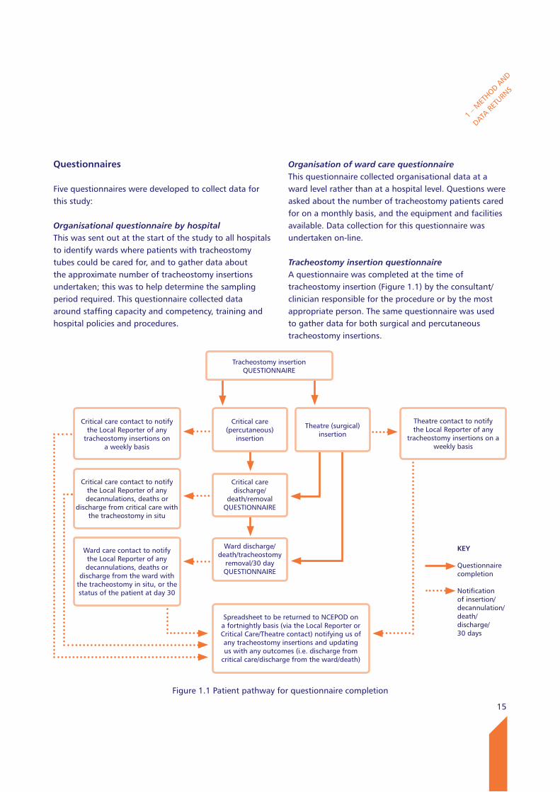
Tracheostomy insertion questionnaireA questionnaire was completed at the time of tracheostomy insertion (Figure 1.1) by the consultant/clinician responsible for the procedure or by the most appropriate person. The same questionnaire was used to gather data for both surgical and percutaneous tracheostomy insertions.
Figure 1.1 Patient pathway for questionnaire completion
1 – M
ETHOD A
ND
DATA R
ETURN
S
Critical care (percutaneous)
insertion
Critical care discharge/
death/removal QUESTIONNAIRE
Ward discharge/death/tracheostomy
removal/30 day QUESTIONNAIRE
Spreadsheet to be returned to NCEPOD on a fortnightly basis (via the Local Reporter or Critical Care/Theatre contact) notifying us of any tracheostomy insertions and updating us with any outcomes (i.e. discharge from
critical care/discharge from the ward/death)
Critical care contact to notify the Local Reporter of any
tracheostomy insertions on a weekly basis
Tracheostomy insertion QUESTIONNAIRE
Critical care contact to notify the Local Reporter of any decannulations, deaths or
discharge from critical care with the tracheostomy in situ
Ward care contact to notify the Local Reporter of any decannulations, deaths or
discharge from the ward with the tracheostomy in situ, or the status of the patient at day 30
Theatre (surgical) insertion
Theatre contact to notify the Local Reporter of any
tracheostomy insertions on a weekly basis
kEy
Questionnaire completion
Notification of insertion/decannulation/death/discharge/30 days
16
Case notes
Photocopied case note extracts were requested for two cases per hospital and these were randomly selected by NCEPOD. The requested extracts included:• Inpatientannotations(maincasenotes)• Nursing/speechandlanguagetherapy/ physiotherapy notes• IntensiveCare(Level3)/HighDependency (Level 2) Unit notes• Anaestheticrecords• Surgical/operationnotes• Observationcharts• Tracheostomycarerecords• Warddischargesummaries
Case notes were requested for the time period up to:• Successfuldecannulation(eitheronthecriticalcare
unit or a general ward); or• Death(onthecriticalcareunitorageneralward);
or• Dischargewiththetracheostomyinsitufromthe
hospital; or• Day30followingadmissiontoageneralward,
whichever occurred first.
Advisor group
A multidisciplinary group of Advisors was recruited to undertake peer review of the case notes and associated questionnaires. This group of Advisors comprised clinicians from a number of specialties including critical care medicine, anaesthetics, general medicine, respiratory medicine, oral and maxillofacial surgery, ear, nose and throat (ENT) surgery, plastic surgery, nursing (critical care, critical care outreach, tracheostomy and ENT), physiotherapy and speech and language therapy (SLT). This group also peer reviewed the findings of the larger questionnaire dataset.
Critical care questionnaireThis questionnaire was completed at the time of discharge from the critical care unit to the ward, tracheostomy removal or death, for all patients who were admitted to (or remained on) the critical care unit following their tracheostomy insertion (Figure 1.1). This included patients who had a tracheostomy inserted whilst in the critical care unit and patients who went to the critical care unit following the insertion of a tracheostomy in theatre. As well as collecting clinical data and information about complications, this questionnaire also collected data about the facilities for tracheostomy care in the critical care unit.
Ward questionnaireThis questionnaire was completed for all patients admitted to a ward either from the critical care unit (both surgical and percutaneous) or directly from theatre (Figure 1.1). This was completed at the time of tracheostomy removal, death, discharge from the ward with the tracheostomy in situ, or 30 days post transfer to ward. Again, as well as collecting clinical data and information about complications, this questionnaire collected data about the ward facilities available.
The clinical questionnaires were sent out in packs; each pack contained an insertion, critical care and ward care questionnaire, and also the instructions for completion. Because not all patients had a critical care stay or a general ward stay with a tracheostomy in situ, the completion of all three questionnaires was not required for each patient (Figure 1.1). These study packs were sent out at the beginning of the study based on the number of insertions undertaken annually at each hospital, so they could be completed at the time of tracheostomy insertion.
1 – M
ETHOD A
ND
DATA R
ETURN
S
17
1 – M
ETHOD A
ND
DATA R
ETURN
S
Case notes were checked on receipt for completeness. In a majority of cases all of the relevant data were returned, however there were a small number of cases where some of the case notes were missing.
All patient identifiers were removed from the case notes and questionnaires prior to review. Neither the coordinators at NCEPOD, nor the Advisors, had access to patient identifiable information.
After being anonymised, each case was reviewed by at least one Advisor and at regular intervals throughout the meeting the Chair allowed a period of discussion for each Advisor to summarise their case and ask for opinions from other specialties or raise aspects of care for discussion.
Advisors completed a semi-structured electronic assessment, and were encouraged to enter free text commentary at various points. Where the Advisor felt that there was insufficient information available in the case note extracts present in order to make a decision, there was the option to select ‘insufficient data’.
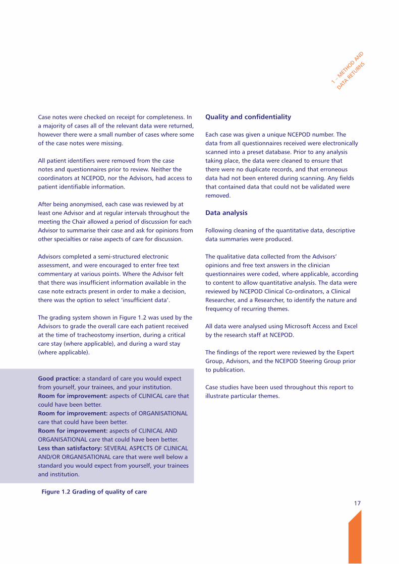
The grading system shown in Figure 1.2 was used by the Advisors to grade the overall care each patient received at the time of tracheostomy insertion, during a critical care stay (where applicable), and during a ward stay (where applicable).
Good practice: a standard of care you would expect from yourself, your trainees, and your institution.Room for improvement: aspects of CLINICAL care that could have been better.Room for improvement: aspects of ORGANISATIONAL care that could have been better.Room for improvement: aspects of CLINICAL AND ORGANISATIONAL care that could have been better.less than satisfactory: SEVERAL ASPECTS OF CLINICAL AND/OR ORGANISATIONAL care that were well below a standard you would expect from yourself, your trainees and institution.
Figure 1.2 Grading of quality of care
Quality and confidentiality
Each case was given a unique NCEPOD number. The data from all questionnaires received were electronically scanned into a preset database. Prior to any analysis taking place, the data were cleaned to ensure that there were no duplicate records, and that erroneous data had not been entered during scanning. Any fields that contained data that could not be validated were removed.
Data analysis
Following cleaning of the quantitative data, descriptive data summaries were produced.
The qualitative data collected from the Advisors’ opinions and free text answers in the clinician questionnaires were coded, where applicable, according to content to allow quantitative analysis. The data were reviewed by NCEPOD Clinical Co-ordinators, a Clinical Researcher, and a Researcher, to identify the nature and frequency of recurring themes.
All data were analysed using Microsoft Access and Excel by the research staff at NCEPOD.
The findings of the report were reviewed by the Expert Group, Advisors, and the NCEPOD Steering Group prior to publication.
Case studies have been used throughout this report to illustrate particular themes.
18
Data returns
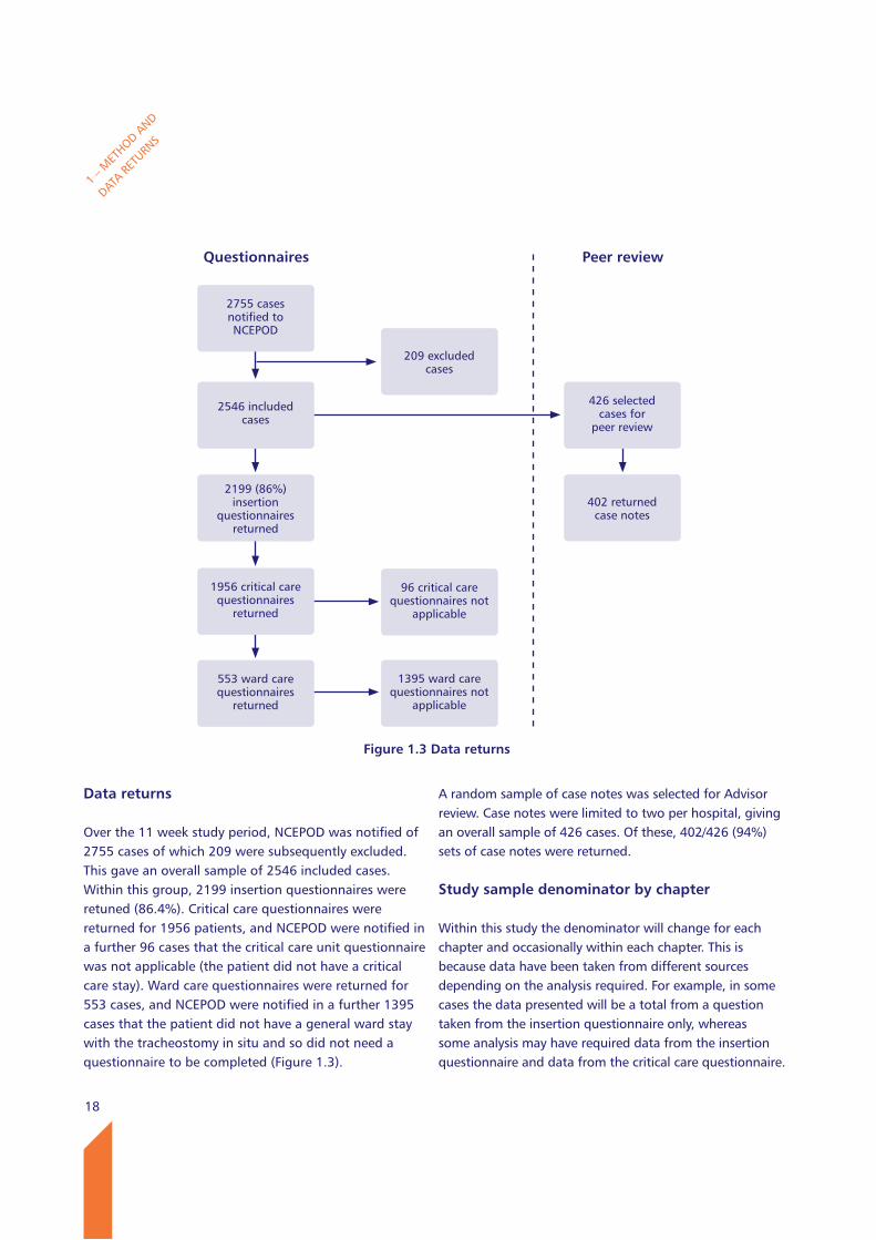
Over the 11 week study period, NCEPOD was notified of 2755 cases of which 209 were subsequently excluded. This gave an overall sample of 2546 included cases. Within this group, 2199 insertion questionnaires were retuned (86.4%). Critical care questionnaires were returned for 1956 patients, and NCEPOD were notified in a further 96 cases that the critical care unit questionnaire was not applicable (the patient did not have a critical care stay). Ward care questionnaires were returned for 553 cases, and NCEPOD were notified in a further 1395 cases that the patient did not have a general ward stay with the tracheostomy in situ and so did not need a questionnaire to be completed (Figure 1.3).
A random sample of case notes was selected for Advisor review. Case notes were limited to two per hospital, giving an overall sample of 426 cases. Of these, 402/426 (94%) sets of case notes were returned.
Study sample denominator by chapter
Within this study the denominator will change for each chapter and occasionally within each chapter. This is because data have been taken from different sources depending on the analysis required. For example, in some cases the data presented will be a total from a question taken from the insertion questionnaire only, whereas some analysis may have required data from the insertion questionnaire and data from the critical care questionnaire.
1 – M
ETHOD A
ND
DATA R
ETURN
S
2546 includedcases
426 selected cases for
peer review
2199 (86%)insertion
questionnairesreturned
Figure 1.3 Data returns
2755 cases notified toNCEPOD
209 excludedcases
1956 critical carequestionnaires
returned
96 critical carequestionnaires not
applicable
553 ward carequestionnaires
returned
1395 ward carequestionnaires not
applicable
402 returned case notes
Questionnaires Peer review
19
Demographics
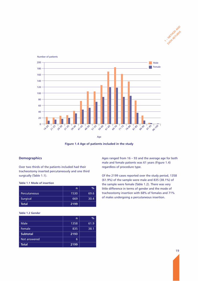
Over two thirds of the patients included had their tracheostomy inserted percutaneously and one third surgically (Table 1.1).
Ages ranged from 16 – 93 and the average age for both male and female patients was 61 years (Figure 1.4) regardless of procedure type.
Of the 2199 cases reported over the study period, 1358 (61.9%) of the sample were male and 835 (38.1%) of the sample were female (Table 1.2). There was very little difference in terms of gender and the mode of tracheostomy insertion with 68% of females and 71% of males undergoing a percutaneous insertion.
1 – M
ETHOD A
ND
DATA R
ETURN
S
Table 1.1 Mode of insertion
n %
Percutaneous 1530 69.6
Surgical 669 30.4
Total 2199
Number of patients
200
180
160
140
120
100
80
60
40
20
0
Figure 1.4 Age of patients included in the study
Age
16-2
021
-25
26-3
031
-35
36-4
041
-45
46-5
051
-55
56-6
061
-65
66-7
071
-75
76-8
081
-85
86-9
091
-95
96-1
00
Male
Female
Table 1.2 Gender
n %
Male 1358 61.9
Female 835 38.1
Subtotal 2193
Not answered 6
Total 2199
20
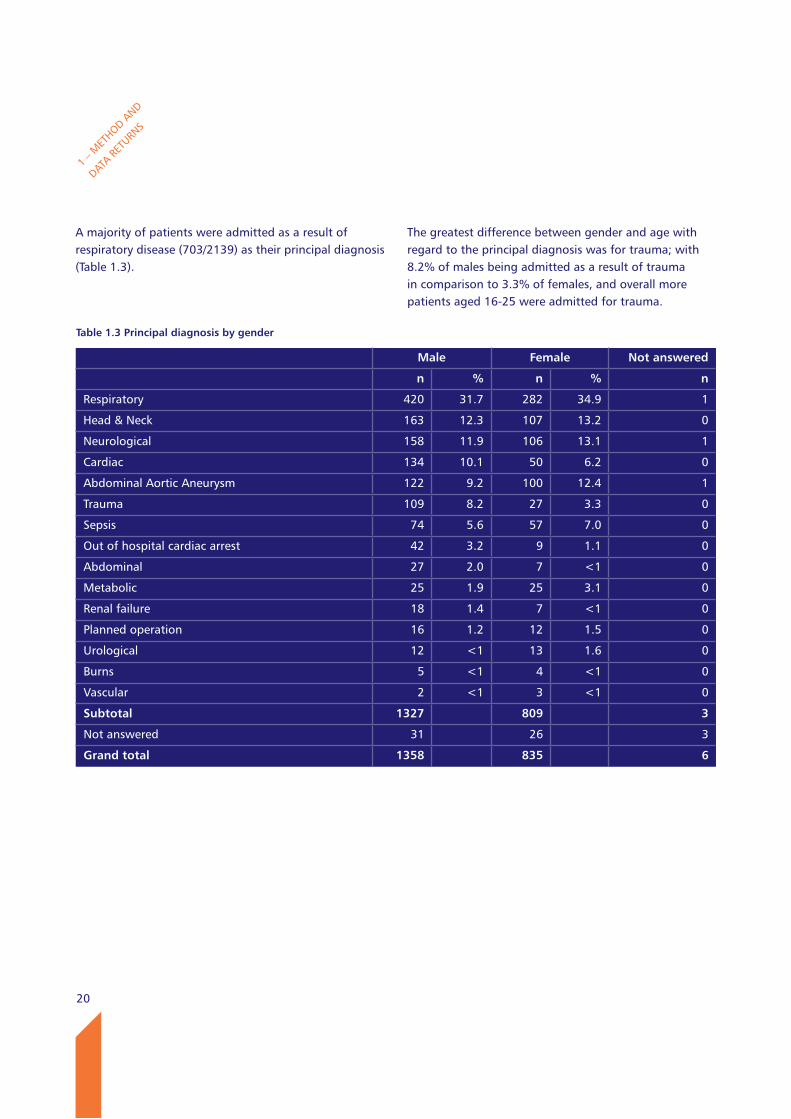
A majority of patients were admitted as a result of respiratory disease (703/2139) as their principal diagnosis (Table 1.3).
The greatest difference between gender and age with regard to the principal diagnosis was for trauma; with 8.2% of males being admitted as a result of trauma in comparison to 3.3% of females, and overall more patients aged 16-25 were admitted for trauma.
1 – M
ETHOD A
ND
DATA R
ETURN
S
Table 1.3 Principal diagnosis by gender
Male Female Not answered
n % n % n
Respiratory 420 31.7 282 34.9 1
Head & Neck 163 12.3 107 13.2 0
Neurological 158 11.9 106 13.1 1
Cardiac 134 10.1 50 6.2 0
Abdominal Aortic Aneurysm 122 9.2 100 12.4 1
Trauma 109 8.2 27 3.3 0
Sepsis 74 5.6 57 7.0 0
Out of hospital cardiac arrest 42 3.2 9 1.1 0
Abdominal 27 2.0 7 <1 0
Metabolic 25 1.9 25 3.1 0
Renal failure 18 1.4 7 <1 0
Planned operation 16 1.2 12 1.5 0
Urological 12 <1 13 1.6 0
Burns 5 <1 4 <1 0
Vascular 2 <1 3 <1 0
Subtotal 1327 809 3
Not answered 31 26 3
Grand total 1358 835 6
21
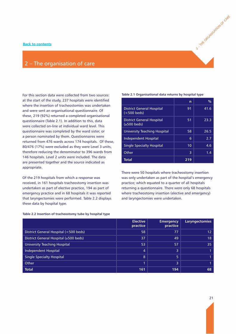
For this section data were collected from two sources: at the start of the study, 237 hospitals were identified where the insertion of tracheostomies was undertaken and were sent an organisational questionnaire. Of these, 219 (92%) returned a completed organisational questionnaire (Table 2.1). In addition to this, data were collected on-line at individual ward level. This questionnaire was completed by the ward sister, or a person nominated by them. Questionnaires were returned from 476 wards across 174 hospitals. Of these, 80/476 (17%) were excluded as they were Level 3 units, therefore reducing the denominator to 396 wards from 146 hospitals. Level 2 units were included. The data are presented together and the source indicated as appropriate.
Of the 219 hospitals from which a response was received, in 161 hospitals tracheostomy insertion was undertaken as part of elective practice, 194 as part of emergency practice and in 68 hospitals it was reported that laryngectomies were performed. Table 2.2 displays these data by hospital type.
There were 50 hospitals where tracheostomy insertion was only undertaken as part of the hospital’s emergency practice; which equated to a quarter of all hospitals returning a questionnaire. There were only 68 hospitals where tracheostomy insertion (elective and emergency) and laryngectomies were undertaken.
2 – The organisation of care
2 – T
HE ORG
ANISATIO
N OF C
ARE
Table 2.1 Organisational data returns by hospital type
n %
District General Hospital (<500 beds)
91 41.6
District General Hospital (≥500 beds)
51 23.3
University Teaching Hospital 58 26.5
Independent Hospital 6 2.7
Single Specialty Hospital 10 4.6
Other 3 1.4
Total 219
Table 2.2 Insertion of tracheostomy tube by hospital type
Elective practice
Emergency practice
laryngectomies
District General Hospital (<500 beds) 58 77 12
District General Hospital (≥500 beds) 37 49 18
University Teaching Hospital 53 57 35
Independent Hospital 4 3 1
Single Specialty Hospital 8 5 1
Other 1 3 1
Total 161 194 68
Back to contents
22
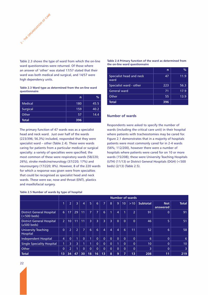
Table 2.3 shows the type of ward from which the on-line ward questionnaires were returned. Of those where an answer of ‘other’ was stated 17/57 stated that their ward was both medical and surgical, and 14/57 were high dependency units.
The primary function of 47 wards was as a specialist head and neck ward. Just over half of the wards (223/396; 56.3%) included, responded that they were specialist ward – other (Table 2.4). These were wards caring for patients from a particular medical or surgical specialty: a variety of specialties were specified; the most common of these were respiratory wards (58/220; 26%), stroke medicine/neurology (37/220; 17%) and neurosurgery (17/220; 8%). However, 8 of the 220 wards for which a response was given were from specialties that could be recognised as specialist head and neck wards. These were ear, nose and throat (ENT), plastics and maxillofacial surgery.
Number of wards
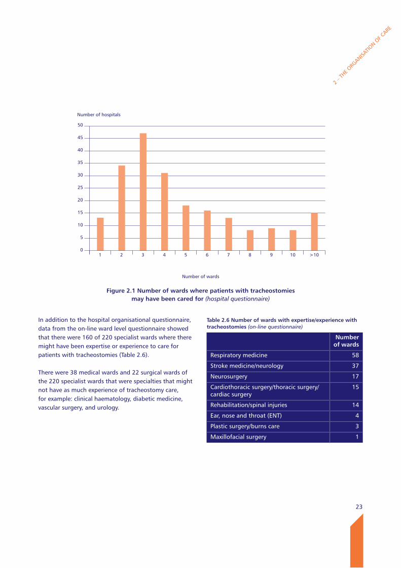
Respondents were asked to specify the number of wards (including the critical care unit) in their hospital where patients with tracheostomies may be cared for. Figure 2.1 demonstrates that in a majority of hospitals patients were most commonly cared for in 2-4 wards (56%; 112/200), however there were a number of hospitals where patients were cared for on 10 or more wards (15/208); these were University Teaching Hospitals (UTH) (11/13) or District General Hospitals (DGH) (<500 beds) (2/13) (Table 2.5).
2 – T
HE ORG
ANISATIO
N OF C
ARE
Table 2.3 ward type as determined from the on-line ward questionnaire
n %
Medical 180 45.5
Surgical 159 40.2
Other 57 14.4
Total 396
Table 2.4 Primary function of the ward as determined from the on-line ward questionnaire
n %
Specialist head and neck ward
47 11.9
Specialist ward - other 223 56.3
General ward 71 17.9
Other 55 13.9
Total 396
Table 2.5 Number of wards by type of hospital
Number of wards
1 2 3 4 5 6 7 8 9 10 >10 Subtotal Not answered
Total
District General Hospital (<500 beds)
6 17 29 11 7 7 6 1 4 1 2 91 0 91
District General Hospital (≥500 beds)
2 10 11 11 3 3 3 3 0 0 0 46 5 51
University Teaching Hospital
0 2 2 7 6 6 4 4 4 6 11 52 6 58
Independent Hospital 4 0 1 0 1 0 0 0 0 0 0 6 0 6
Single Speciality Hospital 1 3 3 1 1 0 0 0 1 0 0 10 0 10
Other 0 2 1 0 0 0 0 0 0 0 0 3 0 3
Total 13 34 47 30 18 16 13 8 9 7 13 208 11 219
23
In addition to the hospital organisational questionnaire, data from the on-line ward level questionnaire showed that there were 160 of 220 specialist wards where there might have been expertise or experience to care for patients with tracheostomies (Table 2.6).
There were 38 medical wards and 22 surgical wards of the 220 specialist wards that were specialties that might not have as much experience of tracheostomy care, for example: clinical haematology, diabetic medicine, vascular surgery, and urology.
2 – T
HE ORG
ANISATIO
N OF C
ARE
Number of hospitals
50
45
40
35
30
25
20
15
10
5
0
Figure 2.1 Number of wards where patients with tracheostomies may have been cared for (hospital questionnaire)
Number of wards
1 2 3 4 5 6 7 8 9 10 >10
Table 2.6 Number of wards with expertise/experience with tracheostomies (on-line questionnaire)
Number of wards
Respiratory medicine 58
Stroke medicine/neurology 37
Neurosurgery 17
Cardiothoracic surgery/thoracic surgery/cardiac surgery
15
Rehabilitation/spinal injuries 14
Ear, nose and throat (ENT) 4
Plastic surgery/burns care 3
Maxillofacial surgery 1
24
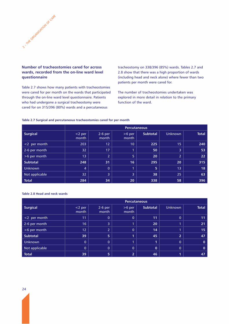
Table 2.7 Surgical and percutaneous tracheostomies cared for per month
Percutaneous
Surgical <2 per month
2-6 per month
>6 per month
Subtotal Unknown Total
<2 per month 203 12 10 225 15 240
2-6 per month 32 17 1 50 3 53
>6 per month 13 2 5 20 2 22
Subtotal 248 31 16 295 20 315
Unknown 4 0 1 5 13 18
Not applicable 32 3 3 38 25 63
Total 284 34 20 338 58 396
Number of tracheostomies cared for across wards, recorded from the on-line ward level questionnaire
Table 2.7 shows how many patients with tracheostomies were cared for per month on the wards that participated through the on-line ward level questionnaire. Patients who had undergone a surgical tracheostomy were cared for on 315/396 (80%) wards and a percutaneous
tracheostomy on 338/396 (85%) wards. Tables 2.7 and 2.8 show that there was a high proportion of wards (including head and neck alone) where fewer than two patients per month were cared for.
The number of tracheostomies undertaken was explored in more detail in relation to the primary function of the ward.
2 – T
HE ORG
ANISATIO
N OF C
ARE
Table 2.8 Head and neck wards
Percutaneous
Surgical <2 per month
2-6 per month
>6 per month
Subtotal Unknown Total
<2 per month 11 0 0 11 0 11
2-6 per month 16 3 1 20 1 21
>6 per month 12 2 0 14 1 15
Subtotal 39 5 1 45 2 47
Unknown 0 0 1 1 0 0
Not applicable 0 0 0 0 0 0
Total 39 5 2 46 1 47
25
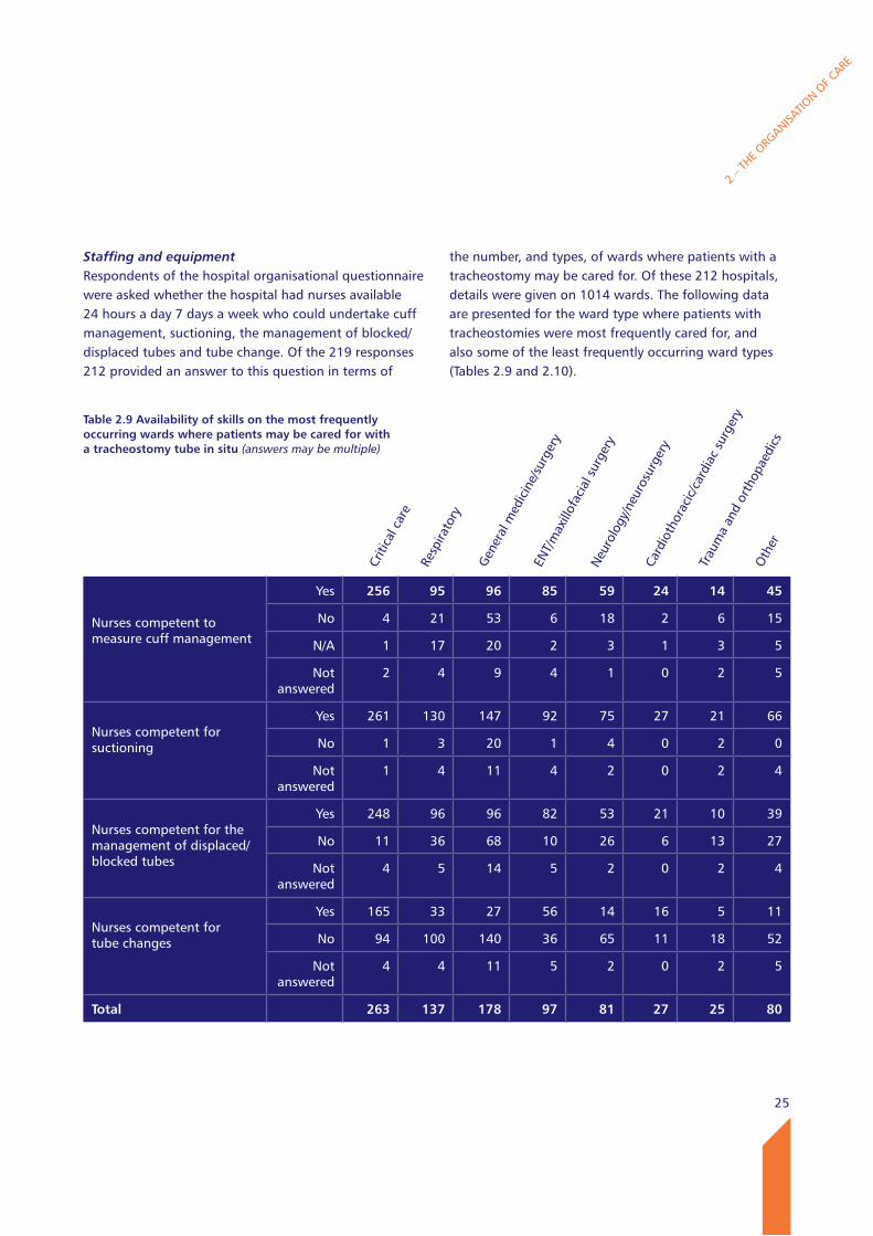
Staffing and equipmentRespondents of the hospital organisational questionnaire were asked whether the hospital had nurses available 24 hours a day 7 days a week who could undertake cuff management, suctioning, the management of blocked/displaced tubes and tube change. Of the 219 responses 212 provided an answer to this question in terms of
the number, and types, of wards where patients with a tracheostomy may be cared for. Of these 212 hospitals, details were given on 1014 wards. The following data are presented for the ward type where patients with tracheostomies were most frequently cared for, and also some of the least frequently occurring ward types (Tables 2.9 and 2.10).
2 – T
HE ORG
ANISATIO
N OF C
ARE
Nurses competent to measure cuff management
Yes 256 95 96 85 59 24 14 45
No 4 21 53 6 18 2 6 15
N/A 1 17 20 2 3 1 3 5
Not answered
2 4 9 4 1 0 2 5
Nurses competent for suctioning
Yes 261 130 147 92 75 27 21 66
No 1 3 20 1 4 0 2 0
Not answered
1 4 11 4 2 0 2 4
Nurses competent for the management of displaced/blocked tubes
Yes 248 96 96 82 53 21 10 39
No 11 36 68 10 26 6 13 27
Not answered
4 5 14 5 2 0 2 4
Nurses competent for tube changes
Yes 165 33 27 56 14 16 5 11
No 94 100 140 36 65 11 18 52
Not answered
4 4 11 5 2 0 2 5
Total 263 137 178 97 81 27 25 80
Criti
cal c
are
Resp
irato
ry
Gen
eral
med
icin
e/su
rger
yEN
T/m
axill
ofac
ial s
urge
ryN
euro
logy
/neu
rosu
rger
yCa
rdio
thor
acic
/car
diac
sur
gery
Trau
ma
and
orth
opae
dics
Oth
er
Table 2.9 Availability of skills on the most frequently occurring wards where patients may be cared for with a tracheostomy tube in situ (answers may be multiple)
26
2 – T
HE ORG
ANISATIO
N OF C
ARE
Nurses competent to measure cuff management
Yes 4 2 2 3 2 3 1 1 0 2
No 1 3 1 1 1 0 1 1 2 0
N/A 1 0 1 0 0 0 0 0 0 0
Not answered
0 0 1 0 0 0 1 0 0 0
Nurses competent for suctioning
Yes 5 2 3 4 3 3 2 2 2 2
No 1 3 1 0 0 0 0 0 0 0
Not answered
0 0 1 0 0 0 1 0 0 0
Nurses competent for the management of displaced/blocked tubes
Yes 5 1 1 3 1 3 0 1 1 2
No 1 4 3 1 2 0 2 1 1 0
Not answered
0 0 1 0 0 0 1 0 0 0
Nurses competent for tube changes
Yes 1 1 1 2 1 2 0 2 1 2
No 5 4 3 2 2 1 2 0 1 0
Not answered
0 0 1 0 0 0 1 0 0 0
Total 6 5 5 4 3 3 3 2 2 2
Uppe
r gas
troi
ntes
tinal
sur
gery
Gas
troe
nter
olog
y
Vasc
ular
sur
gery
Haem
atol
ogy
Urol
ogy
Hepa
tobi
liary
and
pan
crea
tic s
urge
ry
Diab
etic
med
icin
eBr
east
sur
gery
Rheu
mat
olog
yG
ynae
colo
gy
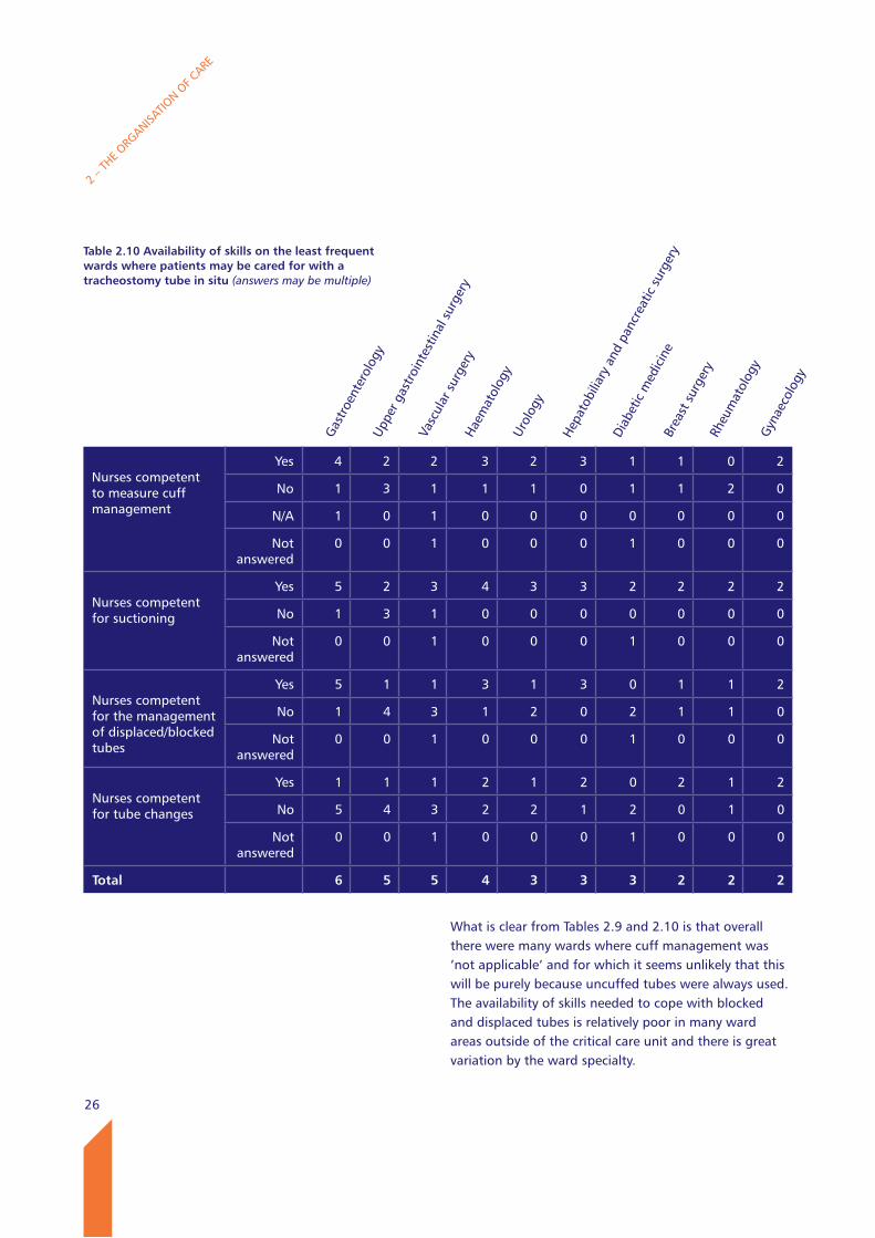
Table 2.10 Availability of skills on the least frequent wards where patients may be cared for with a tracheostomy tube in situ (answers may be multiple)
What is clear from Tables 2.9 and 2.10 is that overall there were many wards where cuff management was ’not applicable’ and for which it seems unlikely that this will be purely because uncuffed tubes were always used. The availability of skills needed to cope with blocked and displaced tubes is relatively poor in many ward areas outside of the critical care unit and there is great variation by the ward specialty.
27
Availability of equipment reported by the on-line ward-level questionnaire
Cuff pressure measurementThe National Tracheostomy Safety Project (NTSP)1 list standards and key performance indicators, which could be used for monitoring compliance to a tracheostomy policy; within this they state that an adequate supply of all necessary equipment should be available on the receiving ward. The availability of equipment to measure cuff pressure on the ward was assessed. Only just over half, (233/396, 59%) of wards from which an on-line ward level questionnaire was received had the equipment to measure cuff pressure on the ward. It was found that the availability of equipment to measure cuff pressure was more frequent in wards where a greater number of patients with tracheostomies were cared for. Table 2.11 shows that, based on proportion, specialist head and neck wards had more equipment available to measure cuff pressure (41/47).
Table 2.11 Availability of equipment to measure cuff pressure
yes No Total
Specialist head and neck ward
41 6 47
Specialist ward - other
137 86 223
General ward 30 41 71
Other 25 30 55
Total 233 163 396
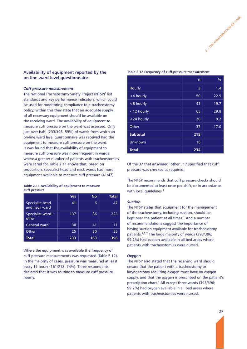
Where the equipment was available the frequency of cuff pressure measurements was requested (Table 2.12). In the majority of cases, pressure was measured at least every 12 hours (161/218; 74%). Three respondents declared that it was routine to measure cuff pressure hourly.
Of the 37 that answered ‘other’, 17 specified that cuff pressure was checked as required.
The NTSP recommends that cuff pressure checks should be documented at least once per shift, or in accordance with local guidelines.1
SuctionThe NTSP states that equipment for the management of the tracheostomy, including suction, should be kept near the patient at all times.1 And a number of recommendations suggest the importance of having suction equipment available for tracheostomy patients.1,2,7 The large majority of wards (393/396; 99.2%) had suction available in all bed areas where patients with tracheostomies were nursed.
Oxygen The NTSP also stated that the receiving ward should ensure that the patient with a tracheostomy or laryngectomy requiring oxygen must have an oxygen supply, and that the oxygen is prescribed on the patient’s prescription chart.1 All except three wards (393/396; 99.2%) had oxygen available in all bed areas where patients with tracheostomies were nursed.
2 – T
HE ORG
ANISATIO
N OF C
ARE
Gyn
aeco
logy
Table 2.12 Frequency of cuff pressure measurement
n %
Hourly 3 1.4
<4 hourly 50 22.9
<8 hourly 43 19.7
<12 hourly 65 29.8
<24 hourly 20 9.2
Other 37 17.0
Subtotal 218
Unknown 16
Total 234
28
Equipment checks371/396; (93.7%) of wards had systems in place for checking and recording equipment function. The NTSP recommends that equipment must be checked and that a means of documenting this must be in place.1
A relatively large number of wards (356/396, 90%) checked equipment (suction and oxygen) on at least a daily basis on the ward.
Number of tracheostomies performed annually
The number of tracheostomy insertions that were undertaken on an annual basis was requested for each hospital. For the 187/219 (85.4%) hospitals where an answer was given, this figure ranged between 1-375, with an average of 64 between 1st April 2011 – 31st March 2012.
In half of cases this number was an estimate (54.3%, 101/186), with the actual number undertaken being given in 44.8% of hospitals (82/183). This was broken down further to look at how many were undertaken in the critical care unit and theatre. The number undertaken in the critical care unit ranged between 1 – 275 with an average of 44, and in theatre between 1 – 226 with an average of 25 annually. Again in a majority of cases this number was an estimate. The number of tracheostomy insertions undertaken was unknown or not answered by 32 hospitals.
It was reassuring to see from the data provided that the majority of tracheostomies that were performed during this period were likely to have been captured ((64 per month x 219 hospitals)/52 weeks in a year) x 11 week period of data collection). This total of 2964 is similar to the total reported to NCEPOD. This study has also provided a clearer picture of the total number of tracheostomies performed annually, rather than just using estimates.
Organisation of tracheostomy care
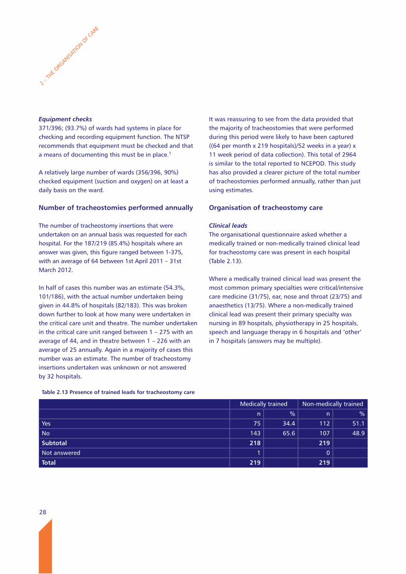
Clinical leadsThe organisational questionnaire asked whether a medically trained or non-medically trained clinical lead for tracheostomy care was present in each hospital (Table 2.13).
Where a medically trained clinical lead was present the most common primary specialties were critical/intensive care medicine (31/75), ear, nose and throat (23/75) and anaesthetics (13/75). Where a non-medically trained clinical lead was present their primary specialty was nursing in 89 hospitals, physiotherapy in 25 hospitals, speech and language therapy in 6 hospitals and ‘other’ in 7 hospitals (answers may be multiple).
2 – T
HE ORG
ANISATIO
N OF C
ARE
Table 2.13 Presence of trained leads for tracheostomy care
Medically trained Non-medically trained
n % n %
Yes 75 34.4 112 51.1
No 143 65.6 107 48.9
Subtotal 218 219
Not answered 1 0
Total 219 219
29
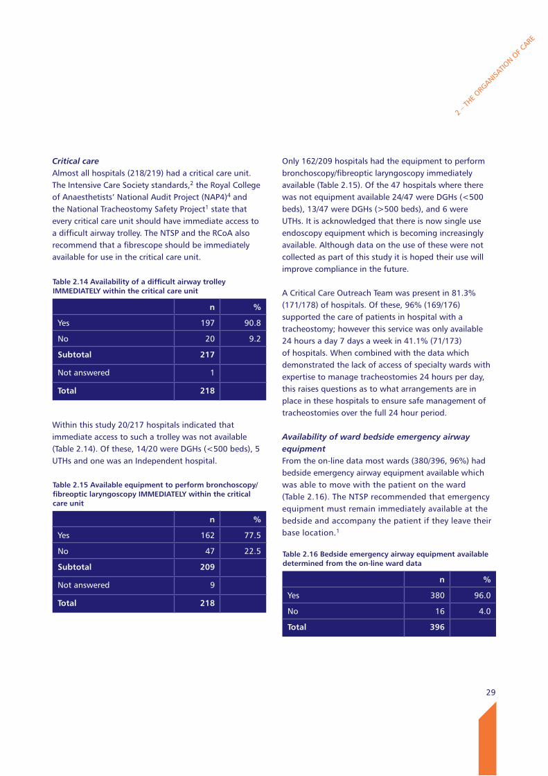
Critical care Almost all hospitals (218/219) had a critical care unit. The Intensive Care Society standards,2 the Royal College of Anaesthetists’ National Audit Project (NAP4)4 and the National Tracheostomy Safety Project1 state that every critical care unit should have immediate access to a difficult airway trolley. The NTSP and the RCoA also recommend that a fibrescope should be immediately available for use in the critical care unit.
Within this study 20/217 hospitals indicated that immediate access to such a trolley was not available (Table 2.14). Of these, 14/20 were DGHs (<500 beds), 5 UTHs and one was an Independent hospital.
2 – T
HE ORG
ANISATIO
N OF C
ARE
Only 162/209 hospitals had the equipment to perform bronchoscopy/fibreoptic laryngoscopy immediately available (Table 2.15). Of the 47 hospitals where there was not equipment available 24/47 were DGHs (<500 beds), 13/47 were DGHs (>500 beds), and 6 were UTHs. It is acknowledged that there is now single use endoscopy equipment which is becoming increasingly available. Although data on the use of these were not collected as part of this study it is hoped their use will improve compliance in the future.
A Critical Care Outreach Team was present in 81.3% (171/178) of hospitals. Of these, 96% (169/176) supported the care of patients in hospital with a tracheostomy; however this service was only available 24 hours a day 7 days a week in 41.1% (71/173) of hospitals. When combined with the data which demonstrated the lack of access of specialty wards with expertise to manage tracheostomies 24 hours per day, this raises questions as to what arrangements are in place in these hospitals to ensure safe management of tracheostomies over the full 24 hour period.
Availability of ward bedside emergency airway equipment From the on-line data most wards (380/396, 96%) had bedside emergency airway equipment available which was able to move with the patient on the ward (Table 2.16). The NTSP recommended that emergency equipment must remain immediately available at the bedside and accompany the patient if they leave their base location.1
Table 2.14 Availability of a difficult airway trolley IMMEDIATEly within the critical care unit
n %
Yes 197 90.8
No 20 9.2
Subtotal 217 Not answered 1 Total 218
Table 2.15 Available equipment to perform bronchoscopy/fibreoptic laryngoscopy IMMEDIATEly within the critical care unit
n %
Yes 162 77.5
No 47 22.5
Subtotal 209 Not answered 9 Total 218
Table 2.16 Bedside emergency airway equipment available determined from the on-line ward data
n %
Yes 380 96.0
No 16 4.0
Total 396
30
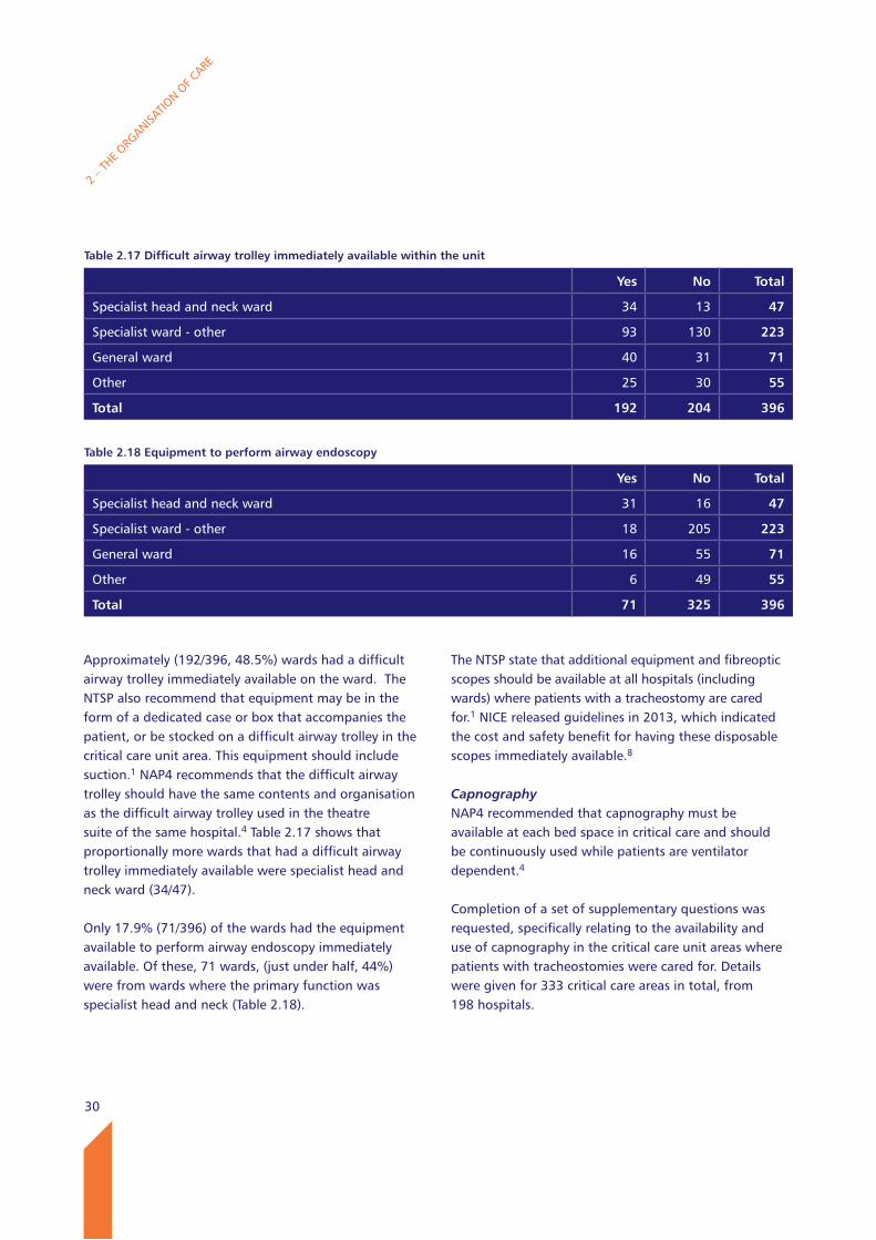
Approximately (192/396, 48.5%) wards had a difficult airway trolley immediately available on the ward. The NTSP also recommend that equipment may be in the form of a dedicated case or box that accompanies the patient, or be stocked on a difficult airway trolley in the critical care unit area. This equipment should include suction.1 NAP4 recommends that the difficult airway trolley should have the same contents and organisation as the difficult airway trolley used in the theatre suite of the same hospital.4 Table 2.17 shows that proportionally more wards that had a difficult airway trolley immediately available were specialist head and neck ward (34/47).
Only 17.9% (71/396) of the wards had the equipment available to perform airway endoscopy immediately available. Of these, 71 wards, (just under half, 44%) were from wards where the primary function was specialist head and neck (Table 2.18).
2 – T
HE ORG
ANISATIO
N OF C
ARE
The NTSP state that additional equipment and fibreoptic scopes should be available at all hospitals (including wards) where patients with a tracheostomy are cared for.1 NICE released guidelines in 2013, which indicated the cost and safety benefit for having these disposable scopes immediately available.8
CapnographyNAP4 recommended that capnography must be available at each bed space in critical care and should be continuously used while patients are ventilator dependent.4
Completion of a set of supplementary questions was requested, specifically relating to the availability and use of capnography in the critical care unit areas where patients with tracheostomies were cared for. Details were given for 333 critical care areas in total, from 198 hospitals.
Table 2.17 Difficult airway trolley immediately available within the unit
yes No Total
Specialist head and neck ward 34 13 47
Specialist ward - other 93 130 223
General ward 40 31 71
Other 25 30 55
Total 192 204 396
Table 2.18 Equipment to perform airway endoscopy
yes No Total
Specialist head and neck ward 31 16 47
Specialist ward - other 18 205 223
General ward 16 55 71
Other 6 49 55
Total 71 325 396
31
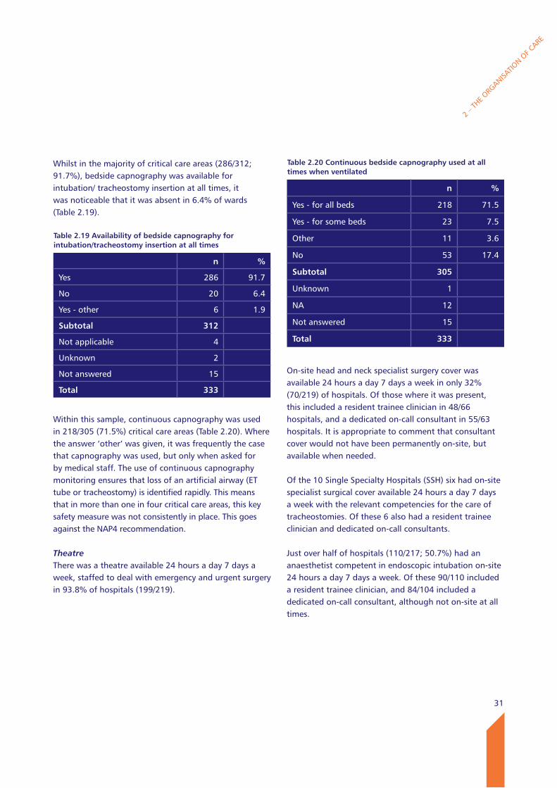
Whilst in the majority of critical care areas (286/312; 91.7%), bedside capnography was available for intubation/ tracheostomy insertion at all times, it was noticeable that it was absent in 6.4% of wards (Table 2.19).
Within this sample, continuous capnography was used in 218/305 (71.5%) critical care areas (Table 2.20). Where the answer ‘other’ was given, it was frequently the case that capnography was used, but only when asked for by medical staff. The use of continuous capnography monitoring ensures that loss of an artificial airway (ET tube or tracheostomy) is identified rapidly. This means that in more than one in four critical care areas, this key safety measure was not consistently in place. This goes against the NAP4 recommendation.
TheatreThere was a theatre available 24 hours a day 7 days a week, staffed to deal with emergency and urgent surgery in 93.8% of hospitals (199/219).
On-site head and neck specialist surgery cover was available 24 hours a day 7 days a week in only 32% (70/219) of hospitals. Of those where it was present, this included a resident trainee clinician in 48/66 hospitals, and a dedicated on-call consultant in 55/63 hospitals. It is appropriate to comment that consultant cover would not have been permanently on-site, but available when needed.
Of the 10 Single Specialty Hospitals (SSH) six had on-site specialist surgical cover available 24 hours a day 7 days a week with the relevant competencies for the care of tracheostomies. Of these 6 also had a resident trainee clinician and dedicated on-call consultants.
Just over half of hospitals (110/217; 50.7%) had an anaesthetist competent in endoscopic intubation on-site 24 hours a day 7 days a week. Of these 90/110 included a resident trainee clinician, and 84/104 included a dedicated on-call consultant, although not on-site at all times.
2 – T
HE ORG
ANISATIO
N OF C
ARE
Table 2.19 Availability of bedside capnography for intubation/tracheostomy insertion at all times
n %
Yes 286 91.7
No 20 6.4
Yes - other 6 1.9
Subtotal 312
Not applicable 4
Unknown 2
Not answered 15
Total 333
Table 2.20 Continuous bedside capnography used at all times when ventilated
n %
Yes - for all beds 218 71.5
Yes - for some beds 23 7.5
Other 11 3.6
No 53 17.4
Subtotal 305
Unknown 1
NA 12
Not answered 15
Total 333
32
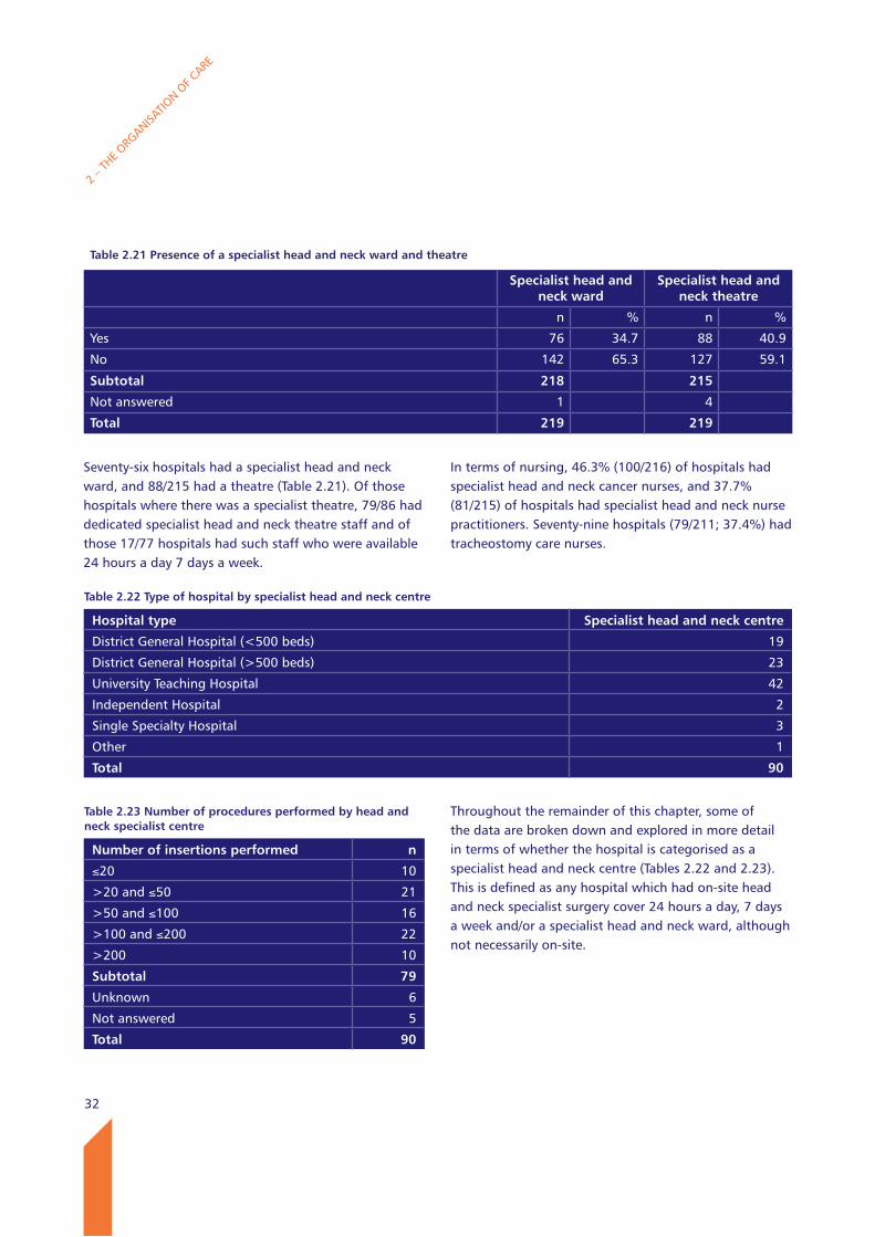
Seventy-six hospitals had a specialist head and neck ward, and 88/215 had a theatre (Table 2.21). Of those hospitals where there was a specialist theatre, 79/86 had dedicated specialist head and neck theatre staff and of those 17/77 hospitals had such staff who were available 24 hours a day 7 days a week.
In terms of nursing, 46.3% (100/216) of hospitals had specialist head and neck cancer nurses, and 37.7% (81/215) of hospitals had specialist head and neck nurse practitioners. Seventy-nine hospitals (79/211; 37.4%) had tracheostomy care nurses.
Throughout the remainder of this chapter, some of the data are broken down and explored in more detail in terms of whether the hospital is categorised as a specialist head and neck centre (Tables 2.22 and 2.23). This is defined as any hospital which had on-site head and neck specialist surgery cover 24 hours a day, 7 days a week and/or a specialist head and neck ward, although not necessarily on-site.
2 – T
HE ORG
ANISATIO
N OF C
ARE
Table 2.21 Presence of a specialist head and neck ward and theatre
Specialist head and neck ward
Specialist head and neck theatre
n % n %
Yes 76 34.7 88 40.9
No 142 65.3 127 59.1
Subtotal 218 215
Not answered 1 4
Total 219 219
Table 2.22 Type of hospital by specialist head and neck centre
Hospital type Specialist head and neck centre
District General Hospital (<500 beds) 19
District General Hospital (>500 beds) 23
University Teaching Hospital 42
Independent Hospital 2
Single Specialty Hospital 3
Other 1
Total 90
Table 2.23 Number of procedures performed by head and neck specialist centre
Number of insertions performed n
≤20 10
>20 and ≤50 21
>50 and ≤100 16
>100 and ≤200 22
>200 10
Subtotal 79
Unknown 6
Not answered 5
Total 90
33
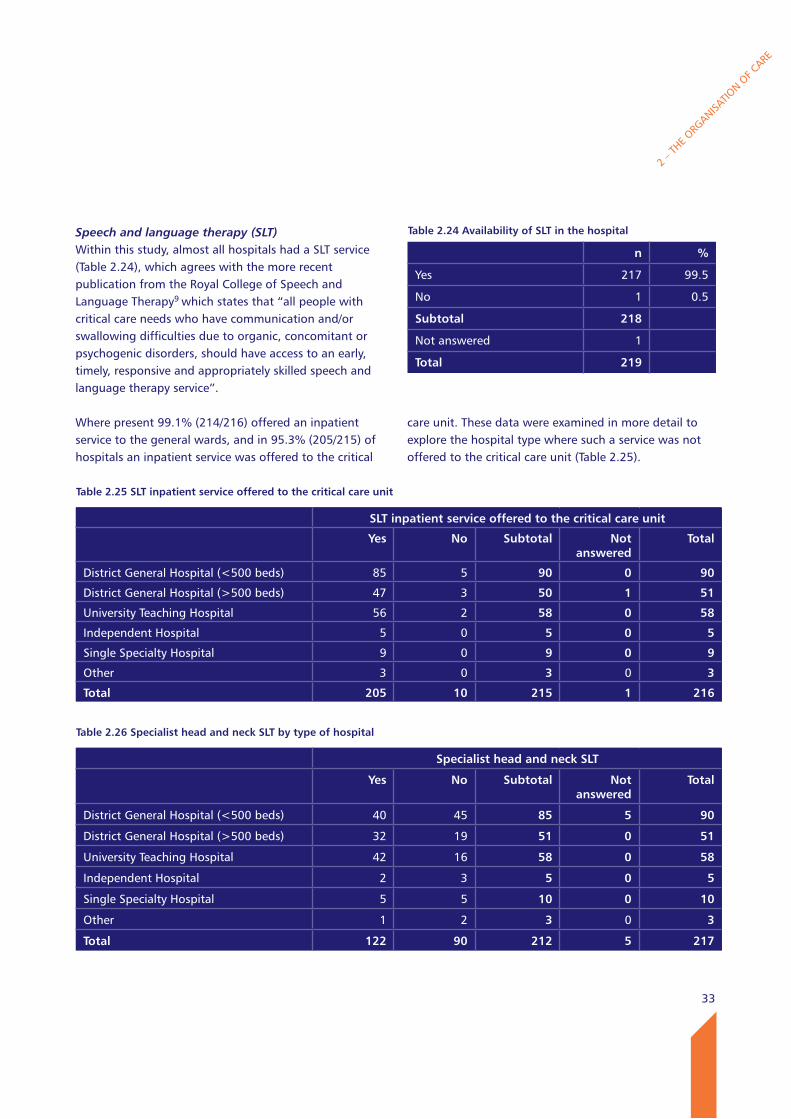
Speech and language therapy (SLT)Within this study, almost all hospitals had a SLT service (Table 2.24), which agrees with the more recent publication from the Royal College of Speech and Language Therapy9 which states that “all people with critical care needs who have communication and/or swallowing difficulties due to organic, concomitant or psychogenic disorders, should have access to an early, timely, responsive and appropriately skilled speech and language therapy service”.
Where present 99.1% (214/216) offered an inpatient service to the general wards, and in 95.3% (205/215) of hospitals an inpatient service was offered to the critical
care unit. These data were examined in more detail to explore the hospital type where such a service was not offered to the critical care unit (Table 2.25).
2 – T
HE ORG
ANISATIO
N OF C
ARE
Table 2.24 Availability of SlT in the hospital
n %
Yes 217 99.5
No 1 0.5
Subtotal 218
Not answered 1
Total 219
Table 2.25 SlT inpatient service offered to the critical care unit
SlT inpatient service offered to the critical care unit
yes No Subtotal Not answered
Total
District General Hospital (<500 beds) 85 5 90 0 90
District General Hospital (>500 beds) 47 3 50 1 51
University Teaching Hospital 56 2 58 0 58
Independent Hospital 5 0 5 0 5
Single Specialty Hospital 9 0 9 0 9
Other 3 0 3 0 3
Total 205 10 215 1 216
Table 2.26 Specialist head and neck SlT by type of hospital
Specialist head and neck SlT
yes No Subtotal Not answered
Total
District General Hospital (<500 beds) 40 45 85 5 90
District General Hospital (>500 beds) 32 19 51 0 51
University Teaching Hospital 42 16 58 0 58
Independent Hospital 2 3 5 0 5
Single Specialty Hospital 5 5 10 0 10
Other 1 2 3 0 3
Total 122 90 212 5 217
34
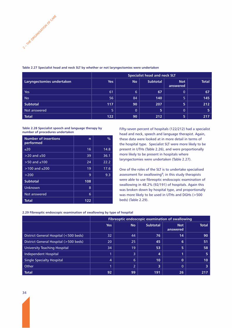
Fifty-seven percent of hospitals (122/212) had a specialist head and neck, speech and language therapist. Again, these data were looked at in more detail in terms of the hospital type. Specialist SLT were more likely to be present in UTHs (Table 2.26), and were proportionally more likely to be present in hospitals where laryngectomies were undertaken (Table 2.27).
One of the roles of the SLT is to undertake specialised assessment for swallowing9; in this study therapists were able to use fibreoptic endoscopic examination of swallowing in 48.2% (92/191) of hospitals. Again this was broken down by hospital type, and proportionally was more likely to be used in UTHs and DGHs (>500 beds) (Table 2.29).
2 – T
HE ORG
ANISATIO
N OF C
ARE
Table 2.27 Specialist head and neck SlT by whether or not laryngectomies were undertaken
Specialist head and neck SlT
laryngectomies undertaken yes No Subtotal Not answered
Total
Yes 61 6 67 0 67
No 56 84 140 5 145
Subtotal 117 90 207 5 212
Not answered 5 0 5 0 5
Total 122 90 212 5 217
Table 2.28 Specialist speech and language therapy by number of procedures undertaken
Number of insertions performed
n %
≤20 16 14.8
>20 and ≤50 39 36.1
>50 and ≤100 24 22.2
>100 and ≤200 19 17.6
>200 9 9.3
Subtotal 108
Unknown 8
Not answered 6
Total 122
2.29 Fibreoptic endoscopic examination of swallowing by type of hospital
Fibreoptic endoscopic examination of swallowing
yes No Subtotal Not answered
Total
District General Hospital (<500 beds) 32 44 76 14 90
District General Hospital (>500 beds) 20 25 45 6 51
University Teaching Hospital 34 19 53 5 58
Independent Hospital 1 3 4 1 5
Single Specialty Hospital 4 6 10 0 10
Other 1 2 3 0 3
Total 92 99 191 26 217
35
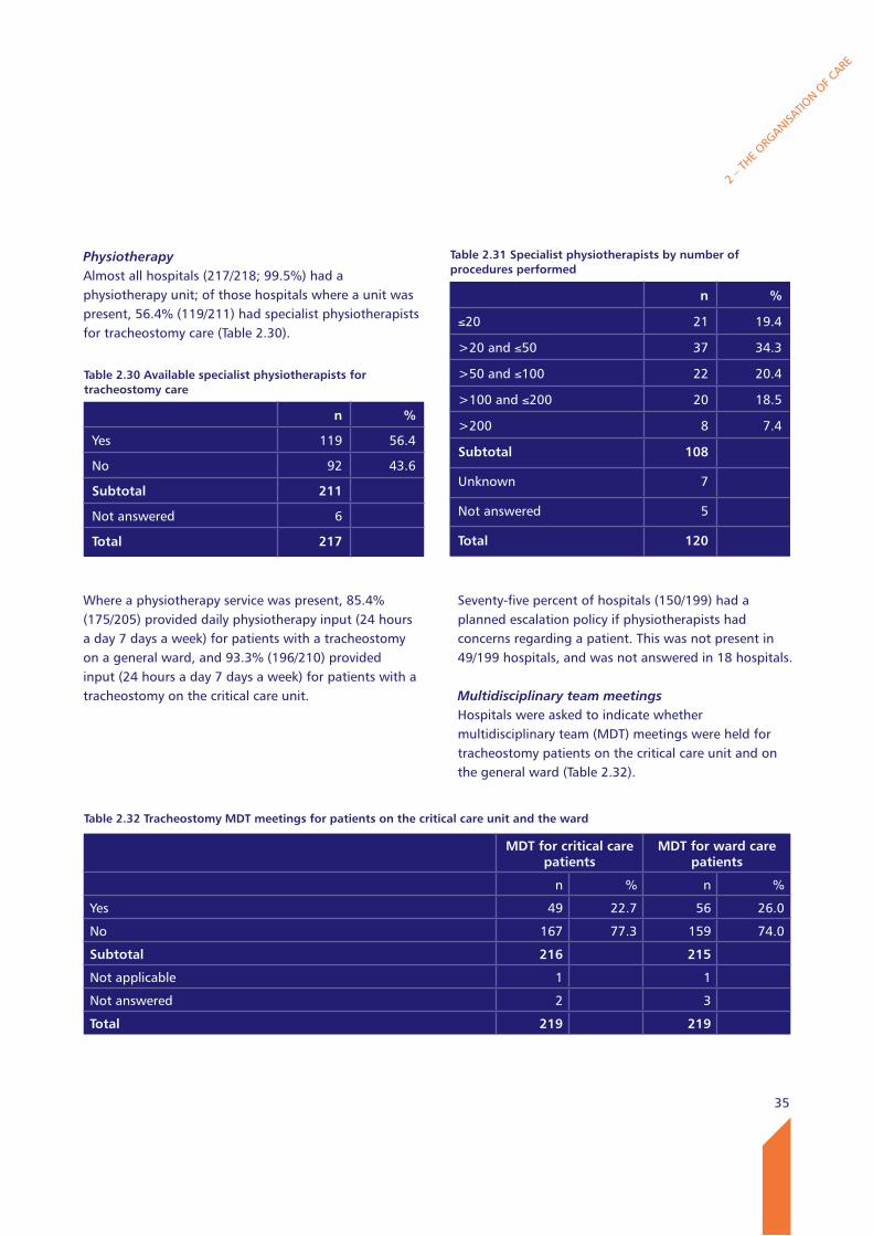
PhysiotherapyAlmost all hospitals (217/218; 99.5%) had a physiotherapy unit; of those hospitals where a unit was present, 56.4% (119/211) had specialist physiotherapists for tracheostomy care (Table 2.30).
Where a physiotherapy service was present, 85.4% (175/205) provided daily physiotherapy input (24 hours a day 7 days a week) for patients with a tracheostomy on a general ward, and 93.3% (196/210) provided input (24 hours a day 7 days a week) for patients with a tracheostomy on the critical care unit.
Seventy-five percent of hospitals (150/199) had a planned escalation policy if physiotherapists had concerns regarding a patient. This was not present in 49/199 hospitals, and was not answered in 18 hospitals.
Multidisciplinary team meetingsHospitals were asked to indicate whether multidisciplinary team (MDT) meetings were held for tracheostomy patients on the critical care unit and on the general ward (Table 2.32).
2 – T
HE ORG
ANISATIO
N OF C
ARE
Table 2.30 Available specialist physiotherapists for tracheostomy care
n %
Yes 119 56.4
No 92 43.6
Subtotal 211
Not answered 6
Total 217
Table 2.31 Specialist physiotherapists by number of procedures performed
n %
≤20 21 19.4
>20 and ≤50 37 34.3
>50 and ≤100 22 20.4
>100 and ≤200 20 18.5
>200 8 7.4
Subtotal 108 Unknown 7 Not answered 5 Total 120
Table 2.32 Tracheostomy MDT meetings for patients on the critical care unit and the ward
MDT for critical care patients
MDT for ward care patients
n % n %
Yes 49 22.7 56 26.0
No 167 77.3 159 74.0
Subtotal 216 215
Not applicable 1 1
Not answered 2 3
Total 219 219
36
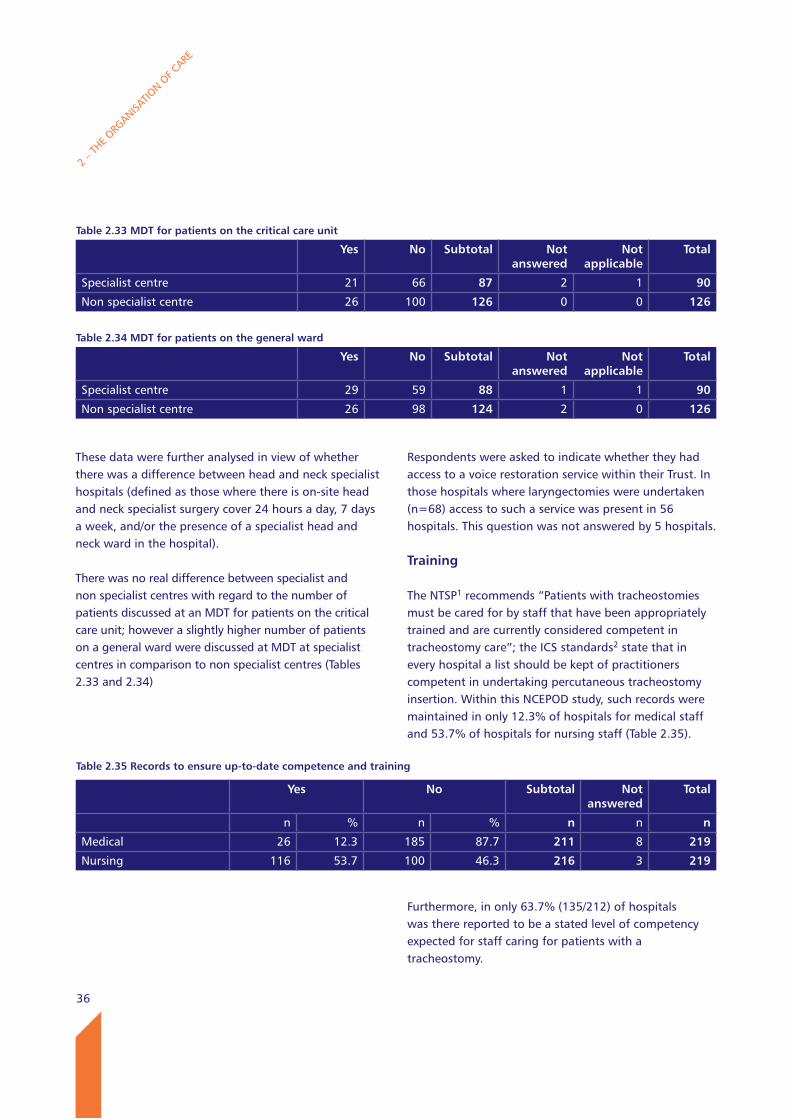
These data were further analysed in view of whether there was a difference between head and neck specialist hospitals (defined as those where there is on-site head and neck specialist surgery cover 24 hours a day, 7 days a week, and/or the presence of a specialist head and neck ward in the hospital).
There was no real difference between specialist and non specialist centres with regard to the number of patients discussed at an MDT for patients on the critical care unit; however a slightly higher number of patients on a general ward were discussed at MDT at specialist centres in comparison to non specialist centres (Tables 2.33 and 2.34)
Respondents were asked to indicate whether they had access to a voice restoration service within their Trust. In those hospitals where laryngectomies were undertaken (n=68) access to such a service was present in 56 hospitals. This question was not answered by 5 hospitals.
Training
The NTSP1 recommends “Patients with tracheostomies must be cared for by staff that have been appropriately trained and are currently considered competent in tracheostomy care”; the ICS standards2 state that in every hospital a list should be kept of practitioners competent in undertaking percutaneous tracheostomy insertion. Within this NCEPOD study, such records were maintained in only 12.3% of hospitals for medical staff and 53.7% of hospitals for nursing staff (Table 2.35).
Furthermore, in only 63.7% (135/212) of hospitals was there reported to be a stated level of competency expected for staff caring for patients with a tracheostomy.
2 – T
HE ORG
ANISATIO
N OF C
ARE
Table 2.33 MDT for patients on the critical care unit
yes No Subtotal Not answered
Not applicable
Total
Specialist centre 21 66 87 2 1 90
Non specialist centre 26 100 126 0 0 126
Table 2.34 MDT for patients on the general ward
yes No Subtotal Not answered
Not applicable
Total
Specialist centre 29 59 88 1 1 90
Non specialist centre 26 98 124 2 0 126
Table 2.35 Records to ensure up-to-date competence and training
yes No Subtotal Not answered
Total
n % n % n n n
Medical 26 12.3 185 87.7 211 8 219
Nursing 116 53.7 100 46.3 216 3 219
37
Table 2.36 Records maintained with head and neck specialist hospital and number of insertions.
Number of insertions performed
Medical Nursing Total
≤20 3 4 7
>20 and ≤50 1 10 11
>50 and ≤100 3 11 14
>100 and ≤200 3 13 16
>200 2 6 8
Subtotal 12 44 56
Unknown 0 1 1
Not answered 0 2 2
Total 12 47 59
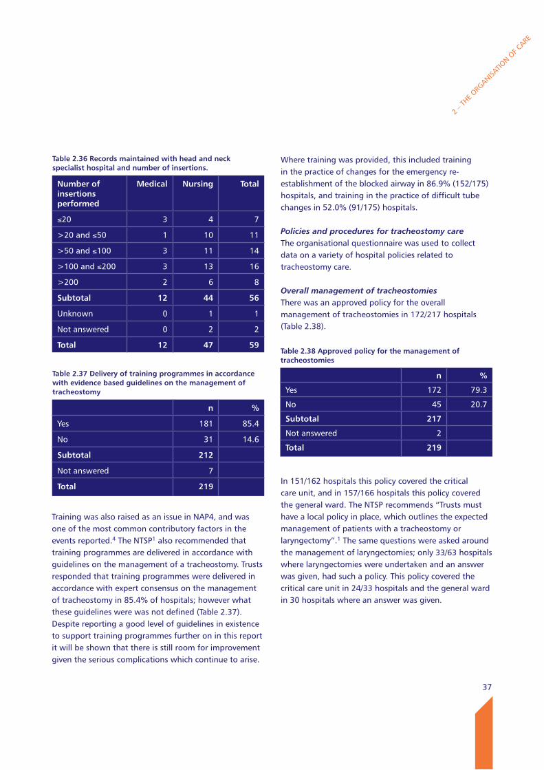
Training was also raised as an issue in NAP4, and was one of the most common contributory factors in the events reported.4 The NTSP1 also recommended that training programmes are delivered in accordance with guidelines on the management of a tracheostomy. Trusts responded that training programmes were delivered in accordance with expert consensus on the management of tracheostomy in 85.4% of hospitals; however what these guidelines were was not defined (Table 2.37). Despite reporting a good level of guidelines in existence to support training programmes further on in this report it will be shown that there is still room for improvement given the serious complications which continue to arise.
Where training was provided, this included training in the practice of changes for the emergency re-establishment of the blocked airway in 86.9% (152/175) hospitals, and training in the practice of difficult tube changes in 52.0% (91/175) hospitals.
Policies and procedures for tracheostomy careThe organisational questionnaire was used to collect data on a variety of hospital policies related to tracheostomy care.
Overall management of tracheostomiesThere was an approved policy for the overall management of tracheostomies in 172/217 hospitals (Table 2.38).
In 151/162 hospitals this policy covered the critical care unit, and in 157/166 hospitals this policy covered the general ward. The NTSP recommends “Trusts must have a local policy in place, which outlines the expected management of patients with a tracheostomy or laryngectomy”.1 The same questions were asked around the management of laryngectomies; only 33/63 hospitals where laryngectomies were undertaken and an answer was given, had such a policy. This policy covered the critical care unit in 24/33 hospitals and the general ward in 30 hospitals where an answer was given.
2 – T
HE ORG
ANISATIO
N OF C
ARE
Table 2.37 Delivery of training programmes in accordance with evidence based guidelines on the management of tracheostomy
n %
Yes 181 85.4
No 31 14.6
Subtotal 212
Not answered 7
Total 219
Table 2.38 Approved policy for the management of tracheostomies
n %
Yes 172 79.3
No 45 20.7
Subtotal 217
Not answered 2
Total 219
38
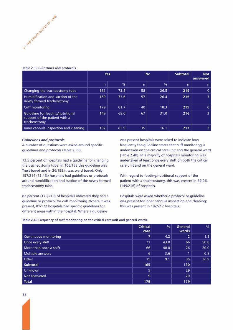
Guidelines and protocolsA number of questions were asked around specific guidelines and protocols (Table 2.39).
73.5 percent of hospitals had a guideline for changing the tracheostomy tube; in 106/158 this guideline was Trust based and in 36/158 it was ward based. Only 157/214 (73.4%) hospitals had guidelines or protocols around humidification and suction of the newly formed tracheostomy tube.
82 percent (179/219) of hospitals indicated they had a guideline or protocol for cuff monitoring. Where it was present, 81/172 hospitals had specific guidelines for different areas within the hospital. Where a guideline
was present hospitals were asked to indicate how frequently the guideline states that cuff monitoring is undertaken on the critical care unit and the general ward (Table 2.40). In a majority of hospitals monitoring was undertaken at least once every shift on both the critical care unit and on the general ward.
With regard to feeding/nutritional support of the patient with a tracheostomy, this was present in 69.0% (149/216) of hospitals.
Hospitals were asked whether a protocol or guideline was present for inner cannula inspection and cleaning; this was present in 182/217 hospitals.
2 – T
HE ORG
ANISATIO
N OF C
ARE
Table 2.39 Guidelines and protocols
yes No Subtotal Not answered
n % n % n n
Changing the tracheostomy tube 161 73.5 58 26.5 219 0
Humidification and suction of the newly formed tracheostomy
159 73.6 57 26.4 216 3
Cuff monitoring 179 81.7 40 18.3 219 0
Guideline for feeding/nutritional support of the patient with a tracheostomy
149 69.0 67 31.0 216 3
Inner cannula inspection and cleaning 182 83.9 35 16.1 217 2
Table 2.40 Frequency of cuff monitoring on the critical care unit and general wards
Critical care
% General wards
%
Continuous monitoring 7 4.2 2 1.5
Once every shift 71 43.0 66 50.8
More than once a shift 66 40.0 26 20.0
Multiple answers 6 3.6 1 0.8
Other 15 9.1 35 26.9
Subtotal 165 130
Unknown 5 29
Not answered 9 20
Total 179 179
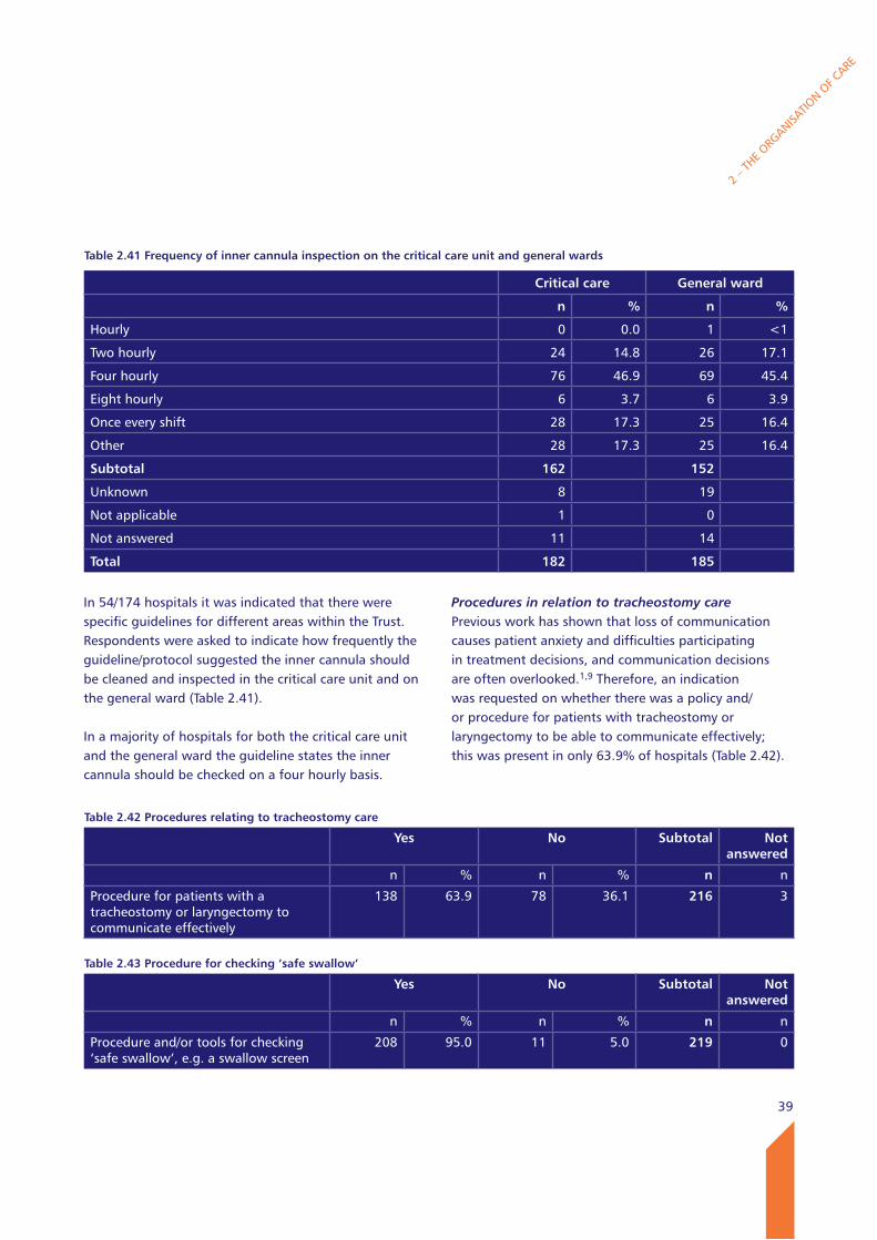
39
In 54/174 hospitals it was indicated that there were specific guidelines for different areas within the Trust. Respondents were asked to indicate how frequently the guideline/protocol suggested the inner cannula should be cleaned and inspected in the critical care unit and on the general ward (Table 2.41).
In a majority of hospitals for both the critical care unit and the general ward the guideline states the inner cannula should be checked on a four hourly basis.
Procedures in relation to tracheostomy care Previous work has shown that loss of communication causes patient anxiety and difficulties participating in treatment decisions, and communication decisions are often overlooked.1,9 Therefore, an indication was requested on whether there was a policy and/or procedure for patients with tracheostomy or laryngectomy to be able to communicate effectively; this was present in only 63.9% of hospitals (Table 2.42).
2 – T
HE ORG
ANISATIO
N OF C
ARE
Table 2.41 Frequency of inner cannula inspection on the critical care unit and general wards
Critical care General ward
n % n %
Hourly 0 0.0 1 <1
Two hourly 24 14.8 26 17.1
Four hourly 76 46.9 69 45.4
Eight hourly 6 3.7 6 3.9
Once every shift 28 17.3 25 16.4
Other 28 17.3 25 16.4
Subtotal 162 152
Unknown 8 19
Not applicable 1 0
Not answered 11 14
Total 182 185
Table 2.42 Procedures relating to tracheostomy care
yes No Subtotal Not answered
n % n % n n
Procedure for patients with a tracheostomy or laryngectomy to communicate effectively
138 63.9 78 36.1 216 3
Table 2.43 Procedure for checking ‘safe swallow’
yes No Subtotal Not answered
n % n % n n
Procedure and/or tools for checking ‘safe swallow’, e.g. a swallow screen
208 95.0 11 5.0 219 0
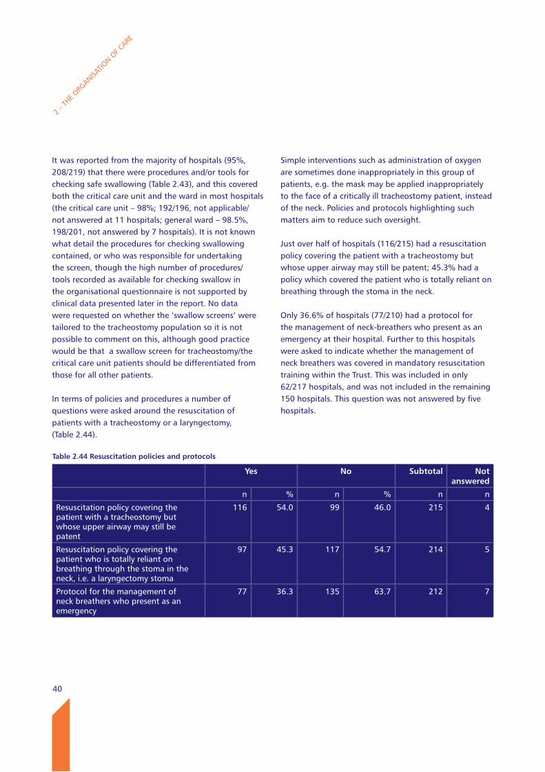
40
It was reported from the majority of hospitals (95%, 208/219) that there were procedures and/or tools for checking safe swallowing (Table 2.43), and this covered both the critical care unit and the ward in most hospitals (the critical care unit – 98%; 192/196, not applicable/not answered at 11 hospitals; general ward – 98.5%, 198/201, not answered by 7 hospitals). It is not known what detail the procedures for checking swallowing contained, or who was responsible for undertaking the screen, though the high number of procedures/tools recorded as available for checking swallow in the organisational questionnaire is not supported by clinical data presented later in the report. No data were requested on whether the ‘swallow screens’ were tailored to the tracheostomy population so it is not possible to comment on this, although good practice would be that a swallow screen for tracheostomy/the critical care unit patients should be differentiated from those for all other patients.
In terms of policies and procedures a number of questions were asked around the resuscitation of patients with a tracheostomy or a laryngectomy, (Table 2.44).
Simple interventions such as administration of oxygen are sometimes done inappropriately in this group of patients, e.g. the mask may be applied inappropriately to the face of a critically ill tracheostomy patient, instead of the neck. Policies and protocols highlighting such matters aim to reduce such oversight.
Just over half of hospitals (116/215) had a resuscitation policy covering the patient with a tracheostomy but whose upper airway may still be patent; 45.3% had a policy which covered the patient who is totally reliant on breathing through the stoma in the neck.
Only 36.6% of hospitals (77/210) had a protocol for the management of neck-breathers who present as an emergency at their hospital. Further to this hospitals were asked to indicate whether the management of neck breathers was covered in mandatory resuscitation training within the Trust. This was included in only 62/217 hospitals, and was not included in the remaining 150 hospitals. This question was not answered by five hospitals.
2 – T
HE ORG
ANISATIO
N OF C
ARE
Table 2.44 Resuscitation policies and protocols
yes No Subtotal Not answered
n % n % n n
Resuscitation policy covering the patient with a tracheostomy but whose upper airway may still be patent
116 54.0 99 46.0 215 4
Resuscitation policy covering the patient who is totally reliant on breathing through the stoma in the neck, i.e. a laryngectomy stoma
97 45.3 117 54.7 214 5
Protocol for the management of neck breathers who present as an emergency
77 36.3 135 63.7 212 7
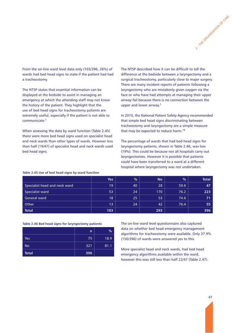
41
From the on-line ward level data only (103/396, 26%) of wards had bed head signs to state if the patient had had a tracheostomy.
The NTSP states that essential information can be displayed at the bedside to assist in managing an emergency at which the attending staff may not know the history of the patient. They highlight that the use of bed head signs for tracheostomy patients are extremely useful, especially if the patient is not able to communicate.1
When assessing the data by ward function (Table 2.45) there were more bed head signs used on specialist head and neck wards than other types of wards. However less than half (19/47) of specialist head and neck wards used bed head signs.
The NTSP described how it can be difficult to tell the difference at the bedside between a laryngectomy and a surgical tracheostomy, particularly close to major surgery. There are many incident reports of patients following a laryngectomy who are mistakenly given oxygen via the face or who have had attempts at managing their upper airway fail because there is no connection between the upper and lower airway.1
In 2010, the National Patient Safety Agency recommended that simple bed head signs discriminating between tracheostomy and laryngectomy are a simple measure that may be expected to reduce harm.10
The percentage of wards that had bed head signs for laryngectomy patients, shown in Table 2.46, was low (19%). This could be because not all hospitals carry out laryngectomies. However it is possible that patients could have been transferred to a ward at a different hospital where laryngectomy was not undertaken.
The on-line ward level questionnaire also captured data on whether bed head emergency management algorithms for tracheostomy were available. Only 37.9% (150/396) of wards were answered yes to this.
More specialist head and neck wards, had bed head emergency algorithms available within the ward, however this was still less than half 22/47 (Table 2.47).
2 – T
HE ORG
ANISATIO
N OF C
ARE
Table 2.45 Use of bed head signs by ward function
yes % No % Total
Specialist head and neck ward 19 40 28 59.6 47
Specialist ward 53 24 170 76.2 223
General ward 18 25 53 74.6 71
Other 13 24 42 76.4 55
Total 103 293 396
Table 2.46 Bed head signs for laryngectomy patients
n %
Yes 75 18.9
No 321 81.1
Total 396
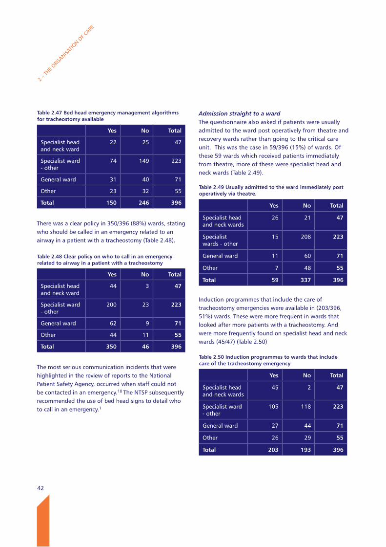
42
There was a clear policy in 350/396 (88%) wards, stating who should be called in an emergency related to an airway in a patient with a tracheostomy (Table 2.48).
The most serious communication incidents that were highlighted in the review of reports to the National Patient Safety Agency, occurred when staff could not be contacted in an emergency.10 The NTSP subsequently recommended the use of bed head signs to detail who to call in an emergency.1
Admission straight to a wardThe questionnaire also asked if patients were usually admitted to the ward post operatively from theatre and recovery wards rather than going to the critical care unit. This was the case in 59/396 (15%) of wards. Of these 59 wards which received patients immediately from theatre, more of these were specialist head and neck wards (Table 2.49).
Induction programmes that include the care of tracheostomy emergencies were available in (203/396, 51%) wards. These were more frequent in wards that looked after more patients with a tracheostomy. And were more frequently found on specialist head and neck wards (45/47) (Table 2.50)
2 – T
HE ORG
ANISATIO
N OF C
ARE
Table 2.48 Clear policy on who to call in an emergency related to airway in a patient with a tracheostomy
yes No Total
Specialist head and neck ward
44 3 47
Specialist ward - other
200 23 223
General ward 62 9 71
Other 44 11 55
Total 350 46 396
Table 2.47 Bed head emergency management algorithms for tracheostomy available
yes No Total
Specialist head and neck ward
22 25 47
Specialist ward - other
74 149 223
General ward 31 40 71
Other 23 32 55
Total 150 246 396
Table 2.49 Usually admitted to the ward immediately post operatively via theatre.
yes No Total
Specialist head and neck wards
26 21 47
Specialist wards - other
15 208 223
General ward 11 60 71
Other 7 48 55
Total 59 337 396
Table 2.50 Induction programmes to wards that include care of the tracheostomy emergency
yes No Total
Specialist head and neck wards
45 2 47
Specialist ward - other
105 118 223
General ward 27 44 71
Other 26 29 55
Total 203 193 396
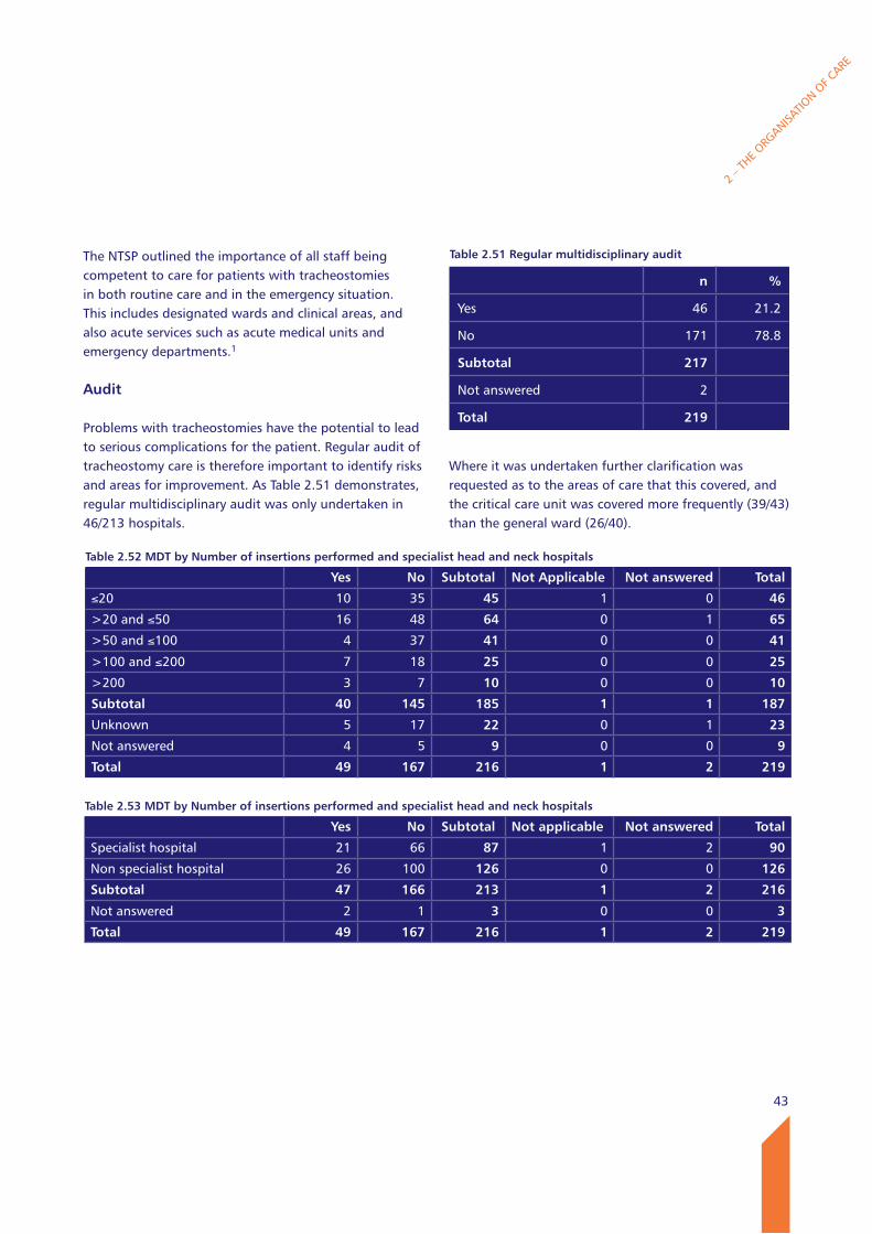
43
The NTSP outlined the importance of all staff being competent to care for patients with tracheostomies in both routine care and in the emergency situation. This includes designated wards and clinical areas, and also acute services such as acute medical units and emergency departments.1
Audit
Problems with tracheostomies have the potential to lead to serious complications for the patient. Regular audit of tracheostomy care is therefore important to identify risks and areas for improvement. As Table 2.51 demonstrates, regular multidisciplinary audit was only undertaken in 46/213 hospitals.
Where it was undertaken further clarification was requested as to the areas of care that this covered, and the critical care unit was covered more frequently (39/43) than the general ward (26/40).
2 – T
HE ORG
ANISATIO
N OF C
ARE
Table 2.51 Regular multidisciplinary audit
n %
Yes 46 21.2
No 171 78.8
Subtotal 217
Not answered 2
Total 219
Table 2.52 MDT by Number of insertions performed and specialist head and neck hospitals
yes No Subtotal Not Applicable Not answered Total
≤20 10 35 45 1 0 46
>20 and ≤50 16 48 64 0 1 65
>50 and ≤100 4 37 41 0 0 41
>100 and ≤200 7 18 25 0 0 25
>200 3 7 10 0 0 10
Subtotal 40 145 185 1 1 187
Unknown 5 17 22 0 1 23
Not answered 4 5 9 0 0 9
Total 49 167 216 1 2 219
Table 2.53 MDT by Number of insertions performed and specialist head and neck hospitals
yes No Subtotal Not applicable Not answered Total
Specialist hospital 21 66 87 1 2 90
Non specialist hospital 26 100 126 0 0 126
Subtotal 47 166 213 1 2 216
Not answered 2 1 3 0 0 3
Total 49 167 216 1 2 219
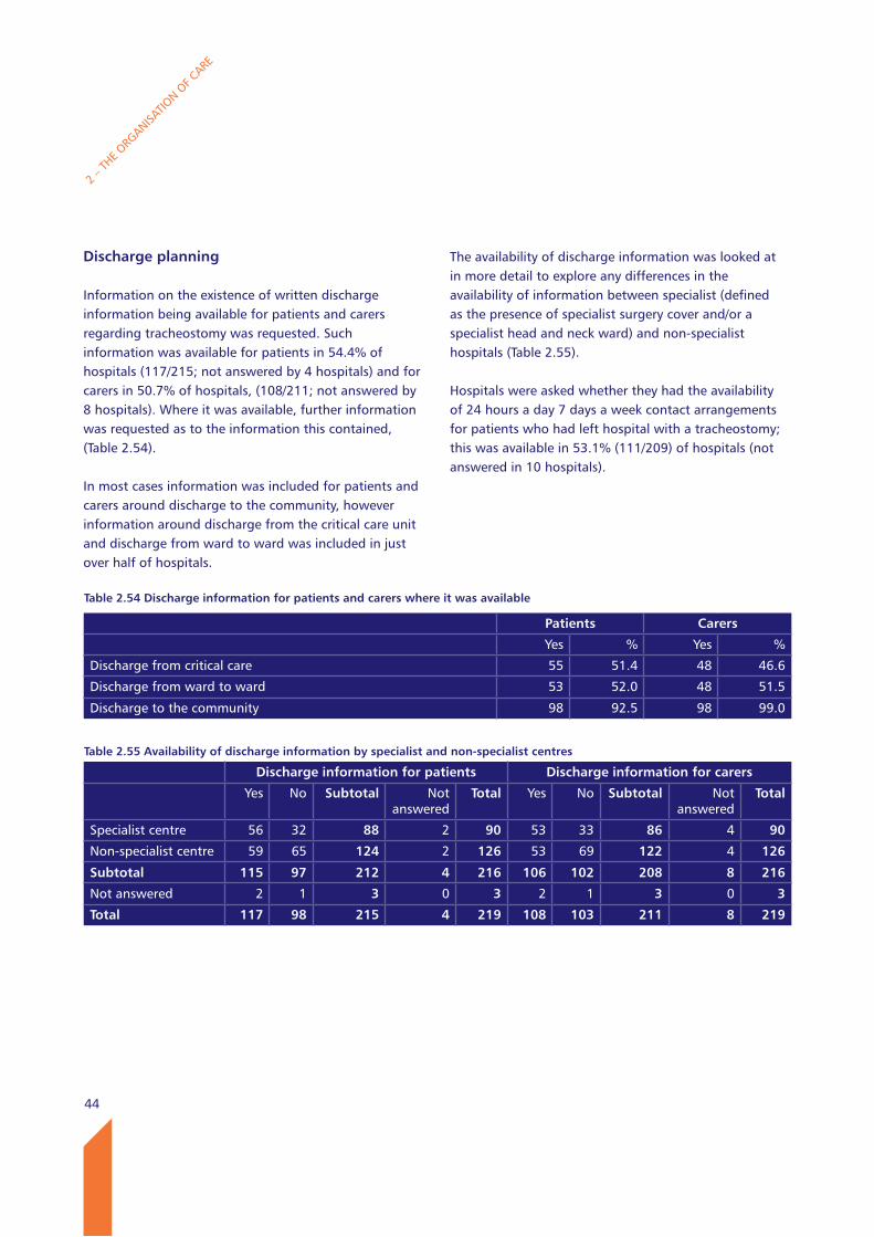
44
The availability of discharge information was looked at in more detail to explore any differences in the availability of information between specialist (defined as the presence of specialist surgery cover and/or a specialist head and neck ward) and non-specialist hospitals (Table 2.55).
Hospitals were asked whether they had the availability of 24 hours a day 7 days a week contact arrangements for patients who had left hospital with a tracheostomy; this was available in 53.1% (111/209) of hospitals (not answered in 10 hospitals).
Discharge planning
Information on the existence of written discharge information being available for patients and carers regarding tracheostomy was requested. Such information was available for patients in 54.4% of hospitals (117/215; not answered by 4 hospitals) and for carers in 50.7% of hospitals, (108/211; not answered by 8 hospitals). Where it was available, further information was requested as to the information this contained, (Table 2.54).
In most cases information was included for patients and carers around discharge to the community, however information around discharge from the critical care unit and discharge from ward to ward was included in just over half of hospitals.
2 – T
HE ORG
ANISATIO
N OF C
ARE
Table 2.55 Availability of discharge information by specialist and non-specialist centres
Discharge information for patients Discharge information for carers
Yes No Subtotal Not answered
Total Yes No Subtotal Not answered
Total
Specialist centre 56 32 88 2 90 53 33 86 4 90
Non-specialist centre 59 65 124 2 126 53 69 122 4 126
Subtotal 115 97 212 4 216 106 102 208 8 216
Not answered 2 1 3 0 3 2 1 3 0 3
Total 117 98 215 4 219 108 103 211 8 219
Table 2.54 Discharge information for patients and carers where it was available
Patients Carers
Yes % Yes %
Discharge from critical care 55 51.4 48 46.6
Discharge from ward to ward 53 52.0 48 51.5
Discharge to the community 98 92.5 98 99.0
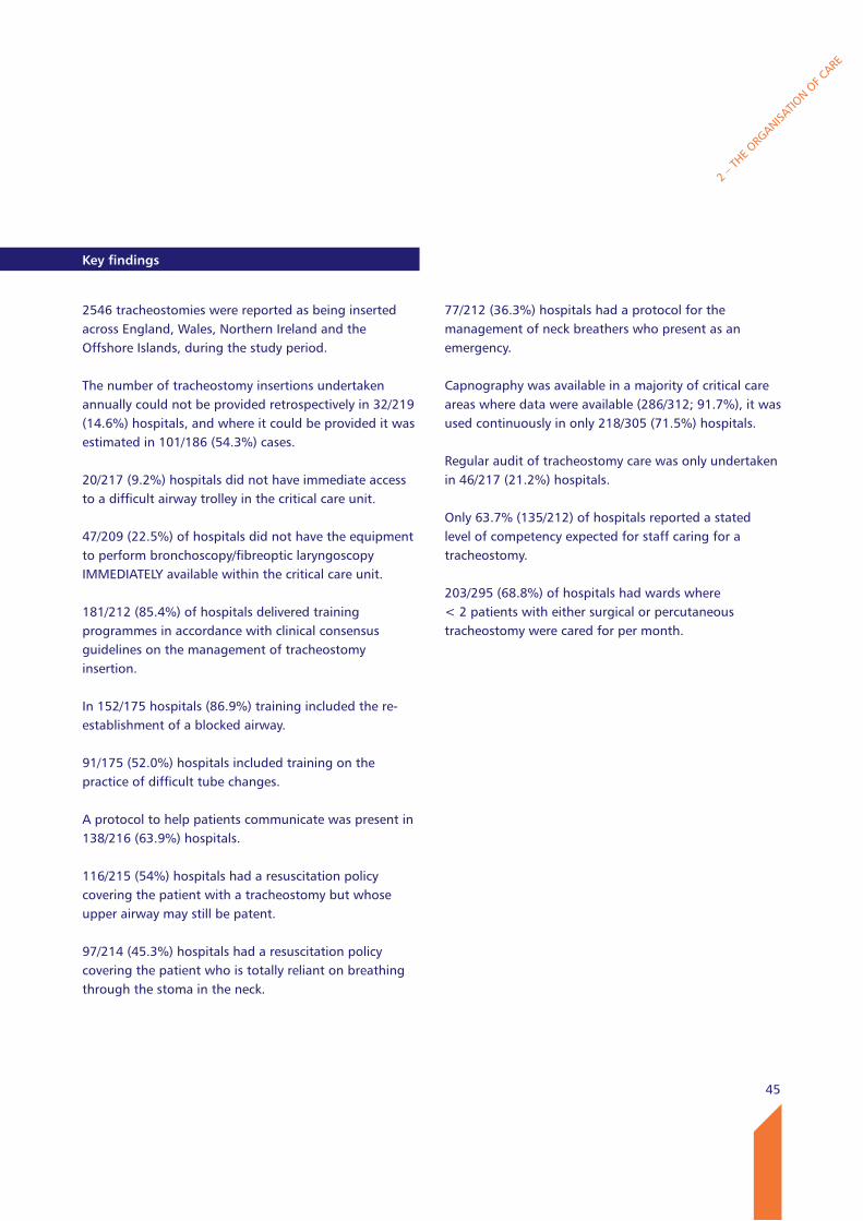
45
2 – T
HE ORG
ANISATIO
N OF C
ARE
key findings
2546 tracheostomies were reported as being inserted across England, Wales, Northern Ireland and the Offshore Islands, during the study period.
The number of tracheostomy insertions undertaken annually could not be provided retrospectively in 32/219 (14.6%) hospitals, and where it could be provided it was estimated in 101/186 (54.3%) cases.
20/217 (9.2%) hospitals did not have immediate access to a difficult airway trolley in the critical care unit.
47/209 (22.5%) of hospitals did not have the equipment to perform bronchoscopy/fibreoptic laryngoscopy IMMEDIATELY available within the critical care unit.
181/212 (85.4%) of hospitals delivered training programmes in accordance with clinical consensus guidelines on the management of tracheostomy insertion.
In 152/175 hospitals (86.9%) training included the re-establishment of a blocked airway.
91/175 (52.0%) hospitals included training on the practice of difficult tube changes.
A protocol to help patients communicate was present in 138/216 (63.9%) hospitals.
116/215 (54%) hospitals had a resuscitation policy covering the patient with a tracheostomy but whose upper airway may still be patent.
97/214 (45.3%) hospitals had a resuscitation policy covering the patient who is totally reliant on breathing through the stoma in the neck.
77/212 (36.3%) hospitals had a protocol for the management of neck breathers who present as an emergency.
Capnography was available in a majority of critical care areas where data were available (286/312; 91.7%), it was used continuously in only 218/305 (71.5%) hospitals.
Regular audit of tracheostomy care was only undertaken in 46/217 (21.2%) hospitals.
Only 63.7% (135/212) of hospitals reported a stated level of competency expected for staff caring for a tracheostomy.
203/295 (68.8%) of hospitals had wards where< 2 patients with either surgical or percutaneous tracheostomy were cared for per month.
46
Recommendations
1. Tracheostomy insertion should be recorded and coded as an operative procedure. Data collection in all locations should be as robust as that for a theatre environment. This will facilitate better care planning and allow for national and local review and audit. (Medical Directors and National Coding Systems)
2. Critical care units need a rapidly available difficult airway trolley/fibreoptic laryngoscopy. This recommendation reinforces the Intensive Care Society and Royal College of Anaesthetists’ recommendations. (Clinical Directors)
3. Training programmes in blocked/displaced tubes/airways and difficult tube changes should be delivered in accordance with clinical consensus guidelines as stated by the National Tracheostomy Safety Project and the Intensive Care Society. (Medical Directors and Directors of Nursing)
4. Capnography must be available at each bed space in critical care and should be continuously used when patients are ventilator dependent. This reinforces the recommendation from NAP4 and others. (Clinical Directors)
5. Core competences for the care of tracheostomy patients, including resuscitation, should be set out by all Trusts using existing national resources available. (Medical Directors and Directors of Nursing)
2 – T
HE ORG
ANISATIO
N OF C
ARE
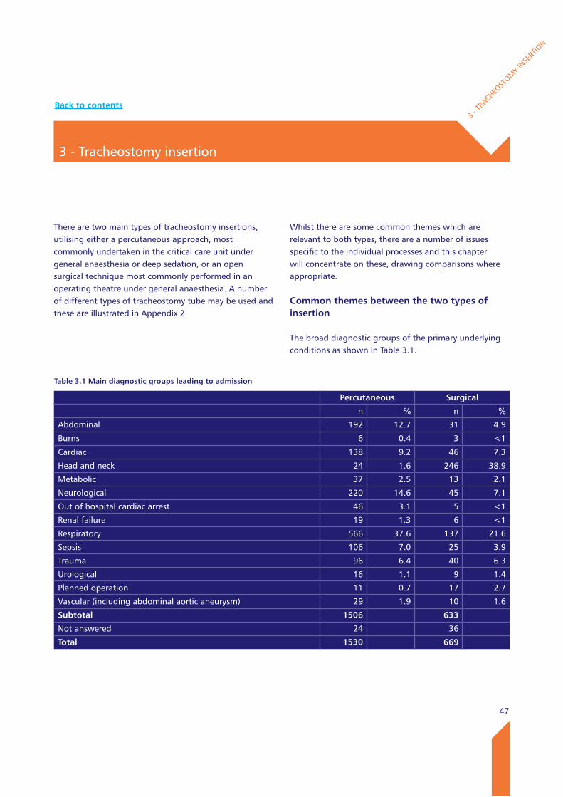
47
3 - Tracheostomy insertion
3 - T
RACHEO
STOM
Y IN
SERT
ION
There are two main types of tracheostomy insertions, utilising either a percutaneous approach, most commonly undertaken in the critical care unit under general anaesthesia or deep sedation, or an open surgical technique most commonly performed in an operating theatre under general anaesthesia. A number of different types of tracheostomy tube may be used and these are illustrated in Appendix 2.
Whilst there are some common themes which are relevant to both types, there are a number of issues specific to the individual processes and this chapter will concentrate on these, drawing comparisons where appropriate.
Common themes between the two types of insertion
The broad diagnostic groups of the primary underlying conditions as shown in Table 3.1.
Table 3.1 Main diagnostic groups leading to admission
Percutaneous Surgical
n % n %
Abdominal 192 12.7 31 4.9
Burns 6 0.4 3 <1
Cardiac 138 9.2 46 7.3
Head and neck 24 1.6 246 38.9
Metabolic 37 2.5 13 2.1
Neurological 220 14.6 45 7.1
Out of hospital cardiac arrest 46 3.1 5 <1
Renal failure 19 1.3 6 <1
Respiratory 566 37.6 137 21.6
Sepsis 106 7.0 25 3.9
Trauma 96 6.4 40 6.3
Urological 16 1.1 9 1.4
Planned operation 11 0.7 17 2.7
Vascular (including abdominal aortic aneurysm) 29 1.9 10 1.6
Subtotal 1506 633
Not answered 24 36
Total 1530 669
Back to contents
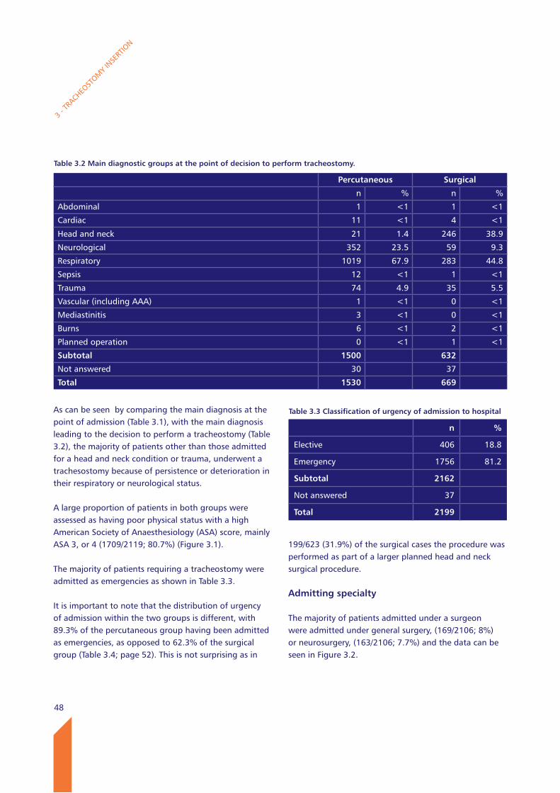
48
As can be seen by comparing the main diagnosis at the point of admission (Table 3.1), with the main diagnosis leading to the decision to perform a tracheostomy (Table 3.2), the majority of patients other than those admitted for a head and neck condition or trauma, underwent a trachesostomy because of persistence or deterioration in their respiratory or neurological status.
A large proportion of patients in both groups were assessed as having poor physical status with a high American Society of Anaesthesiology (ASA) score, mainly ASA 3, or 4 (1709/2119; 80.7%) (Figure 3.1).
The majority of patients requiring a tracheostomy were admitted as emergencies as shown in Table 3.3.
It is important to note that the distribution of urgency of admission within the two groups is different, with 89.3% of the percutaneous group having been admitted as emergencies, as opposed to 62.3% of the surgical group (Table 3.4; page 52). This is not surprising as in
199/623 (31.9%) of the surgical cases the procedure was performed as part of a larger planned head and neck surgical procedure.
Admitting specialty
The majority of patients admitted under a surgeon were admitted under general surgery, (169/2106; 8%) or neurosurgery, (163/2106; 7.7%) and the data can be seen in Figure 3.2.
3 - T
RACHEO
STOM
Y IN
SERT
ION
Table 3.2 Main diagnostic groups at the point of decision to perform tracheostomy.
Percutaneous Surgical
n % n %
Abdominal 1 <1 1 <1
Cardiac 11 <1 4 <1
Head and neck 21 1.4 246 38.9
Neurological 352 23.5 59 9.3
Respiratory 1019 67.9 283 44.8
Sepsis 12 <1 1 <1
Trauma 74 4.9 35 5.5
Vascular (including AAA) 1 <1 0 <1
Mediastinitis 3 <1 0 <1
Burns 6 <1 2 <1
Planned operation 0 <1 1 <1
Subtotal 1500 632
Not answered 30 37
Total 1530 669
Table 3.3 Classification of urgency of admission to hospital
n %
Elective 406 18.8
Emergency 1756 81.2
Subtotal 2162
Not answered 37
Total 2199
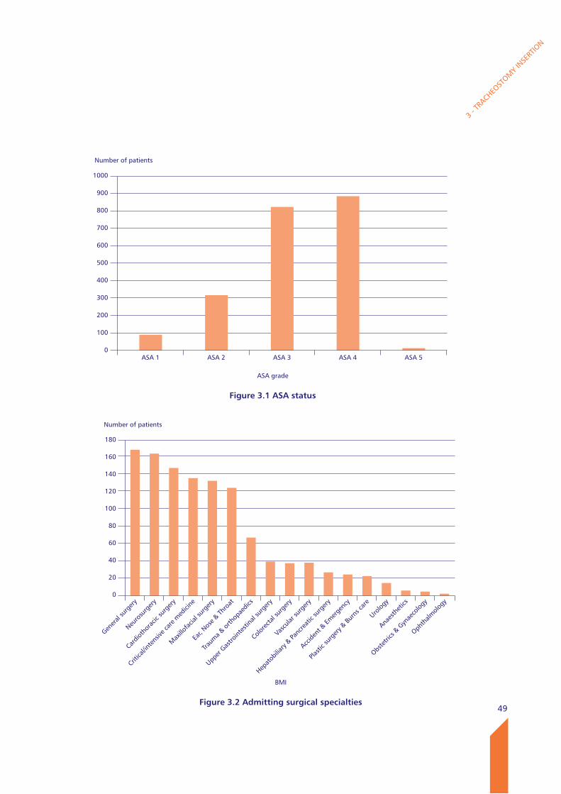
49
3 - T
RACHEO
STOM
Y IN
SERT
ION
Number of patients
1000
900
800
700
600
500
400
300
200
100
0
Figure 3.1 ASA status
ASA grade
ASA 1 ASA 2 ASA 3 ASA 4 ASA 5
Number of patients
180
160
140
120
100
80
60
40
20
0
Figure 3.2 Admitting surgical specialties
BMI
Gener
al su
rger
y
Neuro
surg
ery
Cardio
thor
acic
surg
ery
Critica
l/inte
nsive
care
med
icine
Max
illofa
cial s
urge
ry
Ear, N
ose &
Thro
at
Traum
a & o
rthop
aedic
s
Upper
Gas
troint
estin
al su
rger
y
Colore
ctal s
urge
ry
Vasc
ular s
urge
ry
Hepat
obilia
ry & Pa
ncre
atic
surg
ery
Accide
nt &
Emer
genc
y
Plasti
c sur
gery
& Bur
ns ca
re
Urolog
y
Anaes
thet
ics
Obste
trics
& G
ynae
colog
y
Ophth
almolo
gy
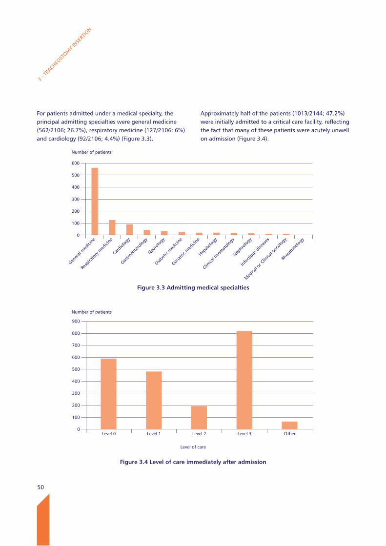
50
For patients admitted under a medical specialty, the principal admitting specialties were general medicine (562/2106; 26.7%), respiratory medicine (127/2106; 6%) and cardiology (92/2106; 4.4%) (Figure 3.3).
Approximately half of the patients (1013/2144; 47.2%) were initially admitted to a critical care facility, reflecting the fact that many of these patients were acutely unwell on admission (Figure 3.4).
3 - T
RACHEO
STOM
Y IN
SERT
ION
Figure 3.4 level of care immediately after admission
Number of patients
900
800
700
600
500
400
300
200
100
0Level 0 Level 1 Level 2 Level 3 Other
Level of care
Number of patients
600
500
400
300
200
100
0
Figure 3.3 Admitting medical specialties
Gener
al m
edici
ne
Resp
irato
ry m
edici
ne
Cardio
logy
Gastro
ente
rolog
y
Neuro
logy
Diabet
ic m
edici
ne
Geriat
ric m
edici
ne
Hepat
ology
Clinica
l hae
mat
ology
Nephr
ology
Infe
ctiou
s dise
ases
Med
ical o
r Clin
ical o
ncolo
gy
Rheu
mat
ology
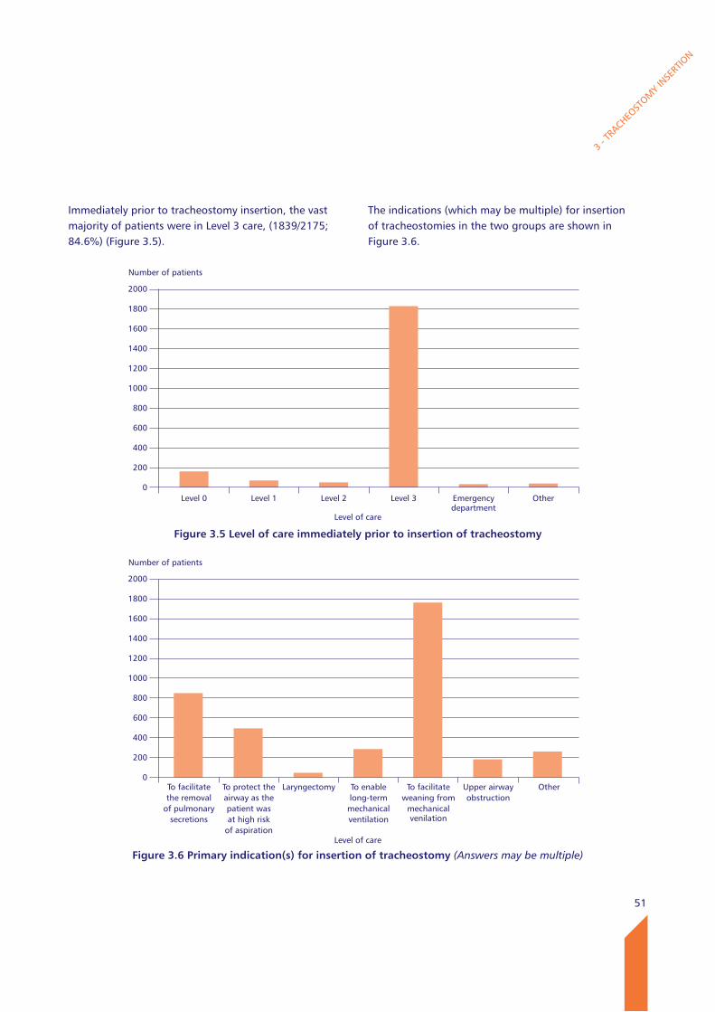
51
Immediately prior to tracheostomy insertion, the vast majority of patients were in Level 3 care, (1839/2175; 84.6%) (Figure 3.5).
The indications (which may be multiple) for insertion of tracheostomies in the two groups are shown in Figure 3.6.
3 - T
RACHEO
STOM
Y IN
SERT
ION
Number of patients
2000
1800
1600
1400
1200
1000
800
600
400
200
0Level 0 Level 1 Level 2 Level 3 Emergency
departmentOther
Figure 3.5 level of care immediately prior to insertion of tracheostomy
Level of care
Number of patients
2000
1800
1600
1400
1200
1000
800
600
400
200
0To facilitate the removal
of pulmonary secretions
To protect the airway as the patient was at high risk
of aspiration
Laryngectomy To enablelong-term
mechanical ventilation
To facilitate weaning from
mechanical venilation
Upper airway obstruction
Other
Figure 3.6 Primary indication(s) for insertion of tracheostomy (Answers may be multiple)Level of care
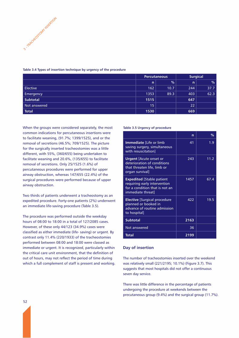
52
When the groups were considered separately, the most common indications for percutaneous insertions were to facilitate weaning, (91.7%; 1399/1525), and or the removal of secretions (46.5%; 709/1525). The picture for the surgically inserted tracheostomies was a little different, with 55%, (360/655) being undertaken to facilitate weaning and 20.6%, (135/655) to facilitate removal of secretions. Only 25/1525 (1.6%) of percutaneous procedures were performed for upper airway obstruction, whereas 147/655 (22.4%) of the surgical procedures were performed because of upper airway obstruction.
Two thirds of patients underwent a tracheostomy as an expedited procedure. Forty-one patients (2%) underwent an immediate life-saving procedure (Table 3.5).
The procedure was performed outside the weekday hours of 08:00 to 18:00 in a total of 127/2085 cases. However, of these only 44/123 (34.9%) cases were classified as either immediate (life- saving) or urgent. By contrast only 11.4% (220/1933) of the tracheostomies performed between 08:00 and 18:00 were classed as immediate or urgent. It is recognized, particularly within the critical care unit environment, that the definition of out of hours, may not reflect the period of time during which a full complement of staff is present and working.
Day of insertion
The number of tracheostomies inserted over the weekend was relatively small (221/2195; 10.1%) (Figure 3.7). This suggests that most hospitals did not offer a continuous seven day service.
There was little difference in the percentage of patients undergoing the procedure at weekends between the precutaneous group (9.4%) and the surgical group (11.7%).
3 - T
RACHEO
STOM
Y IN
SERT
ION
Table 3.4 Types of insertion technique by urgency of the procedure
Percutaneous Surgical
n % n %
Elective 162 10.7 244 37.7
Emergency 1353 89.3 403 62.3
Subtotal 1515 647
Not answered 15 22
Total 1530 669
Table 3.5 Urgency of procedure
n %
Immediate [Life or limb saving surgery, simultaneous with resuscitation]
41 1.9
Urgent [Acute onset or deterioration of conditions that threaten life, limb or organ survival]
243 11.2
Expedited [Stable patient requiring early intervention for a condition that is not an immediate threat]
1457 67.4
Elective [Surgical procedure planned or booked in advance of routine admission to hospital]
422 19.5
Subtotal 2163
Not answered 36
Total 2199
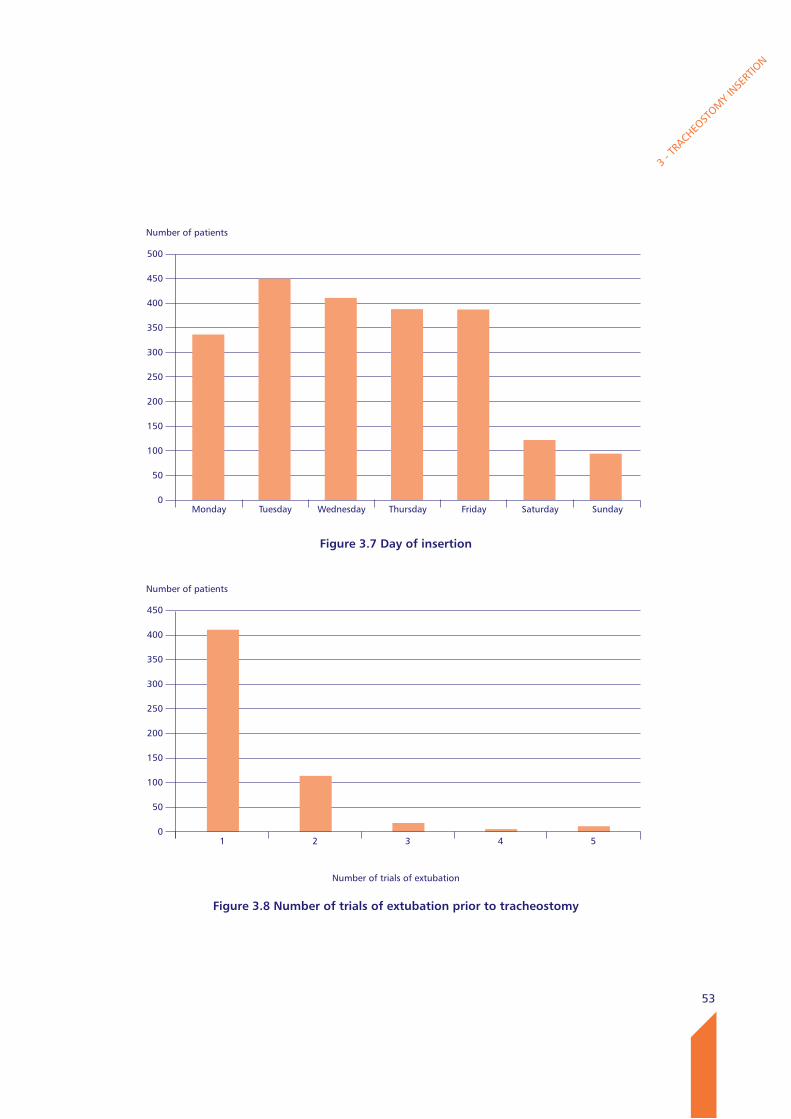
53
3 - T
RACHEO
STOM
Y IN
SERT
ION
Number of patients
450
400
350
300
250
200
150
100
50
01 2 3 4 5
Figure 3.8 Number of trials of extubation prior to tracheostomy
Number of trials of extubation
Number of patients
500
450
400
350
300
250
200
150
100
50
0Monday Tuesday Wednesday Thursday Friday Saturday Sunday
Figure 3.7 Day of insertion
54
3 - T
RACHEO
STOM
Y IN
SERT
ION
Trials of extubation
Prior to insertion of a tracheostomy a trial of extubation occurred in 565/1890 (29.9%) of patients (excluding those having a tracheostomy as part of a planned head and neck procedure) who received a tracheostomy (Figure 3.8). The percentage of patients having a trial of extubation rose a little to 31.3% (539/1721) where an indication was for removal of secretions and/or to facilitate weaning. In the majority of cases, (410/559; 73.3%) only one attempt at extubation was made.
Table 3.6 There was a clear indication for the tracheostomy
n %
Yes 367 97.9
No 8 2.1
Subtotal 375
Insufficient data 18
Not answered 3
Total 396
Case study 1
A middle aged patient with liver cirrhosis was admitted with an upper GI bleed and coma due to a subdural haematoma. The patient underwent endotracheal intubation in the emergency department and was admitted to intensive care for ventilatory support. On admission the patient had a low platelet count (56) and an elevated INR (1.5). On reducing sedation, the patient became agitated. The platelet and clotting abnormalities were not corrected. On day four without any systematic attempt at weaning, a percutaneous tracheostomy was performed. On the 2nd post-operative day the patient was stepped down to HDU. Blood clots and blood stained secretions were repeatedly suctioned from the tracheostomy tube. This was thought to be due to local trauma from the tracheostomy tube, and a laryngoscopy was requested from the ENT department, but was not done. Two weeks post-operatively, following a violent episode of coughing which caused local bleeding, the tracheostomy tube was removed by an intensivist and the patient discharged to a general medical ward. A week after decannulation, the patient suffered a major GI bleed. Endotracheal intubation failed, but it was possible to re-intubate through the tracheostomy site, albeit with significant local bleeding. The patient suffered a cardiac arrest, but was successfully resuscitated and taken to theatre, for exploration of the neck and a formal surgical tracheostomy. A bronchoscopy identified bleeding from the right main bronchus. The patient died eight hours later on the intensive care unit.
Advisors questioned whether the tracheostomy had been performed too soon, and without a reasonable systematic attempt to wean the patient. The source of the bleeding from the trachea did not appear to have been adequately investigated, and the tracheostomy tube was removed whilst the patient was continuing to bleed from the tracheo-bronchial tree.
Indications for insertion
Advisors stated, when reviewing the cases that in the vast majority there was a clear indication for a tracheostomy (Table 3.6).
55
3 - T
RACHEO
STOM
Y IN
SERT
ION
However in 67/300 (22.3%) cases where there was sufficient information to make an assessment, they did not feel that the indication for the tracheostomy had been clearly documented (Table 3.7).
Furthermore of the 291 cases where Advisors had sufficient information to make a judgment, only 183 (62.9%) documented an assessment of airway difficulty (Table 3.8). This assessment is important in order to ensure that an appropriate anaesthetic and operative technique is employed with the appropriate equipment available, and to ensure correct seniority of staff perform the procedure.
The failure to adequately document the difficulty of the airway was judged by the Advisors to have caused delay in three cases, critical airway compromise in one case and in two cases was undertaken by an non-consultant when, due to obesity, a consultant should have performed the procedure.
Table 3.7 The indication for tracheostomy was clearly documented
n %
Yes 287 81.1
No 67 18.9
Subtotal 354
Insufficient data 32
Not answered 10
Total 396
Table 3.8 There was an adequately documented assessment of airway difficulty
n %
Yes 183 62.9
No 108 37.1
Subtotal 291
Insufficient data 82
Not answered 23
Total 396
Obesity
Obesity poses greater technical challenges to the insertion of a tracheostomy tube. The distance between the skin surface and the trachea is greater, and patients often have a short neck, so that access is reduced. The equipment and the type of tracheostomy tube used need to be of an appropriate size, both in length and diameter, for the individual patient, standardised tubes may not be satisfactory.
In 1379 cases a BMI was recorded in the questionnaire (Table 3.9); in 531 cases the BMI was not recorded in the questionnaire but a height and weight were given and the BMI was subsequently calculated (Table 3.10). In 289 patients, the BMI was not recorded and could not be calculated. Therefore, of the 1910 patients for whom the BMI was recorded or calculated, 29.6% of patients were either obese (BMI≥30) or morbidly obese (BMI≥ 40). Recognizing that obese patients are at greater risk of complications, every effort should have been made to obtain weight and height or at the very least make an estimate.
Table 3.9 Classification of BMI where available
n %
Underweight 80 4.2
Normal 643 33.7
Overweight 621 32.5
Obese 474 24.8
Morbidly obese 92 4.8
Total 1910
Table 3.10 BMI was recorded or calculated
n %
Yes 1910 86.9
No 289 13.1
Total 2199
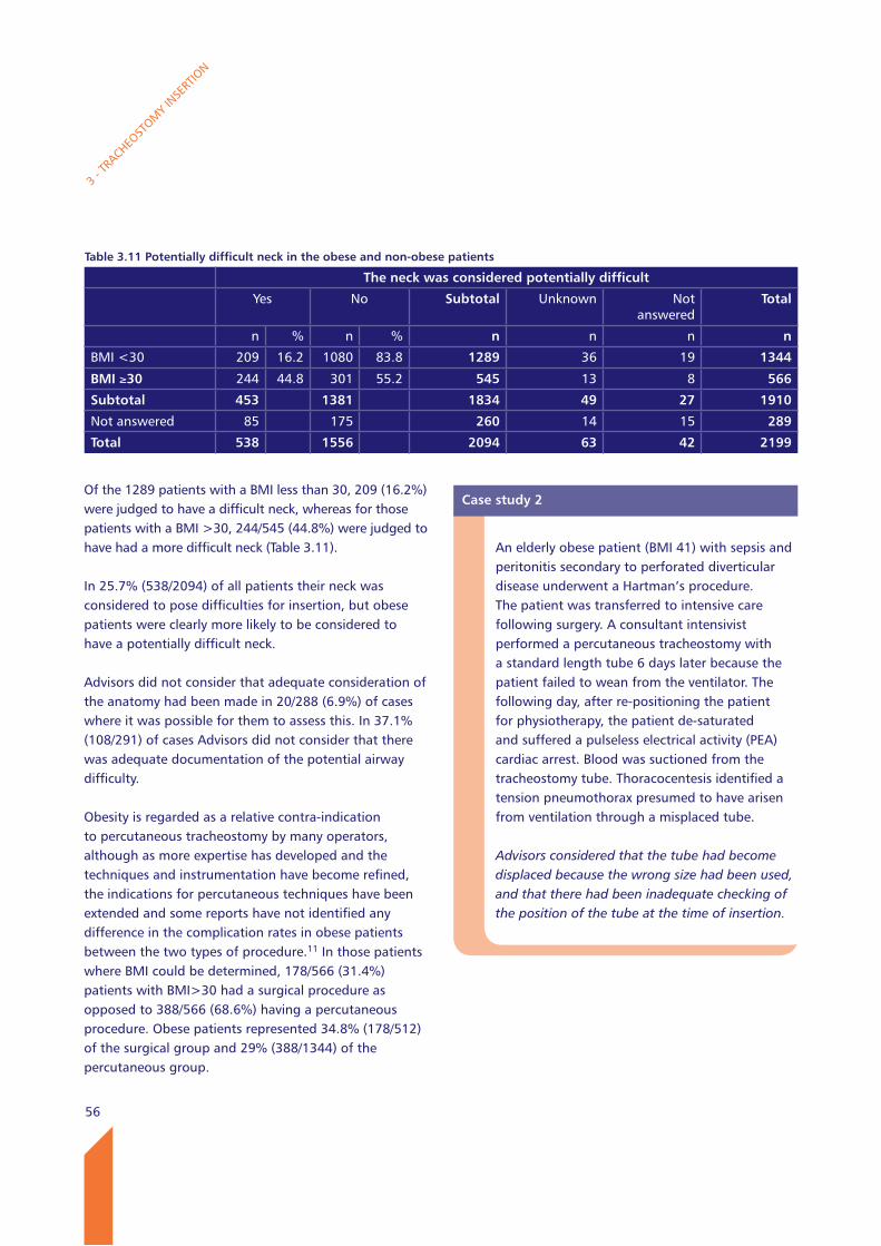
56
Of the 1289 patients with a BMI less than 30, 209 (16.2%) were judged to have a difficult neck, whereas for those patients with a BMI >30, 244/545 (44.8%) were judged to have had a more difficult neck (Table 3.11).
In 25.7% (538/2094) of all patients their neck was considered to pose difficulties for insertion, but obese patients were clearly more likely to be considered to have a potentially difficult neck.
Advisors did not consider that adequate consideration of the anatomy had been made in 20/288 (6.9%) of cases where it was possible for them to assess this. In 37.1% (108/291) of cases Advisors did not consider that there was adequate documentation of the potential airway difficulty.
Obesity is regarded as a relative contra-indication to percutaneous tracheostomy by many operators, although as more expertise has developed and the techniques and instrumentation have become refined, the indications for percutaneous techniques have been extended and some reports have not identified any difference in the complication rates in obese patients between the two types of procedure.11 In those patients where BMI could be determined, 178/566 (31.4%) patients with BMI>30 had a surgical procedure as opposed to 388/566 (68.6%) having a percutaneous procedure. Obese patients represented 34.8% (178/512) of the surgical group and 29% (388/1344) of the percutaneous group.
3 - T
RACHEO
STOM
Y IN
SERT
ION
Table 3.11 Potentially difficult neck in the obese and non-obese patients
The neck was considered potentially difficult
Yes No Subtotal Unknown Not answered
Total
n % n % n n n n
BMI <30 209 16.2 1080 83.8 1289 36 19 1344
BMI ≥30 244 44.8 301 55.2 545 13 8 566
Subtotal 453 1381 1834 49 27 1910
Not answered 85 175 260 14 15 289
Total 538 1556 2094 63 42 2199
Case study 2
An elderly obese patient (BMI 41) with sepsis and peritonitis secondary to perforated diverticular disease underwent a Hartman’s procedure. The patient was transferred to intensive care following surgery. A consultant intensivist performed a percutaneous tracheostomy with a standard length tube 6 days later because the patient failed to wean from the ventilator. The following day, after re-positioning the patient for physiotherapy, the patient de-saturated and suffered a pulseless electrical activity (PEA) cardiac arrest. Blood was suctioned from the tracheostomy tube. Thoracocentesis identified a tension pneumothorax presumed to have arisen from ventilation through a misplaced tube.
Advisors considered that the tube had become displaced because the wrong size had been used, and that there had been inadequate checking of the position of the tube at the time of insertion.
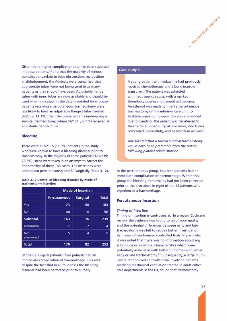
57
Given that a higher complication rate has been reported in obese patients,12 and that the majority of serious complications relate to tube obstruction, malposition or dislodgement, the Advisors were concerned that appropriate tubes were not being used in as many patients as they should have been. Adjustable flange tubes with inner tubes are now available and should be used when indicated. In the data presented here, obese patients receiving a percutaneous tracheostomy were less likely to have an adjustable flanged tube inserted (40/359; 11.1%), than the obese patients undergoing a surgical tracheostomy, where 56/151 (37.1%) received an adjustable flanged tube.
Bleeding
There were 252/2115 (11.9%) patients in the study who were known to have a bleeding disorder prior to tracheostomy. In the majority of these patients (183/239; 76.6%), steps were taken in an attempt to correct the abnormality; of these 183 cases, 123 insertions were undertaken percutaneously and 60 surgically (Table 3.12).
Of the 82 surgical patients, four patients had an immediate complication of haemorrhage. This was despite the fact that in all four cases the bleeding disorder had been corrected prior to surgery.
In the percutaneous group, fourteen patients had an immediate complication of haemorrhage. Within this group the bleeding abnormality had not been corrected prior to the procedure in eight of the 14 patients who experienced a haemorrhage.
Percutaneous Insertion
Timing of insertionTiming of insertion is controversial. In a recent Cochrane review, the evidence was found to be of poor quality and the potential differences between early and late tracheostomy was felt to require better investigation by means of randomized controlled trials. In particular it was noted that there was no information about any subgroups or individual characteristics which were potentially associated with better outcomes with either early or late tracheostomy.13 Subsequently, a large multi-centre randomized controlled trial involving patients receiving mechanical ventilation treated in adult critical care departments in the UK; found that tracheostomy
3 - T
RACHEO
STOM
Y IN
SERT
ION
Table 3.12 Control of bleeding disorder by mode of tracheostomy insertion
Mode of insertion
Percutaneous Surgical Total
Yes 123 60 183
No 40 16 56
Subtotal 163 76 239
Unknown 2 2 4
Not answered
5 4 9
Total 170 82 252
Case study 3
A young patient with leukaemia had previously received chemotherapy and a bone marrow transplant. The patient was admitted with neutropenic sepsis, with a marked thrombocytopenia and generalised oedema. An attempt was made to insert a percutaneous tracheostomy on the intensive care unit, to facilitate weaning, however this was abandoned due to bleeding. The patient was transferred to theatre for an open surgical procedure, which was completed uneventfully, and haemostasis achieved.
Advisors felt that a formal surgical tracheostomy would have been preferable from the outset, following platelet administration.
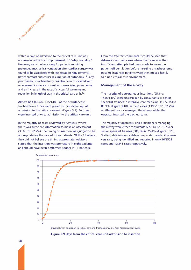
58
within 4 days of admission to the critical care unit was not associated with an improvement in 30-day mortality.5 However, early tracheostomy for patients requiring prolonged mechanical ventilation after cardiac surgery was found to be associated with less sedation requirements, better comfort and earlier resumption of autonomy.14 Early percutaneous tracheostomy has also been associated with a decreased incidence of ventilator associated pneumonia, and an increase in the rate of successful weaning and reduction in length of stay in the critical care unit.15
Almost half (45.4%, 675/1486) of the percutaneous tracheostomy tubes were placed within seven days of admission to the critical care unit (Figure 3.9). Fourteen were inserted prior to admission to the critical care unit.
In the majority of cases reviewed by Advisors, where there was sufficient information to make an assessment (333/361; 92.2%), the timing of insertion was judged to be appropriate for the care of those patients. Of the 28 where they did not believe the timing appropriate, Advisors stated that the insertion was premature in eight patients and should have been performed sooner in 11 patients.
From the free text comments it could be seen that Advisors identified cases where their view was that insufficient attempts had been made to wean the patient off ventilation before inserting a tracheostomy. In some instances patients were then moved hastily to a non-critical care environment.
Management of the airway
The majority of percutaneous insertions (95.1%; 1425/1499) were undertaken by consultants or senior specialist trainees in intensive care medicine, (1272/1516; 83.9%) (Figure 3.10). In most cases (1393/1502 (92.7%) a different doctor managed the airway whilst the operator inserted the tracheostomy.
The majority of operators, and practitioners managing the airway were either consultants (777/1496; 51.9%) or senior specialist trainees (380/1496; 25.4%) (Figure 3.11). Staffing deficiencies or delays due to staff availability were very rare, being identified and reported in only 16/1508 cases and 10/341 cases respectively.
3 - T
RACHEO
STOM
Y IN
SERT
ION
Cumulative percentage
100
90
80
70
60
50
40
30
20
10
0
Figure 3.9 Days from the critical care unit admission to insertion
Days between admission to critical care and tracheostomy insertion (percutaneous only)
0 20 40 60 80
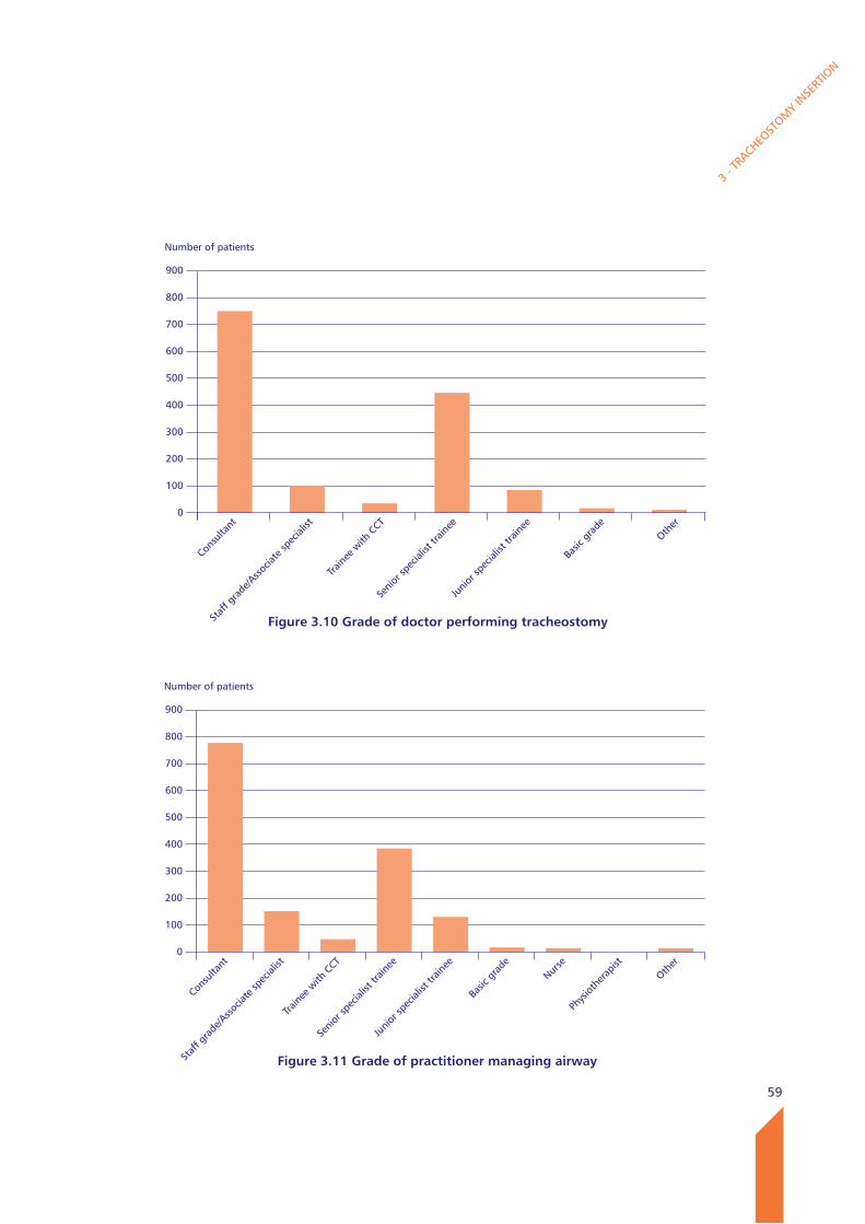
59
3 - T
RACHEO
STOM
Y IN
SERT
ION
Number of patients
900
800
700
600
500
400
300
200
100
0
Figure 3.11 Grade of practitioner managing airway
Consu
ltant
Staf
f gra
de/A
ssocia
te sp
ecial
ist
Traine
e with
CCT
Senio
r spe
cialis
t tra
inee
Junio
r spe
cialis
t tra
inee
Basic
gra
deNur
se
Phys
iothe
rapis
t
Other
Number of patients
900
800
700
600
500
400
300
200
100
0
Figure 3.10 Grade of doctor performing tracheostomy
Consu
ltant
Staf
f gra
de/A
ssocia
te sp
ecial
ist
Traine
e with
CCT
Senio
r spe
cialis
t tra
inee
Junio
r spe
cialis
t tra
inee
Basic
gra
deOth
er
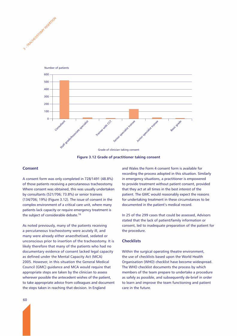
60
Consent
A consent form was only completed in 728/1491 (48.8%) of those patients receiving a percutaneous tracheostomy. Where consent was obtained, this was usually undertaken by consultants (521/706; 73.8%) or senior trainees (134/706; 19%) (Figure 3.12). The issue of consent in the complex environment of a critical care unit, where many patients lack capacity or require emergency treatment is the subject of considerable debate.16
As noted previously, many of the patients receiving a percutaneous tracheostomy were acutely ill, and many were already either anaesthetised, sedated or unconscious prior to insertion of the tracheostomy. It is likely therefore that many of the patients who had no documentary evidence of consent lacked legal capacity as defined under the Mental Capacity Act (MCA) 2005. However, in this situation the General Medical Council (GMC) guidance and MCA would require that appropriate steps are taken by the clinician to assess wherever possible the antecedent wishes of the patient, to take appropriate advice from colleagues and document the steps taken in reaching that decision. In England
and Wales the Form 4 consent form is available for recording the process adopted in this situation. Similarly in emergency situations, a practitioner is empowered to provide treatment without patient consent, provided that they act at all times in the best interest of the patient. The GMC would reasonably expect the reasons for undertaking treatment in these circumstances to be documented in the patient’s medical record.
In 25 of the 299 cases that could be assessed, Advisors stated that the lack of patient/family information or consent, led to inadequate preparation of the patient for the procedure.
Checklists
Within the surgical operating theatre environment, the use of checklists based upon the World Health Organisation (WHO) checklist have become widespread. The WHO checklist documents the process by which members of the team prepare to undertake a procedure as safely as possible, and subsequently de-brief in order to learn and improve the team functioning and patient care in the future.
3 - T
RACHEO
STOM
Y IN
SERT
ION
Number of patients
600
500
400
300
200
100
0
Figure 3.12 Grade of practitioner taking consent
Consu
ltant
Staf
f gra
de/A
ssocia
te sp
ecial
ist
Traine
e with
CCT
Senio
r spe
cialis
t tra
inee
Junio
r spe
cialis
t tra
inee
Basic
gra
de
Grade of clinician taking consent
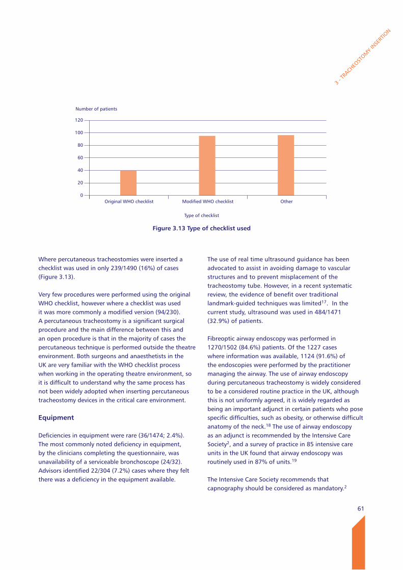
61
The use of real time ultrasound guidance has been advocated to assist in avoiding damage to vascular structures and to prevent misplacement of the tracheostomy tube. However, in a recent systematic review, the evidence of benefit over traditional landmark-guided techniques was limited17. In the current study, ultrasound was used in 484/1471 (32.9%) of patients.
Fibreoptic airway endoscopy was performed in 1270/1502 (84.6%) patients. Of the 1227 cases where information was available, 1124 (91.6%) of the endoscopies were performed by the practitioner managing the airway. The use of airway endoscopy during percutaneous tracheostomy is widely considered to be a considered routine practice in the UK, although this is not uniformly agreed, it is widely regarded as being an important adjunct in certain patients who pose specific difficulties, such as obesity, or otherwise difficult anatomy of the neck.18 The use of airway endoscopy as an adjunct is recommended by the Intensive Care Society2, and a survey of practice in 85 intensive care units in the UK found that airway endoscopy was routinely used in 87% of units.19
The Intensive Care Society recommends that capnography should be considered as mandatory.2
3 - T
RACHEO
STOM
Y IN
SERT
ION
Where percutaneous tracheostomies were inserted a checklist was used in only 239/1490 (16%) of cases (Figure 3.13).
Very few procedures were performed using the original WHO checklist, however where a checklist was used it was more commonly a modified version (94/230). A percutaneous tracheostomy is a significant surgical procedure and the main difference between this and an open procedure is that in the majority of cases the percutaneous technique is performed outside the theatre environment. Both surgeons and anaesthetists in the UK are very familiar with the WHO checklist process when working in the operating theatre environment, so it is difficult to understand why the same process has not been widely adopted when inserting percutaneous tracheostomy devices in the critical care environment.
Equipment
Deficiencies in equipment were rare (36/1474; 2.4%). The most commonly noted deficiency in equipment, by the clinicians completing the questionnaire, was unavailability of a serviceable bronchoscope (24/32). Advisors identified 22/304 (7.2%) cases where they felt there was a deficiency in the equipment available.
Number of patients
120
100
80
60
40
20
0
Figure 3.13 Type of checklist used
Original WHO checklist Modified WHO checklist Other
Type of checklist
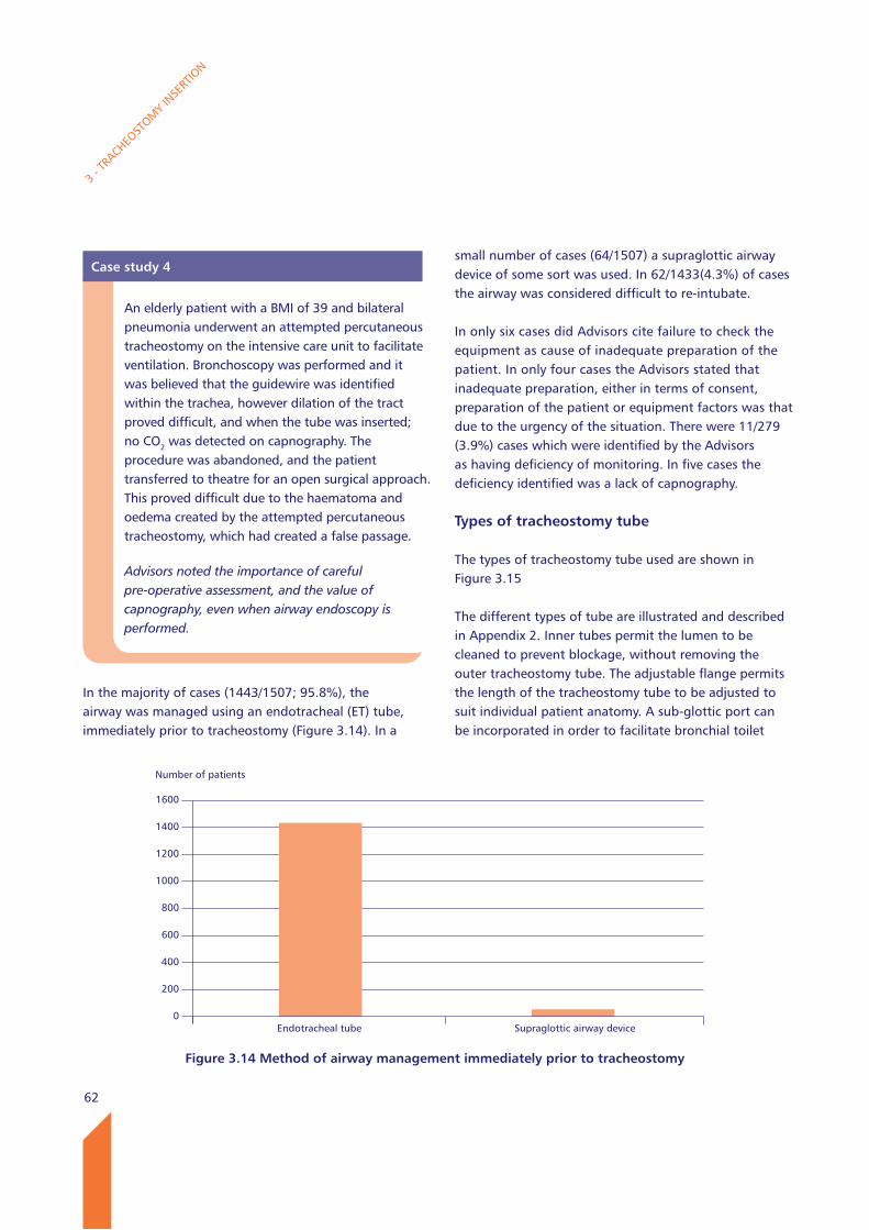
62
In the majority of cases (1443/1507; 95.8%), the airway was managed using an endotracheal (ET) tube, immediately prior to tracheostomy (Figure 3.14). In a
small number of cases (64/1507) a supraglottic airway device of some sort was used. In 62/1433(4.3%) of cases the airway was considered difficult to re-intubate.
In only six cases did Advisors cite failure to check the equipment as cause of inadequate preparation of the patient. In only four cases the Advisors stated that inadequate preparation, either in terms of consent, preparation of the patient or equipment factors was that due to the urgency of the situation. There were 11/279 (3.9%) cases which were identified by the Advisors as having deficiency of monitoring. In five cases the deficiency identified was a lack of capnography.
Types of tracheostomy tube
The types of tracheostomy tube used are shown in Figure 3.15
The different types of tube are illustrated and described in Appendix 2. Inner tubes permit the lumen to be cleaned to prevent blockage, without removing the outer tracheostomy tube. The adjustable flange permits the length of the tracheostomy tube to be adjusted to suit individual patient anatomy. A sub-glottic port can be incorporated in order to facilitate bronchial toilet
3 - T
RACHEO
STOM
Y IN
SERT
ION
Case study 4
An elderly patient with a BMI of 39 and bilateral pneumonia underwent an attempted percutaneous tracheostomy on the intensive care unit to facilitate ventilation. Bronchoscopy was performed and it was believed that the guidewire was identified within the trachea, however dilation of the tract proved difficult, and when the tube was inserted; no CO2 was detected on capnography. The procedure was abandoned, and the patient transferred to theatre for an open surgical approach. This proved difficult due to the haematoma and oedema created by the attempted percutaneous tracheostomy, which had created a false passage.
Advisors noted the importance of careful pre-operative assessment, and the value of capnography, even when airway endoscopy is performed.
Number of patients
1600
1400
1200
1000
800
600
400
200
0
Figure 3.14 Method of airway management immediately prior to tracheostomy
Endotracheal tube Supraglottic airway device
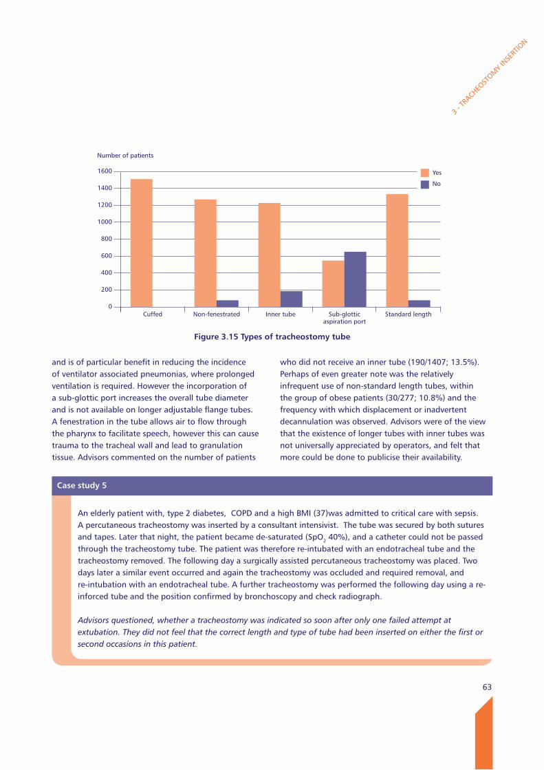
63
and is of particular benefit in reducing the incidence of ventilator associated pneumonias, where prolonged ventilation is required. However the incorporation of a sub-glottic port increases the overall tube diameter and is not available on longer adjustable flange tubes. A fenestration in the tube allows air to flow through the pharynx to facilitate speech, however this can cause trauma to the tracheal wall and lead to granulation tissue. Advisors commented on the number of patients
who did not receive an inner tube (190/1407; 13.5%). Perhaps of even greater note was the relatively infrequent use of non-standard length tubes, within the group of obese patients (30/277; 10.8%) and the frequency with which displacement or inadvertent decannulation was observed. Advisors were of the view that the existence of longer tubes with inner tubes was not universally appreciated by operators, and felt that more could be done to publicise their availability.
3 - T
RACHEO
STOM
Y IN
SERT
ION
Case study 5
An elderly patient with, type 2 diabetes, COPD and a high BMI (37)was admitted to critical care with sepsis. A percutaneous tracheostomy was inserted by a consultant intensivist. The tube was secured by both sutures and tapes. Later that night, the patient became de-saturated (SpO2 40%), and a catheter could not be passed through the tracheostomy tube. The patient was therefore re-intubated with an endotracheal tube and the tracheostomy removed. The following day a surgically assisted percutaneous tracheostomy was placed. Two days later a similar event occurred and again the tracheostomy was occluded and required removal, and re-intubation with an endotracheal tube. A further tracheostomy was performed the following day using a re-inforced tube and the position confirmed by bronchoscopy and check radiograph.
Advisors questioned, whether a tracheostomy was indicated so soon after only one failed attempt at extubation. They did not feel that the correct length and type of tube had been inserted on either the first or second occasions in this patient.
Number of patients
1600
1400
1200
1000
800
600
400
200
0
Figure 3.15 Types of tracheostomy tube
Cuffed Non-fenestrated Inner tube Sub-glottic aspiration port
Standard length
Yes
No
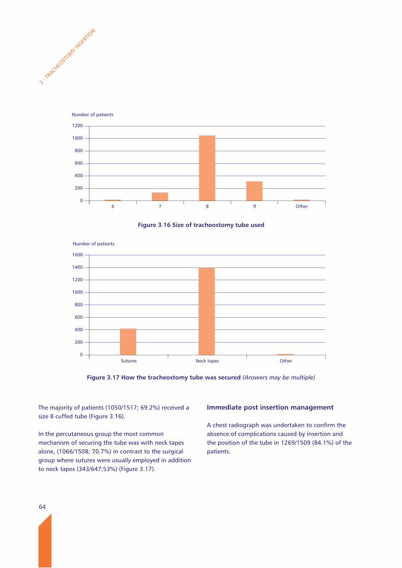
64
The majority of patients (1050/1517; 69.2%) received a size 8 cuffed tube (Figure 3.16).
In the percutaneous group the most common mechanism of securing the tube was with neck tapes alone, (1066/1508; 70.7%) in contrast to the surgical group where sutures were usually employed in addition to neck tapes (343/647;53%) (Figure 3.17).
3 - T
RACHEO
STOM
Y IN
SERT
ION
Immediate post insertion management
A chest radiograph was undertaken to confirm the absence of complications caused by insertion and the position of the tube in 1269/1509 (84.1%) of the patients.
Number of patients
1200
1000
800
600
400
200
0
Figure 3.16 Size of tracheostomy tube used
6 7 8 9 Other
Number of patients
1600
1400
1200
1000
800
600
400
200
0
Figure 3.17 How the tracheostomy tube was secured (Answers may be multiple)
Sutures Neck tapes Other
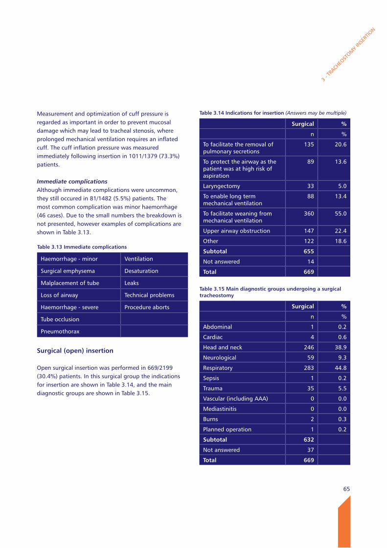
65
3 - T
RACHEO
STOM
Y IN
SERT
ION
Measurement and optimization of cuff pressure is regarded as important in order to prevent mucosal damage which may lead to tracheal stenosis, where prolonged mechanical ventilation requires an inflated cuff. The cuff inflation pressure was measured immediately following insertion in 1011/1379 (73.3%) patients.
Immediate complicationsAlthough immediate complications were uncommon, they still occured in 81/1482 (5.5%) patients. The most common complication was minor haemorrhage (46 cases). Due to the small numbers the breakdown is not presented, however examples of complications are shown in Table 3.13.
Surgical (open) insertion
Open surgical insertion was performed in 669/2199 (30.4%) patients. In this surgical group the indications for insertion are shown in Table 3.14, and the main diagnostic groups are shown in Table 3.15.
Table 3.13 Immediate complications
Haemorrhage - minor Ventilation
Surgical emphysema Desaturation
Malplacement of tube Leaks
Loss of airway Technical problems
Haemorrhage - severe Procedure aborts
Tube occlusion
Pneumothorax
Table 3.14 Indications for insertion (Answers may be multiple)
Surgical %
n %
To facilitate the removal of pulmonary secretions
135 20.6
To protect the airway as the patient was at high risk of aspiration
89 13.6
Laryngectomy 33 5.0
To enable long term mechanical ventilation
88 13.4
To facilitate weaning from mechanical ventilation
360 55.0
Upper airway obstruction 147 22.4
Other 122 18.6
Subtotal 655
Not answered 14
Total 669
Table 3.15 Main diagnostic groups undergoing a surgical tracheostomy
Surgical %
n %
Abdominal 1 0.2
Cardiac 4 0.6
Head and neck 246 38.9
Neurological 59 9.3
Respiratory 283 44.8
Sepsis 1 0.2
Trauma 35 5.5
Vascular (including AAA) 0 0.0
Mediastinitis 0 0.0
Burns 2 0.3
Planned operation 1 0.2
Subtotal 632
Not answered 37
Total 669
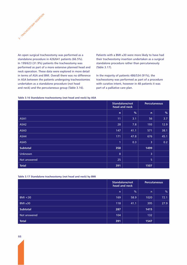
66
An open surgical tracheostomy was performed as a standalone procedure in 426/641 patients (66.5%). In 199/623 (31.9%) patients the tracheostomy was performed as part of a more extensive planned head and neck operation. These data were explored in more detail in terms of ASA and BMI. Overall there was no difference in ASA between the patients undergoing tracheostomies undertaken as a standalone procedure (not head and neck) and the percutaneous group (Table 3.16).
Patients with a BMI ≥30 were more likely to have had their tracheostomy insertion undertaken as a surgical standalone procedure rather than percutaneously (Table 3.17).
In the majority of patients 486/534 (91%), the tracheostomy was performed as part of a procedure with curative intent, however in 48 patients it was part of a palliative care plan.
3 - T
RACHEO
STOM
Y IN
SERT
ION
Table 3.16 Standalone tracheostomy (not head and neck) by ASA
Standalone/not head and neck
Percutaneous
n % n %
ASA1 11 3.1 56 3.7
ASA2 28 7.8 193 12.9
ASA3 147 41.1 571 38.1
ASA4 171 47.8 676 45.1
ASA5 1 0.3 3 0.2
Subtotal 358 1499
Unknown 8 3
Not answered 25 5
Total 391 1507
Table 3.17 Standalone tracheostomy (not head and neck) by BMI
Standalone/not head and neck
Percutaneous
n % n %
BMI <30 169 58.9 1020 72.1
BMI ≥30 118 41.1 395 27.9
Subtotal 287 1415
Not answered 104 132
Total 391 1547
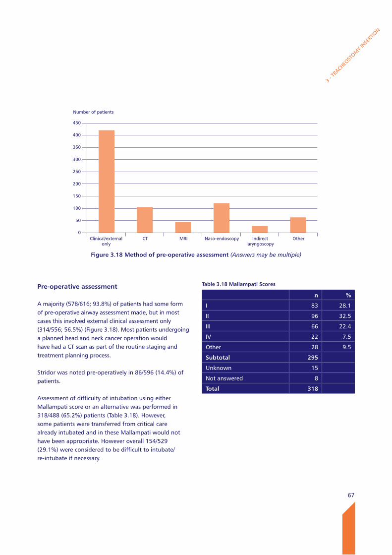
67
Pre-operative assessment
A majority (578/616; 93.8%) of patients had some form of pre-operative airway assessment made, but in most cases this involved external clinical assessment only (314/556; 56.5%) (Figure 3.18). Most patients undergoing a planned head and neck cancer operation would have had a CT scan as part of the routine staging and treatment planning process.
Stridor was noted pre-operatively in 86/596 (14.4%) of patients.
Assessment of difficulty of intubation using either Mallampati score or an alternative was performed in 318/488 (65.2%) patients (Table 3.18). However, some patients were transferred from critical care already intubated and in these Mallampati would not have been appropriate. However overall 154/529 (29.1%) were considered to be difficult to intubate/re-intubate if necessary.
3 - T
RACHEO
STOM
Y IN
SERT
ION
Number of patients
450
400
350
300
250
200
150
100
50
0
Figure 3.18 Method of pre-operative assessment (Answers may be multiple)
Clinical/external only
CT MRI Naso-endoscopy Indirect laryngoscopy
Other
Table 3.18 Mallampati Scores
n %
I 83 28.1
II 96 32.5
III 66 22.4
IV 22 7.5
Other 28 9.5
Subtotal 295
Unknown 15
Not answered 8
Total 318
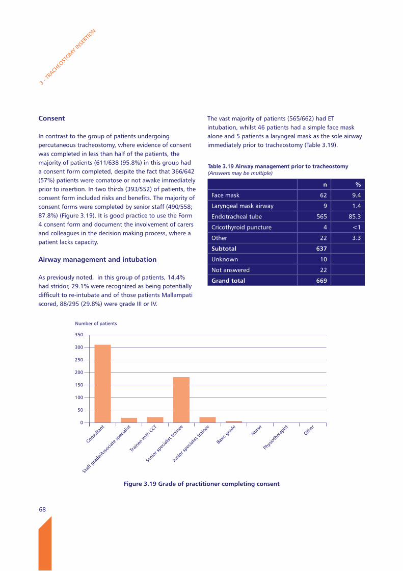
68
Consent
In contrast to the group of patients undergoing percutaneous tracheostomy, where evidence of consent was completed in less than half of the patients, the majority of patients (611/638 (95.8%) in this group had a consent form completed, despite the fact that 366/642 (57%) patients were comatose or not awake immediately prior to insertion. In two thirds (393/552) of patients, the consent form included risks and benefits. The majority of consent forms were completed by senior staff (490/558; 87.8%) (Figure 3.19). It is good practice to use the Form 4 consent form and document the involvement of carers and colleagues in the decision making process, where a patient lacks capacity.
Airway management and intubation
As previously noted, in this group of patients, 14.4% had stridor, 29.1% were recognized as being potentially difficult to re-intubate and of those patients Mallampati scored, 88/295 (29.8%) were grade III or IV.
3 - T
RACHEO
STOM
Y IN
SERT
ION
The vast majority of patients (565/662) had ET intubation, whilst 46 patients had a simple face mask alone and 5 patients a laryngeal mask as the sole airway immediately prior to tracheostomy (Table 3.19).
Table 3.19 Airway management prior to tracheostomy (Answers may be multiple)
n %
Face mask 62 9.4
Laryngeal mask airway 9 1.4
Endotracheal tube 565 85.3
Cricothyroid puncture 4 <1
Other 22 3.3
Subtotal 637
Unknown 10
Not answered 22
Grand total 669
Number of patients
350
300
250
200
150
100
50
0
Figure 3.19 Grade of practitioner completing consent
Consu
ltant
Staf
f gra
de/A
ssocia
te sp
ecial
ist
Traine
e with
CCT
Senio
r spe
cialis
t tra
inee
Junio
r spe
cialis
t tra
inee
Basic
gra
deNur
se
Phys
iothe
rapis
t
Other
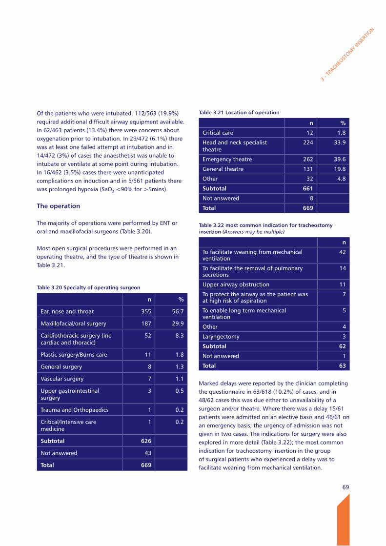
69
Of the patients who were intubated, 112/563 (19.9%) required additional difficult airway equipment available. In 62/463 patients (13.4%) there were concerns about oxygenation prior to intubation. In 29/472 (6.1%) there was at least one failed attempt at intubation and in 14/472 (3%) of cases the anaesthetist was unable to intubate or ventilate at some point during intubation. In 16/462 (3.5%) cases there were unanticipated complications on induction and in 5/561 patients there was prolonged hypoxia (SaO2 <90% for >5mins).
The operation
The majority of operations were performed by ENT or oral and maxillofacial surgeons (Table 3.20).
Most open surgical procedures were performed in an operating theatre, and the type of theatre is shown in Table 3.21.
3 - T
RACHEO
STOM
Y IN
SERT
ION
Marked delays were reported by the clinician completing the questionnaire in 63/618 (10.2%) of cases, and in 48/62 cases this was due either to unavailability of a surgeon and/or theatre. Where there was a delay 15/61 patients were admitted on an elective basis and 46/61 on an emergency basis; the urgency of admission was not given in two cases. The indications for surgery were also explored in more detail (Table 3.22); the most common indication for tracheostomy insertion in the group of surgical patients who experienced a delay was to facilitate weaning from mechanical ventilation.
Table 3.20 Specialty of operating surgeon
n %
Ear, nose and throat 355 56.7
Maxillofacial/oral surgery 187 29.9
Cardiothoracic surgery (inc cardiac and thoracic)
52 8.3
Plastic surgery/Burns care 11 1.8
General surgery 8 1.3
Vascular surgery 7 1.1
Upper gastrointestinal surgery
3 0.5
Trauma and Orthopaedics 1 0.2
Critical/Intensive care medicine
1 0.2
Subtotal 626
Not answered 43
Total 669
Table 3.21 location of operation
n %
Critical care 12 1.8
Head and neck specialist theatre
224 33.9
Emergency theatre 262 39.6
General theatre 131 19.8
Other 32 4.8
Subtotal 661
Not answered 8
Total 669
Table 3.22 most common indication for tracheostomy insertion (Answers may be multiple)
n
To facilitate weaning from mechanical ventilation
42
To facilitate the removal of pulmonary secretions
14
Upper airway obstruction 11
To protect the airway as the patient was at high risk of aspiration
7
To enable long term mechanical ventilation
5
Other 4
Laryngectomy 3
Subtotal 62
Not answered 1
Total 63
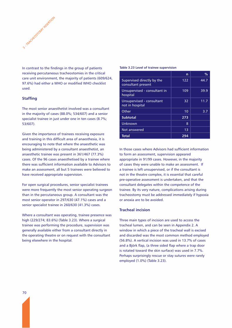
70
In contrast to the findings in the group of patients receiving percutaneous tracheostomies in the critical care unit environment, the majority of patients (609/624, 97.6%) had either a WHO or modified WHO checklist used.
Staffing
The most senior anaesthetist involved was a consultant in the majority of cases (88.0%; 534/607) and a senior specialist trainee in just under one in ten cases (8.7%; 53/607).
Given the importance of trainees receiving exposure and training in this difficult area of anaesthesia, it is encouraging to note that where the anaesthetic was being administered by a consultant anaesthetist, an anaesthetic trainee was present in 361/467 (77.3%) cases. Of the 96 cases anaesthetised by a trainee where there was sufficient information available to Advisors to make an assessment, all but 5 trainees were believed to have received appropriate supervision.
For open surgical procedures, senior specialist trainees were more frequently the most senior operating surgeon than in the percutaneous group. A consultant was the most senior operator in 297/630 (47.1%) cases and a senior specialist trainee in 260/630 (41.3%) cases.
Where a consultant was operating, trainee presence was high (229/274; 83.6%) (Table 3.23). Where a surgical trainee was performing the procedure, supervision was generally available either from a consultant directly in the operating theatre or on request with the consultant being elsewhere in the hospital.
3 - T
RACHEO
STOM
Y IN
SERT
ION
In those cases where Advisors had sufficient information to form an assessment, supervision appeared appropriate in 91/99 cases. However, in the majority of cases they were unable to make an assessment. If a trainee is left unsupervised, or if the consultant is not in the theatre complex, it is essential that careful pre-operative assessment is undertaken, and that the consultant delegates within the competence of the trainee. By its very nature, complications arising during tracheostomy must be addressed immediately if hypoxia or anoxia are to be avoided.
Tracheal incision
Three main types of incision are used to access the tracheal lumen, and can be seen in Appendix 2. A window in which a piece of the tracheal wall is excised and discarded was the most common method employed (56.8%). A vertical incision was used in 13.7% of cases and a Björk flap, (a three sided flap where a trap door is rotated toward the skin surface) was used in 7.7%. Perhaps surprisingly rescue or stay sutures were rarely employed (1.0%) (Table 3.23).
Table 3.23 level of trainee supervision
n %
Supervised directly by the consultant present
122 44.7
Unsupervised - consultant in hospital
109 39.9
Unsupervised - consultant not in hospital
32 11.7
Other 10 3.7
Subtotal 273
Unknown 8
Not answered 13
Total 294
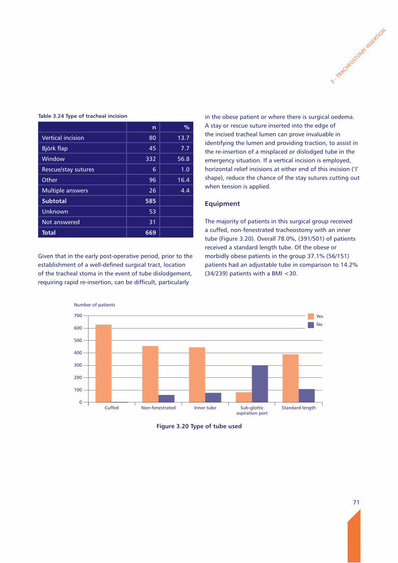
71
Given that in the early post-operative period, prior to the establishment of a well-defined surgical tract, location of the tracheal stoma in the event of tube dislodgement, requiring rapid re-insertion, can be difficult, particularly
in the obese patient or where there is surgical oedema. A stay or rescue suture inserted into the edge of the incised tracheal lumen can prove invaluable in identifying the lumen and providing traction, to assist in the re-insertion of a misplaced or dislodged tube in the emergency situation. If a vertical incision is employed, horizontal relief incisions at either end of this incision (‘I’ shape), reduce the chance of the stay sutures cutting out when tension is applied.
Equipment
The majority of patients in this surgical group received a cuffed, non-fenestrated tracheostomy with an inner tube (Figure 3.20). Overall 78.0%, (391/501) of patients received a standard length tube. Of the obese or morbidly obese patients in the group 37.1% (56/151) patients had an adjustable tube in comparison to 14.2% (34/239) patients with a BMI <30.
3 - T
RACHEO
STOM
Y IN
SERT
ION
Table 3.24 Type of tracheal incision
n %
Vertical incision 80 13.7
Björk flap 45 7.7
Window 332 56.8
Rescue/stay sutures 6 1.0
Other 96 16.4
Multiple answers 26 4.4
Subtotal 585
Unknown 53
Not answered 31
Total 669
Number of patients
700
600
500
400
300
200
100
0
Figure 3.20 Type of tube used
Cuffed Non-fenestrated Inner tube Sub-glottic aspiration port
Standard length
Yes
No
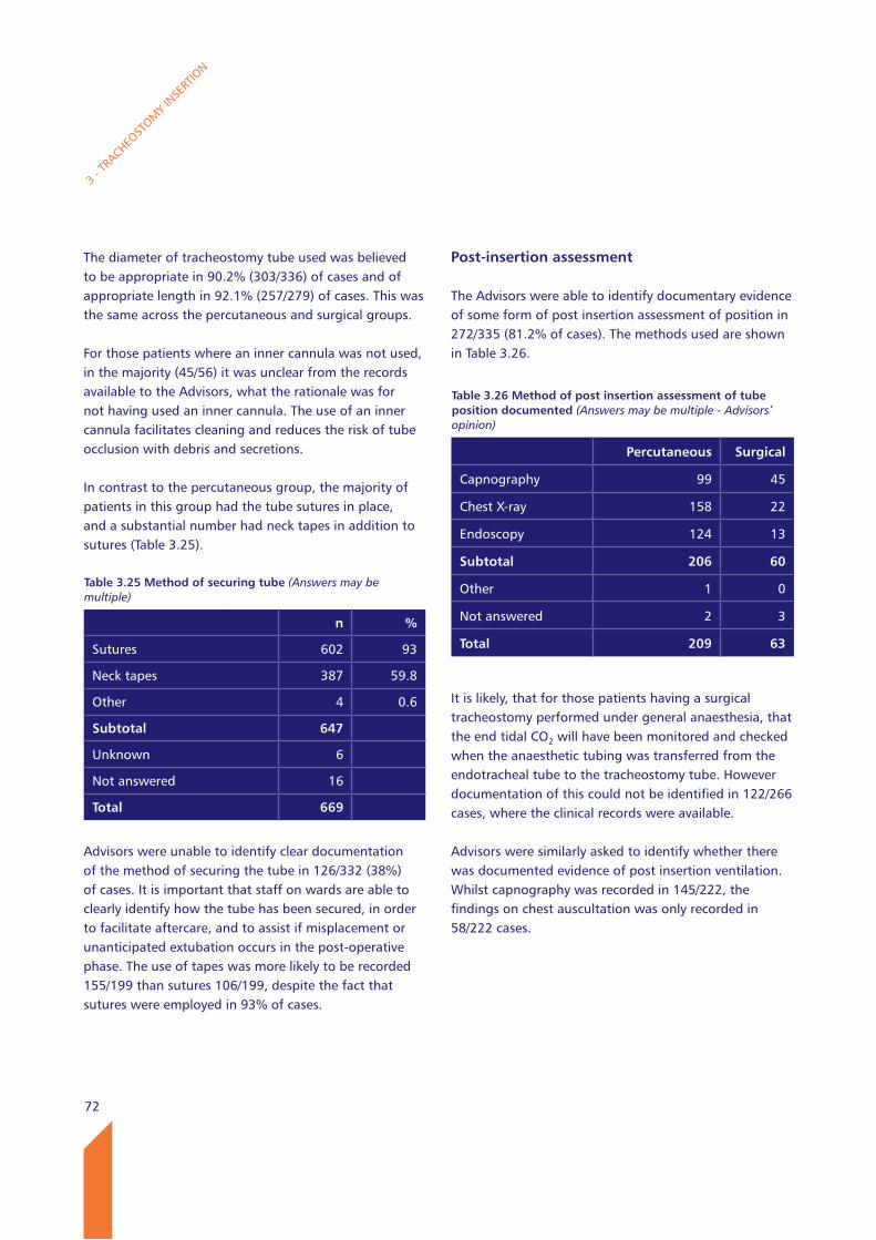
72
3 - T
RACHEO
STOM
Y IN
SERT
ION
The diameter of tracheostomy tube used was believed to be appropriate in 90.2% (303/336) of cases and of appropriate length in 92.1% (257/279) of cases. This was the same across the percutaneous and surgical groups.
For those patients where an inner cannula was not used, in the majority (45/56) it was unclear from the records available to the Advisors, what the rationale was for not having used an inner cannula. The use of an inner cannula facilitates cleaning and reduces the risk of tube occlusion with debris and secretions.
In contrast to the percutaneous group, the majority of patients in this group had the tube sutures in place, and a substantial number had neck tapes in addition to sutures (Table 3.25).
Advisors were unable to identify clear documentation of the method of securing the tube in 126/332 (38%) of cases. It is important that staff on wards are able to clearly identify how the tube has been secured, in order to facilitate aftercare, and to assist if misplacement or unanticipated extubation occurs in the post-operative phase. The use of tapes was more likely to be recorded 155/199 than sutures 106/199, despite the fact that sutures were employed in 93% of cases.
Post-insertion assessment
The Advisors were able to identify documentary evidence of some form of post insertion assessment of position in 272/335 (81.2% of cases). The methods used are shown in Table 3.26.
It is likely, that for those patients having a surgical tracheostomy performed under general anaesthesia, that the end tidal CO2 will have been monitored and checked when the anaesthetic tubing was transferred from the endotracheal tube to the tracheostomy tube. However documentation of this could not be identified in 122/266 cases, where the clinical records were available.
Advisors were similarly asked to identify whether there was documented evidence of post insertion ventilation. Whilst capnography was recorded in 145/222, the findings on chest auscultation was only recorded in 58/222 cases.
Table 3.25 Method of securing tube (Answers may be multiple)
n %
Sutures 602 93
Neck tapes 387 59.8
Other 4 0.6
Subtotal 647
Unknown 6
Not answered 16
Total 669
Table 3.26 Method of post insertion assessment of tube position documented (Answers may be multiple - Advisors’ opinion)
Percutaneous Surgical
Capnography 99 45
Chest X-ray 158 22
Endoscopy 124 13
Subtotal 206 60
Other 1 0
Not answered 2 3
Total 209 63
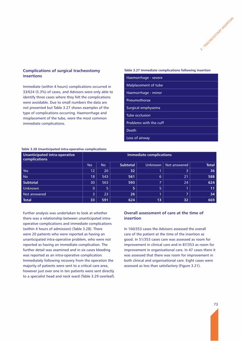
73
3 - T
RACHEO
STOM
Y IN
SERT
ION
Complications of surgical tracheostomy insertions
Immediate (within 4 hours) complications occurred in 33/624 (5.3%) of cases, and Advisors were only able to identify three cases where they felt the complications were avoidable. Due to small numbers the data are not presented but Table 3.27 shows examples of the type of complications occurring. Haemorrhage and misplacement of the tube, were the most common immediate complications.
Further analysis was undertaken to look at whether there was a relationship between unanticipated intra-operative complications and immediate complications (within 4 hours of admission) (Table 3.28). There were 20 patients who were reported as having an unanticipated intra-operative problem, who were not reported as having an immediate complication. The further detail was examined and in six cases bleeding was reported as an intra-operative complication. Immediately following recovery from the operation the majority of patients were sent to a critical care area, however just over one in ten patients were sent directly to a specialist head and neck ward (Table 3.29 overleaf).
Overall assessment of care at the time of insertion
In 160/353 cases the Advisors assessed the overall care of the patient at the time of the insertion as good. In 51/353 cases care was assessed as room for improvement in clinical care and in 87/353 as room for improvement in organisational care. In 47 cases there it was assessed that there was room for improvement in both clinical and organisational care. Eight cases were assessed as less than satisfactory (Figure 3.21).
Table 3.27 Immediate complications following insertion
Haemorrhage - severe
Malplacement of tube
Haemorrhage - minor
Pneumothorax
Surgical emphysema
Tube occlusion
Problems with the cuff
Death
Loss of airway
Table 3.28 Unanticipated intra-operative complications
Unanticipated intra-operative complications
Immediate complications
Yes No Subtotal Unknown Not answered Total
Yes 12 20 32 1 3 36
No 18 543 561 6 21 588
Subtotal 30 563 593 7 24 624
Unknown 0 5 5 5 1 11
Not answered 3 23 26 1 7 34
Total 33 591 624 13 32 669
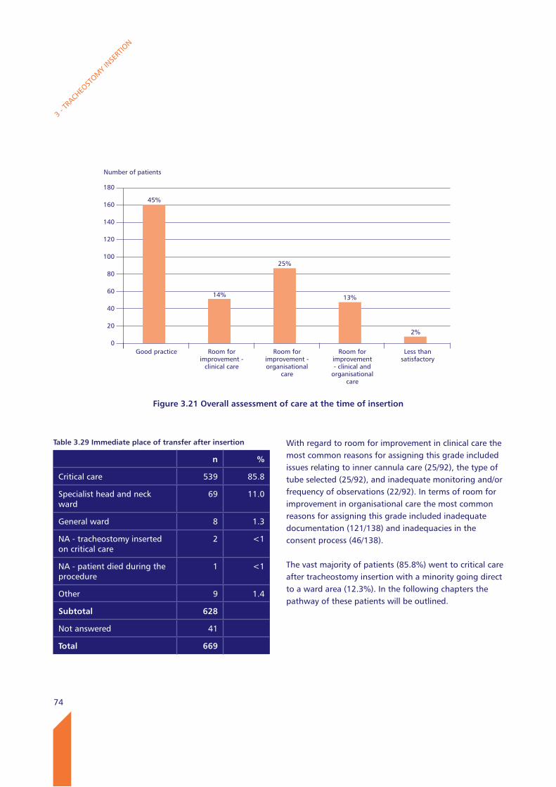
74
With regard to room for improvement in clinical care the most common reasons for assigning this grade included issues relating to inner cannula care (25/92), the type of tube selected (25/92), and inadequate monitoring and/or frequency of observations (22/92). In terms of room for improvement in organisational care the most common reasons for assigning this grade included inadequate documentation (121/138) and inadequacies in the consent process (46/138).
The vast majority of patients (85.8%) went to critical care after tracheostomy insertion with a minority going direct to a ward area (12.3%). In the following chapters the pathway of these patients will be outlined.
3 - T
RACHEO
STOM
Y IN
SERT
ION
Table 3.29 Immediate place of transfer after insertion
n %
Critical care 539 85.8
Specialist head and neck ward
69 11.0
General ward 8 1.3
NA - tracheostomy inserted on critical care
2 <1
NA - patient died during the procedure
1 <1
Other 9 1.4
Subtotal 628
Not answered 41
Total 669
Number of patients
180
160
140
120
100
80
60
40
20
0
Figure 3.21 Overall assessment of care at the time of insertion
Good practice Room for improvement -
clinical care
Room for improvement - organisational
care
Room for improvement - clinical and
organisationalcare
Less than satisfactory
45%
14%
25%
13%
2%
75
key findings
728/1491 (48.8%) patients had consent taken for a percutaneous tracheostomy, compared with 611/638 (95.8%) undergoing a surgical insertion.
239/1490 (16%) patients undergoing a percutaneous tracheostomy had a WHO type (surgical) checklist used.
Adjustable length tracheostomy tubes were used in only 185/1825 (10.1%) of patients. Inner tubes were used in 1661/1931 (86%) of patients.
566/1910 (29.6%) patients included in the study were obese or morbidly obese, but adjustable flanged tubes were only used in 96/510 (18.8%) of patients.
Capnography to assess tube placement was documented in 144/266 (54.1%) of patients.
Post-insertion endoscopy was used in 137/266 (51.5%) of patients.
Recommendations
6. Consent and WHO type (surgical) checklists should be adopted and used prior to tracheostomy insertion, wherever it is performed. (Medical Directors and Clinical Directors)
7. The diameter and length of the tube used should be appropriate for the size and anatomy of the individual patient, therefore an adequate range of tracheostomy tubes needs to be stocked by units. Operators should be aware of the types of tube available and in particular recognize that adjustable flanged tubes are available with inner tubes. Professionals need to continue to work closely with manufacturers to optimise design and tube options for a non standard population. (Consultant Operators, Theatre and Critical Care Managers and Professional Health Care Bodies)
8. Confirmation of tube placement must be obtained using capnography. This should be readily available and the events documented. (All Health Care Professionals)
9. Appropriate positioning of the tube should be made using airway endoscopy. This should be
readily available and the events documented. (All Consultants)
3 - T
RACHEO
STOM
Y IN
SERT
ION

76
4 - T
ERTIA
RY C
ARE
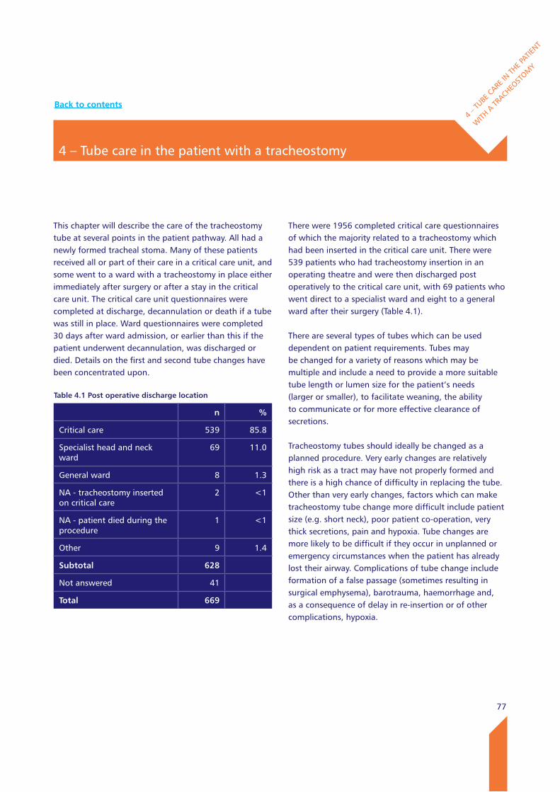
77
This chapter will describe the care of the tracheostomy tube at several points in the patient pathway. All had a newly formed tracheal stoma. Many of these patients received all or part of their care in a critical care unit, and some went to a ward with a tracheostomy in place either immediately after surgery or after a stay in the critical care unit. The critical care unit questionnaires were completed at discharge, decannulation or death if a tube was still in place. Ward questionnaires were completed 30 days after ward admission, or earlier than this if the patient underwent decannulation, was discharged or died. Details on the first and second tube changes have been concentrated upon.
There were 1956 completed critical care questionnaires of which the majority related to a tracheostomy which had been inserted in the critical care unit. There were 539 patients who had tracheostomy insertion in an operating theatre and were then discharged post operatively to the critical care unit, with 69 patients who went direct to a specialist ward and eight to a general ward after their surgery (Table 4.1).
There are several types of tubes which can be used dependent on patient requirements. Tubes may be changed for a variety of reasons which may be multiple and include a need to provide a more suitable tube length or lumen size for the patient’s needs (larger or smaller), to facilitate weaning, the ability to communicate or for more effective clearance of secretions.
Tracheostomy tubes should ideally be changed as a planned procedure. Very early changes are relatively high risk as a tract may have not properly formed and there is a high chance of difficulty in replacing the tube. Other than very early changes, factors which can make tracheostomy tube change more difficult include patient size (e.g. short neck), poor patient co-operation, very thick secretions, pain and hypoxia. Tube changes are more likely to be difficult if they occur in unplanned or emergency circumstances when the patient has already lost their airway. Complications of tube change include formation of a false passage (sometimes resulting in surgical emphysema), barotrauma, haemorrhage and, as a consequence of delay in re-insertion or of other complications, hypoxia.
Table 4.1 Post operative discharge location
n %
Critical care 539 85.8
Specialist head and neck ward
69 11.0
General ward 8 1.3
NA - tracheostomy inserted on critical care
2 <1
NA - patient died during the procedure
1 <1
Other 9 1.4
Subtotal 628
Not answered 41
Total 669
4 – Tube care in the patient with a tracheostomy
4 – T
UBE C
ARE IN
THE P
ATIENT
WITH
A TR
ACHEOST
OMY
Back to contents
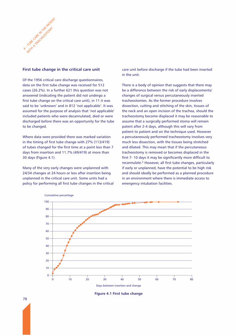
78
First tube change in the critical care unit
Of the 1956 critical care discharge questionnaires, data on the first tube change was received for 512 cases (26.2%). In a further 621 this question was not answered (indicating the patient did not undergo a first tube change on the critical care unit), in 11 it was said to be ‘unknown’ and in 812 ‘not applicable’. It was assumed for the purpose of analysis that ‘not applicable’ included patients who were decannulated, died or were discharged before there was an opportunity for the tube to be changed.
Where data were provided there was marked variation in the timing of first tube change with 27% (113/419) of tubes changed for the first time at a point less than 7 days from insertion and 11.7% (49/419) at more than 30 days (Figure 4.1).
Many of the very early changes were unplanned with 24/34 changes at 24 hours or less after insertion being unplanned in the critical care unit. Some units had a policy for performing all first tube changes in the critical
care unit before discharge if the tube had been inserted in the unit.
There is a body of opinion that suggests that there may be a difference between the risk of early displacements/changes of surgical versus percutaneously inserted tracheostomies. As the former procedure involves dissection, cutting and stitching of the skin, tissues of the neck and an open incision of the trachea, should the tracheostomy become displaced it may be reasonable to assume that a surgically performed stoma will remain patent after 2-4 days, although this will vary from patient to patient and on the technique used. However a percutaneously performed tracheostomy involves very much less dissection, with the tissues being stretched and dilated. This may mean that if the percutaneous tracheostomy is removed or becomes displaced in the first 7- 10 days it may be significantly more difficult to recannulate.2 However, all first tube changes, particularly if early or unplanned, have the potential to be high risk and should ideally be performed as a planned procedure in an environment where there is immediate access to emergency intubation facilities.
4 – T
UBE C
ARE IN
THE P
ATIENT
WITH
A TR
ACHEOST
OMY
Cumulative percentage
100
90
80
70
60
50
40
30
20
10
0
Figure 4.1 First tube change
Days between insertion and change
0 10 20 30 40 50 60 70 80
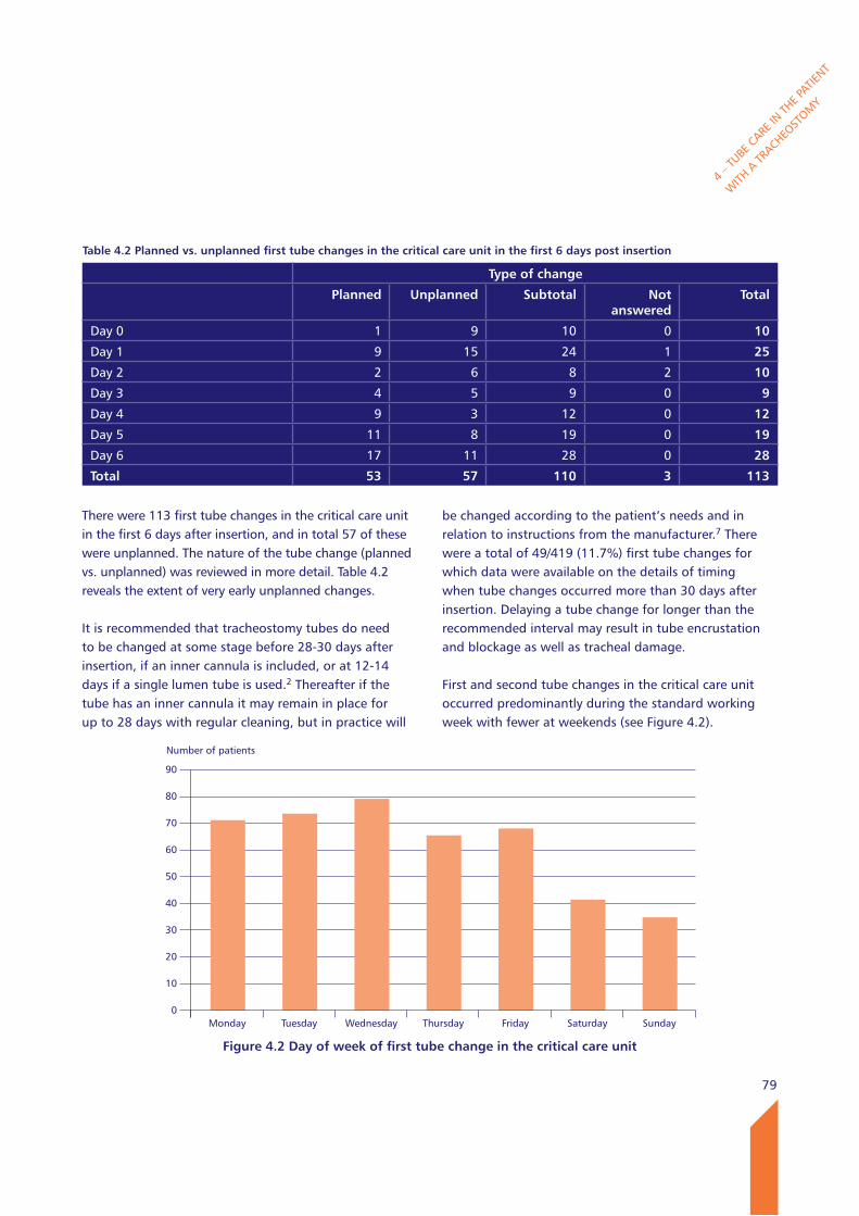
79
be changed according to the patient’s needs and in relation to instructions from the manufacturer.7 There were a total of 49/419 (11.7%) first tube changes for which data were available on the details of timing when tube changes occurred more than 30 days after insertion. Delaying a tube change for longer than the recommended interval may result in tube encrustation and blockage as well as tracheal damage.
First and second tube changes in the critical care unit occurred predominantly during the standard working week with fewer at weekends (see Figure 4.2).
There were 113 first tube changes in the critical care unit in the first 6 days after insertion, and in total 57 of these were unplanned. The nature of the tube change (planned vs. unplanned) was reviewed in more detail. Table 4.2 reveals the extent of very early unplanned changes.
It is recommended that tracheostomy tubes do need to be changed at some stage before 28-30 days after insertion, if an inner cannula is included, or at 12-14 days if a single lumen tube is used.2 Thereafter if the tube has an inner cannula it may remain in place for up to 28 days with regular cleaning, but in practice will
4 – T
UBE C
ARE IN
THE P
ATIENT
WITH
A TR
ACHEOST
OMY
Table 4.2 Planned vs. unplanned first tube changes in the critical care unit in the first 6 days post insertion
Type of change
Planned Unplanned Subtotal Not answered
Total
Day 0 1 9 10 0 10
Day 1 9 15 24 1 25
Day 2 2 6 8 2 10
Day 3 4 5 9 0 9
Day 4 9 3 12 0 12
Day 5 11 8 19 0 19
Day 6 17 11 28 0 28
Total 53 57 110 3 113
Number of patients
90
80
70
60
50
40
30
20
10
0
Figure 4.2 Day of week of first tube change in the critical care unit
Monday Tuesday Wednesday Thursday Friday Saturday Sunday
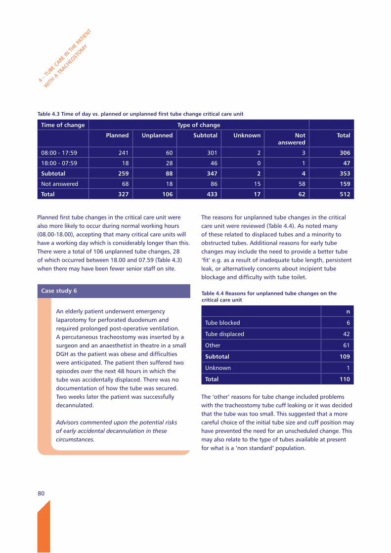
80
The reasons for unplanned tube changes in the critical care unit were reviewed (Table 4.4). As noted many of these related to displaced tubes and a minority to obstructed tubes. Additional reasons for early tube changes may include the need to provide a better tube ‘fit’ e.g. as a result of inadequate tube length, persistent leak, or alternatively concerns about incipient tube blockage and difficulty with tube toilet.
The ‘other’ reasons for tube change included problems with the tracheostomy tube cuff leaking or it was decided that the tube was too small. This suggested that a more careful choice of the initial tube size and cuff position may have prevented the need for an unscheduled change. This may also relate to the type of tubes available at present for what is a ‘non standard’ population.
Planned first tube changes in the critical care unit were also more likely to occur during normal working hours (08.00-18.00), accepting that many critical care units will have a working day which is considerably longer than this. There were a total of 106 unplanned tube changes, 28 of which occurred between 18.00 and 07.59 (Table 4.3) when there may have been fewer senior staff on site.
4 – T
UBE C
ARE IN
THE P
ATIENT
WITH
A TR
ACHEOST
OMY
Table 4.4 Reasons for unplanned tube changes on the critical care unit
n
Tube blocked 6
Tube displaced 42
Other 61
Subtotal 109
Unknown 1
Total 110
Case study 6
An elderly patient underwent emergency laparotomy for perforated duodenum and required prolonged post-operative ventilation. A percutaneous tracheostomy was inserted by a surgeon and an anaesthetist in theatre in a small DGH as the patient was obese and difficulties were anticipated. The patient then suffered two episodes over the next 48 hours in which the tube was accidentally displaced. There was no documentation of how the tube was secured. Two weeks later the patient was successfully decannulated.
Advisors commented upon the potential risks of early accidental decannulation in these circumstances.
Table 4.3 Time of day vs. planned or unplanned first tube change critical care unit
Time of change Type of change
Planned Unplanned Subtotal Unknown Not answered
Total
08:00 - 17:59 241 60 301 2 3 306
18:00 - 07:59 18 28 46 0 1 47
Subtotal 259 88 347 2 4 353
Not answered 68 18 86 15 58 159
Total 327 106 433 17 62 512
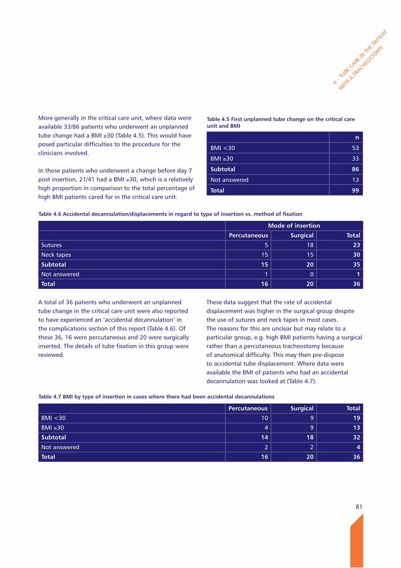
81
More generally in the critical care unit, where data were available 33/86 patients who underwent an unplanned tube change had a BMI ≥30 (Table 4.5). This would have posed particular difficulties to the procedure for the clinicians involved.
In those patients who underwent a change before day 7 post insertion, 21/41 had a BMI ≥30, which is a relatively high proportion in comparison to the total percentage of high BMI patients cared for in the critical care unit.
A total of 36 patients who underwent an unplanned tube change in the critical care unit were also reported to have experienced an ‘accidental decannulation’ in the complications section of this report (Table 4.6). Of these 36, 16 were percutaneous and 20 were surgically inserted. The details of tube fixation in this group were reviewed.
These data suggest that the rate of accidental displacement was higher in the surgical group despite the use of sutures and neck tapes in most cases. The reasons for this are unclear but may relate to a particular group, e.g. high BMI patients having a surgical rather than a percutaneous tracheostomy because of anatomical difficulty. This may then pre-dispose to accidental tube displacement. Where data were available the BMI of patients who had an accidental decannulation was looked at (Table 4.7).
4 – T
UBE C
ARE IN
THE P
ATIENT
WITH
A TR
ACHEOST
OMY
Table 4.6 Accidental decannulation/displacements in regard to type of insertion vs. method of fixation
Mode of insertion
Percutaneous Surgical Total
Sutures 5 18 23
Neck tapes 15 15 30
Subtotal 15 20 35
Not answered 1 0 1
Total 16 20 36
Table 4.7 BMI by type of insertion in cases where there had been accidental decannulations
Percutaneous Surgical Total
BMI <30 10 9 19
BMI ≥30 4 9 13
Subtotal 14 18 32
Not answered 2 2 4
Total 16 20 36
Table 4.5 First unplanned tube change on the critical care unit and BMI
n
BMI <30 53
BMI ≥30 33
Subtotal 86
Not answered 13
Total 99
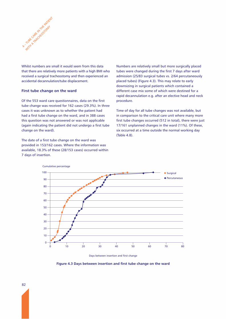
82
Whilst numbers are small it would seem from this data that there are relatively more patients with a high BMI who received a surgical tracheostomy and then experienced an accidental decannulation/tube displacement. First tube change on the ward
Of the 553 ward care questionnaires, data on the first tube change was received for 162 cases (29.3%). In three cases it was unknown as to whether the patient had had a first tube change on the ward, and in 388 cases this question was not answered or was not applicable (again indicating the patient did not undergo a first tube change on the ward).
The date of a first tube change on the ward was provided in 153/162 cases. Where the information was available, 18.3% of these (28/153 cases) occurred within 7 days of insertion.
Numbers are relatively small but more surgically placed tubes were changed during the first 7 days after ward admission (25/83 surgical tubes vs. 2/64 percutaneously placed tubes) (Figure 4.3). This may relate to early downsizing in surgical patients which contained a different case mix some of which were destined for a rapid decannulation e.g. after an elective head and neck procedure.
Time of day for all tube changes was not available, but in comparison to the critical care unit where many more first tube changes occurred (512 in total), there were just 17/161 unplanned changes in the ward (11%). Of these, six occurred at a time outside the normal working day (Table 4.8).
4 – T
UBE C
ARE IN
THE P
ATIENT
WITH
A TR
ACHEOST
OMY
Cumulative percentage
100
90
80
70
60
50
40
30
20
10
0
Figure 4.3 Days between insertion and first tube change on the ward
Days between insertion and first change
0 10 20 30 40 50 60 70 80
Surgical
Percutaneous
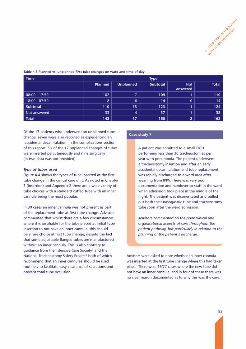
83
Of the 17 patients who underwent an unplanned tube change, seven were also reported as experiencing an ‘accidental decannulation’ in the complications section of this report. Six of the 17 unplanned changes of tubes were inserted percutaneously and nine surgically (in two data was not provided).
Type of tubes usedFigure 4.4 shows the types of tube inserted at the first tube change in the critical care unit. As noted in Chapter 3 (Insertion) and Appendix 2 there are a wide variety of tube choices with a standard cuffed tube with an inner cannula being the most popular.
In 30 cases an inner cannula was not present as part of the replacement tube at first tube change. Advisors commented that whilst there are a few circumstances where it is justifiable for the tube placed at initial tube insertion to not have an inner cannula, this should be a rare choice at first tube change, despite the fact that some adjustable flanged tubes are manufactured without an inner cannula. This is also contrary to guidance from the Intensive Care Society2 and the National Tracheostomy Safety Project1 both of which recommend that an inner cannulae should be used routinely to facilitate easy clearance of secretions and prevent total tube occlusion.
Advisors were asked to note whether an inner cannula was inserted at the first tube change where this had taken place. There were 16/77 cases where the new tube did not have an inner cannula, and in four of these there was no clear reason documented as to why this was the case.
4 – T
UBE C
ARE IN
THE P
ATIENT
WITH
A TR
ACHEOST
OMY
Table 4.8 Planned vs. unplanned first tube changes on ward and time of day
Time Type
Planned Unplanned Subtotal Not answered
Total
08:00 - 17:59 102 7 109 1 110
18:00 - 07:59 8 6 14 0 14
Subtotal 110 13 123 1 124
Not answered 33 4 37 1 38
Total 143 17 160 2 162
Case study 7
A patient was admitted to a small DGH performing less than 30 tracheostomies per year with pneumonia. The patient underwent a tracheostomy insertion and after an early accidental decannulation and tube replacement was rapidly discharged to a ward area after weaning from IPPV. There was very poor documentation and handover to staff in the ward when admission took place in the middle of the night. The patient was disorientated and pulled out both their nasogastric tube and tracheostomy tube soon after the ward admission.
Advisors commented on the poor clinical and organisational aspects of care throughout the patient pathway, but particularly in relation to the planning of the patient’s discharge.
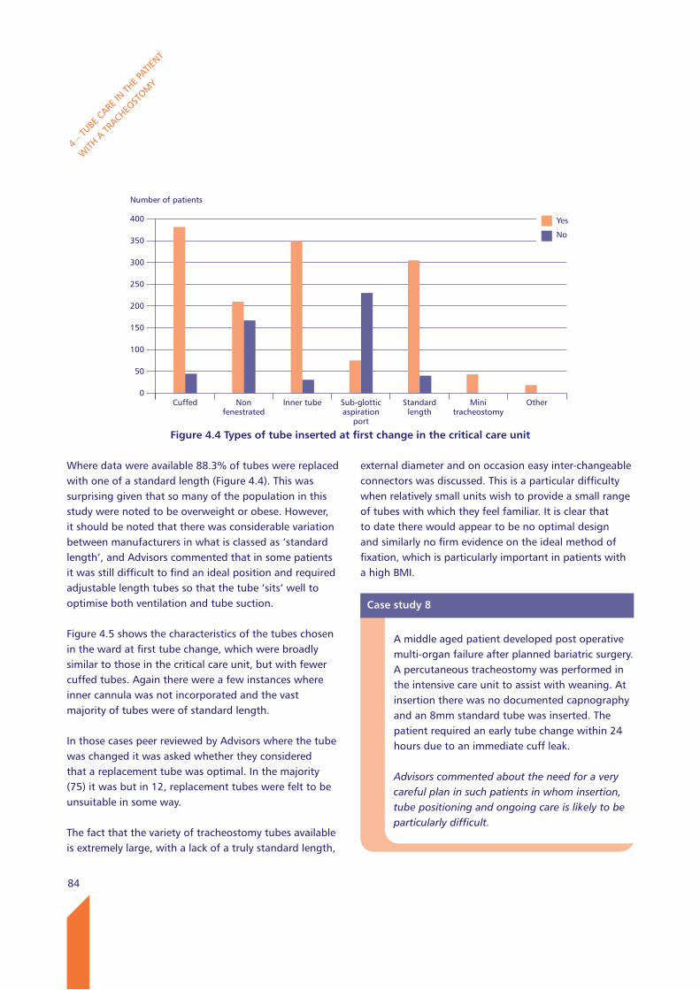
84
Where data were available 88.3% of tubes were replaced with one of a standard length (Figure 4.4). This was surprising given that so many of the population in this study were noted to be overweight or obese. However, it should be noted that there was considerable variation between manufacturers in what is classed as ‘standard length’, and Advisors commented that in some patients it was still difficult to find an ideal position and required adjustable length tubes so that the tube ‘sits’ well to optimise both ventilation and tube suction.
Figure 4.5 shows the characteristics of the tubes chosen in the ward at first tube change, which were broadly similar to those in the critical care unit, but with fewer cuffed tubes. Again there were a few instances where inner cannula was not incorporated and the vast majority of tubes were of standard length.
In those cases peer reviewed by Advisors where the tube was changed it was asked whether they considered that a replacement tube was optimal. In the majority (75) it was but in 12, replacement tubes were felt to be unsuitable in some way.
The fact that the variety of tracheostomy tubes available is extremely large, with a lack of a truly standard length,
external diameter and on occasion easy inter-changeable connectors was discussed. This is a particular difficulty when relatively small units wish to provide a small range of tubes with which they feel familiar. It is clear that to date there would appear to be no optimal design and similarly no firm evidence on the ideal method of fixation, which is particularly important in patients with a high BMI.
4 – T
UBE C
ARE IN
THE P
ATIENT
WITH
A TR
ACHEOST
OMY
Number of patients
400
350
300
250
200
150
100
50
0
Figure 4.4 Types of tube inserted at first change in the critical care unit
Cuffed Non fenestrated
Inner tube Sub-glottic aspiration
port
Standard length
Mini tracheostomy
Other
Yes
No
Case study 8
A middle aged patient developed post operative multi-organ failure after planned bariatric surgery. A percutaneous tracheostomy was performed in the intensive care unit to assist with weaning. At insertion there was no documented capnography and an 8mm standard tube was inserted. The patient required an early tube change within 24 hours due to an immediate cuff leak.
Advisors commented about the need for a very careful plan in such patients in whom insertion, tube positioning and ongoing care is likely to be particularly difficult.
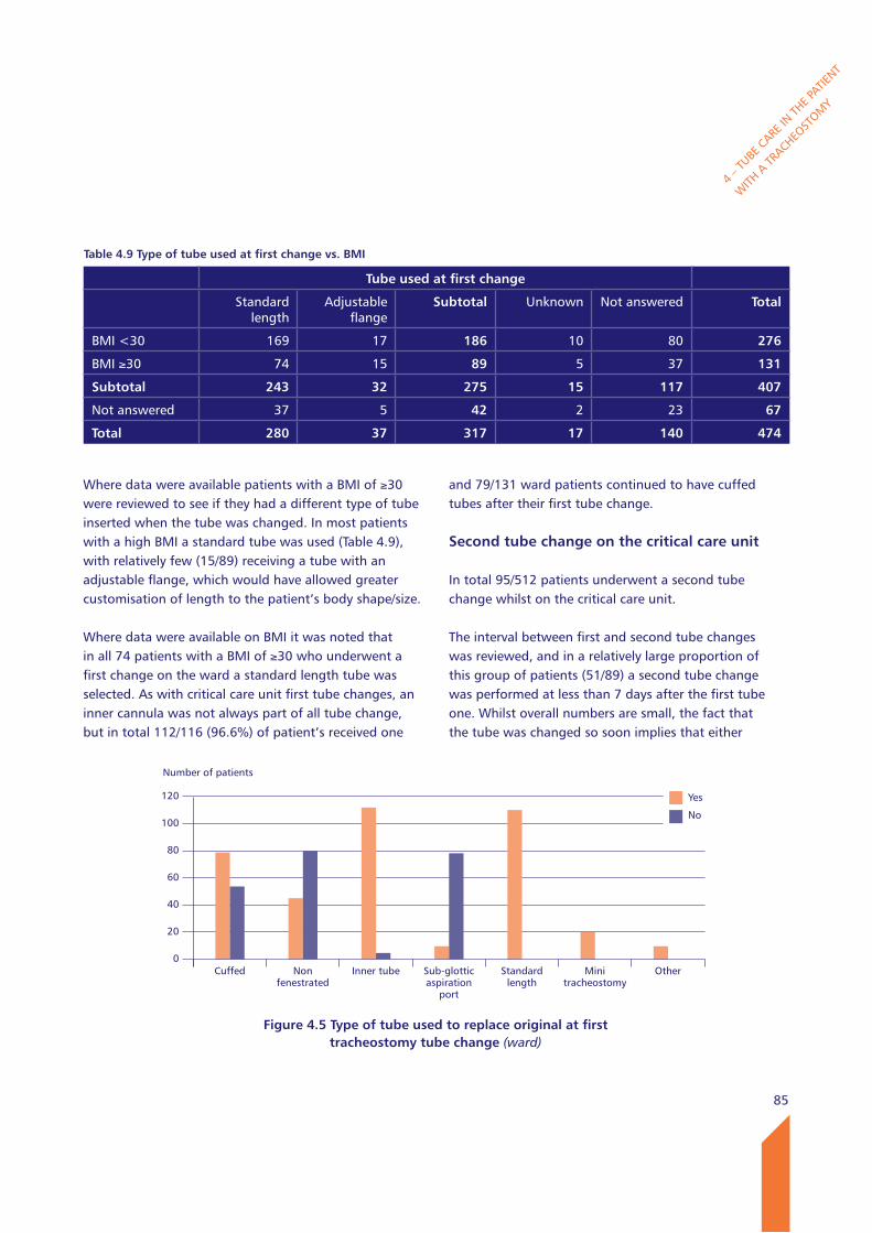
85
Where data were available patients with a BMI of ≥30 were reviewed to see if they had a different type of tube inserted when the tube was changed. In most patients with a high BMI a standard tube was used (Table 4.9), with relatively few (15/89) receiving a tube with an adjustable flange, which would have allowed greater customisation of length to the patient’s body shape/size.
Where data were available on BMI it was noted that in all 74 patients with a BMI of ≥30 who underwent a first change on the ward a standard length tube was selected. As with critical care unit first tube changes, an inner cannula was not always part of all tube change, but in total 112/116 (96.6%) of patient’s received one
and 79/131 ward patients continued to have cuffed tubes after their first tube change.
Second tube change on the critical care unit
In total 95/512 patients underwent a second tube change whilst on the critical care unit.
The interval between first and second tube changes was reviewed, and in a relatively large proportion of this group of patients (51/89) a second tube change was performed at less than 7 days after the first tube one. Whilst overall numbers are small, the fact that the tube was changed so soon implies that either
4 – T
UBE C
ARE IN
THE P
ATIENT
WITH
A TR
ACHEOST
OMY
Table 4.9 Type of tube used at first change vs. BMI
Tube used at first change
Standard length
Adjustable flange
Subtotal Unknown Not answered Total
BMI <30 169 17 186 10 80 276
BMI ≥30 74 15 89 5 37 131
Subtotal 243 32 275 15 117 407
Not answered 37 5 42 2 23 67
Total 280 37 317 17 140 474
Number of patients
120
100
80
60
40
20
0
Figure 4.5 Type of tube used to replace original at first tracheostomy tube change (ward)
Cuffed Non fenestrated
Inner tube Sub-glottic aspiration
port
Standard length
Mini tracheostomy
Other
Yes
No
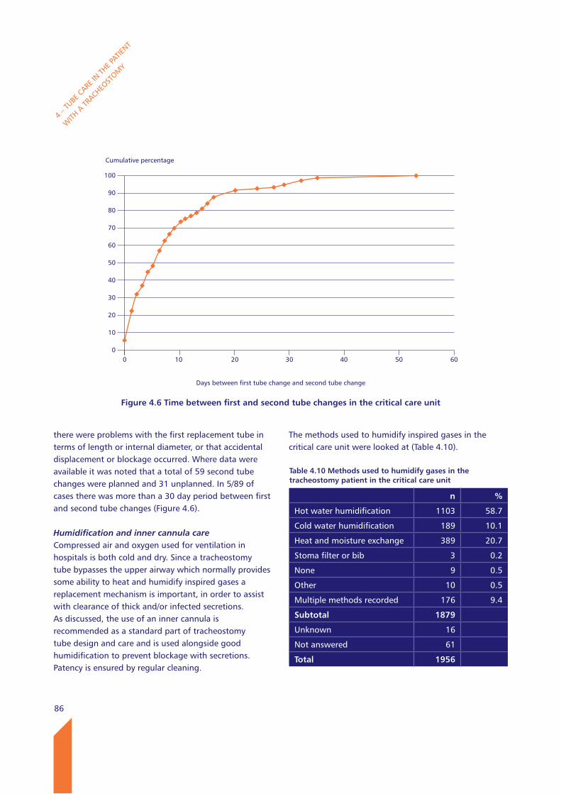
86
there were problems with the first replacement tube in terms of length or internal diameter, or that accidental displacement or blockage occurred. Where data were available it was noted that a total of 59 second tube changes were planned and 31 unplanned. In 5/89 of cases there was more than a 30 day period between first and second tube changes (Figure 4.6).
Humidification and inner cannula careCompressed air and oxygen used for ventilation in hospitals is both cold and dry. Since a tracheostomy tube bypasses the upper airway which normally provides some ability to heat and humidify inspired gases a replacement mechanism is important, in order to assist with clearance of thick and/or infected secretions. As discussed, the use of an inner cannula is recommended as a standard part of tracheostomy tube design and care and is used alongside good humidification to prevent blockage with secretions. Patency is ensured by regular cleaning.
4 – T
UBE C
ARE IN
THE P
ATIENT
WITH
A TR
ACHEOST
OMY
The methods used to humidify inspired gases in the critical care unit were looked at (Table 4.10).
Cumulative percentage
100
90
80
70
60
50
40
30
20
10
0
Figure 4.6 Time between first and second tube changes in the critical care unit
Days between first tube change and second tube change
0 10 20 30 40 50 60
Table 4.10 Methods used to humidify gases in the tracheostomy patient in the critical care unit
n %
Hot water humidification 1103 58.7
Cold water humidification 189 10.1
Heat and moisture exchange 389 20.7
Stoma filter or bib 3 0.2
None 9 0.5
Other 10 0.5
Multiple methods recorded 176 9.4
Subtotal 1879
Unknown 16
Not answered 61
Total 1956
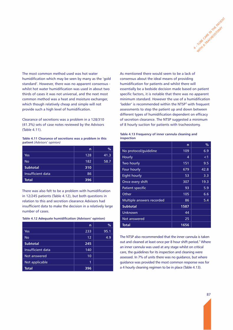
87
The most common method used was hot water humidification which may be seen by many as the ‘gold standard’. However, there was no apparent consensus - whilst hot water humidification was used in about two thirds of cases it was not universal, and the next most common method was a heat and moisture exchanger, which though relatively cheap and simple will not provide such a high level of humidification.
Clearance of secretions was a problem in a 128/310 (41.3%) sets of case notes reviewed by the Advisors(Table 4.11).
There was also felt to be a problem with humidification in 12/245 patients (Table 4.12), but both questions in relation to this and secretion clearance Advisors had insufficient data to make the decision in a relatively large number of cases.
As mentioned there would seem to be a lack of consensus about the ideal means of providing humidification for patients and whilst there will essentially be a bedside decision made based on patient specific factors, it is notable that there was no apparent minimum standard. However the use of a humidification ‘ladder’ is recommended within the NTSP1 with frequent assessments to step the patient up and down between different types of humidification dependent on efficacy of secretion clearance. The NTSP suggested a minimum of 8 hourly suction for patients with tracheostomy.
The NTSP also recommended that the inner cannula is taken out and cleaned at least once per 8 hour shift period.1 Where an inner cannula was used at any stage whilst on critical care, the guidelines for its inspection and cleaning were assessed. In 7% of units there was no guidance, but where guidance was provided the most common response was for a 4 hourly cleaning regimen to be in place (Table 4.13).
4 – T
UBE C
ARE IN
THE P
ATIENT
WITH
A TR
ACHEOST
OMY
Table 4.11 Clearance of secretions was a problem in this patient (Advisors’ opinion)
n %
Yes 128 41.3
No 182 58.7
Subtotal 310
Insufficient data 86
Total 396
Table 4.12 Adequate humidification (Advisors’ opinion)
n %
Yes 233 95.1
No 12 4.9
Subtotal 245
Insufficient data 140
Not answered 10
Not applicable 1
Total 396
Table 4.13 Frequency of inner cannula cleaning and inspection
n %
No protocol/guideline 109 6.9
Hourly 4 <1
Two hourly 151 9.5
Four hourly 679 42.8
Eight hourly 53 3.3
Once every shift 307 19.3
Patient specific 93 5.9
Other 105 6.6
Multiple answers recorded 86 5.4
Subtotal 1587
Unknown 44
Not answered 25
Total 1656
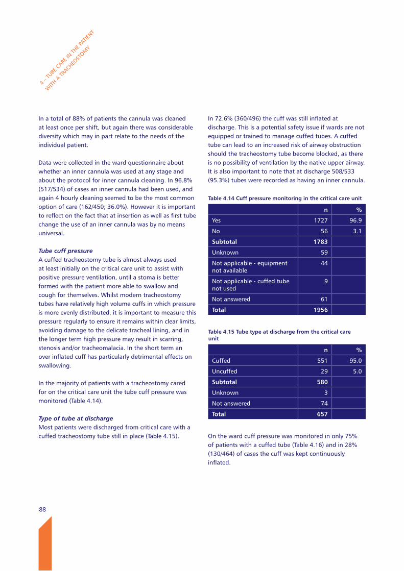
88
4 – T
UBE C
ARE IN
THE P
ATIENT
WITH
A TR
ACHEOST
OMY
In a total of 88% of patients the cannula was cleaned at least once per shift, but again there was considerable diversity which may in part relate to the needs of the individual patient.
Data were collected in the ward questionnaire about whether an inner cannula was used at any stage and about the protocol for inner cannula cleaning. In 96.8% (517/534) of cases an inner cannula had been used, and again 4 hourly cleaning seemed to be the most common option of care (162/450; 36.0%). However it is important to reflect on the fact that at insertion as well as first tube change the use of an inner cannula was by no means universal.
Tube cuff pressureA cuffed tracheostomy tube is almost always used at least initially on the critical care unit to assist with positive pressure ventilation, until a stoma is better formed with the patient more able to swallow and cough for themselves. Whilst modern tracheostomy tubes have relatively high volume cuffs in which pressure is more evenly distributed, it is important to measure this pressure regularly to ensure it remains within clear limits, avoiding damage to the delicate tracheal lining, and in the longer term high pressure may result in scarring, stenosis and/or tracheomalacia. In the short term an over inflated cuff has particularly detrimental effects on swallowing.
In the majority of patients with a tracheostomy cared for on the critical care unit the tube cuff pressure was monitored (Table 4.14).
Type of tube at dischargeMost patients were discharged from critical care with a cuffed tracheostomy tube still in place (Table 4.15).
In 72.6% (360/496) the cuff was still inflated at discharge. This is a potential safety issue if wards are not equipped or trained to manage cuffed tubes. A cuffed tube can lead to an increased risk of airway obstruction should the tracheostomy tube become blocked, as there is no possibility of ventilation by the native upper airway.It is also important to note that at discharge 508/533 (95.3%) tubes were recorded as having an inner cannula.
On the ward cuff pressure was monitored in only 75% of patients with a cuffed tube (Table 4.16) and in 28% (130/464) of cases the cuff was kept continuously inflated.
Table 4.14 Cuff pressure monitoring in the critical care unit
n %
Yes 1727 96.9
No 56 3.1
Subtotal 1783
Unknown 59
Not applicable - equipment not available
44
Not applicable - cuffed tube not used
9
Not answered 61
Total 1956
Table 4.15 Tube type at discharge from the critical care unit
n %
Cuffed 551 95.0
Uncuffed 29 5.0
Subtotal 580
Unknown 3
Not answered 74
Total 657
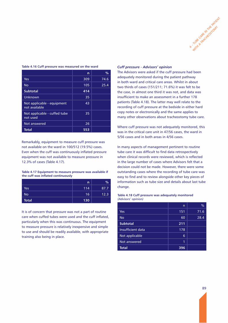
89
4 – T
UBE C
ARE IN
THE P
ATIENT
WITH
A TR
ACHEOST
OMY
Remarkably, equipment to measure cuff pressure was not available on the ward in 100/512 (19.5%) cases. Even when the cuff was continuously inflated pressure equipment was not available to measure pressure in 12.3% of cases (Table 4.17).
It is of concern that pressure was not a part of routine care when cuffed tubes were used and the cuff inflated, particularly when this was continuous. The equipment to measure pressure is relatively inexpensive and simple to use and should be readily available, with appropriate training also being in place.
Cuff pressure - Advisors’ opinionThe Advisors were asked if the cuff pressure had been adequately monitored during the patient pathway in both ward and critical care areas. Whilst in about two thirds of cases (151/211; 71.6%) it was felt to be the case, in almost one third it was not, and data was insufficient to make an assessment in a further 178 patients (Table 4.18). The latter may well relate to the recording of cuff pressure at the bedside in either hard copy notes or electronically and the same applies to many other observations about tracheostomy tube care.
Where cuff pressure was not adequately monitored, this was in the critical care unit in 47/56 cases, the ward in 5/56 cases and in both areas in 4/56 cases.
In many aspects of management pertinent to routine tube care it was difficult to find data retrospectively when clinical records were reviewed, which is reflected in the large number of cases where Advisors felt that a decision could not be made. However, there were some outstanding cases where the recording of tube care was easy to find and to review alongside other key pieces of information such as tube size and details about last tube change.
Table 4.16 Cuff pressure was measured on the ward
n %
Yes 309 74.6
No 105 25.4
Subtotal 414
Unknown 35
Not applicable - equipment not available
43
Not applicable - cuffed tube not used
35
Not answered 26
Total 553
Table 4.17 Equipment to measure pressure was available if the cuff was inflated continuously
n %
Yes 114 87.7
No 16 12.3
Total 130
Table 4.18 Cuff pressure was adequately monitored (Advisors’ opinion)
n %
Yes 151 71.6
No 60 28.4
Subtotal 211
Insufficient data 178
Not applicable 6
Not answered 1
Total 396
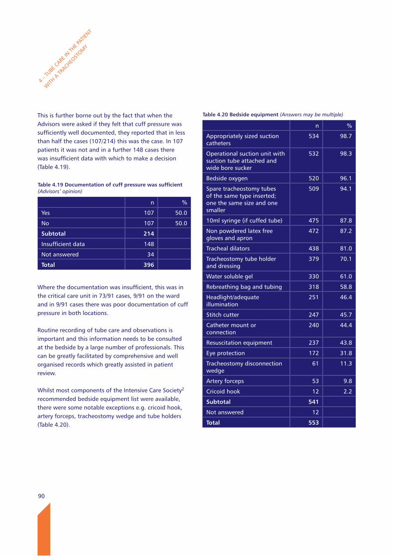
90
4 – T
UBE C
ARE IN
THE P
ATIENT
WITH
A TR
ACHEOST
OMY
This is further borne out by the fact that when the Advisors were asked if they felt that cuff pressure was sufficiently well documented, they reported that in less than half the cases (107/214) this was the case. In 107 patients it was not and in a further 148 cases there was insufficient data with which to make a decision (Table 4.19).
Where the documentation was insufficient, this was in the critical care unit in 73/91 cases, 9/91 on the ward and in 9/91 cases there was poor documentation of cuff pressure in both locations.
Routine recording of tube care and observations is important and this information needs to be consulted at the bedside by a large number of professionals. This can be greatly facilitated by comprehensive and well organised records which greatly assisted in patient review.
Whilst most components of the Intensive Care Society2
recommended bedside equipment list were available, there were some notable exceptions e.g. cricoid hook, artery forceps, tracheostomy wedge and tube holders (Table 4.20).
Table 4.19 Documentation of cuff pressure was sufficient (Advisors’ opinion)
n %
Yes 107 50.0
No 107 50.0
Subtotal 214
Insufficient data 148
Not answered 34
Total 396
Table 4.20 Bedside equipment (Answers may be multiple)
n %
Appropriately sized suction catheters
534 98.7
Operational suction unit with suction tube attached and wide bore sucker
532 98.3
Bedside oxygen 520 96.1
Spare tracheostomy tubes of the same type inserted; one the same size and one smaller
509 94.1
10ml syringe (if cuffed tube) 475 87.8
Non powdered latex free gloves and apron
472 87.2
Tracheal dilators 438 81.0
Tracheostomy tube holder and dressing
379 70.1
Water soluble gel 330 61.0
Rebreathing bag and tubing 318 58.8
Headlight/adequate illumination
251 46.4
Stitch cutter 247 45.7
Catheter mount or connection
240 44.4
Resuscitation equipment 237 43.8
Eye protection 172 31.8
Tracheostomy disconnection wedge
61 11.3
Artery forceps 53 9.8
Cricoid hook 12 2.2
Subtotal 541
Not answered 12
Total 553
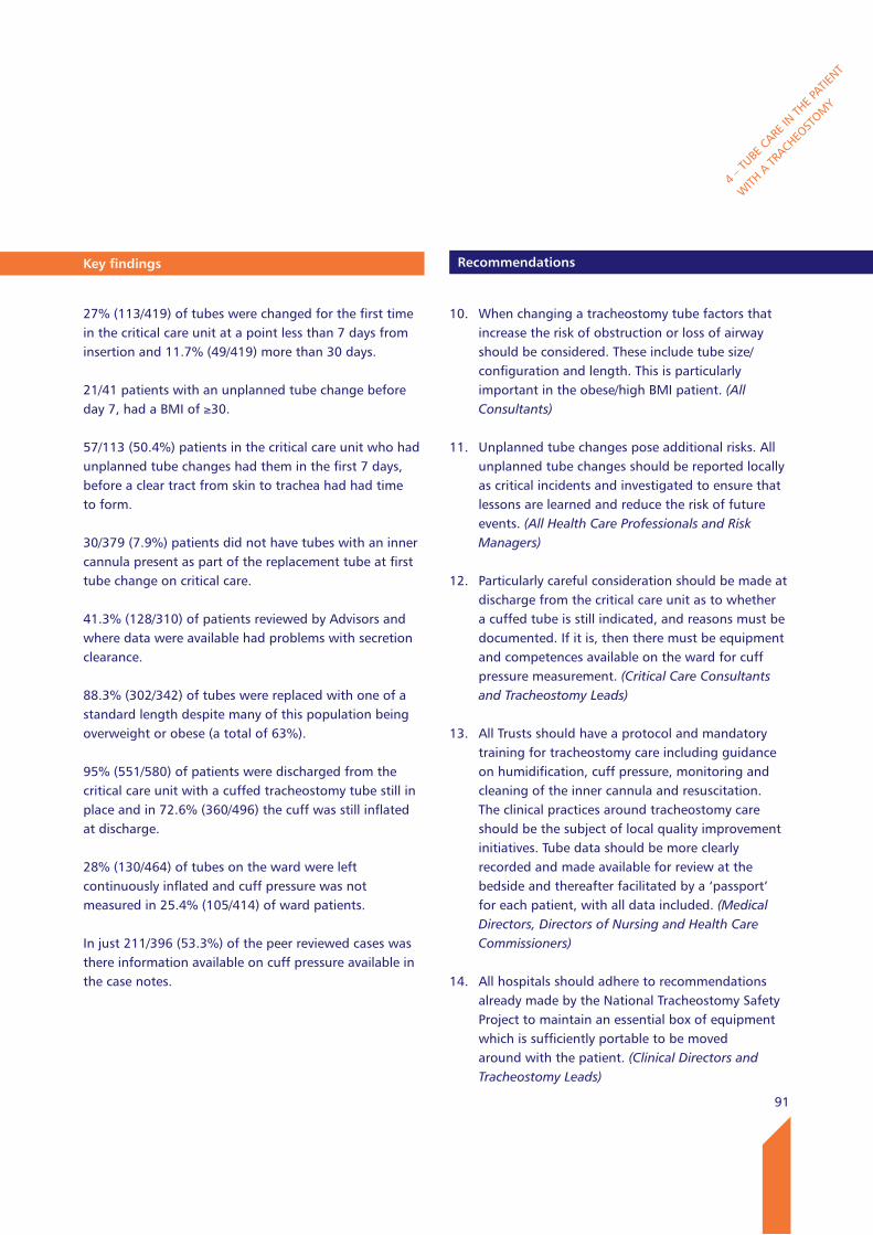
91
key findings
27% (113/419) of tubes were changed for the first time in the critical care unit at a point less than 7 days from insertion and 11.7% (49/419) more than 30 days.
21/41 patients with an unplanned tube change before day 7, had a BMI of ≥30.
57/113 (50.4%) patients in the critical care unit who had unplanned tube changes had them in the first 7 days, before a clear tract from skin to trachea had had time to form.
30/379 (7.9%) patients did not have tubes with an inner cannula present as part of the replacement tube at first tube change on critical care.
41.3% (128/310) of patients reviewed by Advisors and where data were available had problems with secretion clearance.
88.3% (302/342) of tubes were replaced with one of a standard length despite many of this population being overweight or obese (a total of 63%).
95% (551/580) of patients were discharged from the critical care unit with a cuffed tracheostomy tube still in place and in 72.6% (360/496) the cuff was still inflated at discharge. 28% (130/464) of tubes on the ward were left continuously inflated and cuff pressure was not measured in 25.4% (105/414) of ward patients.
In just 211/396 (53.3%) of the peer reviewed cases was there information available on cuff pressure available in the case notes.
4 – T
UBE C
ARE IN
THE P
ATIENT
WITH
A TR
ACHEOST
OMY
Recommendations
10. When changing a tracheostomy tube factors that increase the risk of obstruction or loss of airway should be considered. These include tube size/configuration and length. This is particularly important in the obese/high BMI patient. (All Consultants)
11. Unplanned tube changes pose additional risks. All unplanned tube changes should be reported locally as critical incidents and investigated to ensure that lessons are learned and reduce the risk of future events. (All Health Care Professionals and Risk Managers)
12. Particularly careful consideration should be made at discharge from the critical care unit as to whether a cuffed tube is still indicated, and reasons must be documented. If it is, then there must be equipment and competences available on the ward for cuff pressure measurement. (Critical Care Consultants and Tracheostomy Leads)
13. All Trusts should have a protocol and mandatory training for tracheostomy care including guidance on humidification, cuff pressure, monitoring and cleaning of the inner cannula and resuscitation. The clinical practices around tracheostomy care should be the subject of local quality improvement initiatives. Tube data should be more clearly recorded and made available for review at the bedside and thereafter facilitated by a ‘passport’ for each patient, with all data included. (Medical Directors, Directors of Nursing and Health Care Commissioners)
14. All hospitals should adhere to recommendations already made by the National Tracheostomy Safety Project to maintain an essential box of equipment which is sufficiently portable to be moved around with the patient. (Clinical Directors and Tracheostomy Leads)

92
4 – T
UBE C
ARE IN
THE P
ATIENT
WITH
A TR
ACHEOST
OMY
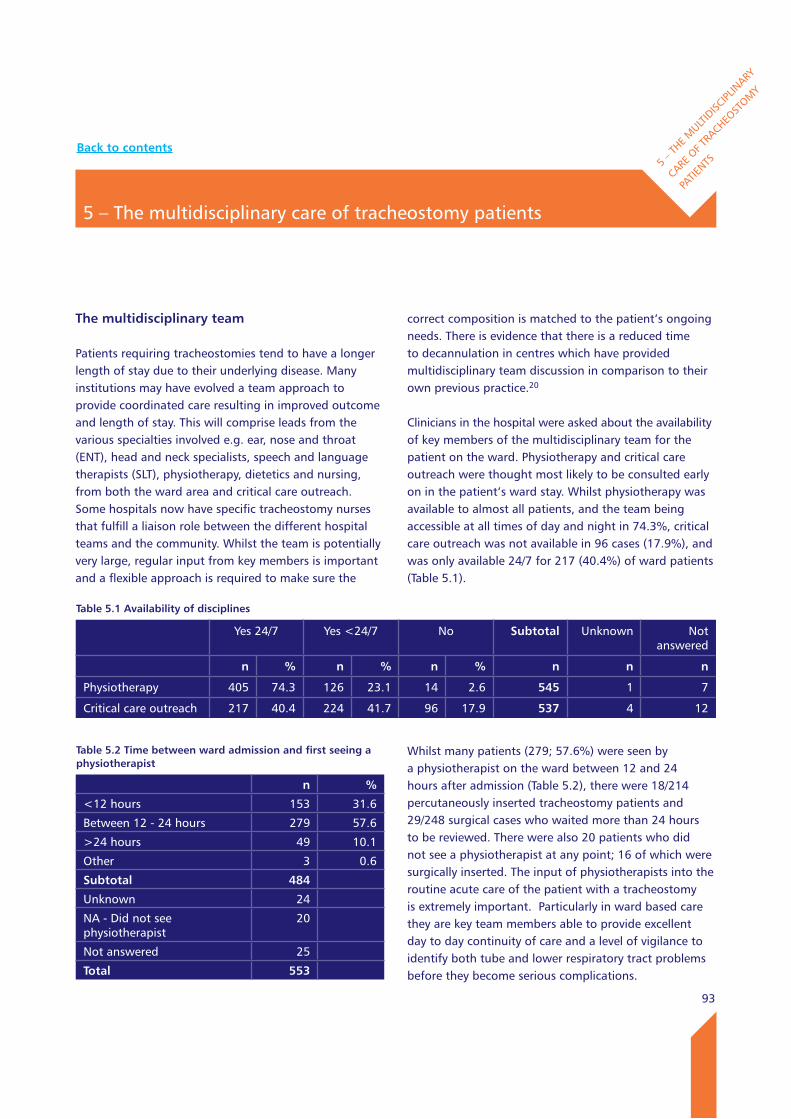
93
The multidisciplinary team
Patients requiring tracheostomies tend to have a longer length of stay due to their underlying disease. Many institutions may have evolved a team approach to provide coordinated care resulting in improved outcome and length of stay. This will comprise leads from the various specialties involved e.g. ear, nose and throat (ENT), head and neck specialists, speech and language therapists (SLT), physiotherapy, dietetics and nursing, from both the ward area and critical care outreach. Some hospitals now have specific tracheostomy nurses that fulfill a liaison role between the different hospital teams and the community. Whilst the team is potentially very large, regular input from key members is important and a flexible approach is required to make sure the
correct composition is matched to the patient’s ongoing needs. There is evidence that there is a reduced time to decannulation in centres which have provided multidisciplinary team discussion in comparison to their own previous practice.20
Clinicians in the hospital were asked about the availability of key members of the multidisciplinary team for the patient on the ward. Physiotherapy and critical care outreach were thought most likely to be consulted early on in the patient’s ward stay. Whilst physiotherapy was available to almost all patients, and the team being accessible at all times of day and night in 74.3%, critical care outreach was not available in 96 cases (17.9%), and was only available 24/7 for 217 (40.4%) of ward patients (Table 5.1).
Whilst many patients (279; 57.6%) were seen by a physiotherapist on the ward between 12 and 24 hours after admission (Table 5.2), there were 18/214 percutaneously inserted tracheostomy patients and 29/248 surgical cases who waited more than 24 hours to be reviewed. There were also 20 patients who did not see a physiotherapist at any point; 16 of which were surgically inserted. The input of physiotherapists into the routine acute care of the patient with a tracheostomy is extremely important. Particularly in ward based care they are key team members able to provide excellent day to day continuity of care and a level of vigilance to identify both tube and lower respiratory tract problems before they become serious complications.
5 – The multidisciplinary care of tracheostomy patients
5 – T
HE MULT
IDISC
IPLIN
ARY
CARE O
F TRA
CHEOST
OMY
PATIE
NTS
Table 5.1 Availability of disciplines
Yes 24/7 Yes <24/7 No Subtotal Unknown Not answered
n % n % n % n n n
Physiotherapy 405 74.3 126 23.1 14 2.6 545 1 7
Critical care outreach 217 40.4 224 41.7 96 17.9 537 4 12
Table 5.2 Time between ward admission and first seeing a physiotherapist
n %
<12 hours 153 31.6
Between 12 - 24 hours 279 57.6
>24 hours 49 10.1
Other 3 0.6
Subtotal 484
Unknown 24
NA - Did not see physiotherapist
20
Not answered 25
Total 553
Back to contents
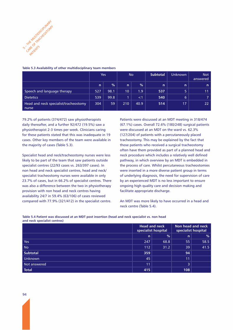
94
5 – T
HE MULT
IDISC
IPLIN
ARY
CARE O
F TRA
CHEOST
OMY
PATIE
NTS
79.2% of patients (374/472) saw physiotherapists daily thereafter, and a further 92/472 (19.5%) saw a physiotherapist 2-3 times per week. Clinicians caring for these patients stated that this was inadequate in 19 cases. Other key members of the team were available in the majority of cases (Table 5.3).
Specialist head and neck/tracheostomy nurses were less likely to be part of the team that saw patients outside specialist centres (22/93 cases vs. 263/397 cases). In non head and neck specialist centres, head and neck/specialist tracheostomy nurses were available in only 23.7% of cases, but in 66.2% of specialist centres. There was also a difference between the two in physiotherapy provision with non head and neck centres having availability 24/7 in 59.4% (63/106) of cases reviewed compared with 77.9% (321/412) in the specialist centre.
Patients were discussed at an MDT meeting in 318/474 (67.1%) cases. Overall 72.6% (180/248) surgical patients were discussed at an MDT on the ward vs. 62.3% (127/204) of patients with a percutaneously placed tracheostomy. This may be explained by the fact that those patients who received a surgical tracheostomy often have them provided as part of a planned head and neck procedure which includes a relatively well defined pathway, in which overview by an MDT is embedded in the process of care. Whilst percutaneous tracheostomies were inserted in a more diverse patient group in terms of underlying diagnosis, the need for supervision of care by an experienced MDT is no less important to ensure ongoing high quality care and decision making and facilitate appropriate discharge.
An MDT was more likely to have occurred in a head and neck centre (Table 5.4).
Table 5.3 Availability of other multidisciplinary team members
Yes No Subtotal Unknown Not answered
n % n % n n n
Speech and language therapy 527 98.1 10 1.9 537 5 11
Dietetics 539 99.8 1 <1 540 6 7
Head and neck specialist/tracheostomy nurse
304 59 210 40.9 514 17 22
Table 5.4 Patient was discussed at an MDT post insertion (head and neck specialist vs. non head and neck specialist centres)
Head and neck specialist hospital
Non head and neck specialist hospital
n % n %
Yes 247 68.8 55 58.5
No 112 31.2 39 41.5
Subtotal 359 94
Unknown 45 11
Not answered 11 3
Total 415 108
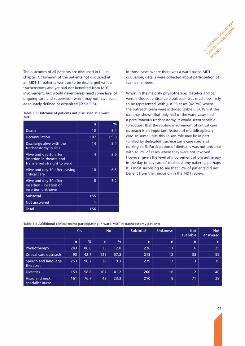
95
5 – T
HE MULT
IDISC
IPLIN
ARY
CARE O
F TRA
CHEOST
OMY
PATIE
NTS
The outcomes of all patients are discussed in full in chapter 7. However, of the patients not discussed at an MDT 14 patients went on to be discharged with a tracheostomy and yet had not benefited from MDT involvement, but would nevertheless need some level of ongoing care and supervision which may not have been adequately defined or organized (Table 5.5).
In those cases where there was a ward based MDT discussion, details were collected about participation of teams members.
Whilst in the majority physiotherapy, dietetics and SLT were included, critical care outreach was much less likely to be represented, with just 93 cases (42.7%) where the outreach team were included (Table 5.6). Whilst the data has shown that only half of the ward cases had a percutaneous tracheostomy, it would seem sensible to suggest that the routine involvement of critical care outreach is an important feature of multidisciplinary care. In some units this liaison role may be in part fulfilled by dedicated tracheostomy care specialist nursing staff. Participation of dietitians was not universal with 41.2% of cases where they were not involved. However given the level of involvement of physiotherapy in the day to day care of tracheostomy patients, perhaps it is most surprising to see that 12% of patients did not benefit from their inclusion in the MDT review.
Table 5.5 Outcome of patients not discussed at a ward MDT
n %
Death 13 8.4
Decannulation 107 69.0
Discharge alive with the tracheostomy in situ
14 8.4
Alive and day 30 after insertion in theatre and transferred straight to ward
4 2.6
Alive and day 30 after leaving critical care
10 6.5
Alive and day 30 after insertion - location of insertion unknown
8 5.2
Subtotal 155
Not answered 1
Total 156
Table 5.6 Additional clinical teams participating in ward MDT in tracheostomy patients
Yes No Subtotal Unknown Not available
Not answered
n % n % n n n n
Physiotherapy 243 88.0 33 12.0 276 11 6 25
Critical care outreach 93 42.7 125 57.3 218 12 33 55
Speech and language therapist
253 90.7 26 9.3 279 17 3 19
Dietetics 153 58.8 107 41.2 260 16 2 40
Head and neck specialist nurse
161 76.7 49 23.3 210 9 71 28
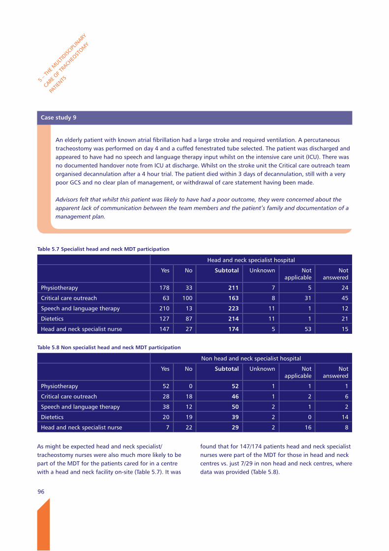
96
As might be expected head and neck specialist/tracheostomy nurses were also much more likely to be part of the MDT for the patients cared for in a centre with a head and neck facility on-site (Table 5.7). It was
5 – T
HE MULT
IDISC
IPLIN
ARY
CARE O
F TRA
CHEOST
OMY
PATIE
NTS
found that for 147/174 patients head and neck specialist nurses were part of the MDT for those in head and neck centres vs. just 7/29 in non head and neck centres, where data was provided (Table 5.8).
Case study 9
An elderly patient with known atrial fibrillation had a large stroke and required ventilation. A percutaneous tracheostomy was performed on day 4 and a cuffed fenestrated tube selected. The patient was discharged and appeared to have had no speech and language therapy input whilst on the intensive care unit (ICU). There was no documented handover note from ICU at discharge. Whilst on the stroke unit the Critical care outreach team organised decannulation after a 4 hour trial. The patient died within 3 days of decannulation, still with a very poor GCS and no clear plan of management, or withdrawal of care statement having been made.
Advisors felt that whilst this patient was likely to have had a poor outcome, they were concerned about the apparent lack of communication between the team members and the patient’s family and documentation of a management plan.
Table 5.7 Specialist head and neck MDT participation
Head and neck specialist hospital
Yes No Subtotal Unknown Not applicable
Not answered
Physiotherapy 178 33 211 7 5 24
Critical care outreach 63 100 163 8 31 45
Speech and language therapy 210 13 223 11 1 12
Dietetics 127 87 214 11 1 21
Head and neck specialist nurse 147 27 174 5 53 15
Table 5.8 Non specialist head and neck MDT participation
Non head and neck specialist hospital
Yes No Subtotal Unknown Not applicable
Not answered
Physiotherapy 52 0 52 1 1 1
Critical care outreach 28 18 46 1 2 6
Speech and language therapy 38 12 50 2 1 2
Dietetics 20 19 39 2 0 14
Head and neck specialist nurse 7 22 29 2 16 8
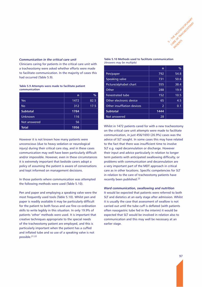
97
Communication in the critical care unitClinicians caring for patients in the critical care unit with a tracheostomy were asked whether efforts were made to facilitate communication. In the majority of cases this had occurred (Table 5.9).
However it is not known how many patients were unconscious (due to heavy sedation or neurological injury) during their critical care stay, and in these cases communication may well have been particularly difficult and/or impossible. However, even in these circumstances it is extremely important that bedside carers adopt a policy of assuming the patient is aware of conversations and kept informed on management decisions.
In those patients where communication was attempted the following methods were used (Table 5.10).
Pen and paper and employing a speaking valve were the most frequently used tools (Table 5.10). Whilst pen and paper is readily available it may be particularly difficult for the patient to both focus and use fine co-ordination skills to write legibly in this situation. In only 19.9% of patients ‘other’ methods were used. It is important that creative techniques appropriate to the special needs of the tracheostomy patient are employed, and this is particularly important when the patient has a cuffed and inflated tube and so use of a speaking valve is not possible.21,22
Whilst in 1472 patients cared for with a new tracheostomy on the critical care unit attempts were made to facilitate communication, in just 456/1693 (26.9%) cases was the advice of SLT sought. In some cases this may have related to the fact that there was insufficient time to involve SLT e.g. rapid decannulation or discharge. However their input and advice particularly in relation to longer term patients with anticipated swallowing difficulty, or problems with communication and decannulation are a very important part of the MDT approach in critical care as in other locations. Specific competencies for SLT in relation to the care of tracheostomy patients have recently been published.23
Ward communication, swallowing and nutritionIt would be expected that patients were referred to both SLT and dietetics at an early stage after admission. Whilst it is usually the case that assessment of swallow is not carried out until the tube cuff is deflated (with patients often nasogastric tube fed in the interim) it would be expected that SLT would be involved in relation also to communication and this may well be necessary at an earlier stage.
5 – T
HE MULT
IDISC
IPLIN
ARY
CARE O
F TRA
CHEOST
OMY
PATIE
NTS
Table 5.9 Attempts were made to facilitate patient communication
n %
Yes 1472 82.5
No 312 17.5
Subtotal 1784
Unknown 116
Not answered 56
Total 1956
Table 5.10 Methods used to facilitate communication (Answers may be multiple)
n %
Pen/paper 792 54.8
Speaking valve 731 50.6
Picture/alphabet chart 555 38.4
Other 288 19.9
Fenestrated tube 152 10.5
Other electronic device 65 4.5
Other insufflation devices 2 0.1
Subtotal 1444
Not answered 28
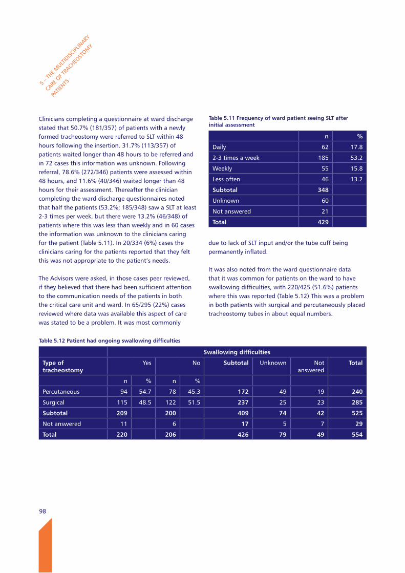
98
Clinicians completing a questionnaire at ward discharge stated that 50.7% (181/357) of patients with a newly formed tracheostomy were referred to SLT within 48 hours following the insertion. 31.7% (113/357) of patients waited longer than 48 hours to be referred and in 72 cases this information was unknown. Following referral, 78.6% (272/346) patients were assessed within 48 hours, and 11.6% (40/346) waited longer than 48 hours for their assessment. Thereafter the clinician completing the ward discharge questionnaires noted that half the patients (53.2%; 185/348) saw a SLT at least 2-3 times per week, but there were 13.2% (46/348) of patients where this was less than weekly and in 60 cases the information was unknown to the clinicians caring for the patient (Table 5.11). In 20/334 (6%) cases the clinicians caring for the patients reported that they felt this was not appropriate to the patient’s needs. The Advisors were asked, in those cases peer reviewed, if they believed that there had been sufficient attention to the communication needs of the patients in both the critical care unit and ward. In 65/295 (22%) cases reviewed where data was available this aspect of care was stated to be a problem. It was most commonly
5 – T
HE MULT
IDISC
IPLIN
ARY
CARE O
F TRA
CHEOST
OMY
PATIE
NTS
due to lack of SLT input and/or the tube cuff being permanently inflated.
It was also noted from the ward questionnaire data that it was common for patients on the ward to have swallowing difficulties, with 220/425 (51.6%) patients where this was reported (Table 5.12) This was a problem in both patients with surgical and percutaneously placed tracheostomy tubes in about equal numbers.
Table 5.11 Frequency of ward patient seeing SlT after initial assessment
n %
Daily 62 17.8
2-3 times a week 185 53.2
Weekly 55 15.8
Less often 46 13.2
Subtotal 348
Unknown 60
Not answered 21
Total 429
Table 5.12 Patient had ongoing swallowing difficulties
Swallowing difficulties
Type of tracheostomy
Yes No Subtotal Unknown Not answered
Total
n % n %
Percutaneous 94 54.7 78 45.3 172 49 19 240
Surgical 115 48.5 122 51.5 237 25 23 285
Subtotal 209 200 409 74 42 525
Not answered 11 6 17 5 7 29
Total 220 206 426 79 49 554
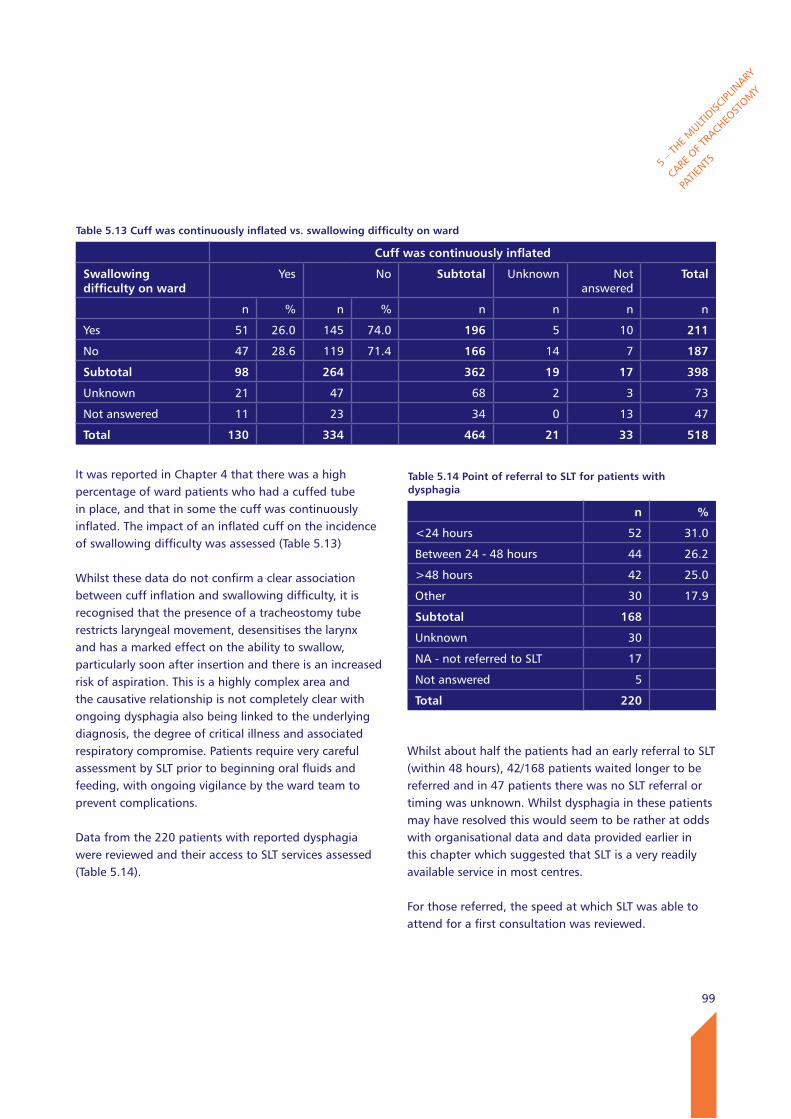
99
5 – T
HE MULT
IDISC
IPLIN
ARY
CARE O
F TRA
CHEOST
OMY
PATIE
NTS
It was reported in Chapter 4 that there was a high percentage of ward patients who had a cuffed tube in place, and that in some the cuff was continuously inflated. The impact of an inflated cuff on the incidence of swallowing difficulty was assessed (Table 5.13)
Whilst these data do not confirm a clear association between cuff inflation and swallowing difficulty, it is recognised that the presence of a tracheostomy tube restricts laryngeal movement, desensitises the larynx and has a marked effect on the ability to swallow, particularly soon after insertion and there is an increased risk of aspiration. This is a highly complex area and the causative relationship is not completely clear with ongoing dysphagia also being linked to the underlying diagnosis, the degree of critical illness and associated respiratory compromise. Patients require very careful assessment by SLT prior to beginning oral fluids and feeding, with ongoing vigilance by the ward team to prevent complications.
Data from the 220 patients with reported dysphagia were reviewed and their access to SLT services assessed (Table 5.14).
Whilst about half the patients had an early referral to SLT (within 48 hours), 42/168 patients waited longer to be referred and in 47 patients there was no SLT referral or timing was unknown. Whilst dysphagia in these patients may have resolved this would seem to be rather at odds with organisational data and data provided earlier in this chapter which suggested that SLT is a very readily available service in most centres.
For those referred, the speed at which SLT was able to attend for a first consultation was reviewed.
Table 5.13 Cuff was continuously inflated vs. swallowing difficulty on ward
Cuff was continuously inflated
Swallowing difficulty on ward
Yes No Subtotal Unknown Not answered
Total
n % n % n n n n
Yes 51 26.0 145 74.0 196 5 10 211
No 47 28.6 119 71.4 166 14 7 187
Subtotal 98 264 362 19 17 398
Unknown 21 47 68 2 3 73
Not answered 11 23 34 0 13 47
Total 130 334 464 21 33 518
Table 5.14 Point of referral to SlT for patients with dysphagia
n %
<24 hours 52 31.0
Between 24 - 48 hours 44 26.2
>48 hours 42 25.0
Other 30 17.9
Subtotal 168
Unknown 30
NA - not referred to SLT 17
Not answered 5
Total 220
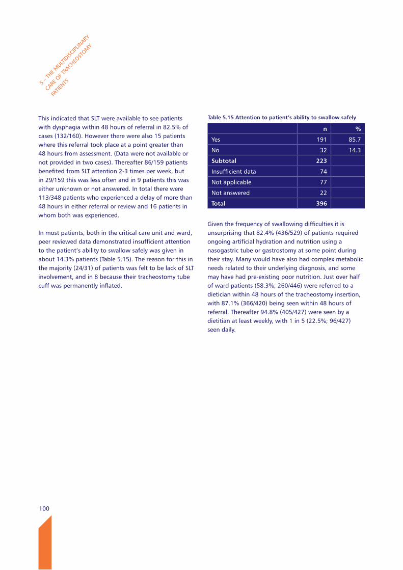
100
5 – T
HE MULT
IDISC
IPLIN
ARY
CARE O
F TRA
CHEOST
OMY
PATIE
NTS
This indicated that SLT were available to see patients with dysphagia within 48 hours of referral in 82.5% of cases (132/160). However there were also 15 patients where this referral took place at a point greater than 48 hours from assessment. (Data were not available or not provided in two cases). Thereafter 86/159 patients benefited from SLT attention 2-3 times per week, but in 29/159 this was less often and in 9 patients this was either unknown or not answered. In total there were 113/348 patients who experienced a delay of more than 48 hours in either referral or review and 16 patients in whom both was experienced.
In most patients, both in the critical care unit and ward, peer reviewed data demonstrated insufficient attention to the patient’s ability to swallow safely was given in about 14.3% patients (Table 5.15). The reason for this in the majority (24/31) of patients was felt to be lack of SLT involvement, and in 8 because their tracheostomy tube cuff was permanently inflated.
Given the frequency of swallowing difficulties it is unsurprising that 82.4% (436/529) of patients required ongoing artificial hydration and nutrition using a nasogastric tube or gastrostomy at some point during their stay. Many would have also had complex metabolic needs related to their underlying diagnosis, and some may have had pre-existing poor nutrition. Just over half of ward patients (58.3%; 260/446) were referred to a dietician within 48 hours of the tracheostomy insertion, with 87.1% (366/420) being seen within 48 hours of referral. Thereafter 94.8% (405/427) were seen by a dietitian at least weekly, with 1 in 5 (22.5%; 96/427) seen daily.
Table 5.15 Attention to patient’s ability to swallow safely
n %
Yes 191 85.7
No 32 14.3
Subtotal 223
Insufficient data 74
Not applicable 77
Not answered 22
Total 396
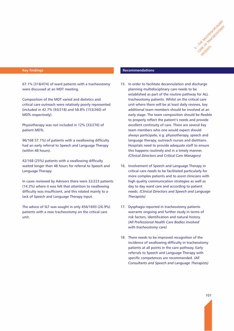
101
5 – T
HE MULT
IDISC
IPLIN
ARY
CARE O
F TRA
CHEOST
OMY
PATIE
NTS
key findings
67.1% (318/474) of ward patients with a tracheostomy were discussed at an MDT meeting.
Composition of the MDT varied and dietetics and critical care outreach were relatively poorly represented (included in 42.7% (93/218) and 58.8% (153/260) of MDTs respectively). Physiotherapy was not included in 12% (33/276) of patient MDTs.
96/168 57.1%) of patients with a swallowing difficulty had an early referral to Speech and Language Therapy (within 48 hours).
42/168 (25%) patients with a swallowing difficulty waited longer than 48 hours for referral to Speech and Language Therapy.
In cases reviewed by Advisors there were 32/223 patients (14.3%) where it was felt that attention to swallowing difficulty was insufficient, and this related mainly to a lack of Speech and Language Therapy input.
The advice of SLT was sought in only 456/1693 (26.9%) patients with a new tracheostomy on the critical care unit.
Recommendations
15. In order to facilitate decannulation and discharge planning multidisciplinary care needs to be established as part of the routine pathway for ALL tracheostomy patients. Whilst on the critical care unit where there will be at least daily reviews, key additional team members should be involved at an early stage. The team composition should be flexible to properly reflect the patient’s needs and provide excellent continuity of care. There are several key team members who one would expect should always participate, e.g. physiotherapy, speech and language therapy, outreach nurses and dietitians. Hospitals need to provide adequate staff to ensure this happens routinely and in a timely manner. (Clinical Directors and Critical Care Managers)
16. Involvement of Speech and Language Therapy in critical care needs to be facilitated particularly for more complex patients and to assist clinicians with high quality communication strategies as well as day to day ward care and according to patient needs. (Clinical Directors and Speech and Language Therapists)
17. Dysphagia reported in tracheostomy patients warrants ongoing and further study in terms of
risk factors, identification and natural history. (All Professional Health Care Bodies involved with tracheostomy care)
18. There needs to be improved recognition of the incidence of swallowing difficulty in tracheostomy patients at all points in the care pathway. Early referrals to Speech and Language Therapy with specific competences are recommended. (All Consultants and Speech and Language Therapists)

102
REFE
RENCES
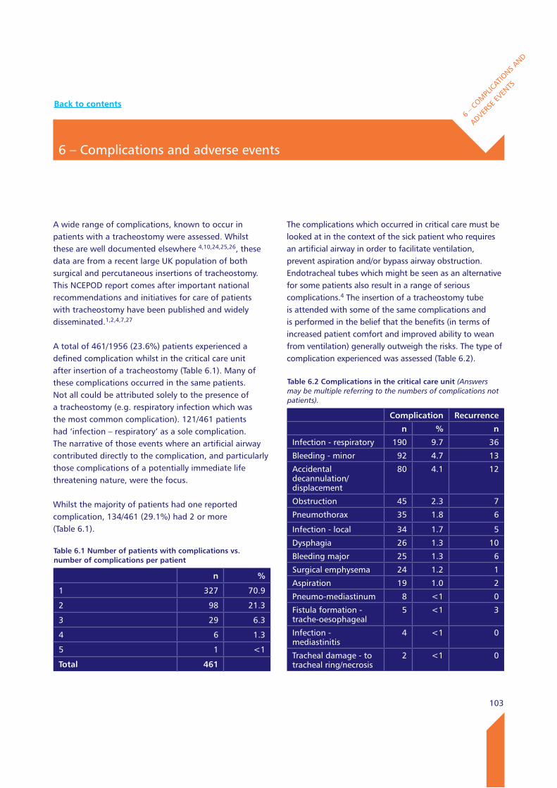
103
A wide range of complications, known to occur in patients with a tracheostomy were assessed. Whilst these are well documented elsewhere 4,10,24,25,26, these data are from a recent large UK population of both surgical and percutaneous insertions of tracheostomy. This NCEPOD report comes after important national recommendations and initiatives for care of patients with tracheostomy have been published and widely disseminated.1,2,4,7,27
A total of 461/1956 (23.6%) patients experienced a defined complication whilst in the critical care unit after insertion of a tracheostomy (Table 6.1). Many of these complications occurred in the same patients. Not all could be attributed solely to the presence of a tracheostomy (e.g. respiratory infection which was the most common complication). 121/461 patients had ‘infection – respiratory’ as a sole complication. The narrative of those events where an artificial airway contributed directly to the complication, and particularly those complications of a potentially immediate life threatening nature, were the focus.
Whilst the majority of patients had one reported complication, 134/461 (29.1%) had 2 or more (Table 6.1).
The complications which occurred in critical care must be looked at in the context of the sick patient who requires an artificial airway in order to facilitate ventilation, prevent aspiration and/or bypass airway obstruction. Endotracheal tubes which might be seen as an alternative for some patients also result in a range of serious complications.4 The insertion of a tracheostomy tube is attended with some of the same complications and is performed in the belief that the benefits (in terms of increased patient comfort and improved ability to wean from ventilation) generally outweigh the risks. The type of complication experienced was assessed (Table 6.2).
6 – Complications and adverse events
6 – C
OMPL
ICATIO
NS AND
ADVERSE
EVEN
TS
Table 6.1 Number of patients with complications vs. number of complications per patient
n %
1 327 70.9
2 98 21.3
3 29 6.3
4 6 1.3
5 1 <1
Total 461
Table 6.2 Complications in the critical care unit (Answers may be multiple referring to the numbers of complications not patients).
Complication Recurrence
n % n
Infection - respiratory 190 9.7 36
Bleeding - minor 92 4.7 13
Accidental decannulation/displacement
80 4.1 12
Obstruction 45 2.3 7
Pneumothorax 35 1.8 6
Infection - local 34 1.7 5
Dysphagia 26 1.3 10
Bleeding major 25 1.3 6
Surgical emphysema 24 1.2 1
Aspiration 19 1.0 2
Pneumo-mediastinum 8 <1 0
Fistula formation - trache-oesophageal
5 <1 3
Infection - mediastinitis
4 <1 0
Tracheal damage - to tracheal ring/necrosis
2 <1 0
Back to contents
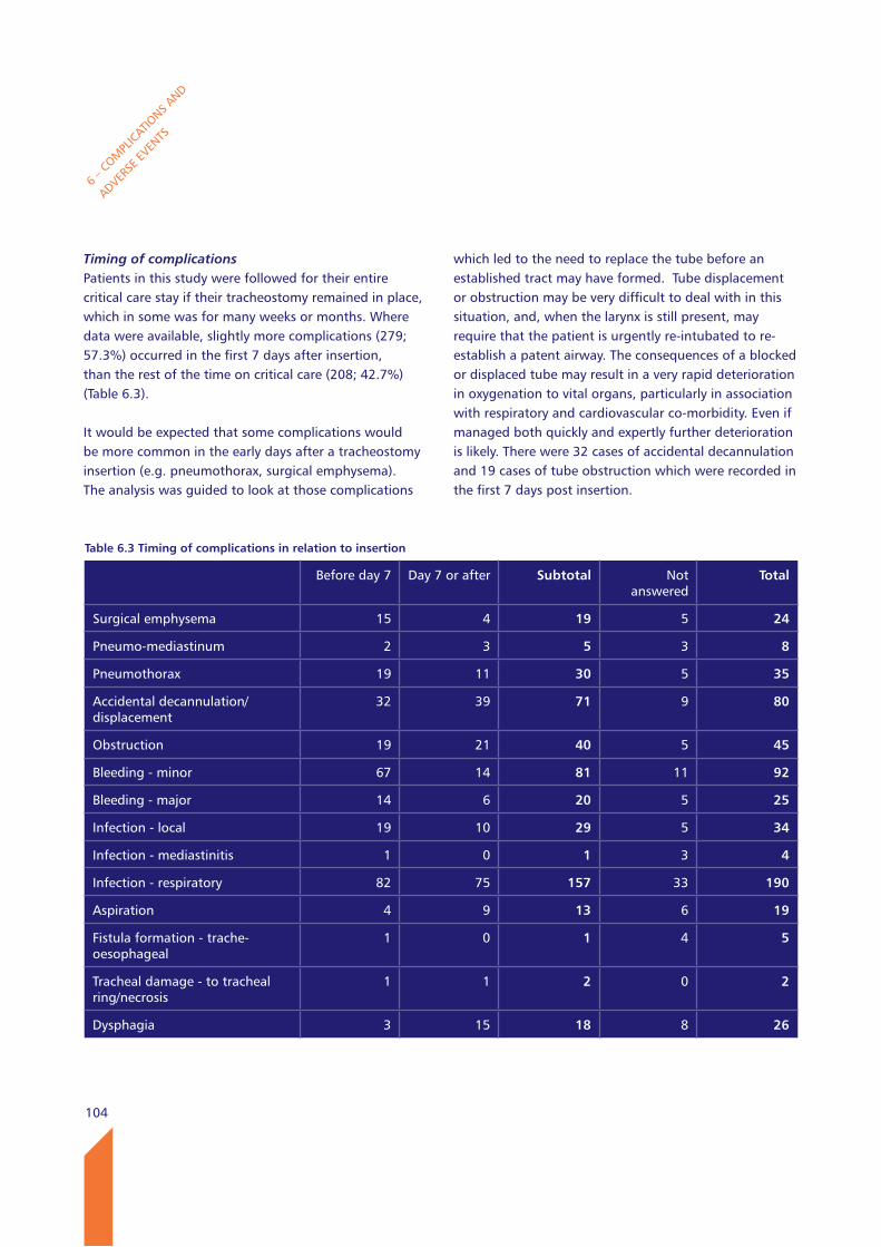
104
6 – C
OMPL
ICATIO
NS AND
ADVERSE
EVEN
TS
Timing of complications Patients in this study were followed for their entire critical care stay if their tracheostomy remained in place, which in some was for many weeks or months. Where data were available, slightly more complications (279; 57.3%) occurred in the first 7 days after insertion, than the rest of the time on critical care (208; 42.7%)(Table 6.3).
It would be expected that some complications would be more common in the early days after a tracheostomy insertion (e.g. pneumothorax, surgical emphysema). The analysis was guided to look at those complications
which led to the need to replace the tube before an established tract may have formed. Tube displacement or obstruction may be very difficult to deal with in this situation, and, when the larynx is still present, may require that the patient is urgently re-intubated to re-establish a patent airway. The consequences of a blocked or displaced tube may result in a very rapid deterioration in oxygenation to vital organs, particularly in association with respiratory and cardiovascular co-morbidity. Even if managed both quickly and expertly further deterioration is likely. There were 32 cases of accidental decannulation and 19 cases of tube obstruction which were recorded in the first 7 days post insertion.
Table 6.3 Timing of complications in relation to insertion
Before day 7 Day 7 or after Subtotal Not answered
Total
Surgical emphysema 15 4 19 5 24
Pneumo-mediastinum 2 3 5 3 8
Pneumothorax 19 11 30 5 35
Accidental decannulation/displacement
32 39 71 9 80
Obstruction 19 21 40 5 45
Bleeding - minor 67 14 81 11 92
Bleeding - major 14 6 20 5 25
Infection - local 19 10 29 5 34
Infection - mediastinitis 1 0 1 3 4
Infection - respiratory 82 75 157 33 190
Aspiration 4 9 13 6 19
Fistula formation - trache-oesophageal
1 0 1 4 5
Tracheal damage - to tracheal ring/necrosis
1 1 2 0 2
Dysphagia 3 15 18 8 26
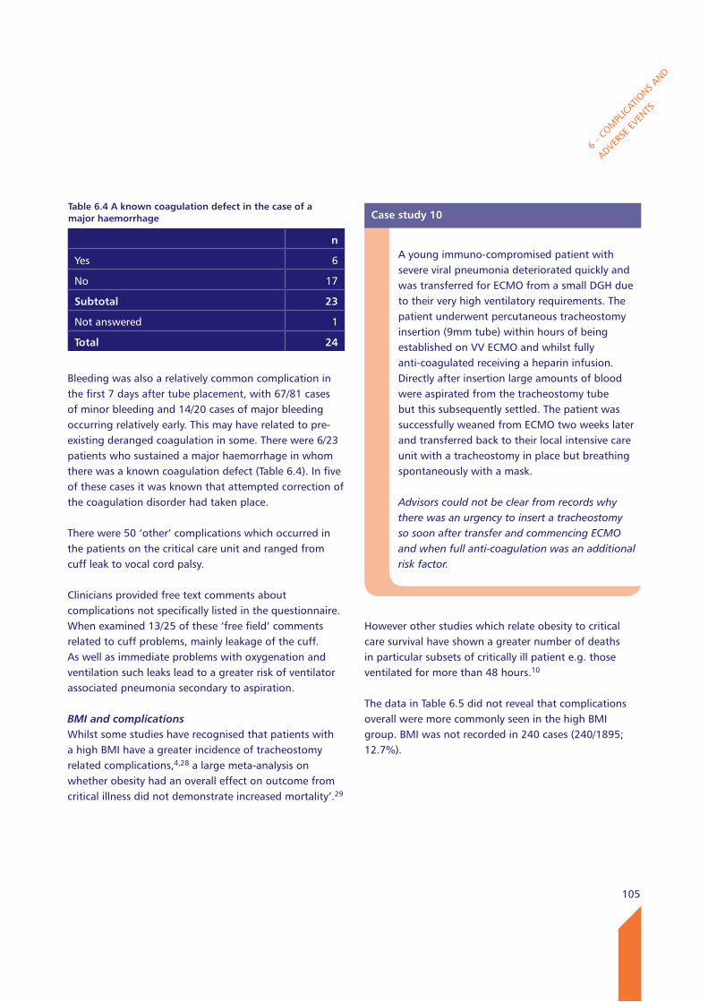
105
Bleeding was also a relatively common complication in the first 7 days after tube placement, with 67/81 cases of minor bleeding and 14/20 cases of major bleeding occurring relatively early. This may have related to pre-existing deranged coagulation in some. There were 6/23 patients who sustained a major haemorrhage in whom there was a known coagulation defect (Table 6.4). In five of these cases it was known that attempted correction of the coagulation disorder had taken place.
There were 50 ‘other’ complications which occurred in the patients on the critical care unit and ranged from cuff leak to vocal cord palsy.
Clinicians provided free text comments about complications not specifically listed in the questionnaire. When examined 13/25 of these ‘free field’ comments related to cuff problems, mainly leakage of the cuff. As well as immediate problems with oxygenation and ventilation such leaks lead to a greater risk of ventilator associated pneumonia secondary to aspiration.
BMI and complicationsWhilst some studies have recognised that patients with a high BMI have a greater incidence of tracheostomy related complications,4,28 a large meta-analysis on whether obesity had an overall effect on outcome from critical illness did not demonstrate increased mortality’.29
However other studies which relate obesity to critical care survival have shown a greater number of deaths in particular subsets of critically ill patient e.g. those ventilated for more than 48 hours.10
The data in Table 6.5 did not reveal that complications overall were more commonly seen in the high BMI group. BMI was not recorded in 240 cases (240/1895; 12.7%).
6 – C
OMPL
ICATIO
NS AND
ADVERSE
EVEN
TS
Table 6.4 A known coagulation defect in the case of a major haemorrhage
n
Yes 6
No 17
Subtotal 23
Not answered 1
Total 24
Case study 10
A young immuno-compromised patient with severe viral pneumonia deteriorated quickly and was transferred for ECMO from a small DGH due to their very high ventilatory requirements. The patient underwent percutaneous tracheostomy insertion (9mm tube) within hours of being established on VV ECMO and whilst fully anti-coagulated receiving a heparin infusion. Directly after insertion large amounts of blood were aspirated from the tracheostomy tube but this subsequently settled. The patient was successfully weaned from ECMO two weeks later and transferred back to their local intensive care unit with a tracheostomy in place but breathing spontaneously with a mask.
Advisors could not be clear from records why there was an urgency to insert a tracheostomy so soon after transfer and commencing ECMO and when full anti-coagulation was an additional risk factor.
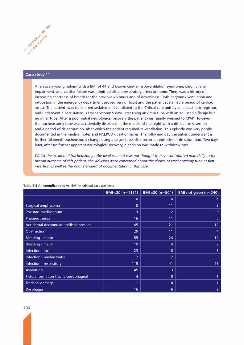
106
6 – C
OMPL
ICATIO
NS AND
ADVERSE
EVEN
TS
Case study 11
A relatively young patient with a BMI of 44 and known central hypoventilation syndrome, chronic renal impairment, and cardiac failure was admitted after a respiratory arrest at home. There was a history of increasing shortness of breath for the previous 48 hours and of drowsiness. Both bag/mask ventilation and intubation in the emergency department proved very difficult and the patient sustained a period of cardiac arrest. The patient was transferred sedated and ventilated to the Critical care unit by an anaesthetic registrar, and underwent a percutaneous tracheostomy 5 days later using an 8mm tube with an adjustable flange but no inner tube. After a poor initial neurological recovery the patient was rapidly weaned to CPAP. However the tracheostomy tube was accidentally displaced in the middle of the night with a difficult re-insertion and a period of de-saturation, after which the patient required re-ventilation. This episode was very poorly documented in the medical notes and NCEPOD questionnaires. The following day the patient underwent a further (planned) tracheostomy change using a larger tube after recurrent episodes of de-saturation. Two days later, after no further apparent neurological recovery, a decision was made to withdraw care.
Whilst the accidental tracheostomy tube displacement was not thought to have contributed materially to the overall outcome of this patient, the Advisors were concerned about the choice of tracheostomy tube at first insertion as well as the poor standard of documentation in this case.
Table 6.5 All complications vs. BMI in critical care patients
BMI<30 (n=1151) BMI ≥30 (n=504) BMI not given (n=240)
n n n
Surgical emphysema 8 11 3
Pneumo-mediastinum 3 2 3
Pneumothorax 16 11 5
Accidental decannulation/displacement 45 21 12
Obstruction 29 11 4
Bleeding - minor 55 24 12
Bleeding - major 19 4 2
Infection - local 23 8 3
Infection - mediastinitis 2 2 0
Infection - respiratory 115 41 26
Aspiration 45 3 3
Fistula formation trache-oesophageal 4 0 1
Tracheal damage 1 0 1
Dysphagia 16 6 2
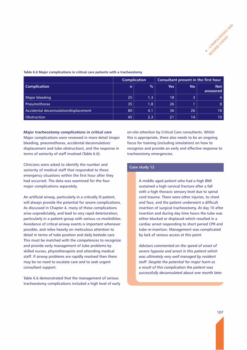
107
6 – C
OMPL
ICATIO
NS AND
ADVERSE
EVEN
TS
Major tracheostomy complications in critical careMajor complications were reviewed in more detail (major bleeding, pneumothorax, accidental decannulation/displacement and tube obstruction), and the response in terms of seniority of staff involved (Table 6.6).
Clinicians were asked to identify the number and seniority of medical staff that responded to these emergency situations within the first hour after they had occurred. The data was examined for the four major complications separately.
An artificial airway, particularly in a critically ill patient, will always provide the potential for severe complications. As discussed in Chapter 4, many of these complications arise unpredictably, and lead to very rapid deterioration, particularly in a patient group with serious co-morbidities. Avoidance of critical airway events is important whenever possible, and relies heavily on meticulous attention to detail in terms of tube position and daily bedside care. This must be matched with the competences to recognize and provide early management of tube problems by skilled nurses, physiotherapists and attending medical staff. If airway problems are rapidly resolved then there may be no need to escalate care and to seek urgent consultant support.
Table 6.6 demonstrated that the management of serious tracheostomy complications included a high level of early
on-site attention by Critical Care consultants. Whilst this is appropriate, there also needs to be an ongoing focus for training (including simulation) on how to recognize and provide an early and effective response to tracheostomy emergencies.
Table 6.6 Major complications in critical care patients with a tracheostomy
Complication Consultant present in the first hour
Complication n % yes No Not answered
Major bleeding 25 1.3 18 3 4
Pneumothorax 35 1.8 26 1 8
Accidental decannulation/displacement 80 4.1 36 26 18
Obstruction 45 2.3 21 14 10
Case study 12
A middle aged patient who had a high BMI sustained a high cervical fracture after a fall with a high thoracic sensory level due to spinal cord trauma. There were other injuries, to chest and face, and the patient underwent a difficult insertion of surgical tracheostomy. At day 10 after insertion and during day time hours the tube was either blocked or displaced which resulted in a cardiac arrest responding to short period CPR and tube re-insertion. Management was complicated by lack of venous access at this point.
Advisors commented on the speed of onset of severe hypoxia and arrest in this patient which was ultimately very well managed by resident staff. Despite the potential for major harm as a result of this complication the patient was successfully decannulated about one month later.
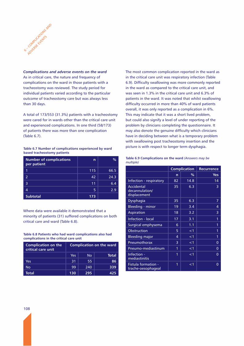
108
6 – C
OMPL
ICATIO
NS AND
ADVERSE
EVEN
TS
Complications and adverse events on the wardAs in critical care, the nature and frequency of complications on the ward in those patients with a tracheostomy was reviewed. The study period for individual patients varied according to the particular outcome of tracheostomy care but was always less than 30 days.
A total of 173/553 (31.3%) patients with a tracheostomy were cared for in wards other than the critical care unit and experienced complications. In one third (58/173) of patients there was more than one complication (Table 6.7).
Where data were available it demonstrated that a minority of patients (31) suffered complications on both critical care and ward (Table 6.8).
The most common complication reported in the ward as in the critical care unit was respiratory infection (Table 6.9). Difficulty swallowing was more commonly reported in the ward as compared to the critical care unit, and was seen in 1.3% in the critical care unit and 6.3% of patients in the ward. It was noted that whilst swallowing difficulty occurred in more than 40% of ward patients overall, it was only reported as a complication in 6%. This may indicate that it was a short lived problem, but could also signify a level of under reporting of the problem by clinicians completing the questionnaire. It may also denote the genuine difficulty which clinicians have in deciding between what is a temporary problem with swallowing post tracheostomy insertion and the picture is with respect to longer term dysphagia. Table 6.7 Number of complications experienced by ward
based tracheostomy patients
Number of complications per patient
n %
1 115 66.5
2 42 24.3
3 11 6.4
4 5 2.9
Subtotal 173
Table 6.8 Patients who had ward complications also had complications in the critical care unit
Complication on the critical care unit
Complication on the ward
Yes No Total
Yes 31 55 86
No 99 240 339
Total 130 295 425
Table 6.9 Complications on the ward (Answers may be multiple)
Complication Recurrence
n % yes
Infection - respiratory 82 14.8 14
Accidental decannulation/displacement
35 6.3 3
Dysphagia 35 6.3 7
Bleeding - minor 19 3.4 4
Aspiration 18 3.2 3
Infection - local 17 3.1 1
Surgical emphysema 6 1.1 1
Obstruction 5 <1 1
Bleeding major 4 <1 1
Pneumothorax 3 <1 0
Pneumo-mediastinum 1 <1 0
Infection - mediastinitis
1 <1 0
Fistula formation - trache-oesophageal
1 <1 0
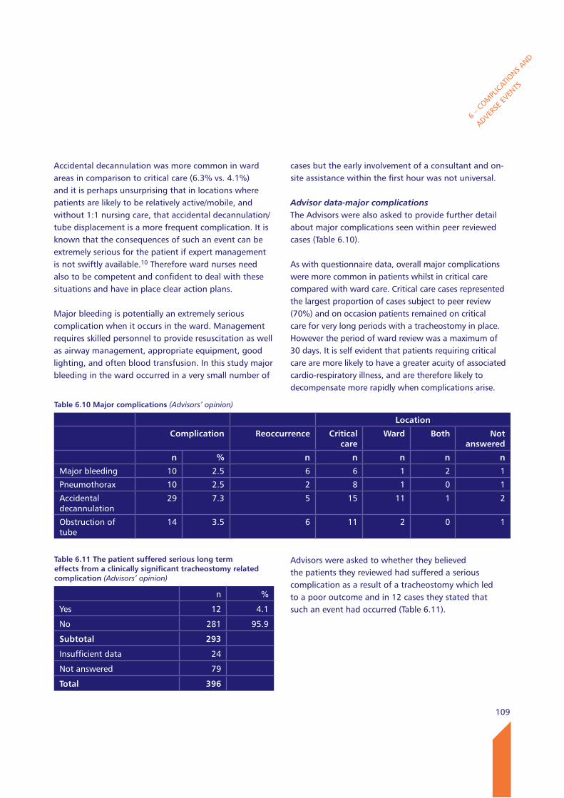
109
6 – C
OMPL
ICATIO
NS AND
ADVERSE
EVEN
TS
Accidental decannulation was more common in ward areas in comparison to critical care (6.3% vs. 4.1%) and it is perhaps unsurprising that in locations where patients are likely to be relatively active/mobile, and without 1:1 nursing care, that accidental decannulation/tube displacement is a more frequent complication. It is known that the consequences of such an event can be extremely serious for the patient if expert management is not swiftly available.10 Therefore ward nurses need also to be competent and confident to deal with these situations and have in place clear action plans.
Major bleeding is potentially an extremely serious complication when it occurs in the ward. Management requires skilled personnel to provide resuscitation as well as airway management, appropriate equipment, good lighting, and often blood transfusion. In this study major bleeding in the ward occurred in a very small number of
cases but the early involvement of a consultant and on-site assistance within the first hour was not universal.
Advisor data-major complicationsThe Advisors were also asked to provide further detail about major complications seen within peer reviewed cases (Table 6.10).
As with questionnaire data, overall major complications were more common in patients whilst in critical care compared with ward care. Critical care cases represented the largest proportion of cases subject to peer review (70%) and on occasion patients remained on critical care for very long periods with a tracheostomy in place. However the period of ward review was a maximum of 30 days. It is self evident that patients requiring critical care are more likely to have a greater acuity of associated cardio-respiratory illness, and are therefore likely to decompensate more rapidly when complications arise.
Advisors were asked to whether they believed the patients they reviewed had suffered a serious complication as a result of a tracheostomy which led to a poor outcome and in 12 cases they stated that such an event had occurred (Table 6.11).
Table 6.10 Major complications (Advisors’ opinion)
location
Complication Reoccurrence Critical care
ward Both Not answered
n % n n n n n
Major bleeding 10 2.5 6 6 1 2 1
Pneumothorax 10 2.5 2 8 1 0 1
Accidental decannulation
29 7.3 5 15 11 1 2
Obstruction of tube
14 3.5 6 11 2 0 1
Table 6.11 The patient suffered serious long term effects from a clinically significant tracheostomy related complication (Advisors’ opinion)
n %
Yes 12 4.1
No 281 95.9
Subtotal 293
Insufficient data 24
Not answered 79
Total 396
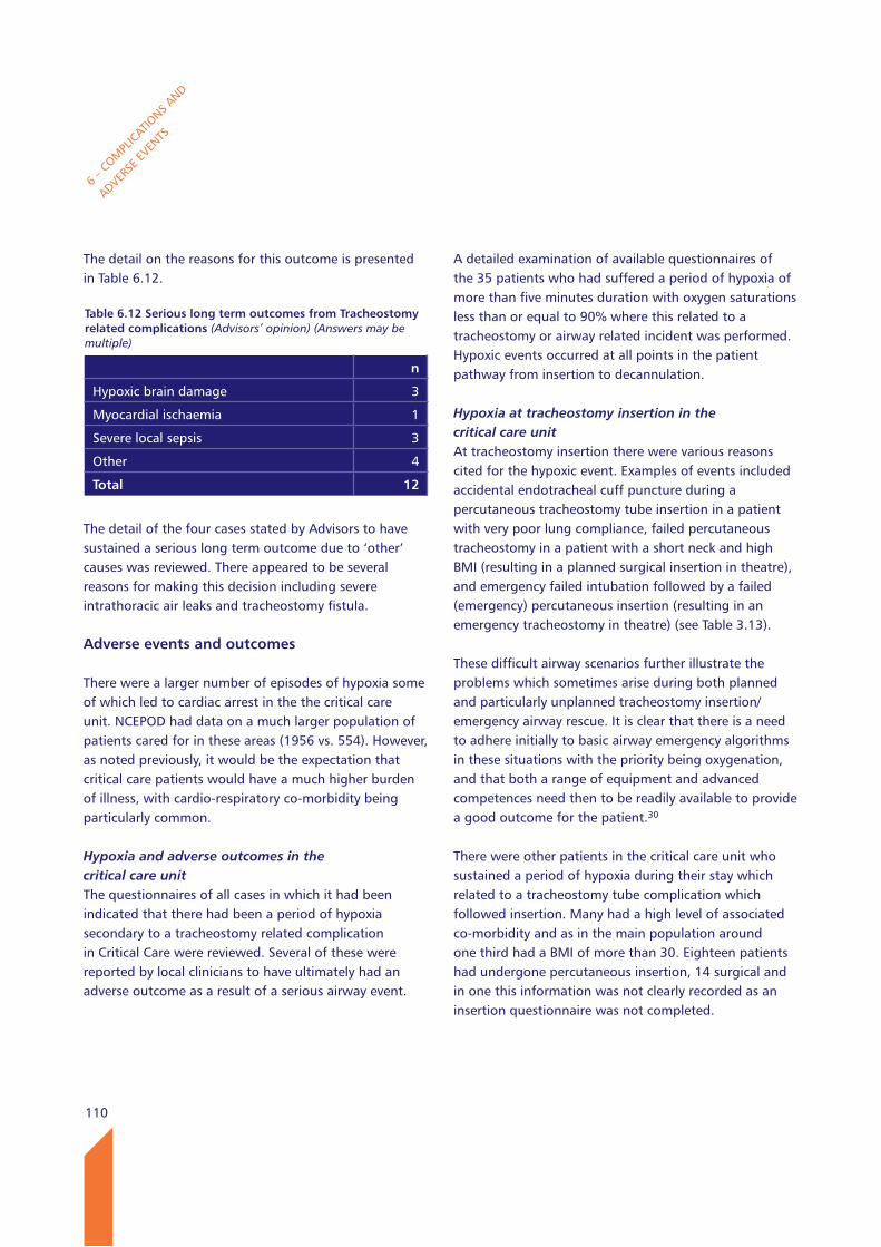
110
6 – C
OMPL
ICATIO
NS AND
ADVERSE
EVEN
TS
The detail on the reasons for this outcome is presented in Table 6.12.
The detail of the four cases stated by Advisors to have sustained a serious long term outcome due to ‘other’ causes was reviewed. There appeared to be several reasons for making this decision including severe intrathoracic air leaks and tracheostomy fistula.
Adverse events and outcomes
There were a larger number of episodes of hypoxia some of which led to cardiac arrest in the the critical care unit. NCEPOD had data on a much larger population of patients cared for in these areas (1956 vs. 554). However, as noted previously, it would be the expectation that critical care patients would have a much higher burden of illness, with cardio-respiratory co-morbidity being particularly common.
Hypoxia and adverse outcomes in the critical care unitThe questionnaires of all cases in which it had been indicated that there had been a period of hypoxia secondary to a tracheostomy related complication in Critical Care were reviewed. Several of these were reported by local clinicians to have ultimately had an adverse outcome as a result of a serious airway event.
A detailed examination of available questionnaires of the 35 patients who had suffered a period of hypoxia of more than five minutes duration with oxygen saturations less than or equal to 90% where this related to a tracheostomy or airway related incident was performed. Hypoxic events occurred at all points in the patient pathway from insertion to decannulation.
Hypoxia at tracheostomy insertion in the critical care unitAt tracheostomy insertion there were various reasons cited for the hypoxic event. Examples of events included accidental endotracheal cuff puncture during a percutaneous tracheostomy tube insertion in a patient with very poor lung compliance, failed percutaneous tracheostomy in a patient with a short neck and high BMI (resulting in a planned surgical insertion in theatre), and emergency failed intubation followed by a failed (emergency) percutaneous insertion (resulting in an emergency tracheostomy in theatre) (see Table 3.13).
These difficult airway scenarios further illustrate the problems which sometimes arise during both planned and particularly unplanned tracheostomy insertion/emergency airway rescue. It is clear that there is a need to adhere initially to basic airway emergency algorithms in these situations with the priority being oxygenation, and that both a range of equipment and advanced competences need then to be readily available to provide a good outcome for the patient.30
There were other patients in the critical care unit who sustained a period of hypoxia during their stay which related to a tracheostomy tube complication which followed insertion. Many had a high level of associated co-morbidity and as in the main population around one third had a BMI of more than 30. Eighteen patients had undergone percutaneous insertion, 14 surgical and in one this information was not clearly recorded as an insertion questionnaire was not completed.
Table 6.12 Serious long term outcomes from Tracheostomy related complications (Advisors’ opinion) (Answers may be multiple)
n
Hypoxic brain damage 3
Myocardial ischaemia 1
Severe local sepsis 3
Other 4
Total 12
111
6 – C
OMPL
ICATIO
NS AND
ADVERSE
EVEN
TS
The reasons for severe and prolonged hypoxia are summarised as follows:
Blocked tracheostomy tubeA minority of cases occurred very early after insertion and were managed appropriately with emergency re-intubation. There were also cases which occurred in association with major haemorrhage. Some patients sustained a cardiac arrest as a consequence of a blocked tube.
Accidental decannulationAccidental decannulation/tube displacement occurred due to the patient removing the tube themselves or whilst being moved to provide care e.g. when rolling the patient to prevent pressure areas, or in moving the patient in or out of bed the tube is dislodged. In this study whilst some cases were managed relatively rapidly without apparent long term effects, all resulted in a period of hypoxia and several cases of displaced tube led to cardiac arrest.
Severe haemorrhageSevere haemorrhage tended to occur relatively early in the first 7 days post tracheostomy insertion, and in a minority was associated with severe hypoxia. Haemorrhage resulted in tube occlusion, and also occurred after accidental decannulation. As well as airway clearance and tube re-insertion, patients also required transfusion and correction of coagulopathy.
Major tube leaks Tube leaks were a relatively common reason for other complications and in some cases there were serious consequences resulting from inadequate ventilation and oxygenation.
Other causes of severe and prolonged hypoxia include major intra-thoracic air leaks producing pneumothorax, aspiration and difficult tube changes in patients with a pre-existing high oxygen requirement which resulted in more prolonged interruptions in oxygenation/ventilation.
Cardiac arrest in the critical care unitThere were 11/121 cases where clinicians indicated that a cardiac arrest had occurred directly as a result of a complication of the tracheostomy and these patient questionnaires were reviewed. As might be expected the vast majority occurred after a period of hypoxia (see discussion of cases discussed in the previous section).
Patients who sustained a cardiac arrest as a result of a tracheostomy related complication were from mixed diagnostic groups, and many had a high BMI (≥30). All had a very high acuity of associated illness with associated co-morbidities which included severe sepsis. Whilst most had been admitted as emergencies some patients had undergone planned (non head and neck) surgery including cardiac and neurosurgery and had developed severe post operative complications which required prolonged stays in critical care and ventilatory support.
The management of blocked and displaced tubes is a recurrent theme within this study and ultimately it was this complication which was the greatest single cause of hypoxia and cardiac arrest in patients with a tracheostomy leading to serious morbidity and mortality. This is not an unexpected finding and has been highlighted in many previous studies, most recently in the UK in the National Audit of Airway complications conducted by the RCoA and Difficult Airway Society and published in 2011.4
A number of questions were asked in the organisational questionnaire around the guidelines, protocols and training around the management of blocked and displaced tubes. The ICS standards2 state “every hospital must have a procedure for managing patients whose tracheostomy is blocked or displaced. Staff must be aware of this and receive appropriate training to manage the problem”. Such an algorithm is reproduced in Appendix 3.
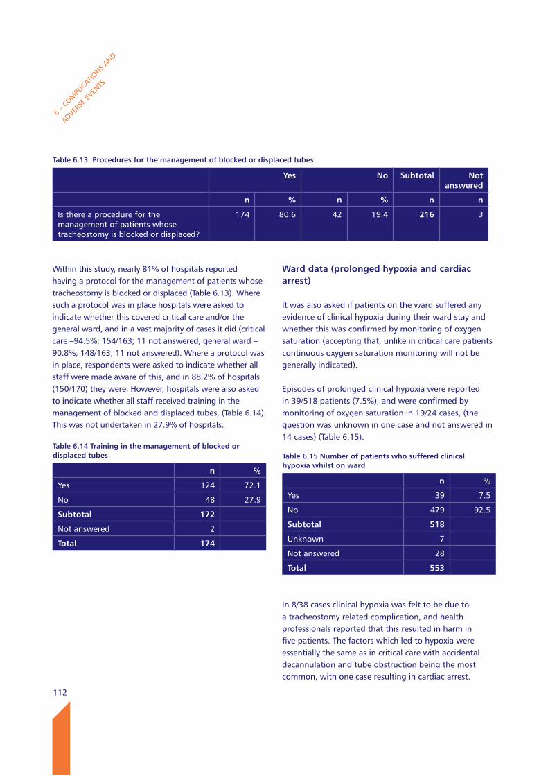
112
ward data (prolonged hypoxia and cardiac arrest)
It was also asked if patients on the ward suffered any evidence of clinical hypoxia during their ward stay and whether this was confirmed by monitoring of oxygen saturation (accepting that, unlike in critical care patients continuous oxygen saturation monitoring will not be generally indicated).
Episodes of prolonged clinical hypoxia were reported in 39/518 patients (7.5%), and were confirmed by monitoring of oxygen saturation in 19/24 cases, (the question was unknown in one case and not answered in 14 cases) (Table 6.15).
In 8/38 cases clinical hypoxia was felt to be due to a tracheostomy related complication, and health professionals reported that this resulted in harm in five patients. The factors which led to hypoxia were essentially the same as in critical care with accidental decannulation and tube obstruction being the most common, with one case resulting in cardiac arrest.
6 – C
OMPL
ICATIO
NS AND
ADVERSE
EVEN
TS
Within this study, nearly 81% of hospitals reported having a protocol for the management of patients whose tracheostomy is blocked or displaced (Table 6.13). Where such a protocol was in place hospitals were asked to indicate whether this covered critical care and/or the general ward, and in a vast majority of cases it did (critical care –94.5%; 154/163; 11 not answered; general ward – 90.8%; 148/163; 11 not answered). Where a protocol was in place, respondents were asked to indicate whether all staff were made aware of this, and in 88.2% of hospitals (150/170) they were. However, hospitals were also asked to indicate whether all staff received training in the management of blocked and displaced tubes, (Table 6.14). This was not undertaken in 27.9% of hospitals.
Table 6.13 Procedures for the management of blocked or displaced tubes
yes No Subtotal Not answered
n % n % n n
Is there a procedure for the management of patients whose tracheostomy is blocked or displaced?
174 80.6 42 19.4 216 3
Table 6.14 Training in the management of blocked or displaced tubes
n %
Yes 124 72.1
No 48 27.9
Subtotal 172
Not answered 2
Total 174
Table 6.15 Number of patients who suffered clinical hypoxia whilst on ward
n %
Yes 39 7.5
No 479 92.5
Subtotal 518
Unknown 7
Not answered 28
Total 553
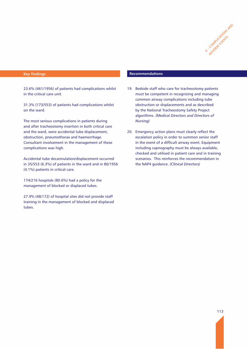
113
6 – C
OMPL
ICATIO
NS AND
ADVERSE
EVEN
TS
key findings
23.6% (461/1956) of patients had complications whilst in the critical care unit. 31.3% (173/553) of patients had complications whilst on the ward. The most serious complications in patients during and after tracheostomy insertion in both critical care and the ward, were accidental tube displacement, obstruction, pneumothorax and haemorrhage. Consultant involvement in the management of these complications was high. Accidental tube decannulation/displacement occurred in 35/553 (6.3%) of patients in the ward and in 80/1956 (4.1%) patients in critical care.
174/216 hospitals (80.6%) had a policy for the management of blocked or displaced tubes.
27.9% (48/172) of hospital sites did not provide staff training in the management of blocked and displaced tubes.
Recommendations
19. Bedside staff who care for tracheostomy patients must be competent in recognizing and managing common airway complications including tube obstruction or displacements and as described by the National Tracheostomy Safety Project algorithms. (Medical Directors and Directors of Nursing)
20. Emergency action plans must clearly reflect the escalation policy in order to summon senior staff in the event of a difficult airway event. Equipment including capnography must be always available, checked and utilised in patient care and in training scenarios. This reinforces the recommendation in the NAP4 guidance. (Clinical Directors)

114
APPEN
DICES
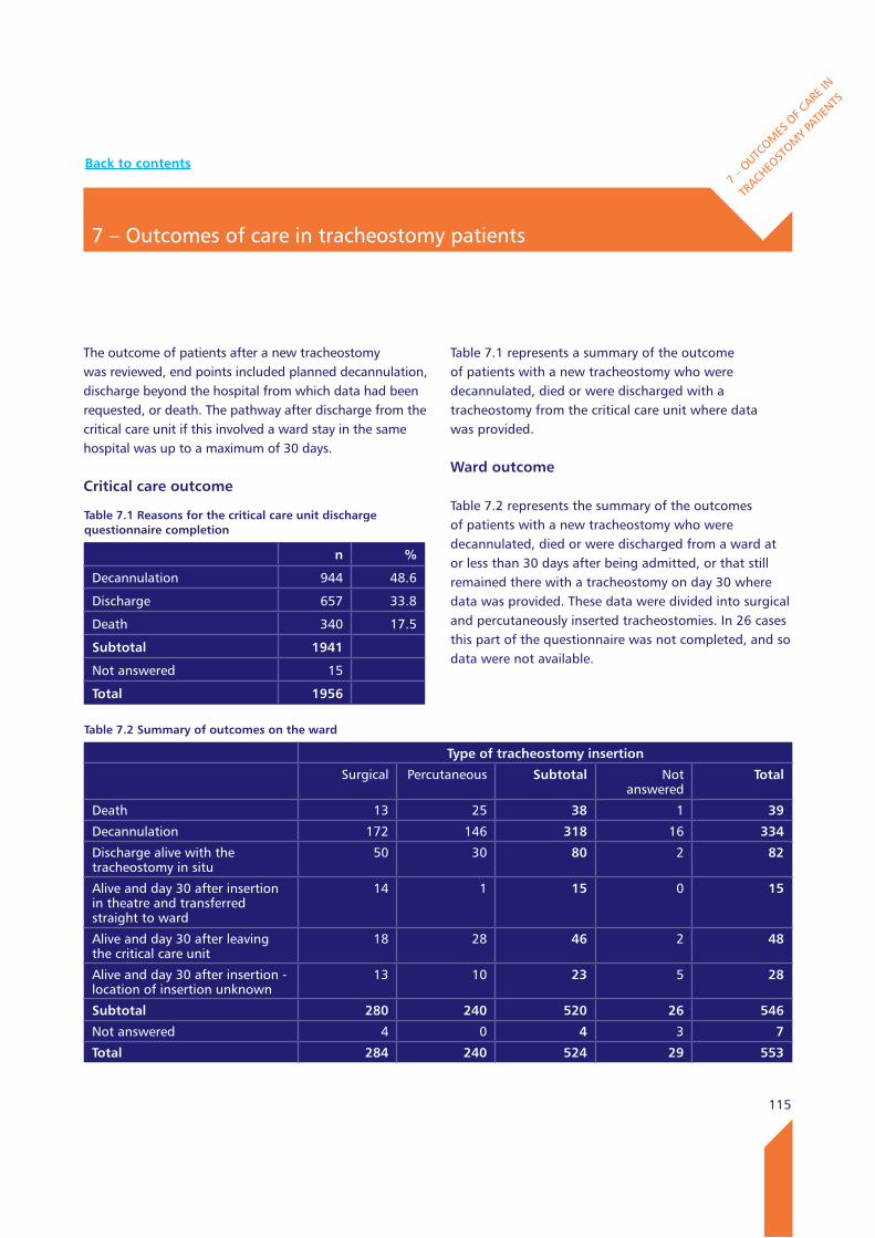
115
7 – Outcomes of care in tracheostomy patients
7 – O
UTCOM
ES O
F CARE
IN
TRACHEO
STOM
Y PA
TIENTS
The outcome of patients after a new tracheostomy was reviewed, end points included planned decannulation, discharge beyond the hospital from which data had been requested, or death. The pathway after discharge from the critical care unit if this involved a ward stay in the same hospital was up to a maximum of 30 days.
Critical care outcome
Table 7.1 represents a summary of the outcome of patients with a new tracheostomy who were decannulated, died or were discharged with a tracheostomy from the critical care unit where data was provided.
ward outcome
Table 7.2 represents the summary of the outcomes of patients with a new tracheostomy who were decannulated, died or were discharged from a ward at or less than 30 days after being admitted, or that still remained there with a tracheostomy on day 30 where data was provided. These data were divided into surgical and percutaneously inserted tracheostomies. In 26 cases this part of the questionnaire was not completed, and so data were not available.
Table 7.1 Reasons for the critical care unit discharge questionnaire completion
n %
Decannulation 944 48.6
Discharge 657 33.8
Death 340 17.5
Subtotal 1941
Not answered 15
Total 1956
Table 7.2 Summary of outcomes on the ward
Type of tracheostomy insertion
Surgical Percutaneous Subtotal Not answered
Total
Death 13 25 38 1 39
Decannulation 172 146 318 16 334
Discharge alive with the tracheostomy in situ
50 30 80 2 82
Alive and day 30 after insertion in theatre and transferred straight to ward
14 1 15 0 15
Alive and day 30 after leaving the critical care unit
18 28 46 2 48
Alive and day 30 after insertion - location of insertion unknown
13 10 23 5 28
Subtotal 280 240 520 26 546
Not answered 4 0 4 3 7
Total 284 240 524 29 553
Back to contents
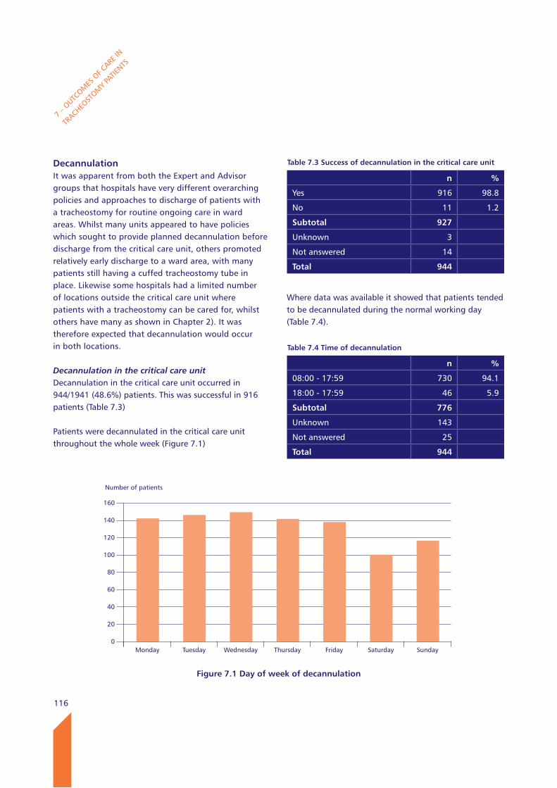
116
7 – O
UTCOM
ES O
F CARE
IN
TRACHEO
STOM
Y PA
TIENTS
DecannulationIt was apparent from both the Expert and Advisor groups that hospitals have very different overarching policies and approaches to discharge of patients with a tracheostomy for routine ongoing care in ward areas. Whilst many units appeared to have policies which sought to provide planned decannulation before discharge from the critical care unit, others promoted relatively early discharge to a ward area, with many patients still having a cuffed tracheostomy tube in place. Likewise some hospitals had a limited number of locations outside the critical care unit where patients with a tracheostomy can be cared for, whilst others have many as shown in Chapter 2). It was therefore expected that decannulation would occur in both locations.
Decannulation in the critical care unitDecannulation in the critical care unit occurred in 944/1941 (48.6%) patients. This was successful in 916 patients (Table 7.3)
Patients were decannulated in the critical care unit throughout the whole week (Figure 7.1)
Where data was available it showed that patients tended to be decannulated during the normal working day (Table 7.4).
Table 7.3 Success of decannulation in the critical care unit
n %
Yes 916 98.8
No 11 1.2
Subtotal 927
Unknown 3
Not answered 14
Total 944
Number of patients
160
140
120
100
80
60
40
20
0
Figure 7.1 Day of week of decannulation
Monday Tuesday Wednesday Thursday Friday Saturday Sunday
Table 7.4 Time of decannulation
n %
08:00 - 17:59 730 94.1
18:00 - 17:59 46 5.9
Subtotal 776
Unknown 143
Not answered 25
Total 944
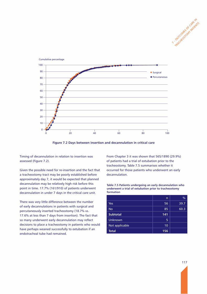
117
7 – O
UTCOM
ES O
F CARE
IN
TRACHEO
STOM
Y PA
TIENTS
Timing of decannulation in relation to insertion was assessed (Figure 7.2).
Given the possible need for re-insertion and the fact that a tracheostomy tract may be poorly established before approximately day 7, it would be expected that planned decannulation may be relatively high risk before this point in time. 17.7% (161/910) of patients underwent decannulation in under 7 days in the critical care unit.
There was very little difference between the number of early decannulations in patients with surgical and percutaneously inserted tracheostomy (18.7% vs. 17.6% at less than 7 days from insertion). The fact that so many underwent early decannulation may reflect decisions to place a tracheostomy in patients who would have perhaps weaned successfully to extubation if an endotracheal tube had remained.
From Chapter 3 it was shown that 565/1890 (29.9%) of patients had a trial of extubation prior to the tracheostomy. Table 7.5 summarises whether it occurred for those patients who underwent an early decannulation.
Cumulative percentage
100
90
80
70
60
50
40
30
20
10
0
Figure 7.2 Days between insertion and decannulation in critical care
0 20 40 60 80 100
Surgical
Percutaneous
Table 7.5 Patients undergoing an early decannulation who underwent a trial of extubation prior to tracheostomy formation
n %
Yes 56 39.7
No 85 60.3
Subtotal 141
Unknown 5
Not applicable 10
Total 156
118
7 – O
UTCOM
ES O
F CARE
IN
TRACHEO
STOM
Y PA
TIENTS
There were 85 patients in total who did not undergo a trial of extubation prior to tracheostomy formation and also underwent early decannulation (See Figure 3.8). Some of these cases were planned surgically inserted tracheostomies as part of a larger head and neck procedure and therefore a trial of extubation would not have been indicated. Of the 156 early decannulations, 38 patients had undergone a surgical tracheostomy and in 13 of these it was part of a planned head and neck procedure. All percutaneously inserted tracheostomies where early decannulation occurred were reviewed with respect to how many patients had had a trial of extubation prior to tracheostomy formation. This revealed that 48/116 patients had not undergone a trial. So of the 85 patients who did not have a trial of extubation the majority (68) were percutaneous. In some groups of patients it is known that a trial of extubation may have been regarded as unwarranted e.g. those who had suffered a serious head injury.
Whilst the decision about timing of tracheostomy is notoriously difficult and there are obvious advantages in terms of providing early tracheostomy in terms of greater patient comfort, and an expected reduction in sedation requirements, it is now known from Trachman data31,35 that early tracheostomy does not result in improved patient outcomes. Serious complications may arise from a tracheostomy and it important that it is not necessarily regarded as being a safer alternative to an endotracheal tube. In most patients admitted to critical care and requiring respiratory support the ultimate aim is still to make them independent of an artificial airway. Whilst patients can often be managed with a tracheostomy in a wider number of hospital locations, there must also be very good levels of competence and equipment available to do so safely.
Airway assessmentAirway assessment with or without airway endoscopy is recommended in preparation for decannulation.2,27 Most tracheostomies in critical care are carried out for weaning and patients do not have an abnormal upper airway per se. However upper airway endoscopy may be useful to check upper airway anatomy in preparation for decannulation to assess for e.g. granulation tissue at the tip of the tube and other potential causes of obstruction and/or bleeding once the tracheostomy tube is removed (Table 7.6).32 A functional assessment of the upper airway may also be particularly useful made by speech and language therapists to look at the patient’s ability to deal with saliva, and therefore to more accurately quantify aspiration risk.
In this study upper airway endoscopy was a procedure which was rarely undertaken, with less than 2% (16) patients having this assessment recorded. Even if an upper airway endoscopy is not performed it would be good practice to document that it has been considered. It is possible that this finding reflect the fact that it may only be routine to perform endoscopy if there is a doubt about the success of decannulation. Advisors commented that it may also be that this very low rate also reflects a lack of routine involvement of SLT at this point in the care pathway, and/or a lack of access to an appropriate (fine) fibrescope suitable for nasendoscopy in many critical care units. Despite this investigation not being carried out, the vast majority of decannulations in the critical care unit were successful.
Table 7.6 Airway endoscopy was performed prior to decannulation
n %
Yes 16 1.9
No 845 98.1
Subtotal 861
Unknown 65
Not answered 18
Total 944
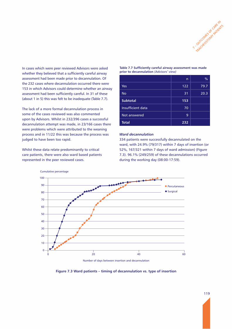
119
7 – O
UTCOM
ES O
F CARE
IN
TRACHEO
STOM
Y PA
TIENTS
In cases which were peer reviewed Advisors were asked whether they believed that a sufficiently careful airway assessment had been made prior to decannulation. Of the 232 cases where decannulation occurred there were 153 in which Advisors could determine whether an airway assessment had been sufficiently careful. In 31 of these (about 1 in 5) this was felt to be inadequate (Table 7.7).
The lack of a more formal decannulation process in some of the cases reviewed was also commented upon by Advisors. Whilst in 232/396 cases a successful decannulation attempt was made, in 23/166 cases there were problems which were attributed to the weaning process and in 11/22 this was because the process was judged to have been too rapid.
Whilst these data relate predominantly to critical care patients, there were also ward based patients represented in the peer reviewed cases.
Ward decannulation334 patients were successfully decannulated on the ward, with 24.9% (79/317) within 7 days of insertion (or 52%, 167/321 within 7 days of ward admission) (Figure 7.3). 96.1% (249/259) of these decannulations occurred during the working day (08:00-17:59).
Table 7.7 Sufficiently careful airway assessment was made prior to decannulation (Advisors’ view)
n %
Yes 122 79.7
No 31 20.3
Subtotal 153
Insufficient data 70
Not answered 9
Total 232
Cumulative percentage
100
90
80
70
60
50
40
30
20
10
0
Figure 7.3 ward patients – timing of decannulation vs. type of insertion
0 20 40 60
Percutaneous
Surgical
Number of days between insertion and decannulation
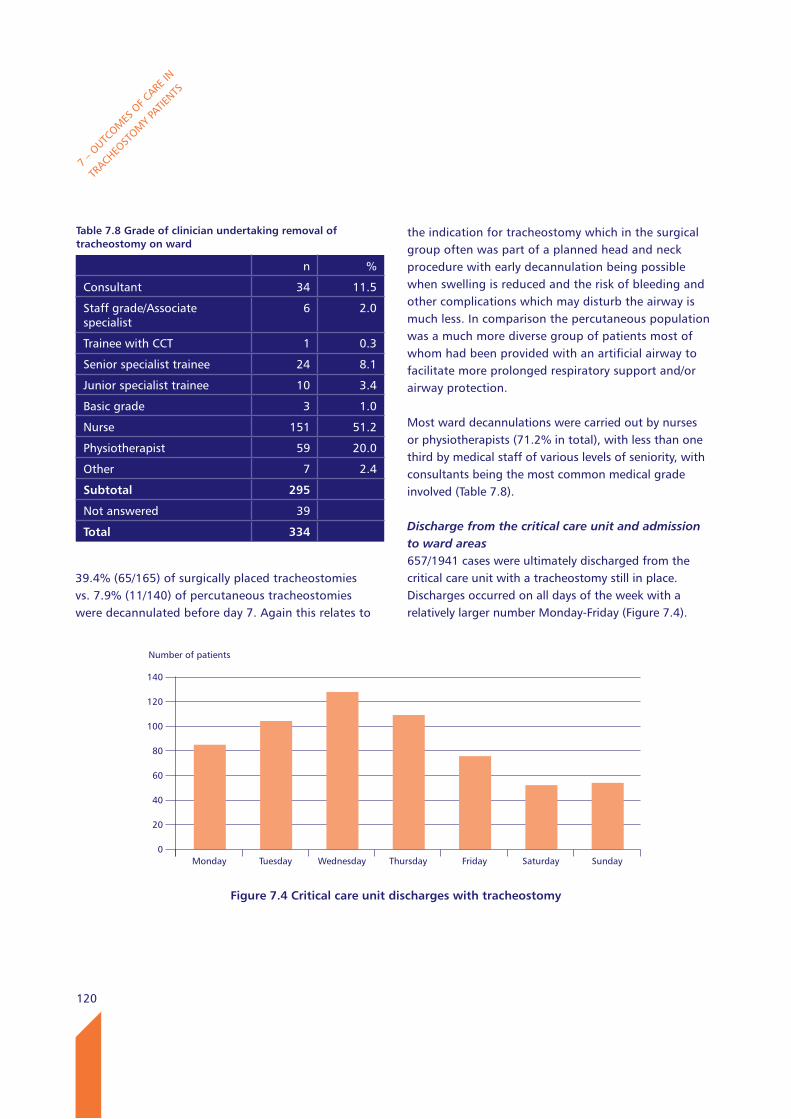
120
7 – O
UTCOM
ES O
F CARE
IN
TRACHEO
STOM
Y PA
TIENTS
39.4% (65/165) of surgically placed tracheostomies vs. 7.9% (11/140) of percutaneous tracheostomies were decannulated before day 7. Again this relates to
the indication for tracheostomy which in the surgical group often was part of a planned head and neck procedure with early decannulation being possible when swelling is reduced and the risk of bleeding and other complications which may disturb the airway is much less. In comparison the percutaneous population was a much more diverse group of patients most of whom had been provided with an artificial airway to facilitate more prolonged respiratory support and/or airway protection.
Most ward decannulations were carried out by nurses or physiotherapists (71.2% in total), with less than one third by medical staff of various levels of seniority, with consultants being the most common medical grade involved (Table 7.8).
Discharge from the critical care unit and admission to ward areas657/1941 cases were ultimately discharged from the critical care unit with a tracheostomy still in place. Discharges occurred on all days of the week with a relatively larger number Monday-Friday (Figure 7.4).
Table 7.8 Grade of clinician undertaking removal of tracheostomy on ward
n %
Consultant 34 11.5
Staff grade/Associate specialist
6 2.0
Trainee with CCT 1 0.3
Senior specialist trainee 24 8.1
Junior specialist trainee 10 3.4
Basic grade 3 1.0
Nurse 151 51.2
Physiotherapist 59 20.0
Other 7 2.4
Subtotal 295
Not answered 39
Total 334
Number of patients
140
120
100
80
60
40
20
0
Figure 7.4 Critical care unit discharges with tracheostomy
Monday Tuesday Wednesday Thursday Friday Saturday Sunday
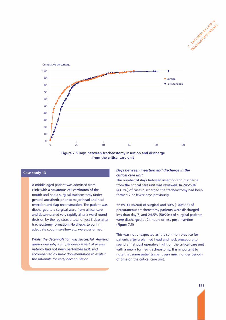
121
7 – O
UTCOM
ES O
F CARE
IN
TRACHEO
STOM
Y PA
TIENTS
Case study 13
A middle aged patient was admitted from clinic with a squamous cell carcinoma of the mouth and had a surgical tracheostomy under general anesthetic prior to major head and neck resection and flap reconstruction. The patient was discharged to a surgical ward from critical care and decannulated very rapidly after a ward round decision by the registrar, a total of just 3 days after tracheostomy formation. No checks to confirm adequate cough, swallow etc. were performed.
Whilst the decannulation was successful, Advisors questioned why a simple bedside test of airway patency had not been performed first, and accompanied by basic documentation to explain the rationale for early decannulation.
Days between insertion and discharge in the critical care unitThe number of days between insertion and discharge from the critical care unit was reviewed. In 245/594 (41.2%) of cases discharged the tracheostomy had been formed 7 or fewer days previously.
56.6% (116/204) of surgical and 30% (100/333) of percutaneous tracheostomy patients were discharged less than day 7, and 24.5% (50/204) of surgical patients were discharged at 24 hours or less post insertion (Figure 7.5)
This was not unexpected as it is common practice for patients after a planned head and neck procedure to spend a first post operative night on the critical care unit with a newly formed tracheostomy. It is important to note that some patients spent very much longer periods of time on the critical care unit.
Cumulative percentage
100
90
80
70
60
50
40
30
20
10
0
Figure 7.5 Days between tracheostomy insertion and discharge from the critical care unit
0 20 40 60 80 100
Surgical
Percutaneous
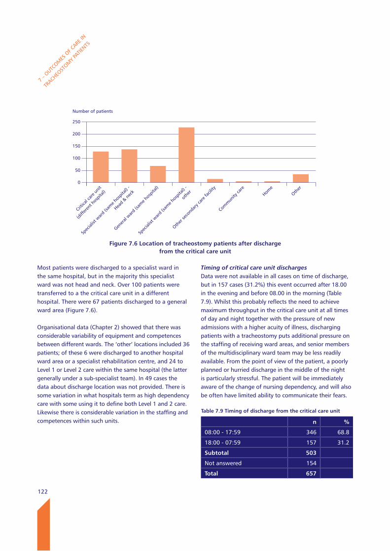
122
7 – O
UTCOM
ES O
F CARE
IN
TRACHEO
STOM
Y PA
TIENTS
Number of patients
250
200
150
100
50
0
Figure 7.6 location of tracheostomy patients after discharge from the critical care unit
Critica
l car
e unit
(diff
eren
t hos
pital)
Spec
ialist
war
d (sa
me h
ospit
al) -
Head
& nec
k
Gener
al war
d (sa
me h
ospit
al)
Spec
ialist
war
d (sa
me h
ospit
al) -
othe
r
Other
seco
ndar
y car
e fac
ility
Comm
unity
care
Home
Other
Most patients were discharged to a specialist ward in the same hospital, but in the majority this specialist ward was not head and neck. Over 100 patients were transferred to a the critical care unit in a different hospital. There were 67 patients discharged to a general ward area (Figure 7.6).
Organisational data (Chapter 2) showed that there was considerable variability of equipment and competences between different wards. The ‘other’ locations included 36 patients; of these 6 were discharged to another hospital ward area or a specialist rehabilitation centre, and 24 to Level 1 or Level 2 care within the same hospital (the latter generally under a sub-specialist team). In 49 cases the data about discharge location was not provided. There is some variation in what hospitals term as high dependency care with some using it to define both Level 1 and 2 care. Likewise there is considerable variation in the staffing and competences within such units.
Table 7.9 Timing of discharge from the critical care unit
n %
08:00 - 17:59 346 68.8
18:00 - 07:59 157 31.2
Subtotal 503
Not answered 154
Total 657
Timing of critical care unit dischargesData were not available in all cases on time of discharge, but in 157 cases (31.2%) this event occurred after 18.00 in the evening and before 08.00 in the morning (Table 7.9). Whilst this probably reflects the need to achieve maximum throughput in the critical care unit at all times of day and night together with the pressure of new admissions with a higher acuity of illness, discharging patients with a tracheostomy puts additional pressure on the staffing of receiving ward areas, and senior members of the multidisciplinary ward team may be less readily available. From the point of view of the patient, a poorly planned or hurried discharge in the middle of the night is particularly stressful. The patient will be immediately aware of the change of nursing dependency, and will also be often have limited ability to communicate their fears.
123
There were a total of 157 discharges at or after 18.00 and before 08.00. Table 7.10 provides a summary of discharge location.
The additional need for discharge to a ward with the correct equipment and competences has already been noted, and the undesirability of night time discharges from the critical care unit is addressed in generic ICS standards for organisation of care from 2013.33
Of the 20/155 patients transferred to a critical care unit in another hospital, 6 were between the hours of 21.00 and 06.00 in the morning.
7 – O
UTCOM
ES O
F CARE
IN
TRACHEO
STOM
Y PA
TIENTS
Table 7.10 Destination after the critical care unit discharge
n %
Critical care unit (different hospital)
20 12.9
Specialist ward - head and neck (same hospital)
26 16.8
General ward (same hospital) 21 13.5
Specialist ward - other (same hospital)
74 47.7
Other secondary care facility 1 0.6
Other 13 8.4
Subtotal 155
Not answered 2
Total 157
Table 7.11 Out of hours discharge to a location designated for patients with tracheostomy
n %
Yes 151 96.8
No 5 3.2
Subtotal 156
Not answered 1
Total 157
There were 5/156 patients discharged out of hours from the critical care unit to locations which were not designated to provide routine tracheostomy care (Table 7.11). Whilst it was felt that in the vast majority of cases the discharge location was appropriate, in nine cases concerns was expressed about care. Free field comments about these concerns were given in 4/9 cases with concern about staff training, numbers and/or risk assessment for tracheostomy care being the overwhelming theme.
Clinicians on the critical care unit were asked whether a discharge summary formed part of the record which accompanied the patient. Whilst in 90.9% of cases (541/595) it was provided, in 9.1% (54/595) it was not and in many it did not contain several important elements which would be expected to facilitate a high standard of ongoing tracheostomy care.
In particular clinicians themselves indicated that the following details were included in only a minority of summaries: • Carerequirementsforthetracheostomy – included in 35.3% (174/494)• Followupplanforthetracheostomy – included in 31.5% (155/493)• Weaningplanforthetracheostomy – included in 26.6% (131/493)• Whotocontactifproblemswiththetracheostomy
– included in 31.8% (158/497)• Responsibilityfordecisionsaboutthetracheostomy
– included in 25.0% (122/488)
124
7 – O
UTCOM
ES O
F CARE
IN
TRACHEO
STOM
Y PA
TIENTS
From the critical care unit discharge questionnaire it is known that 568 patients were discharged to a further ward area. Whilst not all these discharges would have prompted the completion of a subsequent ward questionnaire NCEPOD received 553 ward questionnaires and data on type of insertion was completed in 524, with 240 percutaneous and 284 surgically inserted tracheostomies (Table 7.12). These patients were a mixture from both the critical care unit (the majority) or the operating theatre.
Admission to ward careWhere data was available from ward questionnaires 165/348 (47.4%) admissions were after 18.00 and before 08.00 (Table 7.13). Whilst some wards had specialist head and neck facilities with a high level of dependency and enhanced competencies in care of tracheostomy, some patients went to general wards. Seventy-three patients were admitted between 21:00 – 06:00.
It was questioned as to whether comprehensive risk assessment(s) relating to the care of the tracheostomy were undertaken on the patient before admission (Table 7.14).
Table 7.13 Time of admission to ward
n %
08:00 - 17:59 219 57.0
18:00 - 07:59 165 43.0
Subtotal 384
Unknown 126
Not answered 43
Total 553
Table 7.14 Risk assessments carried out before ward admission
n %
Yes 341 73.2
No 125 26.8
Subtotal 466
Unknown 68
Not answered 19
Total 553
Case study 14
An elderly patient who previously lived independently had several falls and was admitted to hospital with a chest infection. This was initially managed with non invasive ventilation, before intubation and ventilation. The patient had a percutaneous tracheostomy 5 days after intubation, was weaned and decannulated rapidly and sent to a ward 2 days later. Over the next 2 weeks the patient’s chest deteriorated and they died on the ward without any clear discussion about re-escalation of care.
Advisors commented that they felt care had effectively been withdrawn and that the patient was being allowed to die. After initial aggressive and successful treatment this was not easy to understand without a clear documented rationale or plan in the notes.
Table 7.12 Type of insertion of tracheostomy-patients discharged to ward
n %
Surgical 284 54.2
Percutaneous 240 45.8
Subtotal 524
Not answered 29
Total 553
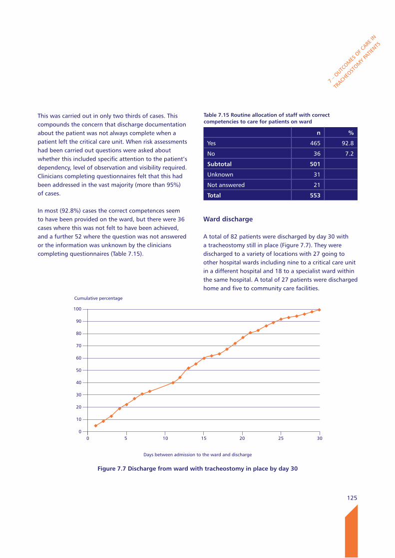
125
7 – O
UTCOM
ES O
F CARE
IN
TRACHEO
STOM
Y PA
TIENTS
This was carried out in only two thirds of cases. This compounds the concern that discharge documentation about the patient was not always complete when a patient left the critical care unit. When risk assessments had been carried out questions were asked about whether this included specific attention to the patient’s dependency, level of observation and visibility required. Clinicians completing questionnaires felt that this had been addressed in the vast majority (more than 95%) of cases.
In most (92.8%) cases the correct competences seem to have been provided on the ward, but there were 36 cases where this was not felt to have been achieved, and a further 52 where the question was not answered or the information was unknown by the clinicians completing questionnaires (Table 7.15).
ward discharge
A total of 82 patients were discharged by day 30 with a tracheostomy still in place (Figure 7.7). They were discharged to a variety of locations with 27 going to other hospital wards including nine to a critical care unit in a different hospital and 18 to a specialist ward within the same hospital. A total of 27 patients were discharged home and five to community care facilities.
Table 7.15 Routine allocation of staff with correct competencies to care for patients on ward
n %
Yes 465 92.8
No 36 7.2
Subtotal 501
Unknown 31
Not answered 21
Total 553
Cumulative percentage
100
90
80
70
60
50
40
30
20
10
0
Figure 7.7 Discharge from ward with tracheostomy in place by day 30
Days between admission to the ward and discharge
0 5 10 15 20 25 30
126
7 – O
UTCOM
ES O
F CARE
IN
TRACHEO
STOM
Y PA
TIENTS
Eleven ward discharges occurred after 18.00 in the evening and before 08.00 in the morning. This would be a particular concern in relation to those patients discharged home as GP services and community nursing would not generally have been so readily available. The timing of discharge and locations of the 11 patients who left the ward were reviewed (Table 7.16).
These data are important, and other authors have highlighted the risks of managing tracheostomy beyond the hospital setting.34 Whilst moving patients around the same Trust is necessary on occasion (e.g. to provide barrier nursing or highly specialist services such as dialysis), this can be highly disruptive to care, especially when it occurs at night/in the very early morning. It is also very stressful to the patient. Some of these discharges would have been arranged at very short notice, and this may reduce the ability to provide a smooth pathway of care for the patient e.g. clear and detailed handover, correct competences in receiving area for tracheostomy care, and correct equipment being available. Moving patients to the community did not tend to occur as late in the day but would still have posed problems if there had been a need to discuss with services such as GPs and community based nursing staff.
Ward based clinicians were asked if they had concerns about the discharge and in the majority (69/73) this was not the case. However there were four patients where concern was expressed and this related to the ability of the part of the discharge location to provide routine tracheostomy care (n=2), and to recognize (n=1) and manage (n=1) tracheostomy complications.
Patients on the ward at day 30
A total of 91 patients remained on the ward with a tracheostomy still in place at day 30 after insertion. The reasons for this are outlined in Table 7.17. Whilst many had an ongoing need for secondary care, in a total of 29 instances there were difficulties in securing adequate rehabilitation and/or community care facilities. In just 41/67 there was a plan for discharge in place.
Advisors commented that the competences for tracheostomy care are not widely available in the community, outside of selected settings. At this stage there is a need on the ward for continued multidisciplinary team input to provide co-ordinated discharge planning and provide a comprehensive package of care.
Table 7.16 Tracheostomy in place-discharge time (out of hours) and location from ward areas
n
Critical care unit (different hospital) 1
General ward (same hospital) 1
Specialist ward (same hospital) - other 6
Other secondary care facility 1
Home 2
Total 11 Table 7.17 Reasons for patients with tracheostomy being still on ward at day 30 after insertion (Answers may be multiple)
n
Ongoing need for secondary medical care 45
Difficulties in securing appropriate community care
14
Difficulties in finding a specialist rehabilitation unit
16
Other 9
Subtotal 84
Not answered 20
127
7 – O
UTCOM
ES O
F CARE
IN
TRACHEO
STOM
Y PA
TIENTS
Death in the critical care unit
340 patients died in the critical care unit with a tracheostomy still in place. The majority of these deaths were expected by the clinicians completing the critical care questionnaire (Table 7.18). However there were 35 cases of unexpected deaths on the critical care unit, and of these three were felt to relate directly to a tracheostomy related complication.
Deaths on the ward
Thirty-nine patients died before day 30 on the ward with a tracheostomy in place. Of these three were felt to be unexpected. One of these was attributed to a tracheostomy related complication.
There were 284 surgical and 240 percutaneous tracheostomy patients cared for on the ward so there was a slight excess of deaths in the percutaneous tracheostomy group (Table 7.19). This can be explained by case mix, with the percutaneous group including mainly patients who had been admitted as an emergency (89.3%), whereas the surgical group contained a relatively large number of patients (31.9%) who had a tracheostomy as part of a planned head and neck procedure.
Advisor data on deaths
In those 396 cases from both critical care and the ward reviewed by Advisors, 80 deaths were identified in clinical notes. Of these there were three deaths which were identified as a result of a tracheostomy related complication, and of these two were felt to be potentially avoidable. Some of the findings from these cases are presented throughout this report but have not been presented in full to avoid loss of anonymisation.
Case study 15
A young patient had a major stroke and needed airway support/protection. After initial intubation a percutaneous tracheostomy was performed on critical care to facilitate ongoing needs. Whilst the patient received good SLT and physiotherapy input on the ward to which they were discharged, there were several problems with humidification during the ward stay. Ultimately the patient was prepared for discharge to a nursing home and there was evidence of good levels of training of receiving staff.
Advisors commented upon the general lack of provision for such training in many parts of the country which often caused major delays in hospital discharge.
Table 7.18 Death in the critical care unit
n %
Expected 290 89.2
Not expected 35 10.8
Subtotal 325
Not answered 15
Total 340
Table 7.19 Deaths in ward surgical vs. percutaneous
n
Surgical 13
Percutaneous 25
Subtotal 38
Not answered 1
Total 39
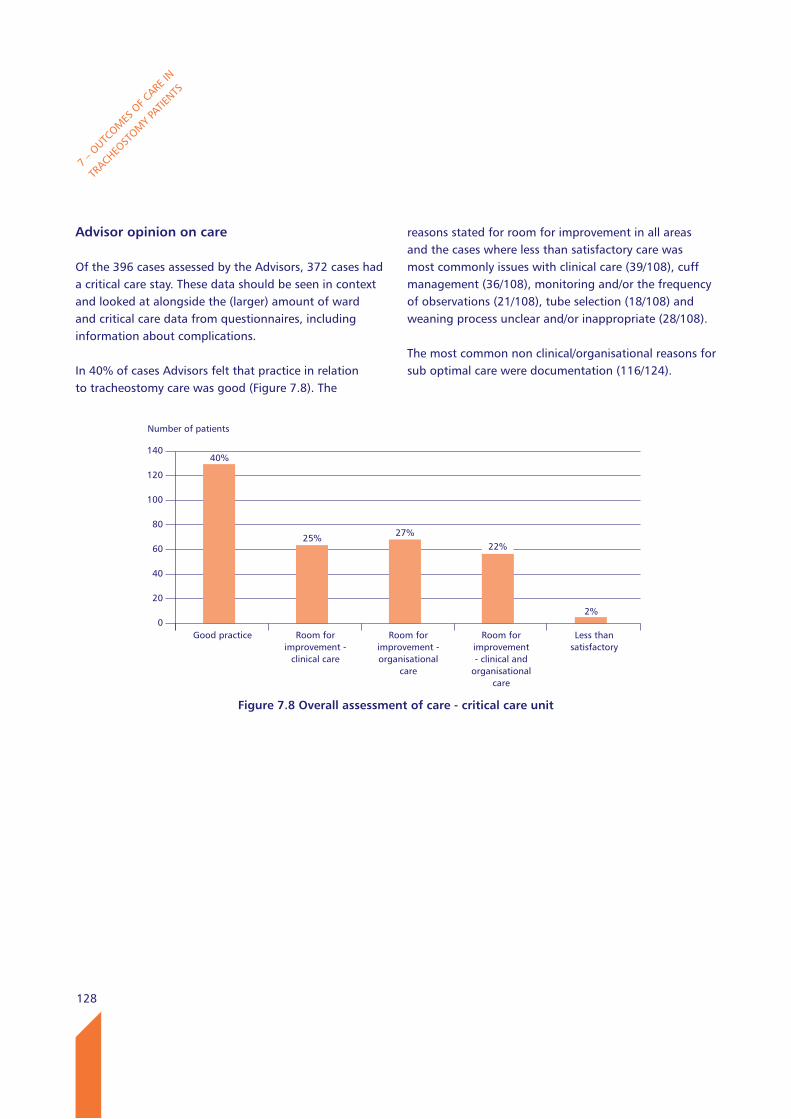
128
7 – O
UTCOM
ES O
F CARE
IN
TRACHEO
STOM
Y PA
TIENTS
Advisor opinion on care
Of the 396 cases assessed by the Advisors, 372 cases had a critical care stay. These data should be seen in context and looked at alongside the (larger) amount of ward and critical care data from questionnaires, including information about complications.
In 40% of cases Advisors felt that practice in relation to tracheostomy care was good (Figure 7.8). The
reasons stated for room for improvement in all areas and the cases where less than satisfactory care was most commonly issues with clinical care (39/108), cuff management (36/108), monitoring and/or the frequency of observations (21/108), tube selection (18/108) and weaning process unclear and/or inappropriate (28/108).
The most common non clinical/organisational reasons for sub optimal care were documentation (116/124).
Number of patients
140
120
100
80
60
40
20
0
Figure 7.8 Overall assessment of care - critical care unit
Good practice Room for improvement -
clinical care
Room for improvement - organisational
care
Room for improvement - clinical and
organisationalcare
Less than satisfactory
40%
25% 27%22%
2%
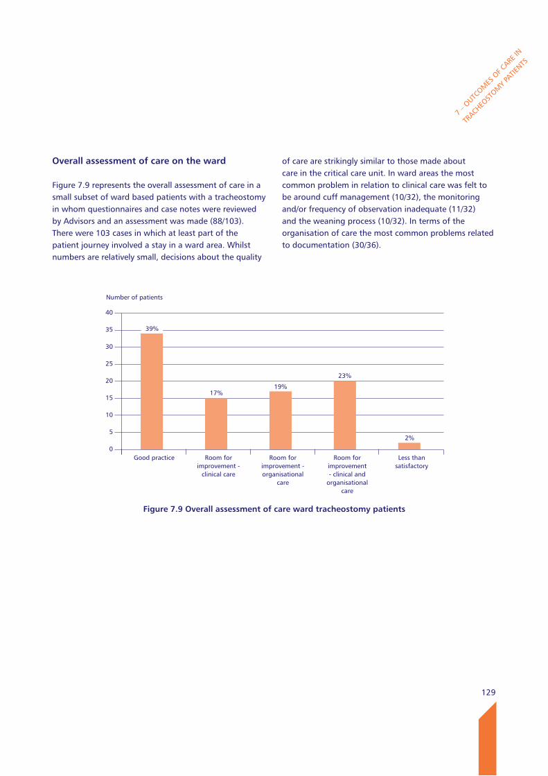
129
7 – O
UTCOM
ES O
F CARE
IN
TRACHEO
STOM
Y PA
TIENTS
Overall assessment of care on the ward
Figure 7.9 represents the overall assessment of care in a small subset of ward based patients with a tracheostomy in whom questionnaires and case notes were reviewed by Advisors and an assessment was made (88/103). There were 103 cases in which at least part of the patient journey involved a stay in a ward area. Whilst numbers are relatively small, decisions about the quality
of care are strikingly similar to those made about care in the critical care unit. In ward areas the most common problem in relation to clinical care was felt to be around cuff management (10/32), the monitoring and/or frequency of observation inadequate (11/32) and the weaning process (10/32). In terms of the organisation of care the most common problems related to documentation (30/36).
Number of patients
40
35
30
25
20
15
10
5
0
Figure 7.9 Overall assessment of care ward tracheostomy patients
Good practice Room for improvement -
clinical care
Room for improvement - organisational
care
Room for improvement - clinical and
organisationalcare
Less than satisfactory
39%
17%19%
23%
2%
130
7 – O
UTCOM
ES O
F CARE
IN
TRACHEO
STOM
Y PA
TIENTS
key findings
18% (161/910) of patients underwent decannulation in under 7 days in the critical care unit.
85/141 patients who had an early decannulation did not undergo a trial of extubation before tracheostomy insertion. 68 of these were percutaneous insertions.
157/503 discharges of patients from the critical care unit occurred after 18.00 in the evening and before 08.00 in the morning. 165/348 (47.4%) ward admissions occurred after 18.00 and before 08.00.
46 patients were discharged from a critical care unit to a ward or different critical care unit area after 21.00 at night and before 06.00 in the morning. 5/156 patients were discharged out of hours from the critical care unit to locations which were not designated to provide routine tracheostomy care.
341/466 (73.2%) of patients had a comprehensive risk assessment carried out prior to ward admission. 90.9% (541/595) of patients had a discharge summary provided when they left the critical care unit, but 460/541 (85%) summaries did not contain several important elements such as weaning plans for the tracheostomy and who had responsibility for decisions about the tracheostomy.
27 patients were discharged home from a ward area and 5 to community care facilities.
Discharge from ward areas to other hospital locations and to community care occurred outside the normal working day in 11 cases.
Recommendations
21. In patients undergoing a tracheostomy without a trial of extubation the reason should be clearly documented. (All Health Care Professionals)
22. Unplanned and night time critical care discharge is not recommended, particularly in patients with a newly formed tracheostomy and/or patients recently weaned from respiratory support. This reinforces the Intensive Care Society’s general recommendation about night time discharges. (Clinical Directors and Risk Managers)
23. Wards accepting tracheostomy patients should be in a state of readiness in terms of equipment and competences. (Clinical Directors and Directors of Nursing)
24. Multidisciplinary agreement about minimum airway assessments prior to decannulation needs to be established including availability of equipment and competences. (Professional Health Care Bodies)
25. Quality of discharge documentation should be improved. A structured and detailed summary must be provided between wards and between hospitals and the community at the point of transfer. (All Health Care Professionals and Tracheostomy Leads)
131
This study looked at the pathway of tracheostomy care for patients beginning with a surgical or percutaneous insertion, followed in most by a stay on critical care and/or up to 30 days on a hospital ward. Patients with a new tracheostomy are a high risk population, not just because of potential airway complications but because most have associated major co-morbidity.
At the outset NCEPOD was unable to use existing OPCS codes to provide an accurate estimate of numbers of all new tracheostomies, as only those inserted surgically tend to be coded. An estimate of between 5,000 to 15,000 was made which provided a challenge to our existing method and to everyone taking part in this study. An important lesson for the future is that in order to facilitate care planning and ongoing review of tracheostomy care, both locally and nationally, there is a need for routine coding and data collection to occur for all tracheostomy insertions.
There are many recommended improvements in care at all steps of the patient pathway in this report, some of which do not involve additional expenditure. This includes the use of checklists for tracheostomy insertion which should be performed for patients in intensive care. These checks ensure that preparation for the tracheostomy is equivalent to those performed as a surgical procedure.
There are other opportunities to improve quality of care and reduce complications if hospitals ensure that adequate equipment is available to care for patients with tracheostomies in intensive care and ward areas, including fibreoptic scopes, difficult intubation kit, and capnography. Whilst this recommendation has been made by other authors, NCEPOD has found that in many centres such equipment is still not readily available or in use.
Before insertion and at every tube change tracheostomy tubes need to be selected according to patient anatomy and the position checked to provide as good a “fit” as possible, whilst selecting equipment with essential safety features such as inner cannulae which help prevent unnecessary serious complications. More serious complications arise during the after care of tracheostomies than at insertion, and attending staff must be able to deal with blocked and displaced tubes. When a patient with a tracheostomy requires resuscitation, there is also a need for staff to be able to use correct airway management techniques in patients with both a simple tracheostomy and post laryngectomy.
The involvement of a large multidisciplinary team of nurses, physiotherapists, physicians and surgeons, speech and language therapists and dietitians is essential for the good aftercare of patients with a tracheostomy, and they should be present in a timely fashion. Adequate numbers of these support staff are required to ensure this happens for each and every patient. Discharge arrangements when transferring patients from critical care, and from wards to the community need to include concise but adequate documentation, with good handover in daylight hours to suitably trained staff in the receiving area.
Summary
SUM
MARY
Back to contents

132
133
References
REFE
RENCES
1. UK National Tracheostomy Safety Project. www.tracheostomy.org.uk.
2. Intensive Care Society. Standards for the care of adult patients with temporary tracheostomy. 2008
3. Thomas AN and McGrath BA. Patient safety incidents associated with airway devices in critical care: a review of reports to the UK National Patient Safety Agency. Anaesthesia 2009; 64(4): 358-65
4. Cook TM, Woodall N, Harper J, Benger J; Fourth National Audit Project. Major complications of airway management in the UK: results of the Fourth National Audit Project of the Royal College of Anaesthetists and the Difficult Airway Society. Part 2: intensive care and emergency departments. Br J Anaesth 2011; 106: 632-642
5. Young D, Harrison DA and Cuthbertson BH et al. Effect of early vs late tracheostomy placement on survival in patients receiving mechanical ventilation: the TracMan randomized trial. TracMan Collaborators. JAMA 2013; 309(20): 2121-9
6. The Global Tracheostomy Collaborative. www.http://globaltrach.org/
7. Caring for the Patient with a Tracheostomy Best Practice Statement. 2007 NHS Quality Improvement Scotland http://www.healthcareimprovementscotland.org/previous_resources/best_practice_statement/tracheostomy_care.aspx
8. National Institute for Health and Care Excellence http://guidance.nice.org.uk/MT/158
9. Speech and Language Therapy in Adult Critical Care - Position Paper. Royal College of Speech and Language Therapy. 2006 http://www.rcslt.org/members/publications/publications2/provision_for_critical_care_
10. McGrath BA and Thomas AN. Patient safety incidents associated with tracheostomies occurring in hospital wards: a review of reports to the UK National Patient Safety Agency. Post Graduate Journal 2010; 86: 522-5
11. Heyrosa MG Melniczek DM and Rovito P et al Percutaneous tracheostomy: a safe procedure in
the morbidly obese. J Am Coll Surg 2006; 202(4): 618-22
12. El Solh AA and Jaafar W. A comparative study of the complications of surgical tracheostomy in morbidly obese critically ill patients. Crit Care 2007; 11(1): R3
13. Gomes Silva BN, Andriolo RB and Saconato H et al. Early versus late tracheostomy for critically ill patients. Cochrane Database Syst Rev 2012; 3
14. Trouillet J-L,Luyt C-E and Guiguet M et al. Early percutaneous tracheotomy versus prolonged intubation of mechanically ventilated patients after cardiac surgery: a randomized trial. Ann Intern Med 2011; 154(6): 373-83
15. Zheng Y, Sui F and Chen XK et al. Early versus late percutaneous dilatational tracheostomy in critically ill patients anticipated requiring prolonged mechanical ventilation. Chin Med J 2012; 125(11): 1925-30
Back to contents
134
REFE
RENCES
16. Denney L and White S. Consent, assent and the importance of risk stratification. Br. J. Anaesth 2012; 109(1): 40-46
17. Rudas M and Seppelt I. Safety and efficacy of ultrasonography before and during percutaneous dilatational tracheostomy in adult patients: a systematic review. Crit Care Resusc 2012; 14(4): 297-301
18. Jackson LS, Davis JW and Kaups KL et al. Percutaneous tracheostomy: to bronch or not to bronch - that is the question. J. Trauma 2011; 71(6): 1553-6
19. Sarkar S, Kelly A and Townsend R. Survey of percutaneous tracheostomy practice in UK intensive care units. JICS 2009; 10(2): 138-40
20. Garrubba M, Turner T and Grieveson C. Multidisciplinary care for tracheostomy patients: a systematic review. Crit Care 2009; 13: R177
21. Rabia Khalaila R, Zbidat W and Anwar K et al. Communication difficulty and Psychoemotional distress in patients receiving mechanical ventilation. Am J Crit Care 2011; 20: 470-9
22. Patak L, Gawlinski A and Fung NI et al. Patient’s reports of health care practitioner interventions related to communication during mechanical ventilation. Heart and Lung. The Journal of Acute and Critical Care 2004; 33 (5): 308-320
23. Tracheostomy competences. Royal College of Speech and language therapists. www.rcslt.org
24. Shah RK, Lander L and Berry JG at al. Tracheostomy outcomes and complications: a national perspective Laryngoscope 2012; 122: 25-29
25. Glossop AJ, Meekings TC and Hutchinson SP et al. Complications following tracheostomy insertion in critically ill patients-experience from a large teaching hospital. J ICS 2011; 12: 301-6
26. Dempsey GA, Grant CA and Jones TM. et al Percutaneous tracheostomy: a 6 year prospective evaluation of the single tapered dilator technique BJA 2010; 6: 782-6
27. Tracheostomy Guidelines. St. Georges Healthcare NHS Trust. https://www.stgeorges.nhs.uk/gps-and-clinicians/clinical-resources/tracheostomy-guidelines/ (accessed 15/03/2014)
28. El Solh AA and Jaafar W. A comparative study of the complications of surgical tracheostomy in morbidly obese critically ill patients. Crit Care 2007; 11: R3
29. Hogue CW, Stearns JD and Colantuoni E et al. The impact of obesity on outcomes after critical illness: a meta-analysis. ICM 2009; 35: 1152-1170
30. Henderson JJ, Popat MT and Latto IP et al. Difficult Airway Society. Guidelines for management of the unanticipated difficult intubation Anaesthesia 2004; 59: 675-694
31. Angus DC. When should a mechanically ventilated patient undergo a tracheostomy? JAMA 2013; 309: 2163-4
32. Hales PA, Drinnan MJ and Wilson JA. The added value of fibreoptic endoscopic evaluation of swallowing in tracheostomy weaning. Clin Otolaryngol 2008; 33(4): 319-24
33. Core standards for Intensive Care Units. The Intensive are Society and the Faculty of Intensive Care Medicine. 2013 www.ics.ac.uk
34. Eibling D and Roberson D. Managing tracheostomy risk: time to look beyond hospital discharge. Laryngoscope 2012; 122: 23-24
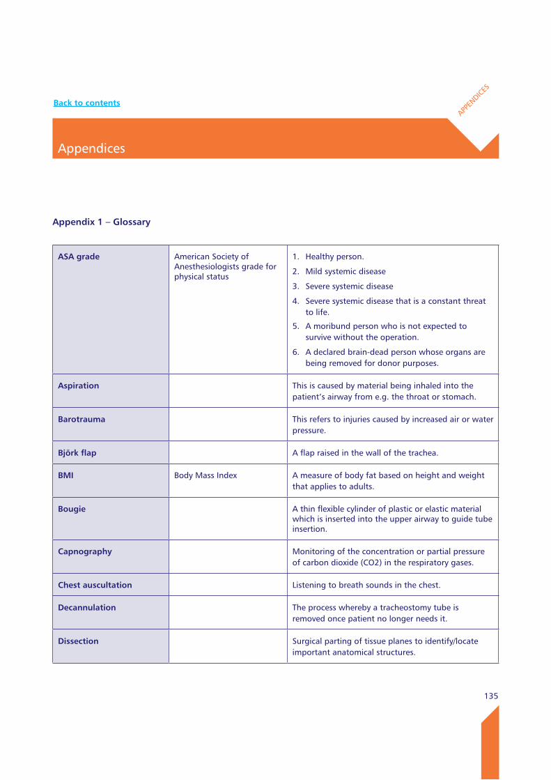
135
Appendix 1 – Glossary
Appendices
APPEN
DICES
ASA grade American Society of Anesthesiologists grade for physical status
1. Healthy person.
2. Mild systemic disease
3. Severe systemic disease
4. Severe systemic disease that is a constant threat to life.
5. A moribund person who is not expected to survive without the operation.
6. A declared brain-dead person whose organs are being removed for donor purposes.
Aspiration This is caused by material being inhaled into the patient’s airway from e.g. the throat or stomach.
Barotrauma This refers to injuries caused by increased air or water pressure.
Björk flap A flap raised in the wall of the trachea.
BMI Body Mass Index A measure of body fat based on height and weight that applies to adults.
Bougie A thin flexible cylinder of plastic or elastic material which is inserted into the upper airway to guide tube insertion.
Capnography Monitoring of the concentration or partial pressure of carbon dioxide (CO2) in the respiratory gases.
Chest auscultation Listening to breath sounds in the chest.
Decannulation The process whereby a tracheostomy tube is removed once patient no longer needs it.
Dissection Surgical parting of tissue planes to identify/locate important anatomical structures.
Back to contents
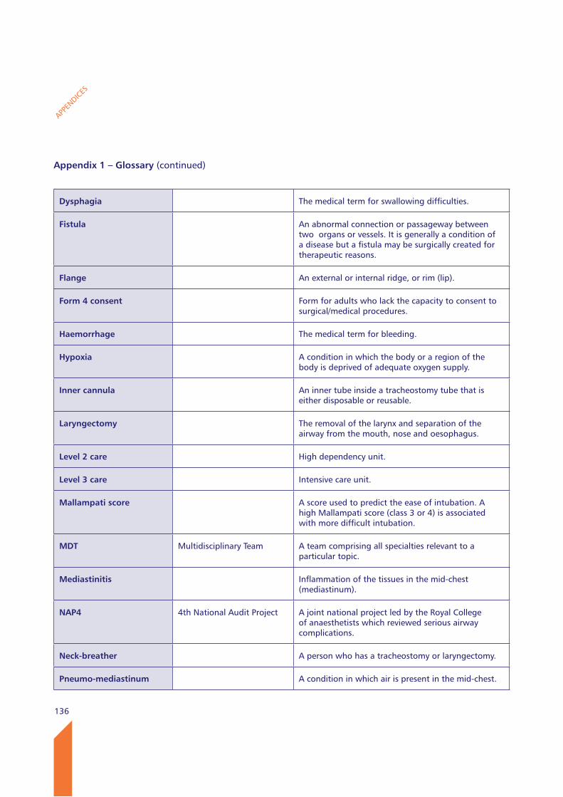
136
APPEN
DICES
Dysphagia The medical term for swallowing difficulties.
Fistula An abnormal connection or passageway between two organs or vessels. It is generally a condition of a disease but a fistula may be surgically created for therapeutic reasons.
Flange An external or internal ridge, or rim (lip).
Form 4 consent Form for adults who lack the capacity to consent to surgical/medical procedures.
Haemorrhage The medical term for bleeding.
Hypoxia A condition in which the body or a region of the body is deprived of adequate oxygen supply.
Inner cannula An inner tube inside a tracheostomy tube that is either disposable or reusable.
laryngectomy The removal of the larynx and separation of the airway from the mouth, nose and oesophagus.
level 2 care High dependency unit.
level 3 care Intensive care unit.
Mallampati score A score used to predict the ease of intubation. A high Mallampati score (class 3 or 4) is associated with more difficult intubation.
MDT Multidisciplinary Team A team comprising all specialties relevant to a particular topic.
Mediastinitis Inflammation of the tissues in the mid-chest (mediastinum).
NAP4 4th National Audit Project A joint national project led by the Royal College of anaesthetists which reviewed serious airway complications.
Neck-breather A person who has a tracheostomy or laryngectomy.
Pneumo-mediastinum A condition in which air is present in the mid-chest.
Appendix 1 – Glossary (continued)
137
APPEN
DICES
Pneumothorax An abnormal collection of air or gas in the space that separates the lungs from the chest wall.
SlT Speech and Language Therapy
SLT assess and treat speech, language and communication problems in people of all ages to help them better communicate.
Stridor A (usually high pitched) sound coming from the upper airway of the patient.
Trachea The medical term for windpipe.
Tracheal stenosis An abnormal narrowing of the central air passageways.
Tracheal stoma A hole in the trachea (windpipe).
Tracheomalacia An abnormal collapse of the tracheal walls.
Tracheostomy A procedure where an opening in the neck at the front of the windpipe and a tube inserted to aid breathing.
wHO checklist World Health Organisation Surgical Checklist
The checklist identifies three phases of an operation, each corresponding to a specific period in the normal flow of work: Before the induction of anaesthesia (“sign in”), before the incision of the skin (“time out”) and before the patient leaves the operating room (“sign out”). In each phase, a checklist coordinator must confirm that the surgery team has completed the listed tasks before it proceeds with the operation.
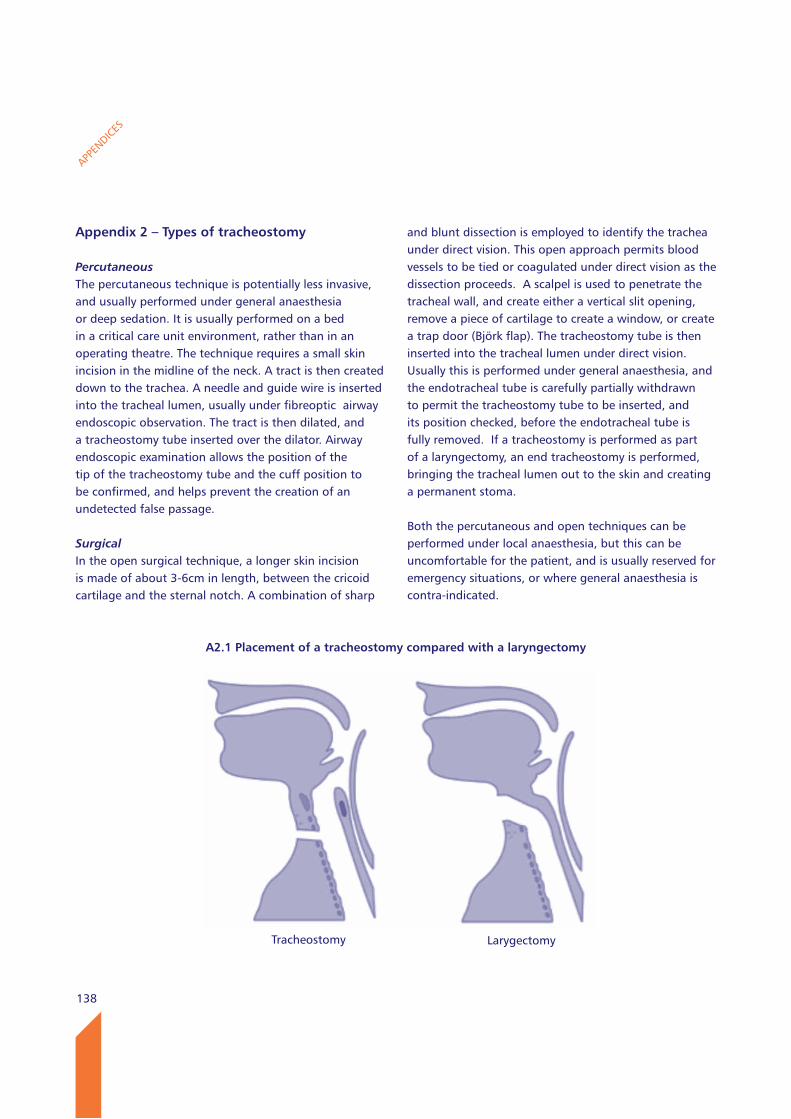
138
APPEN
DICES
Appendix 2 – Types of tracheostomy
PercutaneousThe percutaneous technique is potentially less invasive, and usually performed under general anaesthesia or deep sedation. It is usually performed on a bed in a critical care unit environment, rather than in an operating theatre. The technique requires a small skin incision in the midline of the neck. A tract is then created down to the trachea. A needle and guide wire is inserted into the tracheal lumen, usually under fibreoptic airway endoscopic observation. The tract is then dilated, and a tracheostomy tube inserted over the dilator. Airway endoscopic examination allows the position of the tip of the tracheostomy tube and the cuff position to be confirmed, and helps prevent the creation of an undetected false passage.
SurgicalIn the open surgical technique, a longer skin incision is made of about 3-6cm in length, between the cricoid cartilage and the sternal notch. A combination of sharp
and blunt dissection is employed to identify the trachea under direct vision. This open approach permits blood vessels to be tied or coagulated under direct vision as the dissection proceeds. A scalpel is used to penetrate the tracheal wall, and create either a vertical slit opening, remove a piece of cartilage to create a window, or create a trap door (Björk flap). The tracheostomy tube is then inserted into the tracheal lumen under direct vision. Usually this is performed under general anaesthesia, and the endotracheal tube is carefully partially withdrawn to permit the tracheostomy tube to be inserted, and its position checked, before the endotracheal tube is fully removed. If a tracheostomy is performed as part of a laryngectomy, an end tracheostomy is performed, bringing the tracheal lumen out to the skin and creating a permanent stoma.
Both the percutaneous and open techniques can be performed under local anaesthesia, but this can be uncomfortable for the patient, and is usually reserved for emergency situations, or where general anaesthesia is contra-indicated.
A2.1 Placement of a tracheostomy compared with a laryngectomy
Tracheostomy Larygectomy
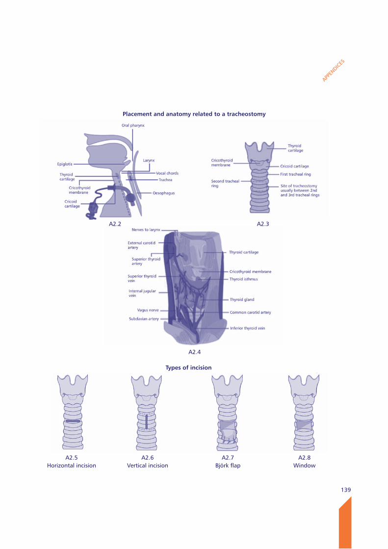
139
APPEN
DICES
Placement and anatomy related to a tracheostomy
A2.5Horizontal incision
A2.6Vertical incision
A2.7Björk flap
A2.8Window
Types of incision
A2.4
A2.2 A2.3
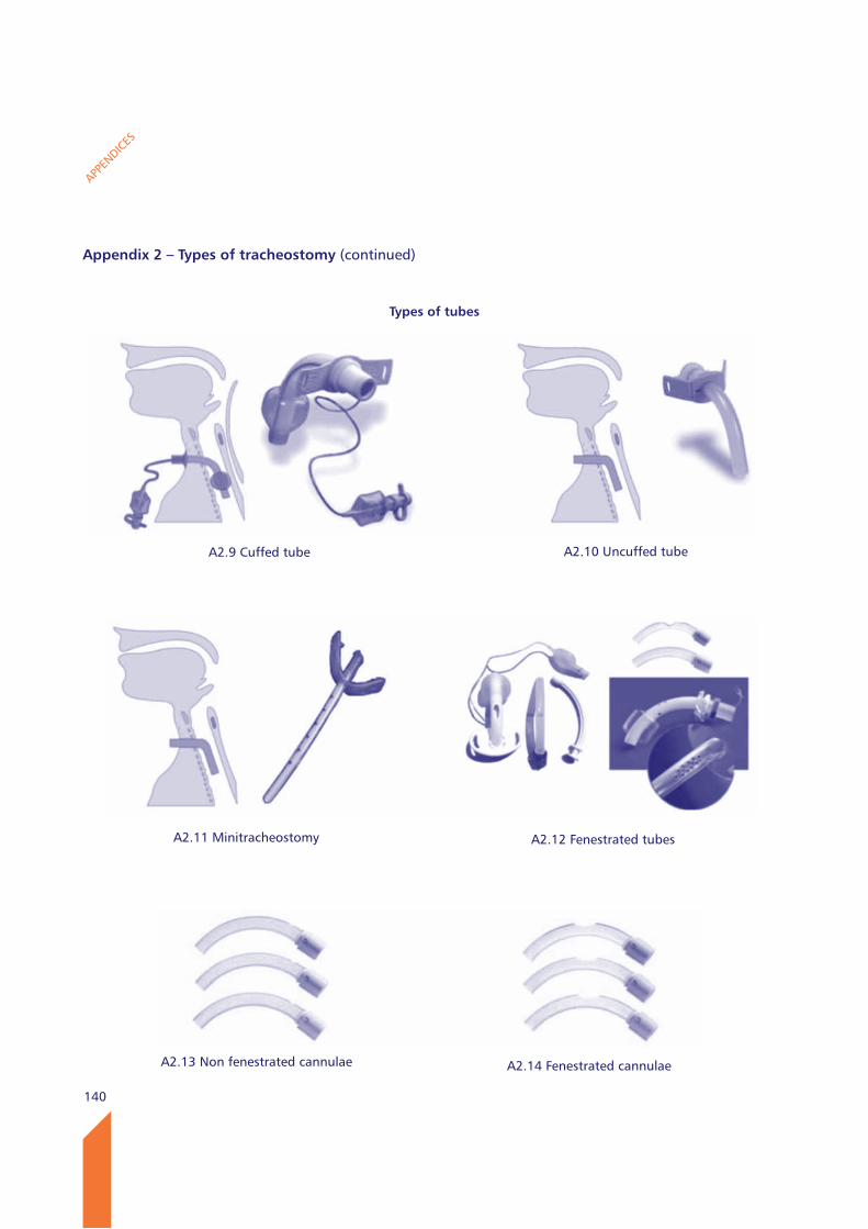
140
APPEN
DICES
Types of tubes
A2.9 Cuffed tube A2.10 Uncuffed tube
A2.11 Minitracheostomy A2.12 Fenestrated tubes
A2.13 Non fenestrated cannulae A2.14 Fenestrated cannulae
Appendix 2 – Types of tracheostomy (continued)
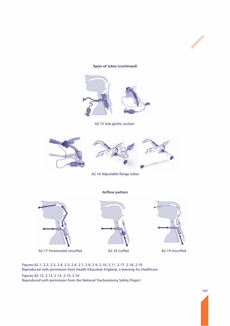
141
APPEN
DICES
A2.19 Uncuffed
A2.15 Sub-glottic suction
A2.16 Adjustable flange tubes
Airflow pattern
A2.17 Fenestrated uncuffed A2.18 Cuffed
Types of tubes (continued)
Figures A2.1, 2.2, 2.3, 2.4, 2.5, 2.6, 2.7, 2.8, 2.9, 2.10, 2.11, 2.17, 2.18, 2.19 Reproduced with permission from Health Education England, e-learning for Healthcare
Figures A2.12, 2.13, 2.14, 2.15, 2.16Reproduced with permission from the National Tracheostomy Safety Project
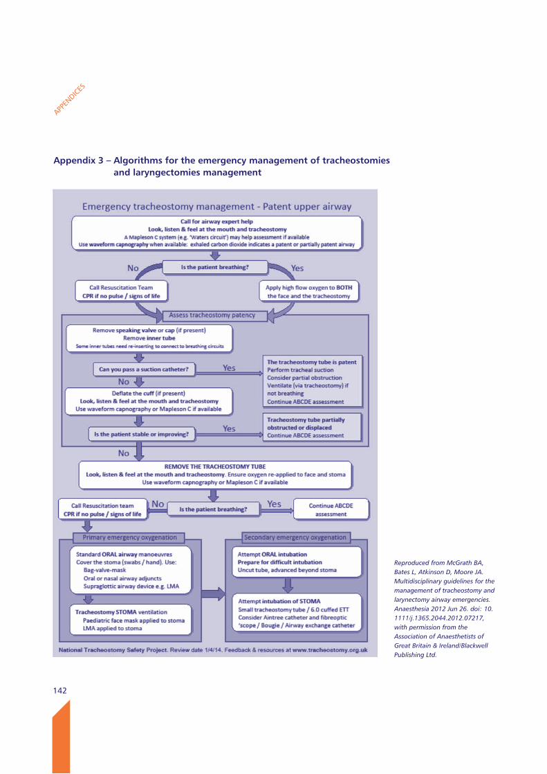
142
APPEN
DICES
Appendix 3 – Algorithms for the emergency management of tracheostomies and laryngectomies management
Reproduced from McGrath BA, Bates L, Atkinson D, Moore JA. Multidisciplinary guidelines for the management of tracheostomy and larynectomy airway emergencies. Anaesthesia 2012 Jun 26. doi: 10.1111/j.1365.2044.2012.07217, with permission from the Association of Anaesthetists of Great Britain & Ireland/Blackwell Publishing Ltd.
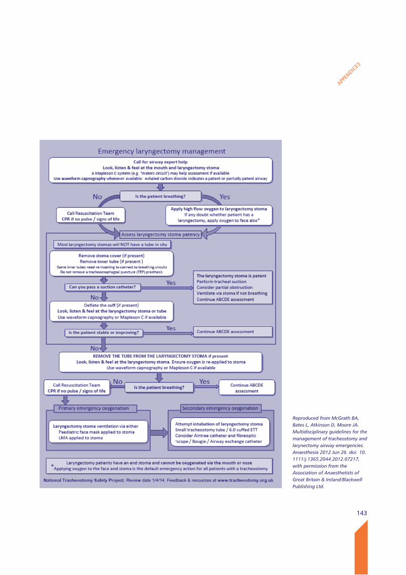
143
APPEN
DICES
Reproduced from McGrath BA, Bates L, Atkinson D, Moore JA. Multidisciplinary guidelines for the management of tracheostomy and larynectomy airway emergencies. Anaesthesia 2012 Jun 26. doi: 10.1111/j.1365.2044.2012.07217, with permission from the Association of Anaesthetists of Great Britain & Ireland/Blackwell Publishing Ltd.
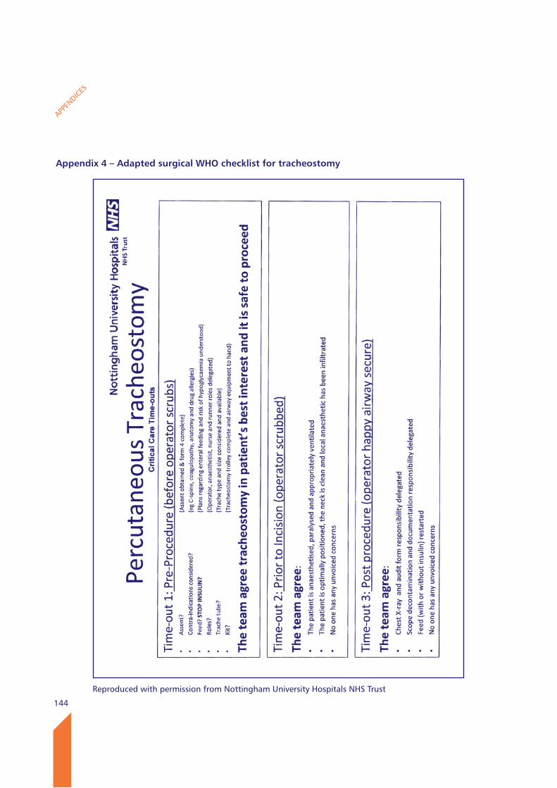
144
APPEN
DICES
Appendix 4 – Adapted surgical wHO checklist for tracheostomy
Reproduced with permission from Nottingham University Hospitals NHS Trust
145
APPEN
DICES
Appendix 5 - The role and structure of NCEPOD
The National Confidential Enquiry into Patient Outcome and Death (NCEPOD) is an independent body to which a corporate commitment has been made by the Medical and Surgical Colleges, Associations and Faculties related to its area of activity. Each of these bodies nominates members on to NCEPOD’s Steering Group.
Steering Group as at 13th June 2014
Dr W Harrop-Griffiths Association of Anaesthetists of Great Britain and IrelandMr F Smith Association of Surgeons of Great Britain and IrelandDr C Mann College of Emergency MedicineVacancy Faculty of Public Health MedicineMs S Payne Lay RepresentativeDr J Fazackerley Royal College of AnaesthetistsDr A Batchelor Royal College of AnaesthetistsDr D Cox Royal College of General PractitionersMrs J Greaves Royal College of NursingDr E Morris Royal College of Obstetricians and GynaecologistsMr W Karwatowski Royal College of OphthalmologistsDr I Doughty Royal College of Paediatrics and Child HealthDr M Osborn Royal College of PathologistsDr A McCune Royal College of PhysiciansDr M Ostermann Royal College of PhysiciansDr M Cusack Royal College of PhysiciansDr T Sabharwal Royal College of RadiologistsMr J Abercrombie Royal College of Surgeons of EnglandMr M Bircher Royal College of Surgeons of EnglandMr K Altman Faculty of Dental Surgery, Royal College of Surgeons of England
Observers
Dr R Hunter Coroners’ Society of England and WalesMrs J Mooney Healthcare Quality in Partnership (HQIP)Dr M Jones Royal College of Physicians of EdinburghMr W Tennant Royal College of Surgeons of Edinburgh
146
APPEN
DICES
NCEPOD is a company, limited by guarantee (Company number: 3019382) and a registered charity (Charity number: 1075588)
Trustees
Mr Bertie Leigh ChairmanDr D Mason Honorary TreasurerProfessor L ReganProfessor R EndacottMr I MartinMr T Hendra
Company Secretary Dr M Mason
Clinical Co-ordinatorsThe Steering Group appoint a Lead Clinical Co-ordinator for a defined tenure. In addition there are six Clinical Co-ordinators who work on each study. All Co-ordinators are engaged in active academic/clinical practice (in the NHS) during their term of office.
Lead Clinical Dr M Juniper (Medicine)Co-ordinator Clinical Co-ordinators Dr K Wilkinson (Anaesthesia) Dr A P L Goodwin (Anaesthesia) Professor M J Gough (Surgery) Mr M Sinclair (Surgery) Dr S McPherson (Radiology) Dr V Srivastava (Medicine)
Supporting organisations
This project was undertaken as part of the Clinical Outcome Review Programme into Medical and Surgical Care.
The Clinical Outcome Review Programme into Medical and Surgical Care is commissioned by the Healthcare Quality Improvement Partnership (HQIP) on behalf of NHS England, NHS Wales, the Northern Ireland Department of Health, Social Services and Public Safety (DHSSPS), the States of Jersey, Guernsey, and the Isle of Man.
The organisations that provided additional funding to cover the cost of this study:Aspen HealthcareBeneden HospitalBMI HealthcareBUPA CromwellEast Kent Medical Services LtdFairfield Independent HospitalHCA InternationalHospital of St John and St ElizabethKing Edward VII’s Hospital Sister AgnesNew Victoria HospitalNuffield HealthRamsay Health Care UKSpire Health CareSt Anthony’s HospitalSt Joseph’s HospitalThe Horder CentreThe London ClinicUlster Independent Clinic
Appendix 5 - The role and structure of NCEPOD (continued)
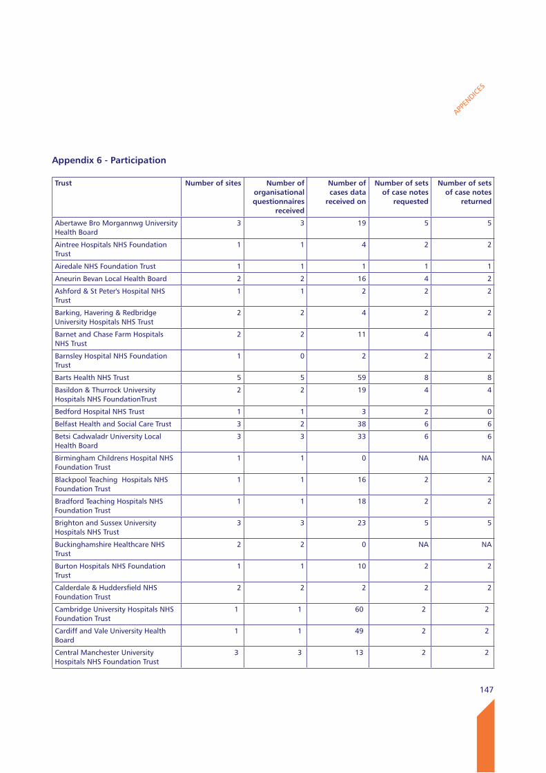
147
APPEN
DICES
Appendix 6 - Participation
Trust Number of sites Number of organisational questionnaires
received
Number of cases data
received on
Number of sets of case notes
requested
Number of sets of case notes
returned
Abertawe Bro Morgannwg University Health Board
3 3 19 5 5
Aintree Hospitals NHS Foundation Trust
1 1 4 2 2
Airedale NHS Foundation Trust 1 1 1 1 1
Aneurin Bevan Local Health Board 2 2 16 4 2
Ashford & St Peter's Hospital NHS Trust
1 1 2 2 2
Barking, Havering & Redbridge University Hospitals NHS Trust
2 2 4 2 2
Barnet and Chase Farm Hospitals NHS Trust
2 2 11 4 4
Barnsley Hospital NHS Foundation Trust
1 0 2 2 2
Barts Health NHS Trust 5 5 59 8 8
Basildon & Thurrock University Hospitals NHS FoundationTrust
2 2 19 4 4
Bedford Hospital NHS Trust 1 1 3 2 0
Belfast Health and Social Care Trust 3 2 38 6 6
Betsi Cadwaladr University Local Health Board
3 3 33 6 6
Birmingham Childrens Hospital NHS Foundation Trust
1 1 0 NA NA
Blackpool Teaching Hospitals NHS Foundation Trust
1 1 16 2 2
Bradford Teaching Hospitals NHS Foundation Trust
1 1 18 2 2
Brighton and Sussex University Hospitals NHS Trust
3 3 23 5 5
Buckinghamshire Healthcare NHS Trust
2 2 0 NA NA
Burton Hospitals NHS Foundation Trust
1 1 10 2 2
Calderdale & Huddersfield NHS Foundation Trust
2 2 2 2 2
Cambridge University Hospitals NHS Foundation Trust
1 1 60 2 2
Cardiff and Vale University Health Board
1 1 49 2 2
Central Manchester University Hospitals NHS Foundation Trust
3 3 13 2 2
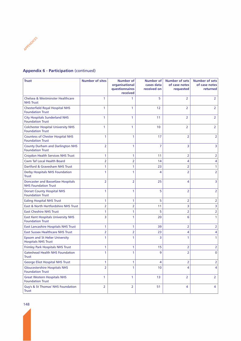
148
APPEN
DICES
Appendix 6 - Participation (continued)
Trust Number of sites Number of organisational questionnaires
received
Number of cases data
received on
Number of sets of case notes
requested
Number of sets of case notes
returned
Chelsea & Westminster Healthcare NHS Trust
1 1 5 2 2
Chesterfield Royal Hospital NHS Foundation Trust
1 1 12 2 2
City Hospitals Sunderland NHS Foundation Trust
1 1 11 2 2
Colchester Hospital University NHS Foundation Trust
1 1 10 2 2
Countess of Chester Hospital NHS Foundation Trust
1 1 17 2 2
County Durham and Darlington NHS Foundation Trust
2 1 7 3 3
Croydon Health Services NHS Trust 1 1 11 2 2
Cwm Taf Local Health Board 2 2 14 4 4
Dartford & Gravesham NHS Trust 1 1 23 2 1
Derby Hospitals NHS Foundation Trust
1 1 4 2 2
Doncaster and Bassetlaw Hospitals NHS Foundation Trust
2 2 25 4 3
Dorset County Hospital NHS Foundation Trust
1 1 5 2 2
Ealing Hospital NHS Trust 1 1 5 2 2
East & North Hertfordshire NHS Trust 2 2 11 3 3
East Cheshire NHS Trust 1 1 5 2 2
East Kent Hospitals University NHS Foundation Trust
3 1 20 6 1
East Lancashire Hospitals NHS Trust 1 1 39 2 2
East Sussex Healthcare NHS Trust 2 2 23 4 4
Epsom and St Helier University Hospitals NHS Trust
1 1 3 1 1
Frimley Park Hospitals NHS Trust 1 1 15 2 2
Gateshead Health NHS Foundation Trust
1 1 9 2 0
George Eliot Hospital NHS Trust 1 1 4 2 2
Gloucestershire Hospitals NHS Foundation Trust
2 1 10 4 4
Great Western Hospitals NHS Foundation Trust
1 1 13 2 2
Guy's & St Thomas' NHS Foundation Trust
2 2 51 4 4
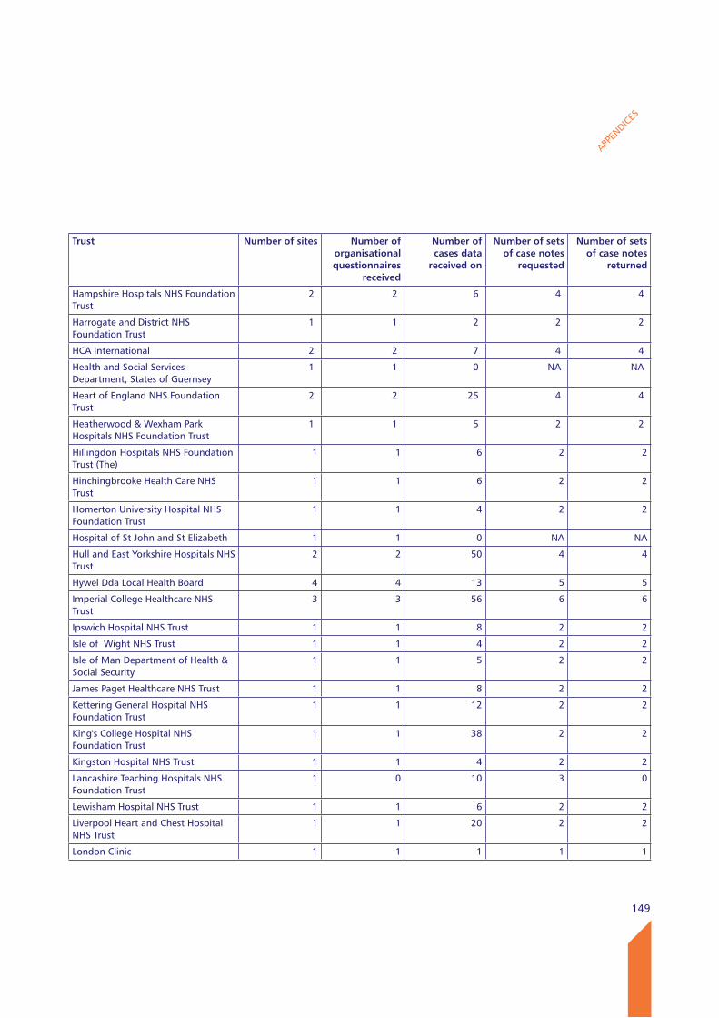
149
APPEN
DICES
Trust Number of sites Number of organisational questionnaires
received
Number of cases data
received on
Number of sets of case notes
requested
Number of sets of case notes
returned
Hampshire Hospitals NHS Foundation Trust
2 2 6 4 4
Harrogate and District NHS Foundation Trust
1 1 2 2 2
HCA International 2 2 7 4 4
Health and Social Services Department, States of Guernsey
1 1 0 NA NA
Heart of England NHS Foundation Trust
2 2 25 4 4
Heatherwood & Wexham Park Hospitals NHS Foundation Trust
1 1 5 2 2
Hillingdon Hospitals NHS Foundation Trust (The)
1 1 6 2 2
Hinchingbrooke Health Care NHS Trust
1 1 6 2 2
Homerton University Hospital NHS Foundation Trust
1 1 4 2 2
Hospital of St John and St Elizabeth 1 1 0 NA NA
Hull and East Yorkshire Hospitals NHS Trust
2 2 50 4 4
Hywel Dda Local Health Board 4 4 13 5 5
Imperial College Healthcare NHS Trust
3 3 56 6 6
Ipswich Hospital NHS Trust 1 1 8 2 2
Isle of Wight NHS Trust 1 1 4 2 2
Isle of Man Department of Health & Social Security
1 1 5 2 2
James Paget Healthcare NHS Trust 1 1 8 2 2
Kettering General Hospital NHS Foundation Trust
1 1 12 2 2
King's College Hospital NHS Foundation Trust
1 1 38 2 2
Kingston Hospital NHS Trust 1 1 4 2 2
Lancashire Teaching Hospitals NHS Foundation Trust
1 0 10 3 0
Lewisham Hospital NHS Trust 1 1 6 2 2
Liverpool Heart and Chest Hospital NHS Trust
1 1 20 2 2
London Clinic 1 1 1 1 1
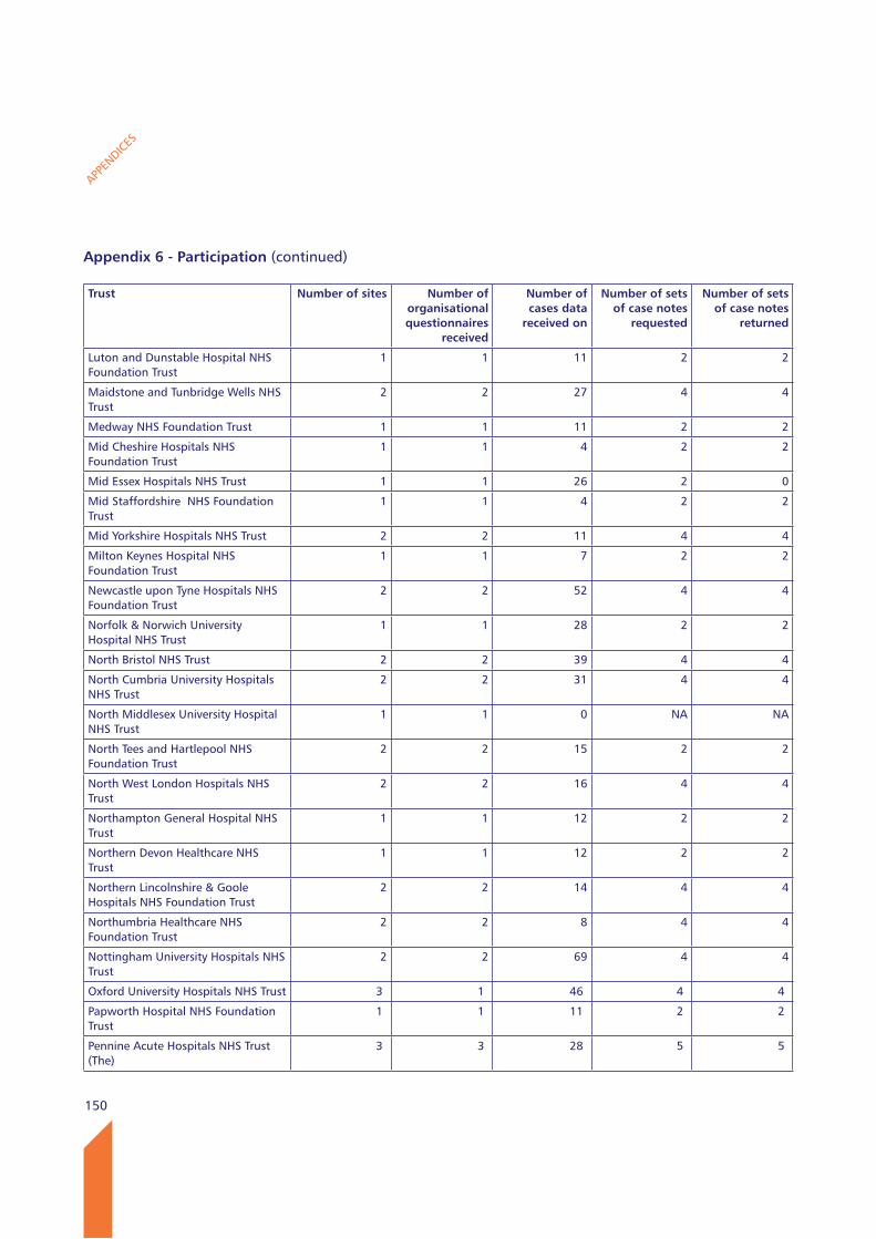
150
APPEN
DICES
Appendix 6 - Participation (continued)
Trust Number of sites Number of organisational questionnaires
received
Number of cases data
received on
Number of sets of case notes
requested
Number of sets of case notes
returned
Luton and Dunstable Hospital NHS Foundation Trust
1 1 11 2 2
Maidstone and Tunbridge Wells NHS Trust
2 2 27 4 4
Medway NHS Foundation Trust 1 1 11 2 2
Mid Cheshire Hospitals NHS Foundation Trust
1 1 4 2 2
Mid Essex Hospitals NHS Trust 1 1 26 2 0
Mid Staffordshire NHS Foundation Trust
1 1 4 2 2
Mid Yorkshire Hospitals NHS Trust 2 2 11 4 4
Milton Keynes Hospital NHS Foundation Trust
1 1 7 2 2
Newcastle upon Tyne Hospitals NHS Foundation Trust
2 2 52 4 4
Norfolk & Norwich University Hospital NHS Trust
1 1 28 2 2
North Bristol NHS Trust 2 2 39 4 4
North Cumbria University Hospitals NHS Trust
2 2 31 4 4
North Middlesex University Hospital NHS Trust
1 1 0 NA NA
North Tees and Hartlepool NHS Foundation Trust
2 2 15 2 2
North West London Hospitals NHS Trust
2 2 16 4 4
Northampton General Hospital NHS Trust
1 1 12 2 2
Northern Devon Healthcare NHS Trust
1 1 12 2 2
Northern Lincolnshire & Goole Hospitals NHS Foundation Trust
2 2 14 4 4
Northumbria Healthcare NHS Foundation Trust
2 2 8 4 4
Nottingham University Hospitals NHS Trust
2 2 69 4 4
Oxford University Hospitals NHS Trust 3 1 46 4 4
Papworth Hospital NHS Foundation Trust
1 1 11 2 2
Pennine Acute Hospitals NHS Trust (The)
3 3 28 5 5
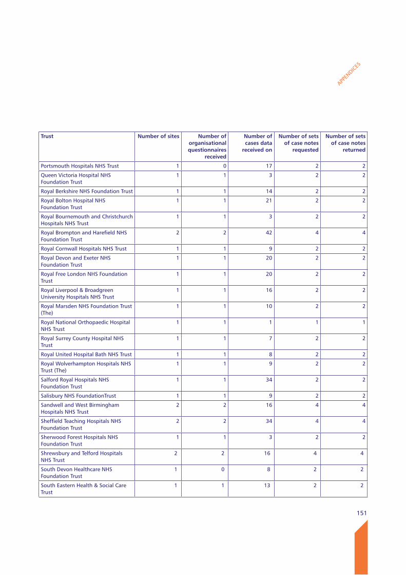
151
APPEN
DICES
Trust Number of sites Number of organisational questionnaires
received
Number of cases data
received on
Number of sets of case notes
requested
Number of sets of case notes
returned
Portsmouth Hospitals NHS Trust 1 0 17 2 2
Queen Victoria Hospital NHS Foundation Trust
1 1 3 2 2
Royal Berkshire NHS Foundation Trust 1 1 14 2 2
Royal Bolton Hospital NHS Foundation Trust
1 1 21 2 2
Royal Bournemouth and Christchurch Hospitals NHS Trust
1 1 3 2 2
Royal Brompton and Harefield NHS Foundation Trust
2 2 42 4 4
Royal Cornwall Hospitals NHS Trust 1 1 9 2 2
Royal Devon and Exeter NHS Foundation Trust
1 1 20 2 2
Royal Free London NHS Foundation Trust
1 1 20 2 2
Royal Liverpool & Broadgreen University Hospitals NHS Trust
1 1 16 2 2
Royal Marsden NHS Foundation Trust (The)
1 1 10 2 2
Royal National Orthopaedic Hospital NHS Trust
1 1 1 1 1
Royal Surrey County Hospital NHS Trust
1 1 7 2 2
Royal United Hospital Bath NHS Trust 1 1 8 2 2
Royal Wolverhampton Hospitals NHS Trust (The)
1 1 9 2 2
Salford Royal Hospitals NHS Foundation Trust
1 1 34 2 2
Salisbury NHS FoundationTrust 1 1 9 2 2
Sandwell and West Birmingham Hospitals NHS Trust
2 2 16 4 4
Sheffield Teaching Hospitals NHS Foundation Trust
2 2 34 4 4
Sherwood Forest Hospitals NHS Foundation Trust
1 1 3 2 2
Shrewsbury and Telford Hospitals NHS Trust
2 2 16 4 4
South Devon Healthcare NHS Foundation Trust
1 0 8 2 2
South Eastern Health & Social Care Trust
1 1 13 2 2
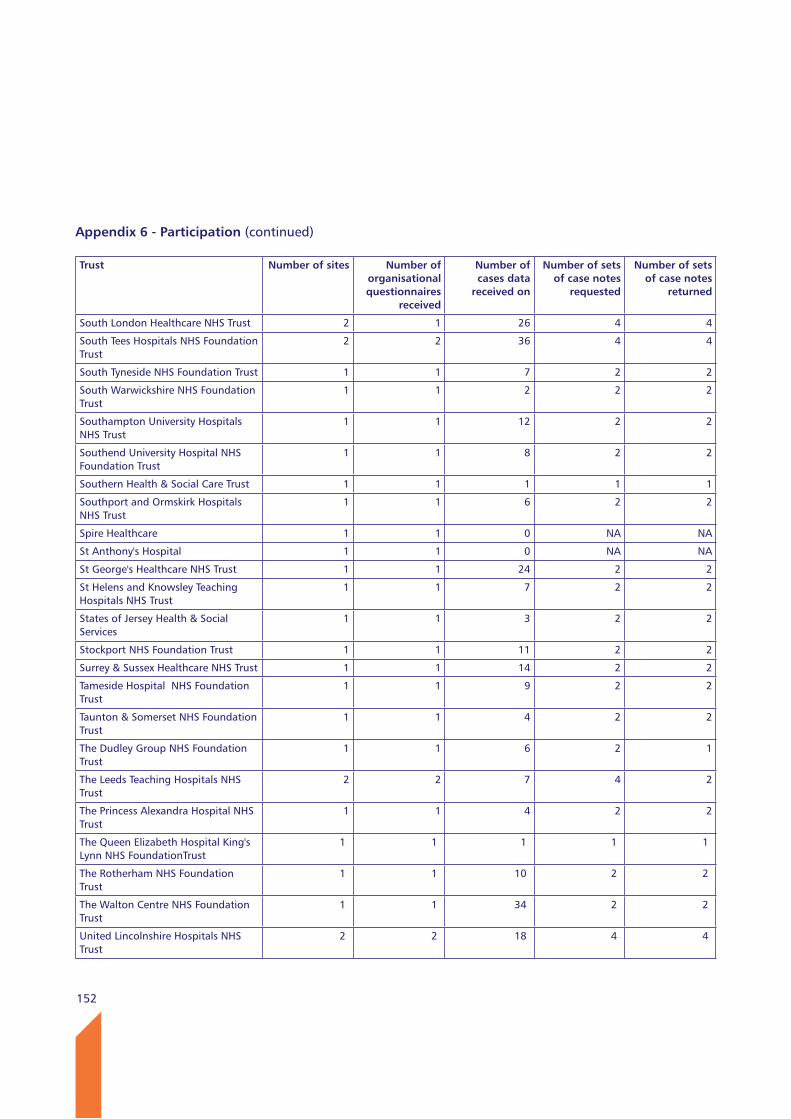
152
Appendix 6 - Participation (continued)
Trust Number of sites Number of organisational questionnaires
received
Number of cases data
received on
Number of sets of case notes
requested
Number of sets of case notes
returned
South London Healthcare NHS Trust 2 1 26 4 4
South Tees Hospitals NHS Foundation Trust
2 2 36 4 4
South Tyneside NHS Foundation Trust 1 1 7 2 2
South Warwickshire NHS Foundation Trust
1 1 2 2 2
Southampton University Hospitals NHS Trust
1 1 12 2 2
Southend University Hospital NHS Foundation Trust
1 1 8 2 2
Southern Health & Social Care Trust 1 1 1 1 1
Southport and Ormskirk Hospitals NHS Trust
1 1 6 2 2
Spire Healthcare 1 1 0 NA NA
St Anthony's Hospital 1 1 0 NA NA
St George's Healthcare NHS Trust 1 1 24 2 2
St Helens and Knowsley Teaching Hospitals NHS Trust
1 1 7 2 2
States of Jersey Health & Social Services
1 1 3 2 2
Stockport NHS Foundation Trust 1 1 11 2 2
Surrey & Sussex Healthcare NHS Trust 1 1 14 2 2
Tameside Hospital NHS Foundation Trust
1 1 9 2 2
Taunton & Somerset NHS Foundation Trust
1 1 4 2 2
The Dudley Group NHS Foundation Trust
1 1 6 2 1
The Leeds Teaching Hospitals NHS Trust
2 2 7 4 2
The Princess Alexandra Hospital NHS Trust
1 1 4 2 2
The Queen Elizabeth Hospital King's Lynn NHS FoundationTrust
1 1 1 1 1
The Rotherham NHS Foundation Trust
1 1 10 2 2
The Walton Centre NHS Foundation Trust
1 1 34 2 2
United Lincolnshire Hospitals NHS Trust
2 2 18 4 4
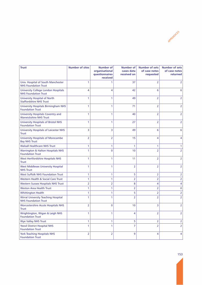
153
APPEN
DICES
Trust Number of sites Number of organisational questionnaires
received
Number of cases data
received on
Number of sets of case notes
requested
Number of sets of case notes
returned
Univ. Hospital of South Manchester NHS Foundation Trust
1 1 37 2 2
University College London Hospitals NHS Foundation Trust
4 4 42 6 6
University Hospital of North Staffordshire NHS Trust
1 1 49 2 2
University Hospitals Birmingham NHS Foundation Trust
1 1 71 2 2
University Hospitals Coventry and Warwickshire NHS Trust
1 1 40 2 2
University Hospitals of Bristol NHS Foundation Trust
1 1 27 2 2
University Hospitals of Leicester NHS Trust
3 3 49 6 6
University Hospitals of Morecambe Bay NHS Trust
2 2 15 4 4
Walsall Healthcare NHS Trust 1 1 1 1 1
Warrington & Halton Hospitals NHS Foundation Trust
1 0 10 2 2
West Hertfordshire Hospitals NHS Trust
1 1 11 2 2
West Middlesex University Hospital NHS Trust
1 1 2 2 2
West Suffolk NHS Foundation Trust 1 1 5 2 2
Western Health & Social Care Trust 1 1 2 2 2
Western Sussex Hospitals NHS Trust 2 2 8 4 4
Weston Area Health Trust 1 1 2 2 0
Whittington Health 1 1 5 2 2
Wirral University Teaching Hospital NHS Foundation Trust
1 1 2 2 2
Worcestershire Acute Hospitals NHS Trust
2 0 10 3 2
Wrightington, Wigan & Leigh NHS Foundation Trust
1 1 4 2 2
Wye Valley NHS Trust 1 1 5 2 2
Yeovil District Hospital NHS Foundation Trust
1 1 7 2 2
York Teaching Hospitals NHS Foundation Trust
2 2 9 4 4

Published June 2014by the National Confidential Enquiry
into Patient Outcome and Death
Ground FloorAbbey House
74-76 St John StreetLondon EC1M 4DZ
T 0207 251 9060F 0207 250 0020
E [email protected] www.ncepod.org.uk
ISBN 978-0-9926184-1-4
A company limited by guarantee Company no. 3019382
Registered charity no. 1075588