on the fine structure of the cambium of fraxinus americana l.
15
ON THE FINE STRUCTURE OF THE CAMBIUM OF FRAXINUS AMERICANA L. LALIT M. SRIVASTAVA From the Department of Biological Sciences, Simon Fraser University, Burnaby, British Colmnbia, Canada ABSTRACT The fine structure of ash cambium was studied after glutaraldehyde-osmium tctroxide fixation. The fusiform and ray initials are essentially alike, and both have the basic comple- ment of organelles and membranes typical of parenchyma cells. The varied behavior of the two types of initials and the role of cambium in oriented production of the xylem and phloem are still unexplained phenomena. Actively growing cambial cells are highly vacuo- late. They are rich in endoplasmic reticulum of the rough cistcrnal form, ribosomes, dictyo- somes, and coated vesicles. Micrombules are present in the peripheral cytoplasm. The plasmalemma appears to be continuous with the endoplasmic reticulum and produces coated vesicles as well as micropinocytotic vesicles with smooth surfaces. The plastids have varying amounts of an intralamellar inclusion which may be a lipoprotein. The quiescent cambium is deficient in rough ER and coated vesicles and has certain structures which may be condensed proteins. INTRODUCTION As the lateral meristem that produces secondary xylem and phloem, cambium has obvious impor- tance in the study of cell differentiation and physi- ology of growth. It has been investigated from several viewpoints, but its cytology is compara- tively unknown. An earlier paper summarized the pertinent literature and described the cambium of Pinus strobus L., a gymnosperm (Srivastava and O'Brien, 1966). The present paper reports on the cambium of Fraxinus americana L. or the common ash, an angiosperm. MATERIAL AND METHODS Material was obtained from young trees of Fraxinus americana L. on the Case Estates, Weston, Massa- chusetts, on two dates: April 3, and June 8, 1965. Cell divisions were well in progress in the June col- lection and most of the results reported here are from that material. The April collection is used to illustrate some features of the quiescent cambium. The preparatory techniques have been described in detail earlier (Srivastava and O'Brien, 1966). In brief, radial slivers of bark, cmnbium, and adjacent wood were extracted from young trunks with the help of a pair of mounted razor blades and a sharp scalpel. The material was fixed initially in 3 % glutaraldehyde (Fisher Scientific Company, Pittsburgh, Biological Grade) in 0.025 M phosphate buffer for 24 hr--the 1st 2 hr at air temperature, the next 22 hr in refriger- ator at approximately 5°C. It was postfixed for 2 hr at room temperature in 2% osmium tetroxide in 0.05 ta phosphate buffer and 0.15 M sucrose. After each fixation, the material was washed thoroughly (3 changes in 6 hr) in a solution containing 0.05 M phos- phate buffer, 0.2 M sucrose, and 0.001 M calcium chloride. The pH of the fixatives and the wash solu- tion was about 6.8-7.0. Dehydration was effected in a graded series of acetone and completed by two changes in porpylene oxide. The material was em- bedded in equal parts of DDSA and Araldite and sectioned on a Porter-Blum MT2 microtome using diamond knives. The grids were stained sequentially 79
-
Upload
phungxuyen -
Category
Documents
-
view
215 -
download
2
Transcript of on the fine structure of the cambium of fraxinus americana l.

O N T H E F I N E S T R U C T U R E OF T H E
C A M B I U M OF F R A X I N U S A M E R I C A N A L.
L A L I T M . S R I V A S T A V A
From the Department of Biological Sciences, Simon Fraser University, Burnaby, British Colmnbia, Canada
A B S T R A C T
The fine structure of ash camb ium was studied after g lu ta ra ldehyde-osmium tctroxide fixation. T he fusiform and ray initials are essentially alike, and bo th have the basic comple- men t of organelles and membranes typical of pa renchyma cells. The varied behavior of the two types of initials and the role of c amb ium in oriented product ion of the xylem and phloem are still unexpla ined phenomena. Actively growing cambial cells are highly vacuo- late. They are rich in endoplasmic ret iculum of the rough cistcrnal form, ribosomes, dictyo- somes, and coated vesicles. Micrombules are present in the per ipheral cytoplasm. The p lasmalemma appears to be continuous with the endoplasmic re t iculum and produces coated vesicles as well as micropinocytotic vesicles with smooth surfaces. The plastids have varying amounts of an in t ra lamel lar inclusion which may be a lipoprotein. The quiescent c a m b i u m is deficient in rough E R and coated vesicles and has certain structures which may be condensed proteins.
I N T R O D U C T I O N
As the lateral meris tem tha t produces secondary xylem and phloem, c a m b i u m has obvious impor- tance in the study of cell differentiat ion and physi- ology of growth. I t has been investigated from several viewpoints, bu t its cytology is compara- tively unknown. An earlier paper summarized the per t inent l i terature and described the c a m b i u m of Pinus strobus L., a gymnosperm (Srivastava and O'Brien, 1966). T he present paper reports on the c a m b i u m of Fraxinus americana L. or the common ash, an angiosperm.
M A T E R I A L A N D M E T H O D S
Material was obtained from young trees of Fraxinus americana L. on the Case Estates, Weston, Massa- chusetts, on two dates: April 3, and June 8, 1965. Cell divisions were well in progress in the June col- lection and most of the results reported here are from that material. The April collection is used to illustrate some features of the quiescent cambium.
The preparatory techniques have been described in detail earlier (Srivastava and O'Brien, 1966). In brief, radial slivers of bark, cmnbium, and adjacent wood were extracted from young trunks with the help of a pair of mounted razor blades and a sharp scalpel. The material was fixed initially in 3 % glutaraldehyde (Fisher Scientific Company, Pittsburgh, Biological Grade) in 0.025 M phosphate buffer for 24 h r - - the 1st 2 hr at air temperature, the next 22 hr in refriger- ator at approximately 5°C. It was postfixed for 2 hr at room temperature in 2% osmium tetroxide in 0.05 ta phosphate buffer and 0.15 M sucrose. After each fixation, the material was washed thoroughly (3 changes in 6 hr) in a solution containing 0.05 M phos- phate buffer, 0.2 M sucrose, and 0.001 M calcium chloride. The pH of the fixatives and the wash solu- tion was about 6.8-7.0. Dehydration was effected in a graded series of acetone and completed by two changes in porpylene oxide. The material was em- bedded in equal parts of DDSA and Araldite and sectioned on a Porter-Blum MT2 microtome using diamond knives. The grids were stained sequentially
79

C, cambium Cr, protein crystal CV, coated vesicle D, dictyosome ER, endoplasmie reticulum F, cytoplasmic fibrils ILl, intralamellar inclusion L, plastid lamella Li, lipid body M, mitoehondrion MT, microtubule N, nucleus Nu, nucleolus
Key to Labeling
Figs. 1 and 18 to ~1 are from tim April collection. Figs. ~2
Ft(~URES 1 and ~ Transections of the cambial zone and adjacent cambial zone are numbered. X 1,050.
P, plastid Ph, phloem P1, plasnlalemma Pld, plasmodesma PV, micropinocytotic vesicle R, ray initial S, plastid stroma St, starch T, tonoplast V, vacuole W, wall WF, wall fibrils Xy, xylem to 17 are from the June collection.
xylem and phloem. Cell layers in the
by uranyl acetate and lead citrate and examined on an RCA-EMU 3D microscope using 50 kv.
R E S U L T S
Although cambium, sensu stricto, is a single layer of cells between xylem and phloem (for reasons, see Barman, 1962), it is difficult to distinguish this layer with certainty from its immediate
derivatives on either side. In the quiescent ma- terial, the undifferentiated derivatives and the initial layer comprise a band of 3 to 4 layers of cells between the mature xylem and phloem (Fig. I). In the actively growing material, in contrast, this zone of undifferentiated cells is much wider (Fig. 2). However, most of these cells are differ- entiating xylem and phloem elements, and with
80 THE ~'OURNAL OF CELL BIOLOGY • VOLUME 31, 1966

FIGI:'rlES S and 4 Radial longitudinal sections of the fusiform cambial cells.
FIG,TaE 3 Tile cells are highly vacuolate and have a thin layer of cytoplasm adjacent to the walls and around the nucleus. The tonoplast is a delicate membrane and is often torn apart. X 13,000.
FIGVRE 4 The nucleus is surrounded by a double membrane which has 500 A pores (arrows). The plastids have a dark intralamellar inclusion. X 29,000.

experience it is possible to delimit the cambium to one of three or four layers of cells (see Srivastava and O'Brien, 1966). Further distinction between these cells in the quiescent or the growing ma- terial is not possible, even at the ultrastructural level. Figs, 1 and 2 show the cells in the cambial zone and, unless otherwise stated, the present paper deals with them only. Differentiation of the phloem derivatives is reported elsewhere (Sriva- stava, in preparation).
Summer Material
The fusiform and ray initials show essentially the same structure. They are highly vacuolate with only a thin layer of cytoplasm around the nucleus and adjacent to the walls (Fig. 3). The nucleus is large and has a single nucleolus. Each nucleolus has several areas of low electron opacity - - commonly though erroneously referred to as vacuoles--surrounded by more heavily stained granular material. The general appearance of the nucleolus is similar to that reported in detail by Lafontaine and Chouinard (1963). The nuclear envelope has well defined pores, approximately 500 A in diameter (Fig. 4). No special electron- opaque areas occur in the cytoplasm opposite the pores, and ribosomes do not invest the outer membrane of the nuclear envelope. The interior of the nucleus does not show a separation of heterochromatin and nucleoplasm or dense and light areas (Figs. 3, 16), a feature typical of aldehyde-fixed interphase nuclei (Sabatini et at,, 1963). A similar situation prevails for the nuclei of the quiescent cambium (Fig. 19) and, to a lesser degree, for those of the bark and wood parenchyma cells.
The ground substance is rich in endoplasmic
reticulum which occurs in the form of typical ribosome-studded cisternae, rough E R (Figs. 6, 8 to t2, 16). Occasional vesiculate structures, especially near the end walls (Fig, 5), may repre- sent tangential sections of these cisternae. A fine fibrillar material is dispersed between the two membranes of the ER (Figs. 5, 8). Profiles of rough ER show spiral and circular patterns of polyribosome aggregations (arrows, Fig. 6). Free ribosomes and aggregates, often in helical pat- terns, are abundant in the ground cytoplasm (arrows, Fig. 5).
Other cytoplasmic inclusions are the so called coated vesicles (Figs. 5, I0, 11). They are more or less spherical and, at the equatorial plane, meas- ure approximately 110 m# in diameter, inclusive of the 20-m~ thick coat. Their size does not seem to very much in different parts of a cell or in different cambial cells. The external sculpting appears alveolar rather than bristly in surface views. Since the cambial cells have only a thin parietal layer of cytoplasm, it is hard to speak of preferred sites of distribution of the coated vesicles. They do occur, however, mostly in the neighbor- hood of the plasmalemma and the dictyosomes. The manner of their origin is not quite clear. In some views, such as one shown in Fig. 5, they seem to arise as surface outgrowths of the plas- malemma.
Mitochondria with well defined cristae, though sometimes only a few in number, and osmiophilic granules are present (Figs. 4, 5, 16). Dictyosomes usually have 5 to 6 cisternae and associated vesicles which contain a fibrillar inclusion similar in density to that present between the two mem- branes of the ER. The resemblance of these inclu- sions to the freshly deposited cell-wall matrix is
FIGUI~ES 5 to 7. Radial sections of the fusiform initials.
FIOURE 5 ER vesicles have fibrillar contents which resemble cell wall material. Helical patterns of ribosome aggregates (arrows) and coated vesicles, approximately 110 mp~ in diameter, are present in the cytoplasm. One coated vesicle, at the lower left corner, may be originating from the plasmalemma. X 47,000.
FIGURE 6 Rough ER shows circular and spiral patterns of polyribosomes (arrows). Microtubules, approximately ~10 A in diameter and several microns long, occur in the peripheral cytoplasm. )< 39,000.
Fm~:~E 7 Enlarged view of part of Fig. 6, to show that mierotublfles are aligned with the wall fibrils. )< 7~,500.
82 THE JOLTRNAL OF CELL BIOLOGY " VOLUME S1, 1966

L. M. Salv,~sv,~vA Fine Structure of Ash Cambium 83

striking (Figs. 5, 8, 10); possibly, both these sources contribute material to the developing walls. Electron-opaque "lines" between the dictyosome cisternae, reported by Turner and Whaley (1965), were not observed, but it is pos- sible that proper views were not examined.
The plastids vary in shape and size (Figs. 4, 12 to 14, 17). Their interior is occupied by a few lamellae, extensions of the inner plastid mem- brane, surrounded by a densely packed fibrillar stroma (Figs. 13, 14). A few osmiophilic bodies, similar to those in mitochondria, are present sometimes, but grana and "chloroplast ribosomes" (Jacobson et al., 1963; Gunning, 1965a) are typically absent. An intralamellar inclusion, which by its amorphous form and staining reaction appears to be a lipoprotein, is common (Figs. 13, 14). It is osmiophilic and stains with uranyl acetate. In the light microscope, it stains red with acid fuchsin. The amount of this material varies in different plastids, often in the same cell, and, depending on the amount, the intralamellar space is irregularly lobed or circular (Figs. 4, 12 to 14). In a few plastids this material has a lami- nated appearance, possibly due to some stacking of the lamellae. A resemblance to grana is obvious, but the lamellar membranes were not discernible in these instances (Figs. 12, 17). Parenchyma cells in the outer bark form typical grana.
Microtubules, approximately 200 to 220 A in diameter and several microns long, are present in the peripheral cytoplasm. They occur indi- vidually as well as in groups of 2 or more. Some of these structures lie parallel to the wall surface, longitudinally and horizontally in respect to the long axis of the cell; others are placed at different angles and extend for varying distances i~ the cytoplasm (Figs. 6, 9, 13, 16). In some views,
microtubules are aligned with the latest fibrillar deposits in the adjacent cell wall (Fig. 7).
In view of the suggested role of microtubules in protoplasmic streaming, it is important to know whether these structures are connected to the plasmalemma. Figs. 7 and 9 suggest such an association, but a clear demonstration is not possible because of the thickness of the sections in relation to the diameter of these organelles. Fig. 9 is an unusual micrograph. It is a longitudinal section, approximately 800 A thick, of a fusiform initial near one of the horizontal end walls. The arrangement of the plasmalemma is explained most satisfactorily by assuming a fold in it such that the section grazes through its surface on the left (gray background) and is normal to it on the right (dark line). An alternative but less convincing explanation would assume the plasmalemma to have been sectioned normally and be represented by the dark line. In this case, there would be no satisfactory answer for the presence of the gray background to the left of the plasmalemma. If the first interpretation is correct, the arrangement of microtubules A and B can be explained in the following two ways : Microtubule A is on the upper surface of the fold; microtubule B, lower down, passes through the plasmalemma and is faintly discernible under the surface. Alternatively, the plasmalemma has a > shaped fold; microtubules A and B are on the upper and lower surfaces, respectively, and neither pierces the plasmalemma. Views such as these depend on a fortuitous plane of sectioning and are extremely rare to find, but they indicate that some microtubules may traverse the plasmalemma. Others may be attached to the surface, and still others lie free in the cyto- plasm.
The plasmalemma is distinct and, in good
FIGURE 8 Transection of a fusiform initial. The section is so mounted that the inner tangential wall of the cell appears on the left. The rough cisternal form of ER is shown. The ER and dictyosome vesicles have fibrillar contents (arrows). X 4%000.
FIGURE 9 Radial section of a fusiform initial near the horizontal end wall. The arrange- ment of the plasmalenmla and microtubules is explained in the text. In the upper half of the micrograph, the plasmalemma appears to be continuous with the ER. X 65,000.
FIGURES 10 and 11 Successive radial sections of a fusiform cambial cell. Dictyosome vesicles (arrows) contain fibrillar contents which resemble wall material. Coated vesicles are seen in equatorial plane as well as in surface views. A micropinocytotic vesicle is shown in Fig. 10. X 37,000.
84 THE JOURNAL OF CELL BIOLOGY • VOLUME 31, 1966

L. M. SRIVASTAV.~ Fine Structure of Ash Cambium 85

views, shows the triple layered structure known for unit membranes. The tonoplast has a similar structure but is thinner (Fig. 15). It seems to be a delicate membrane and is often ruptured and disorganized by the preparatory techniques (Fig. 3). Some views show the plasmalemma to be continuous with the endoplasmic reticulum (Fig. 9). Others illustrate that portions of plasmalemma are pinched off and released into the parietal cytoplasm (Fig. 10) in a manner comparable to the phenomenon of micropinocytosis reported for animal cells (see Fawcett, 1965).
Walls of the cambial cells are thin and show areas of low and high electron opacity. Depending on the plane of section, the electron-opaque areas appear as fibrils of varying lengths or as points (Figs. 5 to 7; 14, 15). Whether these fibrils repre- sent cellulose or polyuronides is not quite clear (see Newcomb and Bonnett, 1965). Plasmodes- mata traverse these wails (Figs. 8, 16). In sec- tional view, each plasmodesma has two dark lines, which are the extensions of the plasma- lemma, and a dark core. There is no evidence of a continuation of the ER of neighboring cells through the plasmodesmata.
Quiescent Cambium
Since cell divisions had not yet started in the April collection--indeed, they did not start until M a y - - i t can be used to illustrate some features of the quiescent cambium. I t should be realized, though, that this material represents a transi- tional stage rather than that of the winter.
No marked differences are evident, between the June and April collections, in the state of the
nucleus, mitochondria, dictyosomes, and micro- tubules (Figs. 18 to 21). Plastids show varying amounts of the dark staining material, interpreted here as lipoprotien, and have numerous starch grains. The rough cisternal form of E R and coated vesicles are present, though perhaps not in as great abundance as in the June collection. The plasmalemma shows a faint degree of con- volution. More noticeable are the following: a microvacuolate cytoplasm which appears rather dense (bigger vacuoles are present in the interior of the cell); the presence of a few membrane- bounded structures with a latticework reminiscent of protein crystals (Fig. 18); and bundles of fibrils or microtubules in the cytoplasm (Fig. 20). A few objects which seem to be lipid droplets are present also (Fig. 18).
The bodies with latticework are seen in a number of cells; they are surrounded by a single membrane, and have an internal matrix of approximately the same density as the surrounding cytoplasm. The crystalline core has two sets of intersecting electron- opaque bands with a center-to-center spacing of about 150 A.
The cytoplasmic fibrils or tubules have been recorded in the winter material of pine cambium also (Srivastava and O'Brien, 1966). In that species, these structures are about 150 A in diam- eter and, in general, much more distinct than those in ash.
D I S C U S S I O N
The eambia of Fraxinus and Pinus are markedly different in an evolutionary sense (Bailey, 1923), yet they show the same fine structure; the differ-
FIGURES 1~ to 15 Radial sections of fusiform initials.
FIGURE 1~ Two plastids in a cell with dark inclusions. The one on the right shows a laminated arrangetnent of the dark material, reminiscent of laInellar stacking in grana formation; the lamellar membranes are not seen, however. X 34,500.
FIGURE 13 A plastid showing the intralamellar origin of the dark nmteriaI. Some micro tubules are cut transversely. X 37,000.
FIGURE 14 Lamellae are extensions of the inner plastid membrane (arrows); tile dark material is surromided by a membrane. X 69,500.
FIGURE 15 The plaslnalemma and tonoplast have the structure of unit membranes-- two dark lines separated by a light space. The vacuolar membrane is thinner than the plasmaleinma. X 10%000.
86 TIIE JOURNAL OF CELL BIOLOGY - VOLUME 31, 1966

L. M. SRIVASTAVA Fine Structure of Ash Cambium 87
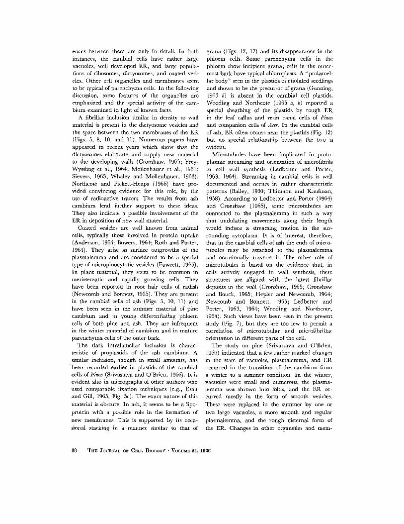
ences between them are only in detail. In both instances, the cambial cells have rather large vacuoles, well developed ER, and large popula- tions of ribosomes, dictyosomes, and coated vesi- cles. Other cell organelles and membranes seem to be typical of parenchyma cells. In the following discussion, some features of the organelles are emphasized and the special activity of the cam- bium examined in light of known facts.
A fibrillar inclusion similar in density to wall material is present in the dictyosome vesicles and the space between the two membranes of the ER (Figs. 5, 8, 10, and 11). Numerous papers have appeared in recent years which show that the dictyosomes elaborate and supply new material to the developing walls (Cronshaw, 1965; Frey- Wyssling et al., 1964; Mollenhauer et al., 1961; Sievers, 1963; Whaley and Mollenhauer, 1963). Northcote and Pickett-Heaps (1966) have pro- vided convincing evidence for this role, by the use of radioactive tracers. The results from ash cambium lend further support to these ideas. They also indicate a possible involvement of the ER in deposition of new wall material.
Coated vesicles are well known from animal cells, typically those involved in protein uptake (Anderson, 1964; Bowers, 1964; Roth and Porter, 1964). They arise as surface outgrowths of the plasmalemma and are considered to be a special type of micropinocytotic vesicles (Fawcett, 1965). In plant material, they seem to be common in meristematic and rapidly growing cells. They have been reported in root hair cells of radish (Newcomb and Bonnett, 1965). They are present in the cambial cells of ash (Figs. 5, 10, 11) and have been seen in the summer material of pine cambium and in young differentiating phloem cells of both pine and ash. They are infrequent in the winter material of cambium and in mature parenchyma cells of the outer bark.
The dark intralamellar inclusion is charac- teristic of proplastids of the ash cambium. A similar inclusion, though in small amounts, has been recorded earlier in plastids of the cambial cells of Pinus (Srivastava and O'Brien, 1966). It is evident also in micrographs of other authors who used comparable fixation techniques (e.g., Esau and Gill, 1965, Fig. 5c). The exact nature of this material is obscure. In ash, it seems to be a lipo- protein with a possible role in the formation of
new membranes. This is supported by its occa- sional stacking in a manner similar to that of
grana (Figs. 12, 17) and its disappearance in the phloem cells. Some parenchyma cells in the phloem show incipient grana; cells in the outer- most bark have typical chloroplasts. A "'prolamel- lar body" seen in the plastids of etiolated seedlings and shown to be the precursor of grana (Gunning, 1965 b) is absent in the cambial cell plastids. Wooding and Northcote (1965 a, b) reported a special sheathing of the plastids by rough ER in the leaf callus and resin canal cells of Pinus and companion ceils of Acer. In the cambial cells of ash, ER often occurs near the plastids (Fig. 12) but no special relationship between the two is evident.
Microtubules have been implicated in proto- plasmic streaming and orientation of microfibrils in cell wall synthesis (Ledbetter and Porter, 1963, 1964). Streaming in cambial cells is well documented and occurs in rather characteristic patterns (Bailey, 1930; Thimann and Kaufman, 1958). According to Ledbetter and Porter (1964) and Cronshaw (1965), some microtubules are connected to the plasmalemma in such a way that undulating movements along their length would induce a streaming motion in the sur- rounding cytoplasm. It is of interest, therefore, that in the cambial ceils of ash the ends of micro- tubules may be attached to the plasmalemma and occasionally traverse it. The other role of microtubules is based on the evidence that, in cells actively engaged in wall synthesis, these structures are aligned with the latest fibrillar deposits in the wall (Cronshaw, 1965; Cronshaw and Bouck, 1965; Hepler and Newcomb, 1964; Newcomb and Bonnett, 1965; Ledbetter and Porter, 1963, 1964; Wooding and Northcote, 1964). Such views have been seen in the present study (Fig. 7), but they are too few to permit a correlation of microtubular and microfibrillar orientation in different parts of the cell.
The study on pine (Srivastava and O'Brien, 1966) indicated that a few rather marked changes in the state of vacuoles, plasmalemma, and ER occurred in the transition of the cambium from a winter to a summer condition. In the winter, vacuoles were small and numerous, the plasma- lemma was thrown into folds, and the ER oc- curred mostly in the form of smooth vesicles. These were replaced in the summer by one or two large vacuoles, a more smooth and regular plasmalemma, and the rough eisternal form of the ER. Changes in other organelles and mere-
88 T~E JOUnNAL OF CELL BIOLOGY • VoLu~ 31, 1966

FIGURE 16 Transect ion of ray initials. Par t of a nucleus appears on the left; other organelles and mem- branes are similar to those in the fusiform initials. Microtubules appear in transectional as well as longi- tudinal views. X 29,500.
FmvR~ 17 Radial section of ray initials. A plastid with the dark inclusion and starch grains. X 29,500.
89

branes were not obvious (see also Parker and Philpott, 1961, 1963). The fine cytoplasmic fibrils were seen only in the winter material. The results from ash are not strictly comparable. However, the microvacuolate condition of the cytoplasm and the presence of fibrils in the quiescent ma- terial of ash parallel the observations on the winter material of pine.
The crystal-containing bodies in the quiescent cambium of ash were not seen in pine. But similar structures are reported from Avena coleoptile, suspension cultures of Eucalyptus, and phloem parenchyma of Acer (Cronshaw, 1964; Thornton and Thimann, 1964). They are reported also in phloem parenchyma of Pisum, but without a crystalline core (Bouck and Cronshaw, 1965). Cronshaw (1964) compared these bodies to lyso- somes and suggested that they may contain storage granules of hydrolytic enzymes. His implication that these bodies are found in "degenerating" systems, however, bears reexamination, in view of their presence in the cambial cells. For the cambium, one can only conclude that the crystal- containing bodies, and perhaps the cytoplasmic fibrils, represent condensed proteins of unknown physiological significance.
From a morphogenetical point of view, it is of interest that the fusiform and ray initials are similar to one another and to parenchyma cells in general. This confirms the earlier observations on Datura (Hohl, 1960) and Pinus (Srivastava and O'Brien, 1966) and illustrates the relatively un- specialized nature of the carnbial cell protoplast. Both types of initials have the usual basic comple- ment of organelles and membranes, and the differences between them are not evident with the techniques used here. Occasionally, ray initials
have a comparatively dilute cytoplasm and show starch in plastids (Figs. 16, 17), but these features are inconsistent and do not constitute a valid distinction. This structural correspondence ex- plains the interconversions between the two types of ini t ials--a phenomenon well documented in the cambial literature (Bannan, 1962; Cheadle and Esau, 1964; Srivastava, 1964). It is not clear, however, why these conversions occur and why these cells behave differently in the intact p l an t - - in respect to their shape, frequency of division in a growth season, and nature of the derivatives. Also, the causal agents for oriented divisions in the cambium and the orderly sequence of differ- entiation of the xylem and phloem remain obscure. As suggested elsewhere (Srivastava and O'Brien, 1966), quantitative and enzymatic differences may be involved and we must await further de- velopments in electron microscopic cytochemistry as applied to botanical material before they are revealed. Already a substantial body of evidence indicates that a special supply of hormones and nutrients is involved in resumption of the cambial growth in the spring and maturation of the xylem and phloem (see reviews by Leopold, 1964; Wareing et al., 1964; Wetmore et al., 1964). In addition, there may be other factors of a more physical nature, dependent largely on the posi- tion this tissue occupies in the plant body. Brown's (1964) experiments show that the cambial cells have the capacity to divide, but the divisions are unoriented and formation of the vascular tissues impossible unless pressure is applied. Oxygen tension may be involved also, for, in fresh, unde- hydrated material of the cambium, intercellular spaces are lacking (I. W. Bailey and R. H. Wet- more, personal communication).
FIGURES 18 to ~1 Radial sections of fusiform initials sampled in April.
FIGURE 18 The cytoplasm is dense and microvacuolate. A membrane-bounded struc- ture with a latticed core (Cr), similar to protein crystals, and a lipid droplet (Li) are present. X 60,000.
FIGURE 19 Part of a nucleus and a dictyosome. X ~5,000.
FIGURE ~0 The cells have snlall amounts of rough ER. The microtubules are cut trans- versely, indicating a horizontal orientation. Thin fibrils (F) or tubules are present in the cytoplasm; their nature is unknown. )< 51,500.
FIGURE ~1 Microtubules adjacent to the horizontal wall. X 37,000.
90 T h E JOUnNMJ OF CELL BIOLOGY " VOLUME 3 | , 1966

L. M. SRIV~STAW~ Fine Structure of Ash Cambium 91

This investigation was carried out at the Biological Laboratories, Harvard University, Cambridge, Mas- sachusetts, while the author was a Maria Moors Cabot Research Fellow. It was supported in part by the United States Public Health Service grant No.
B I B L I O G R A P H Y
ANDERSON, E., 1964, Oocyte differentiation and vitellogenesis in the roach Periplaneta americana, J. Cell Biol., 20, 131.
BAILEY, I. W., 1923, The cambium and its derivative tissues. IV. The increase in girth of the cambium, Am. 3". But., 10, 499.
BAILEY, I. W., 1930, The cambium and its derivative tissues. V. A reconnaissance of the vacuome in liv- ing cells, Sonderdruck Z. Zellforsch. mikr. Anat., 10, 651.
BANNAN, M. W., 1962, The vascular cambium and tree-ring development, in Tree Growth, (T. T. Kozlowski, editor), New York, Ronald Press.
BovcK, G. B., and CRONSHAW, J., 1965, The fine structure of differentiating sieve tube elements, J . Cell Biol., 25, 79.
BOWERS, B., 1964, Coated vesicles in the pericardial cells of the aphid (Myzus persicae Sulz), Proto- plasma, 59, 351.
BROWN, C. L., 1964, The influence of external pres- sure on the differentiation of cells and tissues cultured in vitro, in The Formation of Wood in Forest Trees, (M. H. Zimmermann, editor), New York, Academic Press Inc.
CI-IEADLE, V. I., and ESAU, K., 1964, Secondary phloem of Liriodendron tulipifera, Univ. California Publ. But., 36, 143.
CRONSHAW, J., 1964, Crystal-containing bodies of plant cells, Protoplasma, 59, 318.
CRONSHAW, J., 1965, Cytoplasmic fine structure and cell wall development in differentiating xylem elements, in Cellular Ultrastructure of Woody Plants, (W. A. C6t6, Jr., editor), Syracuse Uni- versity Press.
CRONSHAW, J., and BOUCK, B., 1965, The fine struc- ture of differentiating xylem elements, J. Cell Biol., 24, 415.
ESAU, K., and GILL, R, H., 1965, Observations on cytokinesis, Planta, 67, 168.
FAWCETT, D, W., 1965, Surface specializations of ab- sorbing cells, 3". Histochem. and Cytochem., 13, 75.
FREY-WYssLING, A., LdPEz-SXEz, J. F., and MOH- LETHALER, K., 1964, Formation and development of the cell plate, J . Ultrastruct. Research, 10, 422.
GUNNING, B. E. S., 1965 a, The fine structure of chloroplast stroma following aldehyde osmium- tetroxide fixation, J. Cell Biol., 24, 79.
GUNNING, B. E. S., 1965 b, The greening process in
GM 06637. Grateful acknowledgment is made to the authorities at the Arnold Arboretum, Harvard Uni- versity, for permission to collect the material of ash on their premises.
Received for publication 22 March 1966.
plastids. 1. The structure of the prolamellar body, Protoplasma, 60, 111.
HEPLER, P. K., and NEWCOME, E. H., 1964, Micro- tubules and fibrils in the cytoplasm of Coleus cells undergoing secondary wall deposition, J. Cell Biol., 20, 529.
HOHL, H. R., 1960, /Jber die submikroskopische Struktur normaler und hyperplastischer Gewebe von Datura stramonium L. 1. Normalgewebe, Ber. schweiz, but. Ges., 70, 395.
JACOnSON, A. B., SWIFT, H., and BOGORAD, L., 1963, Cytochemical studies concerning the occurrence and distribution of RNA in plastids of Zea mays, J. Cell Biol., 17, 557.
LAFONTAINE, J. G., and CHOUINARD, L. A., 1963, A correlated light and electron microscope study of the nucleolar material during mitosis in Vicia faba, 3". Cell. Biol, 17, 167.
LEDBETTER, M. C., and PORTER, K. R., 1963, A "microtubule" in plant cell fine structure, J. Cell Biol. 19, 239.
LEDBETTER, M. C., and PORTER, K. R., 1964, Mor- phology of microtubules of plant cells, Science, 144, 872.
LEOPOLD, A. C., 1964, Plant hormones, in The Hor- mones, (G. Pincus, K. V. Thimann, and E. B. Astwood, editors), New York, Academic Press Inc, 4.
MOLLE~HAUER, H. H., WHALEY, W. G., and LEECH, J . H., 1961, A function of the Golgi apparatus in outer root cap cells, J. Ultrastruct. Research, 5, 193.
NEWCOMB, E. H., and BONNETT, H. T., JR., 1965, Cytoplasmic mierotubule and wall microfibril ori- entation in root hairs of radish, J. Cell Biol., 27, 575.
NORTHGOTE, D. H., and PICKETT-HEAPS, J. D., 1966, A function of the Golgi apparatus in polysaccharide synthesis and transport in the root cap cells of wheat, Biochem. 3.., 98, 159.
PARKER, J., and PHILPOTT, D. E., 1961, An electron microscopic study of chloroplast condition in sum- mer and winter in Pinus strobus, Protoplasma, 53, 575.
PARKER, J., and PVlILPOTT, D. E., 1963, Seasonal con- tinuity of chloroplasts in white pine and Rhododen- dron, Protoplasma, 58, 355.
ROTH, T. F., and PORTER, K. R., 1964, Yolk protein uptake in the oocyte of the mosquito Aedes aegypti L., J. Cell Biol., 20, 313.
92 THE JOURNAL OF CELL BIOLOGY • VOLUME 31, 1966

SABATINI, D. D., BENSCH, K., and BARRNETT, R. J., 1963, Cytochemistry and electron microscopy. The preservation of cellular ultrastructure and enzy- matic activity by aldehyde fixation, J. Cell Biol., 17, 19.
SIEVERS, A., 1963, Beteiligung des Golgi-Apparatus bei der Bildung der Zellwand yon Wurzelhaaren, Prot.oplasma, 56, 188.
SRIVASTAVA, L. M., 1964, Anatomy, chemistry, and physiology of hark, in International Review of For- estry Research, (J. A. Romberger and P. Mikola, editors), New York, Academic Press Inc., 1.
SmVASTAVA, L. M., and O'BR~N, T. P., 1966, On the nltrastructure of cambium and its vascular derivatives. I. Cambium of Pinus strobus L., Proto- plasma, 61, 257.
THIMANN, K. V., and KAUFMAN, D., 1958, in The Physiology of Forest Trees (K. V. Thimann, editor), New York, The Ronald Press Co.,
THORNTON, R. M., and TmMANN, K. V., 1964, On a crystal-containing body in cells of the oat coleoptile, J. Cell Biol., 20, 345.
TURNER, F. R., and WHALEY, W. G., 1965, Inter-
cisternal elements of the Golgi apparatus, Science, 147, 1303.
WAREING, P. F., HANNEV, C. E. A., and DIGBV, J., 1964, The role of endogenous hormones in cambial activity and xylem differentiation, in The Forma- tion of Wood in Forest Trees (M. H. Zimmermann, editor), New York, Academic Press Inc.
WETMORE, R. H., DEMAGGIO, A. E. and RmR, J. P., 1964, Contemporary outlook on the differentiation of vascular tissues, Phytomorphology, 14, 203.
WHALEY, W. G., and NIOLL~NHAUER, It. H., 1963, The Golgi apparatus and cell plate formation--a postulate, or. Cell Biol., 17, 216.
WOODING, F. B. P., and NORTHCOTE, D. H., 1964, The development of the secondary wall of the xylem in Acer pseudoplatanus, J. Cell Biol., 23, 327.
WoonxNo, F. B. P., and NORTHCOTE, D. H., 1965 a, Association of the endoplasmic reticulum and the plastids in Acer and Pinus, Am. J. Bot., 52, 526.
WOODINC, F. B. P., and NORTHaOTE, D. H., 1965 b, The fine structure and development of the com- panion cell of the phloem of Acer pseudoplatanus, J. Cell Biol., 24, 117.
L. M. SRIVASTAVA f~ne Structure of Ash Cambium 93