ON SOME CILIATE PARASITES OF FROGS AND TOADS OF...
18
ON SOME CILIATE PARASITES OF FROGS AND TOADS OF KARNATAK, BOMBAY PRESIDENCY* By J. C. UTTANGI, Department of Zoology, Karnatak Oollege, Dharwar. In trod uction Notes on Hosts Systematics Summary References CONTENTS. INTRODUCTION. PAGE. 139 140 140 155 155 The occurrence of binucleate opalinids in Indian Anura has been a subject of great controversy in the past. Bezzenberger (1904), for the first time, described Zelleriella macronucleata (Bezz.) from an Asian toad, Bufo melanostictus Schneider, but his findings were later rendered doubtful by Metcalf (1923) who opened thirty-nine specimens of this toad to find no Zelleriella in them. Bhatia and Gulati (1927) who con- ducted extensive survey of the ciliate parasites of the Anura of the Punjab also reported a complete absence of both Zelleriella and Pro- toopalina in Bufo me!anostictus as as other Anurans they studied. The absence of these ciliates in Indian Anura was, thus,:"more or less accepted, until Nie (1935) again reported Protoopalina caudata microhyl,a Nie, from an Indian frog Microhyla ornata Dumeril and Bibron (Ind. Mus .. Reg. No. 17287), a specimen of which collected at Harnai, Ratnagiri District, Bombay, was supplied to him by the Indian Museum, Calcutta (Met0alf, 1940; p. 472). This discovery of Nie evidently raised the question asked by Metcalf (p. 577) "Why are there no more Protoopa- inas in these Indian Ocean Lands ?", and, thus, led the writer to under- take this interesting piece of work. The present investigation establishes, beyond doubt, the existence of binucleate opalinids in the Anura of Karnatak, and includes the description of five new forms, i.e., Protoopalina indica, P. karnatakensis, P. dlwrwarensis, Zelleriella microhylae and Z. froilanoi. Since the locality is' situated in South India which forms the most ancient part of the Asian continent, the presence of Zelleriella and Protoopalina in this region, may modify the present conclusions in the field of Zoogeography. During the course of this work, the following twelve species of other ciliates found parasitic in the gut of the hosts examined have also been recorded: Nyctotherus macropharyngeus Bezz., N. magnus Bezz., N. magnus rnalabarica De Mello., N .cordiJormis Stein., Balantidium kelenae Bezz., B. duodeni Stein., B. gracile Bezz., Opalina ranarum Purk et Val., Oepedea virg'Ula (Dohell) Metcalf., O. philauti sp. nov., and O. dimidiata • Thesis submitted for the degree of M.Sc. of the University of Bombay. 139
Transcript of ON SOME CILIATE PARASITES OF FROGS AND TOADS OF...

ON SOME CILIATE PARASITES OF FROGS AND TOADS OF KARNATAK, BOMBAY PRESIDENCY*
By J. C. UTTANGI, Department of Zoology, Karnatak Oollege, Dharwar.
In trod uction
Notes on Hosts
Systematics
Summary
References
CONTENTS.
INTRODUCTION.
PAGE.
139
140
140
155
155
The occurrence of binucleate opalinids in Indian Anura has been a subject of great controversy in the past. Bezzenberger (1904), for the first time, described Zelleriella macronucleata (Bezz.) from an Asian toad, Bufo melanostictus Schneider, but his findings were later rendered doubtful by Metcalf (1923) who opened thirty-nine specimens of this toad to find no Zelleriella in them. Bhatia and Gulati (1927) who conducted extensive survey of the ciliate parasites of the Anura of the Punjab also reported a complete absence of both Zelleriella and Protoopalina in Bufo me!anostictus as w~l as other Anurans they studied. The absence of these ciliates in Indian Anura was, thus,:"more or less accepted, until Nie (1935) again reported Protoopalina caudata microhyl,a Nie, from an Indian frog Microhyla ornata Dumeril and Bibron (Ind. Mus .. Reg. No. 17287), a specimen of which collected at Harnai, Ratnagiri District, Bombay, was supplied to him by the Indian Museum, Calcutta (Met0alf, 1940; p. 472). This discovery of Nie evidently raised the question asked by Metcalf (p. 577) "Why are there no more Protoopainas in these Indian Ocean Lands ?", and, thus, led the writer to undertake this interesting piece of work.
The present investigation establishes, beyond doubt, the existence of binucleate opalinids in the Anura of Karnatak, and includes the description of five new forms, i.e., Protoopalina indica, P. karnatakensis, P. dlwrwarensis, Zelleriella microhylae and Z. froilanoi. Since the locality is' situated in South India which forms the most ancient part of the Asian continent, the presence of Zelleriella and Protoopalina in this region, may modify the present conclusions in the field of Zoogeography.
During the course of this work, the following twelve species of other ciliates found parasitic in the gut of the hosts examined have also been recorded: Nyctotherus macropharyngeus Bezz., N. magnus Bezz., N. magnus rnalabarica De Mello., N .cordiJormis Stein., Balantidium kelenae Bezz., B. duodeni Stein., B. gracile Bezz., Opalina ranarum Purk et Val., Oepedea virg'Ula (Dohell) Metcalf., O. philauti sp. nov., and O. dimidiata
• Thesis submitted for the degree of M.Sc. of the University of Bombay.
139
140 Records of the Indian 1J;luseu1i~. [VOL. XLIX,
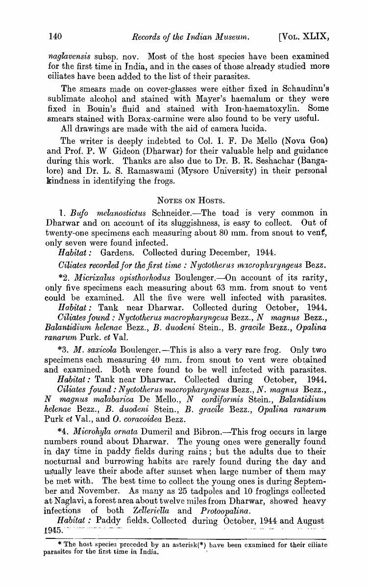
naglavensis subsp. nov. Most of the host species have been examined for the first time in India, and in the cases of those already studied more ciliates have been added to the list of their parasites.
The smears made on cover-glasses were either fixed in Schaudinll's sublimate alcohol and stained with Mayer's haemalum or they were fixed in Bouin's fluid and' stained with Iron-haematoxylin. Some smears stained with Borax-carlnine were also found to be very useful.
All drawings are made ,vith the aid of camera lucida. The writer is deeply indebted to Col. I. F. De Mello (Nova Goa)
and Prof. P. W Gideon (Dharwar) for their valuable help and guidance during this work. Thanks are also due to Dr. B. R. Seshachar (Bangalore) and Dr. L. S. Ramaswami (Mysore University) in their personal kindness in identifying the frogs.
NOTES ON HOSTS.
1. Bufo melanostictus Schneider .-The toad is very COlnmon in Dharwar and on account of its sluggishness, is easy to collect. Out of twenty-one specimens each measuring about 80 lum. from snout to vent, only seven were found infected.
Habitat: Gardens. Collected during Deceluber, 1944.
Ciliates -recorded for the first time: N yctotherus '1nxcrophr:tryngeus Bezz. *2. Micrixalus opisthorhodus Boulenger.-On account of .its rarity,
only five specimens each measuring about 63 mm. from snout to vent could be examined. All the five were well infected with parasites.
Habitat: Tank near Dharwar. Collected during October, 1944. Oiliates found: Nyctotherus rnacropharyngeus Bezz., N magnus Bezz.,
Balantidium helenae Bezz., B. duodeni Stein., B. gracile Bezz., Opalina ranarum Purk. et Val.
*3. M. saxicola Boulenger.-This is also a very rare frog. Only two specimens each measuring 40 mm. from snout to vent were ob~ained and examined. Both were found to be well infected with parasites,
Habitat: Tank near Dharwar. Collected during October, 1944. Oiliates found: Nyctotherus rnacrophal'yngeus Bezz., N. rrnagnus Bezz.,
N magnus ma:labal'ica De Mello., N cordifonnis Stein., Balant·idiurn helenae Bezz., B. duodeni Stein., B. gracile Bezz., Opal'ina ranarum Purk et Val., and O. cOl'acoidea Bezz.
*4. Microhyla ornata Dumeril and Bibron.-This frog occurs in large numbers round about Dharwar. The young ones were generally found in day time in paddy fields during rains; but the adults due to their nocturnal and burrowing habits are rarely found during the day and u~ally leave their abode after sunset when large number of thelu may be met with. The best time to collect the young ones is during September and November. As many as 25 tadpoles and 10 froglings collected at Naglavi, a forest area about twelve Illiles from Dharwar, showed heavy infections of both Zelleriella and Protoopalina.
Habitat: Paddy fields. Collected during October, 1944 and August 1945.'-- ------.-- . -.- - ,- --- ,-, -" -.". -
* The host speciea preceded by an asterisk(*) have been examined for their ciliate parasites for the first time in Indja. \
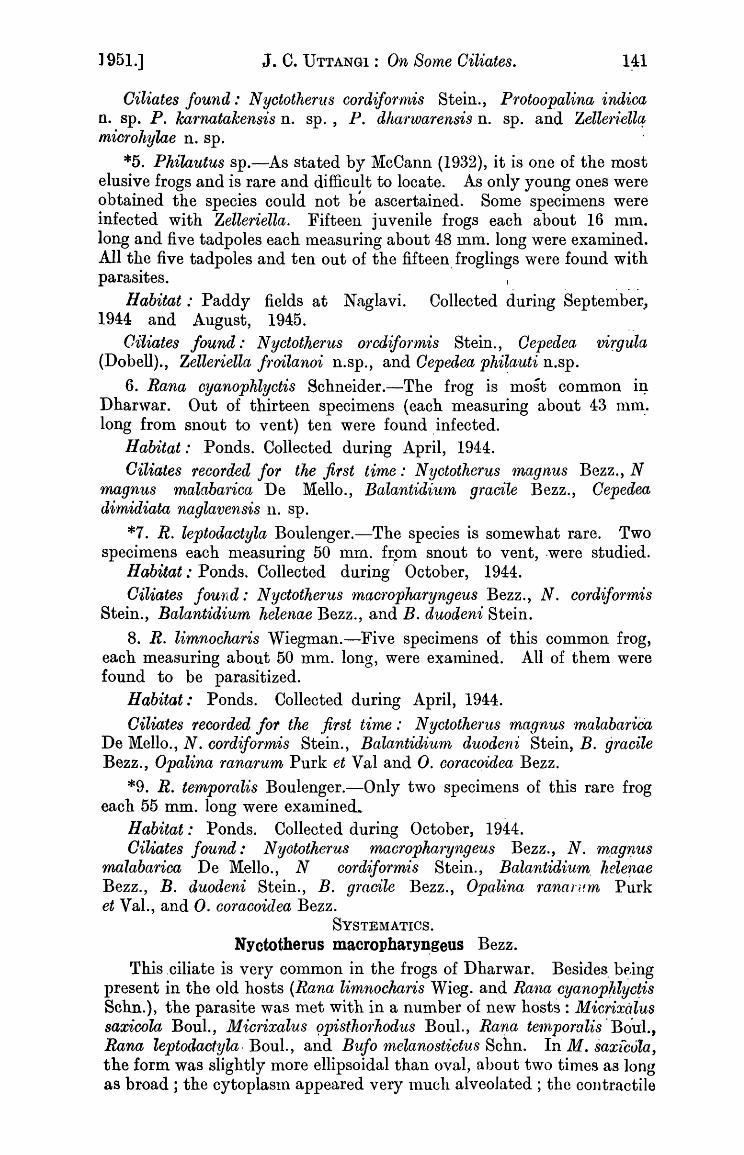
] 951.] J. C. UTTANGI: On SOlne Oiliates. 141
Oiliates found: N yctotheru s cordijor'mis Stein., Protoopalina indica n. sp. P. ka1'natakensis n. sp., P. dha1'warensis n. sp. and Zeller1~ellCf microhylae n. sp.
*5. Philautus sp.-As stated by McCann (1932), it is one of the most elusive frogs and is rare and difficult to locate. As only young ones were obtained the species could not be ascertained. Some specimens were infected with Zelleriella. Fifteen juvenile frogs each about 16 mm. long and five tadpoles each measuring about 48 mm. long were examined. All the five tadpoles and ten out of the fifteen, froglings were found with parasites.
Habitat: Paddy fields at Naglavi. Collected during September" 1944 and August, 1945. ..
Vitiates found: N yctotherus orrx1ifol'mis Stein., Oepedea virgula (Dobell)., Zelleriella froilanoi n.sp., and Oepedea phi~a'uti n.sp.
6. Rana oyanophlyctis Schneider.-The frog is most common iJ? Dhar,var. Out of thirteen specimens (each measuring about 43 nlnl. long from snout to vent) ten were found ,infected.
Ilabitat: Ponds. Collected during April, 1944. Oiliates recorded for the first time: N yctotherus 1nagnus Bezz., N
m,agnus malabarica De Mello., Balantidium gracile Bezz., Cepedea dimidiata naglavensis n. sp.
*7. R. leptodactyla Boulenger.-The species is somewhat rare. Two specimens each nleasuring 50 mm. frpm snout to vent, ·were studied.
Habitat: Ponds. Collected during' October, 1944. Oiliates found: N yctotherus maoropharyngeus Bezz., N. cordif01'mis
Stein., Balantidium helenae Bezz., and B. duodeni Stein.
8. R. limnocharis Wiegman.-Five specimens of this common frog, each measuring about 50 mm. long, were examined. All of them were found to be parasitized.
Habitat: Ponds. Collected during April, 1944.
Oiliates recofded for the first time: N yctotherus magnus malabariC'a De Mello., N. cordifor,mis Stein., Balantidium duodeni Stein, B. gracile Bezz., Opalina ranarum Purk et Val and O. coracoidea Bezz.
*9. R. temporalis Boulenger.-Only two specimens of this rare frog each 55 mm. long were exalninecL
Habitat: Ponds. Collected during October, 1944. Oiliates found: Nyototherus macropharyngeus Bezz., N. mag'f!-us
malabarioa De Mello., N cordiformis Stein., Balantidium, heleTl,ae Bezz., B. duodeni Stein., B. gracile Bezz., Opalina Tanarum Purk et Val., and O. coracoidea Bezz.
SYSTEMATICS.
NYctotherus macrophary~geus Bezz.
This ,ciliate is very common in the frogs of Dharwar. Besides, b~.ing present in the old hosts (Rana limnocharis Wieg. and Rana cyanoplllyc~is Sehn.), the parasite was met with in a number of new hosts: Micrixalus saxicola BouI., Micrixalus 9pisthorhodus BouI., Raf!a te'lnporalis' BouI., Rana leptodactyla. BouI., and Bufo melanostictus Schn. IIi M. saxicvla, the form was slightly more ellipsoidal than oval, about two tinles as long as broad; the cytoplasln appeared very luuch alveoJated ; the coutractile
142 ReC01'ds of the Indian Museum. [VOL. XLIX,
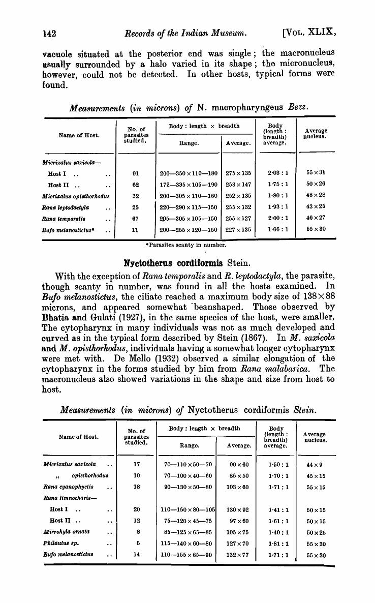
vacuole situated at the posterior end was single; the macronucleus usually surrounded by a halo varied in its shape; the micronucleus, however, could not be detected. In other hosts, typical forms were found.
Measurements (in microns) oj' N. macropharyngeus Bezz.
No. of Body: length x breadth Body Average (length:
Name of Host. parasites breadth) nucleus. studied. Range. Average. average.
MicrixalllB 8axicola-
Host I .. .. 91 200-350 x 110-180 275 x 135 2,03 : 1 65x31
Host II .. · . 62 172-335 x 105-190 253 x 147 1'75: 1 50x26
Micrizalu8 opisthorhoduB 32 200-305 x 110-160 252 x 135 1·80: 1 48x28
Rana leptodtktyla · . 25 220-290 x 115-150 255 x 132 1·93: 1 43x25
Rana temporaris · . 67 2P5-305 x 105-150 255 x 127 2'00: 1 46x27
Bu!o melanostictus· .. 11 200-255 x 120-150 227 x 135 1·66: 1 65 x80
.Parasites scanty in number.
Nyctotherus cordiformis Stein. With the exception of RQtna temporalis and R. leptodactyla, the parasite,
though scanty in number, was found in all the hosts examined. In Bufo melanostict'Us, the ciliate reached a maximum body size of 138x 88 microns, and appeared somewhat "beanshaped. Those observed by Bhatia and Gulati (1927), in the same species of the host, were smaller. The cytopharynx in many individuals was not as much developed and curved as in the typical form described by Stein (1867). In M. saX'icola and M. opisth01'hodus, individuals having a somewhat longer cytopharynx were met with. De Mello (1932) observed a similar elongation of the cytopharynx in the forms studied by him from Rana malabatrica. The macronucleus also showed variations in th~ shape and size from host to host.
Measurements (in microns) of Nyctotherus cordiformis Stein. ,
No. of Body: length x breadth Body Name of Host. parasites (length: Average
studied. breadth) nucleus. Range. Average. average.
Micrixalus Baricola .. 17 70-110 x 60-70 90x60 1'60: 1 44x9
" opi8thorhodu8 10 70-100 x 40-60 85x50 1'70: 1 45 x15
Rana cyanophycti8 · " 18 90-130 x 50-80 103 x60 1'71: 1 55x15
Rana limnochari.-
Host I .. .. 20 110-150 x 80-105 130x 92 1'41 : 1 60x15
Host II .. .. 12 75-120 x 45-75 97x60 1'61: 1 60x15
Mirf'ohyla ornata .. 8 85-125 x 65-85 105 x 76 1'40: 1 60x25
PhilDutWl ap. · . 6 115-140 x 60-80 127 x 70 1'81: 1 65x30
Bu!o melanoBtictUB · . 14 110-155 x 65-90 132x77 1·71 : 1 65x30
1951.] J. C. UTTANGI: On Some Oiliates. 143
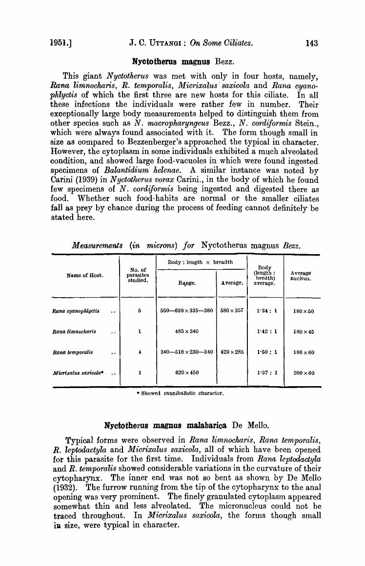
Nyctothems magnus Bezz.
This giant N yctotherus was met with only in four hosts, namely, Rana limnocharis, R. temporalis, M im'ixalus' saxicola and Rana cyanophlyctis of which the first three are new hosts for this ciliate. In all these infections the individuals were rather few in number. Their exceptionally large body measurements helped to distinguish them from other species such as N. macropharyngeus Bezz., N, cordiJormis Stein., which were always found associated with it. The form though small in size as compared to Bezzenberger's approached the typical in character. However, the cytoplasm in some individuals exhibited a much alveolated condition, and showed large food-vacuoles in which were found ingested specimens of Balantidium helenae. A similar instance was noted by Carini (1939) in Nyctotlterus vorax Carini., in the body of which he found few specimens of N. cordiJormis being ingested and digested there as food. Whether such food-habits are normal or the smaller ciliates fall as prey by chance during the process of feeding cannot definitely be stated here.
Measurements (in microns) for Nyctotherus magnus Bezz.
Body: length x breadth
No. of Body Name of Host. parasites (length: Average
studied. bredth) nuc1eus. Ritnge. Average. average.
Rana cyanophlyctis .. 6 650-610 x 335-380 580 x 357 1'34: 1 180x50
Rana limnochaTi3 .. 1 485 x 340 1'42 : 1 180 x 46
Rana temporalis .. 4 340-518 x 230-340 429 x285 1'60: 1 186 x60
Micrixalus saxicola* o. 1 620 x 450 1'37 : 1 200 x60
• Showed canniba.listic cha.racter.
Nyctotherus magnUS malabarica De Mello.
Typical forms were observed in Rana limnocharis, Rana temporalis, R. leptodactyla and Micrixalus saxicola, all of which have been opened for this parasite for the first time. Individuals from Rana leptodactyla and R. temporalis showed considerable variations in the curvature of their cytopharynx. The inner end was not so bent as shown by De Mello (1932). The furrow running from the tip of the cytopharynx to the anal opening was very prominent. The finely granulated cytoplasm appeared somewhat thin and less alveolated. The lllicronucleus could not be traced throughout. In M icrixalus saxicola, the forIns though small ill size, were typical in character.

144 Records of tIle Indian Museum. [VOL. XLIX,
Measurements (in miClfons) for Nyctotherus magnus malabarica De Mello.
No. of Body : length x breadth Body (length: Average Name of Host. parasites breadth) nucleus. studied. Range. AYerage. average.
])1 icrixalu8 opistlwrilOdu8 6 120-150 x 90-110 135 x 100 2'00: 1 55x20
Rana temporalis .. 10 135-190 x 90-130 162 x 110 1·56 : 1 50x25
Rana leptodactllla-
Host I .. - - 21 140-200 x 95-145 170 x 120 1·41: 1 55x30
Host II - . .. 25 200-280 x 110-170 240 x 140 1·91 : 1 55 x30
Rana limnocharis -- 17 105-170 x 75-110 137 x 92 1·48: 1 60x30
Balantidium belenae Bezz.
Though Microhyla ornata, Philautus sp., and Bufo 'lnelanostictus were entirely devoid of this ciliate, it was abundantly present in other host species. In M icrixalus saxicola, the form varied fronl elongately oval to broadly oval. The position of the macronucleus varied in many individuals. Out of 75 specimens 32 had their nuoJeus in about the middle of the body, while in the remaining 43, it was found to lie in the posterior half of the body. The cytoplasm contained some irregular bodies ,vhich were stained like the nucleus. The anal opening situated at the posterior pole was clearly visible. The boring apparatus as reported by Ray, (1932) and Chakravorti (1933) ,vas not detected. In M. opisthorhodus, Rana temp ora lis, R. leptodaotyla, R. limnooharis, and R. cyanophlyctis typical forms were found.
The broadly oval forms have been described under a new species B. ovale (DobeIl1910, p. 74). After studying these forms in the Lahore frogs, Bhatia and Gulati (1927, p. 107) came to the conclusion that they were identical in strncture with B. helenae and differed only in size; and should, therefore, be treated as one and the same species. Similar forms having been met with in the hosts of Dharwar, the writer is of the opinion that Bhatia and Gulati are probably correct.
Measurements (in microns) for Balantidium helenae Be~·z.
No. of Body: length x breadth Body Name of Host. parasites (length: Average
studied. breadth) - nucleus. Range. Average. average.
--M icrixalus saxicola .. 75 80-140 x 40-150 110 x 95 1-10: 1 30x12
},I. opisthorhodus · . 10 4.3-60 x 28-32 52x30 1'73 : 1 20xlO
Rana temporalis · . 22 55-115 x 30-50 85x40 2-12: 1 30xl0
R . leptodactyla · . 34 50-105 x 30-05 77 x47 1'50: 1 25x10
R. limnocllaris · . 20 40-90 x 30-45 65 x37 1-71 : 1 30x9
R _ cyanoplllyctis · . 49 50-85 x 25-50 "-
67 x 37 1'81 : 1 30xl0

,1951.] J. C. UTTANGI: On Some Ciliates. 145
Balantidium gracile Bezz. With the exception of Microhyla ornata, Bufo rnelanostictus and
Philautus sp. the parasite occurred in all the hosts examined at Dharwar. ,It was usually located in the intestine. Bezzenberger (1904) found the macronucleus lying mostly at the hinder end and seldom in the middle. In Dharwar ~orlns it was usually in the middle.
Measure'ments (in '1nic;rons)for Balantidium gracile Bezz.
Name of Host. No. of parasites studied.
-Micr-ixalus saxicola* .. 27
" M. oputhorhodu8 .. 46
Rana temporalut .. 4
Rana leptodactyla .. 44
Rana limnocharu .. 65
Rana cyanophlyctis .. 24
• Pure culture in small intestine. t Parasites scanty in number.
Body: length x bread th
Range. Average.
145-210 x 25-30 177 x 27
95-185 x 20-35 140 x 27
130-155 x 20-35 142x25
170-285 x 20-35 227 x 27
90-195 x 30-40 142 x 35
120-260 x 20-25 190 x 22 I
Balantidium duodeni Stein.
Body (length: breadth) average.
6'55: 1
5'18: 1
5'60: 1
8·40: 1
4'05: 1
8'60: 1 I
Average nucleus.
20xlO
20x9
20xll
36xlO
20x10
30~10
The ciliate occurred in the new hosts, Rana ternporalis, R. leptodactyla, M icrixalus saxicola, and M. opisthorhodus as well as in the old hosts R. limnochwi'is and R. cyanophlyctis. ~ Typical specimens were met with in all the hosts.
Comparing the characters of B. rotundum Bezz. with B. duodeni Stein, it is found that the former differs from the latter in having a straight dorsal surface (which, perhaps, is due to the much rounded sides) that gives the parasite an appearance of an egg. In other characters like the nucleus, the triangular are.i1 in the cytoplasm and the peristome they are quite identical. In the specimens studied at Dharwar, it was noted that both oval and egg-shaped forms were present. Those that were small in size appeared egg-shaped, while the larger forms appeared langeI and oval. B. rotundU1n, therefore, may well be conlpared to these smaller forms of B. duodeni. And since B. rotundum is not structurally very different from B. duodeni, I am inclined to think that both these duodena] forms are identical.
Measurements (in microns) for Balantidium doudeni Stein.
No. of Body: length x breadth Body Name of Host. parasites (length: Average
studied. breadth) nucleus. Range. Average. average.
- -Micrixalus saxicola* .. 19 25-43 x 14-30 34 x22 1'54: 1 lOx 7
M. opisthorhodus .. 53 30-55 x 20-30 42x25 1'68: 1 11 x 8
Rana temporalu .. 23 30-48 x 20-24 39x22 1'77 : 1 10x8
R. limnocharist .. 7 40-50 x 20-30 45x25 1'80: 1 9'5 x!)
R. C'ljan(}pltlyctis .. 16 35-45 x 25-30 40x27 1'58: 1 10x8
• Abundant in duodenum. t Parasites scanty in number.
146 Records of the Indian j;l useurn. [VOL. XLIX,
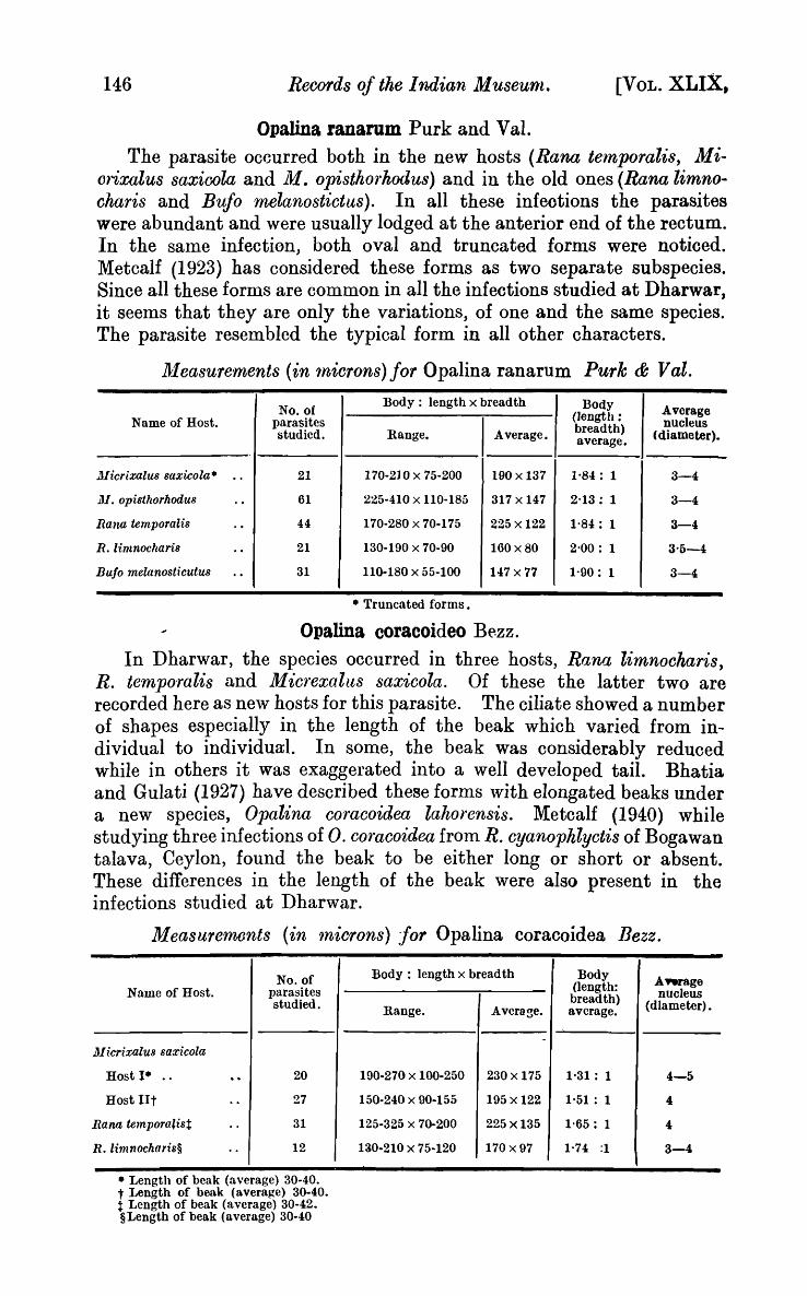
Opalina ran arum Purk and Val. The parasite occurred both in the new hosts (Rana temporalis, Mi
orixalus saxioola and M. opisthorkoilus) and in the old ones (Rana limno .. charis and Bufo melanostictus). In all these infeotions the parasites were abundant and were usually lodged at the anterior end of the rectum. In the same infection, both oval and truncated forms were noticed. Metcalf (1923) has considered these forms as two separate subspecies. Since all these forms are common in all the infections studied at Dharwar, it seems that they are only the variations, of one and the same species. The parasite resembled the typical form in all other characters.
~leasurements {in 1nicrons)for Opalina ranarum Purk & Val.
No. of Body: length x breadth Body Average (length: Name of Host. parasites I Average.
breadth) nucleus studied. Range. average. ( diameter).
l1Iicrixalus saxicola· .. 21 170-210 x 75-200 190 x 137 1,84: 1 3-4
1)1. opisthorhodu8 .. 61 225-410 x 110-185 317x147 2'13: 1 3-4
Rana temp ora lis .. 44 170-280 x 70-175 225 x 122 1'84: 1 3-4
R. limnocharis .. 21 130-100 x 70-90 160 x 80 2'00: 1 3'5-4
Bujo melanosticutus .. 31 110-180 x 55-100 147 x 77 1'90: 1 3-4
• Truncated forms.
Opalina coracoideo Bezz. In D harwar, the species occurred in three hosts, Rana limnockaris,
R. temporalis and Micrexalus saxicola. Of these the latter two are recorded here as new hosts for this parasite. The ciliate showed a number of shapes especially in the length of the beak which varied from individual to individual. In some, the beak was considerably reduced while in others it was exaggerated into a well developed tail. Bhatia and Gulati (1927) have described these forms with elongated beaks under a new species, Opalina coracoidea lahorensis. Metcalf (1940) while studying three infections of O. c014acoidea from R. cyanophlyctis of Bogawan talava, Ceylon, found the beak to be either long or short or absent. These differences in the length of the beak were also present in the infections studied at Dharwar.
Measurements (in '1nicrons) for Opalina coracoidea Bezz.
No. of Name of Host. parasites
studied.
lJ-licrixalu8 saxicola
Host I· . . · . 20
Host lIt · . 27
Rana tempora~ist · . 31
R. limnocharis§ · . 12
• Length of beak (average) 30-40. t I~ength of b~\k (average) 30-40. t Length of beak (average) 30-42. §Length of beak (average) 30-40
Body : length x breadth Body Aftrage (length: nucleus breadth) (dIameter) • Range. Average. average.
-
190-270 x 100-250 230 x 175 1'31: 1 4-5
150-240 x 90-155 195 x 122 1·51: 1 4
125-325 x 70-200 225 x135 1'65: 1 " 1:l0-210 x 75-120 170 x 97 1·74 :1 3-4

1951.] J. C. UTTANGI: On Some Oiliates. 147
Cepedea virgula (Doh ell) Metcalf. The ciliate was met with in Pltilautus sp. which was heavily infected.
The horizontally placed endospherules were well stained with ironhaematoxylin.
Measurements in microns.-35 parasites. Body (length x breadth): range, 80-125 X 35-50; average, 102 x 42 ;
length: breadth (average), 2·33: 1 ; average nucleus (diameter), 5.
Cepede~ philanti, sp. nov. The body, clothed with short cilia, is cylindrical and somewhat
fusiform with the posterior middle of the hody having the greatest width. The anterior and the posterior ends are broadly rounded. The coarsely alveolated cytoplasm is not differentiated into ectosarc and endosarc and appears somewhat compact near the anterior pole. The features that are characteristic in this new species are the numerous large and oval nuclei spread irregularly in the cytoplasm. Incomplete transverse constrictions similar to those seen in O. formosae Met. were observed in some individuals.
Host.-Philautus sp. Measurements in microns.-19 individuals. Body (length x breadth): range, 255-360 x 50-80 ; average, 307 x 65;
Average nucleus (diameter), 4-5. Dividing nucleus (oval), 10 X 7. Cilia line interval, anterior region 1, posterior region, 2. Diameter of endospherule, 3-4. ..
Key to the species of Cepedea. I (3) Anterior end with a spine-like projection 2 Body triangular in cross-~ection. Posterior end
bluntly rounded. Length, 82 II 3 (l) Anterior end without a spine-like projection 4 (15) Body greatly elongated 5 (10) Body with transverl'1e constrictions .. 6 (9) With one or two incomplete constrictions 7 (8) Spindle-shaped body with elliptical nuclei.Length,
179 {L
8 (7) Fusiform body with spherical nuclei. Length, 307 V-9 (6) 'Vith many complete constrictions. Length,365 (.L
10 (5) Body without any constrictions II (12) Nuclei ellipsoidal. Length, 680-1000 V-12 (1 i) N udei spherical 13 (14) Cilia short. Length, 84S .. 14 (13) Cilia. long. Length, 500 V-15 (4) Body not greatly elongated 16 (25) Body cylindrical 17 (24) Sides of the body curved, anterior end rounded and
posterior tapering to a point 18 (19) With a naked spine at the posterior pole small round
nuclei (dia.meter) 3·8 EL, Length, 124 II 19 (IS) 'Vithout a naked spine at the posterior polo 20 (23) Nuclei spherical 21 (22) Endospherulc3 round and irregularly placed
Length, 180 (.t 22 (21) Endospherules elongated and trarwversely placed
Length, 98 V- •• . ,
2
c. punjabensis Bh. & G. 4 5 6 7
C. formosae Met. O. philauti sp. n. C. segmentata. Met. 11 O. longa Bezz. 13 C. dolicosoma Met. C. ophis Met. 16 17
IS
o. spinifera Met. 20 21
O. dimidiata Stein.
(1. virgula (Dobel!)
148 Records of the Indian Museum.
23 (20) Nuclei dumbcll-shaped. Length, 131 EL 24 (17) Sides of the body straight. Anterior end presenting
a vacuolate d appearance. osterior eod rounded, sometime~ IJointed. Length,] 25-440 ,.,. ..
25 (16) Body lanceolate. Anterior end broad and rounded. Posterior, slender and tapering to a point. Nuclei only 4 or 5, Length, 82 (.L ••
26 (25) Body obovate. Length, 315 (.L 27 (26) Body globose .. 28 (29) N llclei elliptical. Cilia dense and long. Length,
170 (1. • 29 (28) Nuclei spherical. Cilia sparse and short. Length,
87 (1. 30.(27) Body sub-cylindrical .. 31 (32) Both ends rounded or anterior end less pointed than
the p03terior. Length, 35-250 (1.
[VOL. XLIX,
a. celebensis Met.
C. th.iagi De Mello.
a. lanceolata Met. a. obovoidea Met. 28
o. globosa l\let.
O. bandivii Met. 31
a. subcylindrica De Mello.
32 (31) Both ends pointed. Length, 64-89 (.L a. sialkoti Bh. & G.
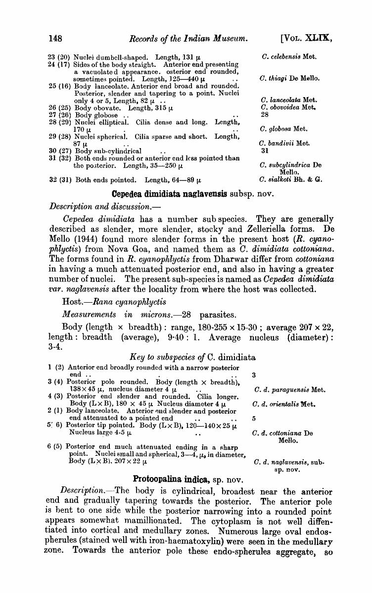
Cepedea dimidiata naglavensis subsp. nov. Description and discussion.-
Oepedea dimidiata has a number sub species. They are geneT&lly described as slender, more slender, stocky and Zelleriella forms. De ~Iello (1944) found more slender forms in the present host (R. cyanophlyctis) from Nova Goa, and named them as O. dimidiata cottoniana. The forms found in R. cyanophlyctis from Dharwar differ from cottoniana in having a much attenuated posterior end, and also in having a greater number of nuclei. The present sub-species is named as Oepedea dimUliata 'l.'ar. naglavensis after the locality from where the host was collected.
Host.-Rana cyanophlyctis
Measurements in microns.-28 parasites. Body (length x breadth): range, 180-255 x 15-30; average 207 x 22,
length: breadth (average), 9·40: 1. Average riucleus (diameter): 3-4.
Key to subspecies of c. dimidiata 1 (2) Anterior end broadly rounded with a narrow posterior
end.. . 3 (4) Posterior pole rounded. Body (length X breadth).
138 X 45 (1., nucleus diameter 4 (1. 4 (3) Posterior end slender and rounded. Cilia longer.
Body (L X B), 180 X 45 (1. Nucleus diameter 4 (1. 2 (1) Body lanceolate. Anterior ~lld slender and posterior
end attenuated to a pointed end 5: 6) Posterior tip pointed. Body (L X B), 120-140 X 25 (l.
Nucleus la!ge 4-5 [J. ••
6 (5) Post~rior end much attenuated ending in a Rharp point. Nuclei s!nall and spherical, 3-4, (1., in diameter, Body (L>< B). 207 X 22 (1.
Protoopalina indica, sp. nov.
3
a. d. paraguensis Met.
a. d. oriental is Met.
5
a. d. cottoniana De Mello.
a. d. naglavensis, subsp. nov.
Description.--The hody is cylindrical, broadest near the anterior end and gradually tapering towards the posterior. The anterior pole is bent to one side while the posterior narrowing into a rounded point appeats somewhat mamillionated. The cytoplasm is not well diffentiated into C9rtieal and medullary zones. Numerous large oval endospherules (stained well with iron-haematoxylin) were seen in the medullary zone. Towards the anterior pole these endo-spherules aggregate, 80
1951.:1 J. C. lJTrANGl: On Some Oiliates.
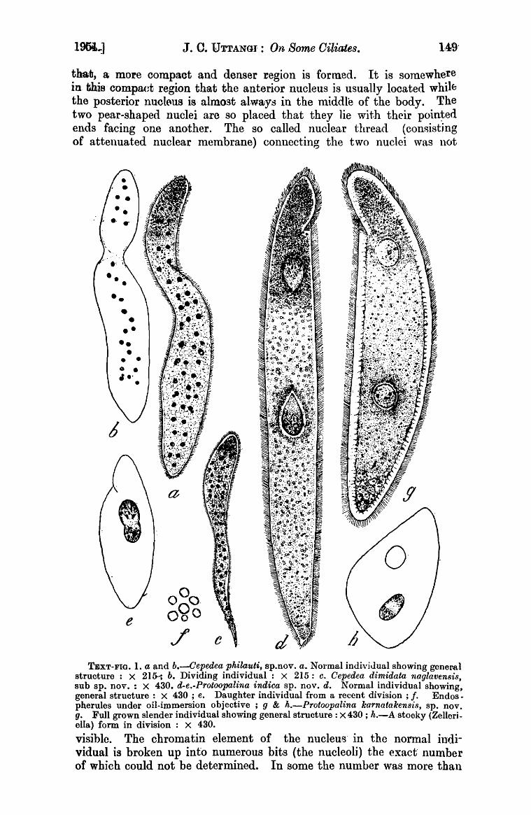
thafi, a more compact and denser region is formed. It is somewhere in this compaet region that the anterior nucleus is usually located whilb the posterior nucl(\us is· almo3t always in the middle of the body. The two pear-shaped nuclei are so placed that they lie wit.h their poin~ed ends facing one another. The so called nuclear thread (conRistjng of attenuated nuclear membrane) connecting the two nuclei was ilot
•• •• • • • • • .e' •
e
TEXT-FlO. 1. a and b.---Cepf!-dea pkilauti, sp.nov. a. Normal indivjJual showing gcneool structure : X 215-; b. Dividing individual : X 215: c. Cepedea dimidata naglavensis, sub Spa nov. : X 430. d-e.-Protoopalina indica Spa nov. d. Normal individual showing, general structure : X 430 ; e. Daughter individual from a recent division ; f. Endos -pherules under oil-immersion objective; g & h.-Protoopalina karnata ken s·is, Spa nov. g. Full grown slender individual showing general structure: X 430; It.-Astooky (Zelleriella) form in division: X 430.
visible. The chromatin element of the nucleus' in the nonnal individual is broken up into numerous bits (the nucleoli) the exact" number of which could not be determined. In some the number was more than

150 Records of the Indian ~luseum. [VOL. XLIX,
twenty. The cilia were long and thick. Individuals undergoing clivision (both transverse and longitudina)), were present. The daugltter cells come from transverse division ,vere all uninucleated.
Host.-Microhyla ornata Measurements in rnicrons.-61 individuals. Body (length X breadth): range, l!l5-415 X 40-75 ; average (LxB)
275 X 57 ; length: breadth (a.verage) 4·82 : 1 ; nucleus (length X breadth) : range, 27 .. 33 X 14-17. Endospherule (length X breadth) : range, 2·5-3·5 X 1·5-2. f;ilia line interval, ant.erior region, 1·8 ; middle region 2·10 ; posterior region, 3·00. Width of ectosarc, 3·00.
Cornparison.-On account of its slender pointed posterior end, as well as the body mea.surements and the shape of the nucleus, P. indica is classed under the sub-generic group II (Metcalf 1940, p. 571-576).
The following table shows the specific differences of the group.
Key to subgeneric group II. 1 (2) Posterior pole tapering to a sharp point and sometimes
with a posterior process 3 3 (6) Posterior end broad and abruptly ending in a point 4 4 (5) Posterior end not curved. Nucleoli 6 in number
Nucleus (L x B) 40 x 26 (.t. Body (L X B) 200 X 95 (L P. caudata (Stein).
5 (4) Posterior end curved. Tail large and blunt. Nuc-leoli 8 in number. Nucleus (L X B) 30 X 14 tL. Body (L X B) 90 X 74 (1. P. macrocaudata. Met.
6 (3) Posterior end elongated and gradually tapering to a narrow point 7
7 (10) Posterior pole rounded or sometimes pointed 8 8 (9) Nucleoli 8 in number. Nucleus pear-5haped (L X B)
32 X 10 lL. Body (L X B) 330 X 68 (1. P. intestinalis. (Stein). 9 (8) Number of nucleoli undetermined. Oval nucleus,
(L X B) 23 X 13 (L Body (L X B) 288 X 63lL . • P. orientalis Met. 10 (7) Posterior pole mamillionated. Nucleoli 20 in
number. Nucleus pear-shaped, (L X B) 27 X 14 (1.. Body (L X B) 275 X 57 lL P. indica sp. nov.
2 (1) Posterior pole tapering to a broadly rounded point 11 11 (12) Nuclei close together in centre. Nucleus oval
(L X B) 18 X 10 lL. Body (L X B) 230 X 46 ll. Nucleoli number not mentioned P. yunnanensis l\Iet.
12 (11) Nuclei far apart 13 ]3 (14) With definite number of nucleoli.-
(a) Nucleoli 6 in number. Body (L X B) 170 X 24lL-Nucleus (L X B) 14.. X 9 (1. • • •• P. stejnegeri Met.
(b) Nucleoli 8 in number. Slender Body (L X B) 97 X 18 (1.. Nucleus ellipsoidal (L X B), 10 X 6 (1. P. pelobatides Met.
(e) Nucleoli 4 in number. Body (L X B) 240 X 68 (L. Nucleus (L X B) 25 X 8·9 (L • • •• P. dorso.lis (Raff).
(d) Nucleoli 3 in number. Nucleus (L X B) 6 X 211, (anaphase). Body (L X B) 63 X 11 (1. P. caccosterni Fantham.
14 (13) With an undetermined number of nucleoli 15 15 (16) Nucleus ellipsoidal (L X B) 10 X 6 (1.. Body
(L X B) 160 X 32 (1. P. peronii Met. 16 (15) Nucleus rounded 17 17 (18) Nucleus smaller (diameter) 10 (1.. Body (L X B)
572 X 70 (1. P. hylarum (Raff). 18 (17) Nucleus large (diameter) 19 (L. Body (L X B)
313 X 70 P. luzonensis Met.
1951.] J. C. UTTANGI: On Some Ciliates. 151
Protoopalina karnatakensis, 81\. nov.
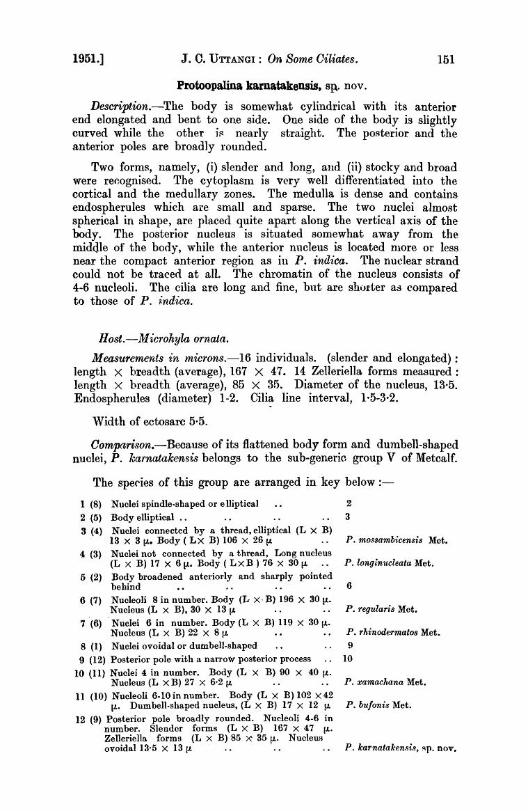
Description.-The body is somewhat cylindrical with its anterior end elongated and bent to one side. One side of the body is slightly curved while the other i~ nearly straight. The pOl'Jterior and the anterior poles are broadly rounded.
Two forms, namely, (i) slender and long, and (ii) stocky and broad were re~ognised. The cyt.oplasnl is very well differentiated into the cortical and the medullary zones. The medulla is dense and contains endospherules which are small and sparse. The two nuclei almost spherical in shape, are placed quite apart along the vertical axis of the body. The posterior nucleus is situated somewhat away from the middle of the body, while the anterior nucleus is located more or less near the compact ant.erior region as in P. ind~·ca. The nuelear strand could not be traced at all. The chromatin of the nucleus consists of 4-6 nucleoli. The cilia are long and fine, but are shorter as cOlnpared to those of P. 1:ndica.
Host.-M icrohyla ornata.
Measurements in microns.-16 individuals. (slender and elongated) : length X breadth (average), 167 X 47. 14 Zelleriella forms measured: length X breadth (average), 85 X 35. Diameter of the nucleus, 13·5. Endospherules (diameter) 1-2. Cilia line interval, 1·5-3,2.
'Vidth of ectosarc 5·5.
Oomparison.-Because of its flattened body form and dumbell-sbaped nuclei, P. karnatakensis belongs to the sub-generic group V of Metcalf.
The spe(lies of this group are arranged in key below :-
1 (8) Nuclei spindle-shaped or elliptical 2
2 (5) Body elliptical .. 3
3 (4) Nuclei connected by a thread, elliptical (L X B) 13 X 3 ~. Body ( L X B) 106 X 26 ~ P. mossambicensis Met. Nuclei not connected by a thread, Long nucleus 4 (3) (L X B) 17 X 6 (1.. Body ( Lx B ) 76 X 30 (1. P. longinucleata Met.
5 (2) Body broadened anteriorly and sharply pointed 6 behind
6 (7) Nucleoli 8 in number. Body (L X - B) 196 X 30 [.1.. P. regularis Met. Nucleus (L X B), 30 X 13 (1.
7 (6) Nuclei 6 in number. Body (L X B) 119 X 30~. Nucleus (L X B) 22 X 8 ~ P. rhinodermatos Met.
8 (1) Nuclei ovoidal or dutnbell-shaped 9
9 (12) Posterior pole with a narroW posterior process . . 10
10 (11) Nuclei 4 in number. Body (L X B) 90 X 40~. Nucleus (L X B) 27 X 6·2 ~ .. .. P. xamachana Met.
11 (10) Nucleoli 6-10 in number. Body (L X B) 102 X 42 ~. Dumbell-shaped nucleus, (L X B) 17 X 12 ~ P. bufonis Met.
12 (9) Posterior pole broadly rounded. Nucleoli 4-6 in number. Slender forms (L X B) 167 X 47 ~. Zelleriella forms (L X B) 85 X 35~. Nucleus ovoidal 13·5 X 13 lL P. karnataken8is, ~p. nov.
152 /teGO'fds of tke Indian M usettrlt. [VOL. XLIX,
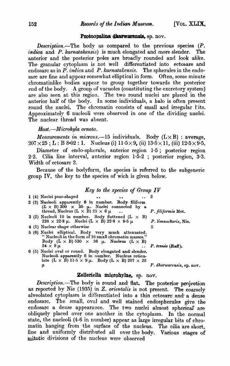
Pmioopaliua tUaarwarensis, sp. nov.
Description.-The body as compared to the previous species (P. indica and P. karnatak.ensis) is much elongated and more slender. Theanterior and the posterior poles are broadly rounded anrl look a·like. The granular cytoplasm is not well differentiated into ecto~are andendosa.rc as in P. indica and P. karnatal~ensis. The spherules in the endosarc are fine and appear 80nlewbat elliptical in form. Often, sonle minute chronlatinlike bodies appear to group t.ogether towards the posterior end of the body. A group of vacuoles (constituting the excret.ory systenlJ are also seen a.t this region. The two round nuclei are placed in the anterior half of the body. In some individuals) a halo is often present round the nuclei. The chromatin consists of snlall and irregular -tits. Approxinlately 6- nucleoli ,vere observed in one of the dividing nuclei. The nuclear thread wa.s absent.
/It()st.-IJI icrohyla ornata.
Measurernents in miC'fons.-15 individuals. Body (J.J X B) : a-verage, 207x25; IJ: B 8-02: 1. Nucleus (i) 11·5x9, (ii) 13·5x11, (iii) 12·5x9·5.
Diameter of enflo-spherule, anterior region 1·5; posterior region 2·2. Cilia line interval, a.nterior region 1·5-2 ; posterior region, 3·3. 'Vidth of ectosarc 2.
Because of the bodyform, the' species is referred to the subgeneric group IV, the key to the species of wich is given below.
Key to the species of flrO'ltp IV 1 (4) Nuclei pear-shaped • • 2 2 (3) N-uoleoli apparently 6 in number. Body. filiform
(L X B)' 300 X 35t lJ... Nuolei connected by a. thread, Nucleus (L X B) 25 X 6 (L P. filiformis Met.
a (2) Nucleoli 10 in number. Body flattened (L X B) 238 X 22·8 fJ.. Nuclei (L X B) 22~8 X 9·5 fJ. P. limnocharis, Nie.
4 (1) Nuclear shape otherwise 5 5 (6) Nuclei elliptical. Body very much attenuated.
" Nucleoli in the form of 16 small chromatin masses." Body (L X B) 530 X 36 (L. Nuoleus (L X B) 24- X 9 (L P. tenms (Baff',.
6 (5) Nuclei oval or round. Body elongated· and: slender .. Nucleoli. apparently 6 in number. Nucleus reticu .. late (L X B) 11·5 X 9lJ.. )lody; (L X B) 207 X 25 l.t P. dharwarensi8, Spa nov.
Zelleriella microhylae~ sp. nov.-
Description.-The body is round and Hat. The posterior pl!ojeotion as reported by Nie (1935) in Z. orientalis is Dot present. The coarse)v alveolated cytoplasm is differentiated into a thin ectosaTC' andl a denB~ endosa,rc. The small, oval and well stained endospherule8' give the endosarc a dense appearance. The two nuclei almost spherieal~ are obliquely placed over one another in the cytoplasm. In the normal state, the nucleoli (4-6 in numbeD) appear as large irregular bits of chro:.. matin hanging from the su;rface of the nucleus. The cilia are short, fine and uniformly distributed all over the body. Various stages of mitotic divisions of the nucleus were observed "

19i1.J J. :0. UTTANGI : ()n, Some Oiliates. 153
Host.-Microhyla ornata.
lfle18·urements iIn miC1008.-34 individua;ls. Body (1e~gth X breadth) : range 70-11iO x60-80 ; average (L X B) X 70, Length: BTeadth(average) 1-28 : 1 ; Dividing individunl (1<1 X B),
~~ (f)fl ~' ~ ~cf) o ~ 0.' : 0 . o·
: 0' 0 ,0
~~ ~/-", ~
~~\:!~:{:i ~" .t·,.·.·.: ~l,:·· .. .,; ... : ... ;-,.~(.~
!l
~";f.,_:.-.... ;~), ... ~~(;~ .. ~~~~.! . .;£.~~ 1(~~~.:~,{, ~t.:}i~)" lCf~~~~~~11
a TEXT-FIG. 2. a and b.-Protoopalina dharwarensis sp. nov. a. Normalindividual
showing general structure: X 430 ; b. Da.ughter cell from a longitudinal division showing dumbell-shaped nucleus : X 430. c-d.-Zelleriella microhylae, ap. nov. c. Normal individual showing general structure: X 430; d. Nuclei in prophase: X 430; e. Dividing individuals with nuclei in telophase: X 430. f. & g.-Zelleriella froilanoi, sp. nov. f. Normal individual showing' general structure: . X 430; g. Dividing individuals under low power: X 100.
150 X 90. Diameter of the resting nucleus, 10-13. Cilia line interval: anteriur region, 3 ; posterjor- region, 4. Length of the ~i1ia, 8. Width
f ectosa.l'C, 4-4·5.

154 Records of the Indian Museum. [VOL. XLIX,
Zelleriella froilanoi, sp. nov.
Description.-The body is thin, flat a.nd bluntly rounded at the posterior end. The anterior pole is broad and round. The posterior one is tapering to a rounded point. The small and stocky forms have somewhat a wedge-shaped body. lJnlike Z. microllylae, the cytoplasm is not. well differentiated into the usual zones. The protoplasm is very finely granulated and is evenly distrihuted. Cytoplasmic inclusions in the form of very fine granules which stain like chromatin 'v ere seen in some individuals. The oval endospherules a.re small and sparsely distributed in the cytoplasm. Some small and large vacuoles are often seen to group together at the posterior pole of the body. The t·,~o spherical nuclei are pla.ced one over the other in the vertical axis of the body. The chromatin is found to be in the form of minute granules and not in large bits as seen in Z. m,icrohylae. In the dividing nucleus the nucleoli were approximately 8 in number but u~ually varied frOID
6-8. Host.-Pllilautus sp.
Measurements in rnicrons.-71 individuals.
Body (length X breadth) : range 80-150 X 2n-55 ; average (L X B), 115 X 4-0 : I.length : Breadth (average) 2·87 : 1 ; diameter of the nucleus 10-12. Distance between the two nuclei 15-30. I.length of the cilia, 12. Cilia line interval, 3.
Key to the specie.s of Zelleriella. 1 (13) Body without a posterior curved process
2 (7) Body with a posterior end narrowly elongated and pointed.-
(a) Body, trumpet-shaped, posterior end elongated. Body (L X B), 180-220 X 75-100 (1.. Nucleus spherical, 20-22 f1. diameter ..
(b) Body cone-shaped. Posterior pole rounded. N uclens spherical 15-17 lL in diameter ; distance between two nuclei 20-25 lL. Body (L X B), 100 X !l0 lL ••
(c) Body comma-shaped. Posterior pole' pulled to a point. Body 70-90 {.I. in length. Diameter of the spherical nuclei, 8-10 lL distance between two nuclei, 10 tJ.
(d). Body wedge-shaped
3 (4) Nuclei ellipsoidal or pear-shaped (number of nucleoli not mentioned); Body (L X B) 207 X 130~. Nucleus (L X B) 32 X 22 ~ ..
4 (3) Nuclei spherical
5 (6) Body very thin and broa~ly wedge-shaped; L X B, 115 X 40 11-. Nucleus diameter 10-12 {.I.. Nucleoli 6-8 in number. Distance between two nuclei, 15-30 (J.
6 (5) Body narrowly we~ge-~haped. Body (L X B),~\ 93 X 50 11-. N ucleoh 4 In n um ber. N uclens diameter 10 f1.
7 (2) Body with a posterior end broadly rounded.
(a) Body irregularly rounded, thin and leaf-like. L X B,300 X 200 11-. Nucleoli 8 in number. Spherical nucleus 25 (l.
2
z. cornucopia Carini.
z. falcata Carini.
Z. corniola Carini.
3
z. magna ~Iet. 5
z. froilanoi, sp. nov.
z. intermedia 1\Iet.
z. foliacea Carini.

1931.] J. C. UT'rANGl: On Some Oiliate,.
(6) Body triangular with a tl'uncated posterior and. Spherical nuclei 10·12 1'. Body (L X B), 80·150 X 45·65 (J. • • •• Z. trunca.ta Carini.
(e) Body oval and greatly flattened
8 (12) Nuclei rounded-
(a) Nuclei large, 20 l1. in diameter
9 (8) Nuolei sMaller
8
z. binucleam (Raft'.
10
10 (11) Nucleus 12 1" in diameter. Nucleoli 3·4 number .•
11 (10) Nucleus 10·13 (J. in diameter. Body thick (L X B),
Z. macronuclea~, Bczt.
70·110 X 60-80 (1.. Nucleoli 4·6 in numbel' .. Z. microkylae, "p. no~ .. 12 (8) Nllclei ovoidal (r~ X B) 1.3 X 10 Nllcleoli 4 in
number. Body (L X B) 130 X 82 (J. ••
13 (1) ·Body with a posterior curv;ed process
Z. bra8iliensi8 (Pinto).
14
14 (15) Posterior tail long and distinct, Nuclei (L X B) 23 X 13·9 (1.. Body (L X B) 100 X 60 (J.
15 (14) Posterior tail otherwise
16 (17) Posterior end terminates to a minute sharp point.
Z. antanesi Pessoa.
16
Nucleoli 8 (1) in number. Bony leaflike, (L x B) 87'5·120 X 45·70 [J... Nucleus 12·5 !l. in diameter .. Z. orientalis Nie.
17 ,16) Body broad anteriorly. Nucleoli 3 in number. Nucleus 10 (J. in diameter. Body (L X B) 105 X 80 V. •• ·Z. bulonix Met.
SUmIARY.
1. The ciliate parasites of about one hundred and twenty-five specimens of frogs a.nd toads of Karnatak, Bomhay Presidency, comprising nine different species have been studied pa.rticularly with a view to ascertain lJhe presence of binucleated opali· nids in them.
2. Five new species of Zelleriella and Protoopalina have been described, thus establishing, beyond doubt, the presenoe of binucleated opalinids m these anurans.
3. CiIiat~ Pltrasites of six of the host species have been reo lrded for the first time in India; and some more ciliates have been added to the list of parasites of the hosts already examined. Besides one new species and one new sub-species of the gt'DUS Cepedea, ten old species of ciliates found parasitic in the gut of these anura have also been recorded.
REFERENCES.
BEZZENBERGER, E., 1904.-Uber Infusorian aus asiatischen Anuren. A~·ch. Prot·istenkd. III, pp. 138-17/1.
BHATIA, B. I~., 1936.-.Faun. Bn:t. Ind., Protozoa: Ciliophora.
BHATIA, B. IJ., and GULATI, A. N., 1927.- ---On some parasitic ciliates from Indian frogs, toads, earth,,'orms and cockroaches. Arch. Prot islenkd. X,TII, pp. 85-120.
CARINI, A.: 1939.-Contribuica ao est.udo dos nictoteros d08 batraquoB of Brazil. Archiot~ de Biologia ano, S. paulo XXIII, pp. 202 -203.
CHAKRAVORTI, 1\1. M., (1933}.-Boring apparatuF; in Balantiailln~. Sci. I; pp. 345-346, figs. 1-3.
C''W'f. 3
156 Record:, of tke Indian llf useu·rn. [VOL. XLIX,
DB MELLO, A. F., 1944.-FUIther additions to the list of ciliates living in t.he intestine of RanI}, cyanophlycits. Journ. _48. SOt!. Bwg. X, pp. 1-7.
DOBELL, C. C., (1910).-On SOlne parasitic Protozoa fronl Ceylon. Spolia Zeylan. VII, pp. 65-87.
DE MELLO, I. F., 1932.--Contribution ale etude infusorie parasites des anure du malabar. Ree. Ind. Mus. XXXIV, pp. 89-124.
MCCANN, C., 1932.--Notes on Indian Batrachians. Jo1.trn. Bomb. Nal. Hist. Soc. XXXVI, pp. 152-180.
METCAL~'" 1\1. ~I., 1923.-The Opalinid ciliat.e infusorians. U. S. Nat. Mus. B"dl. CXX, pp. 1-484.
--, 1940.-]further studie~ on the Opalinid ciliute infusorians and their hosts. Ibid. LXXXVII, pp. 465-634.
}tAY, H., 1931.-0n the Morphology of B7lantidiurn, s'lJ,sln1lii n. sp. from Rana tigr£na, Daud. ,'ourn. Roy. M1·cr. Soc. LI1, pp. 374-82, j pL, 5 text-figs.
NIE DA SHU, 1935.-Intestina.l ciliates of Amphibia of Nanking Corll,r. B·Z:ol. Lab. Sci. Soc. China, Zool. Sere II, pp. 66-95.
STEIN F.) 1867.-Der organismus der infusoriere nach cigenen forschungen in systematiche Reihenfolge bearbatet. Algen~eine u Heterio Leipzig. II! pp. 309-327.